HAL Id: hal-01521854
https://hal.archives-ouvertes.fr/hal-01521854
Submitted on 12 May 2017
HAL is a multi-disciplinary open access
archive for the deposit and dissemination of
sci-entific research documents, whether they are
pub-lished or not. The documents may come from
teaching and research institutions in France or
abroad, or from public or private research centers.
L’archive ouverte pluridisciplinaire HAL, est
destinée au dépôt et à la diffusion de documents
scientifiques de niveau recherche, publiés ou non,
émanant des établissements d’enseignement et de
recherche français ou étrangers, des laboratoires
publics ou privés.
Muscle Fatigue Analysis Using OpenSim
Jing Chang, Damien Chablat, Fouad Bennis, Liang Ma
To cite this version:
Jing Chang, Damien Chablat, Fouad Bennis, Liang Ma. Muscle Fatigue Analysis Using OpenSim.
Digital Human Modeling. Applications in Health, Safety, Ergonomics, and Risk Management:
Er-gonomics and Design, 10286, Springer, Cham, 2017, 978-3-319-58462-1.
�10.1007/978-3-319-58463-8_9�. �hal-01521854�
Muscle Fatigue Analysis Using OpenSim
Jing Chang1, Damien Chablat1, Fouad Bennis1, and Liang Ma21
Laboratoire des Sciences du Num´erique de Nantes (LS2N), ´Ecole Centrale de Nantes, 44321 Cedex 3, Nantes, France
2
Department of Industrial Engineering, Tsinghua University, Beijing, 100084, P.R.China
{Jing.chang, Damien.Chablat, Fouad.Bennis}@ls2n.fr, liangma@tsinghua.edu.cn
Abstract. In this research, attempts are made to conduct concrete mus-cle fatigue analysis of arbitrary motions on OpenSim, a digital human modeling platform. A plug-in is written on the base of a muscle fa-tigue model, which makes it possible to calculate the decline of force-output capability of each muscle along time. The plug-in is tested on a three-dimensional, 29 degree-of-freedom human model. Motion data is obtained by motion capturing during an arbitrary running at a speed of 3.96 m/s. Ten muscles are selected for concrete analysis. As a result, the force-output capability of these muscles reduced to 60% - 70% after 10 minutes’ running, on a general basis. Erector spinae, which loses 39.2% of its maximal capability, is found to be more fatigue-exposed than the others. The influence of subject attributes (fatigability) is evaluated and discussed.
Keywords: muscle fatigue analysis; digital human modeling; OpenSim; muscle fatigue model; muscle fatigability.
1
Introduction: muscle fatigue and digital human
modeling
1.1 Muscle fatigue
Muscle fatigue is defined as the decrease in maximum force [1]. Work-related muscle fatigue contributes to occupational Musculoskeletal Disorders (MSDs) [2], which makes up the vast proposition of the occupational diseases [3]. As illustrated by Armstrong [4], improper physical work requirements lead to mus-cle fatigue. It is important to quantify fatigue and to determine the limits of acceptable work requirements [2]. Proper work design would reduce the risk of excessive physical workload.
In the effort to involve muscle fatigue analysis into work design, there are two key problems that bother us. First, in actual working scene, the motion adopted by workers to finish a task would be arbitrary rather than routine and repeated. This makes it difficult to evaluate the exact workload carried by a certain muscle. Fatigue analysis, without the exact information about muscle workload, would
be inaccurate. Second, muscle fatigue process varies a lot among human groups. The utilization of fatigue analysis would be limited without proper consideration about demographical human attribute.
1.2 Digital human modeling
Digital human modeling (DHM) technique offers an efficient way to simulate ergonomics issues in the process of work design. The integration of biomechani-cal models with DHM systems allows us to evaluate musculoskeletal workload in manual work simulations. Related software such as Jack [5], Delmia [6], 3DSSPP [7], Anybody [8], OpenSim [9] are available for work design. All these softwares render realistic mannequins to visualize work tasks. Backward or inverse dynam-ics methods are used to calculate the muscle-tendon reaction force [10].
Among the mentioned software, the mannequin used by Jack, Delmia and 3DSSPP shall be settled by the gender and the percentile of body height and weight in a given anthropometric database (USA, Canadian, German, Korean, etc). This makes it easy to apply analysis for a specific group of people. Unfortu-nately, as a vital parameter of work design, muscle force capacity is not included in the database. It is unreasonable to assume the same muscle capacity among different anthropometric groups while the other measures diverse. Further mus-cle analysis on the basis of this musmus-cle capability would be low-effective.
In Jack, Delmia and 3DSSPP, a motive task is simulated by the congregation of a set of static tasks with a certain posture. Each working posture is evaluated separately without considering the history of the motion. The external loads, the duration time and the frequency of the posture is identified. By applying strength models or inverse dynamic models, load of several major joints are calculated. In 3DSSPP and Jack, fatigue analysis is available based on the static joint load, task duration and frequency. This method goes well for simple and repetitive tasks. But when it comes to arbitrary task, there would be a great lack of accuracy.
The simulation and analysis by OpenSim and Anybody are more specified. The mannequin is constituted of concrete bones and muscles where musculoskele-tal geometry is scaled and adaptive to subjects. The motions obtained by a mo-tion capture system or computed along a simulated task permitted us to have the kinematic information, such as positions, velocities and accelerations of a motion. The inertial properties of body segments are estimated. By applying the Newtonian principles, the prediction of the resultant extrinsic forces and moments are then available.
Although the joint reaction force and muscle load are accessible in OpenSim and Anybody, the accumulation effect has not been taken into account; and no accurate fatigue analysis is available.
1.3 Objectives
In this research, a plug-in to OpenSim is written to involve the muscle fatigue analysis to an arbitrary task. Concrete muscle force capability change is specified
and the influence of demographic human attributes is considered. This work is promising to offer a virtual work design platform that helps to predict muscle fatigue.
2
Methodology: OpenSim human modeling and muscle
fatigue analysis
2.1 Human modeling and dynamic simulation in OpenSim
As mentioned above, OpenSim is a digital human modeling platform. It allows users to build and analyze different musculoskeletal models. A model consists of a set of rigid segments connected by joints. Muscles and ligaments span the joints, develop force, and generate movements of the joints. After the build-up of a musculoskeletal model, OpenSim takes experimentally-measured kinematics, reaction forces and moments as input data. This is usually obtained by motion capture system from a subject. The experimental kinematics (i.e., trajectories of markers, joint centers, and joint angles) are used to adjust and scale the musculoskeletal model to match the dimensions of the subject[9].
For dynamic simulation, an inverse kinematics problem is solved to find the model joint angles that best reproduce the experimental kinematics. Then a residual reduction algorithm is used to adjust the kinematics so that they are more dynamically consistent with the experimental reaction forces and moments. Finally, a computed muscle control (CMC) algorithm is used to find a set of muscle excitations and distribute forces across synergistic muscles to generate a forward dynamic simulation that closely tracks the motion [9]. In this way, the workload of each muscle is accessible along an arbitrary motion, which paves way for the fatigue analysis.
2.2 Muscle fatigue analysis
Ma et al. [11] proposed a ”Force-load fatigue model based on mechanical parame-ters. It depicts how muscle force declines with time with consideration of relative workload and intrinsic human attribute. The model was described as a differen-tial function (Eq. 1). According to this model, during a fatiguing process, muscle force capability (Fcem(t)) changes depending on a) Maximal (or initial) muscle
force capability, Fmax; b) External load on the muscle, FLoad(t) and c) Intrinsic
muscle fatigability, k. For detailed explanation of this model was introduced in Ma et al. [11, 12] and [13] for static and dynamic cases, respectively.
dFcem(t)
dt = −k
Fcem(t)
Fmax
FLoad(t) (1)
This model has been mathematical validated in Ma et al. [11] with static MET models and other existing dynamic theoretical models.
In this model, intrinsic human attribute concerning to fatigue rate is taken into consideration, which is referred to fatigability. The definition of fatigability
is proposed by Ma & Chang [14] “Muscle fatigability describes a tendency of a muscle from a given subject to get tired or exhausted, and it should only be determined by the physical and psychological properties of the individual subject”. According to this model, the decrease rate of muscle capability is in proportion with both work load and current muscle capability. The proportion coefficient k quantifies the tendency of muscle strength descending, and is noted as fatigability.
Fatigability varies significantly among human groups. For example, females are found to be more fatigue-resistant than males [15]; and the older groups shows significantly much less force loss than the younger group after a certain exercises [16]. Fatigability k has been determined by comparing the Force-Load muscle fatigue model with the empirical maximal endurance models [12]. The determined value of k varies from 0.87 min−1 to 2.15 min−1 for general muscle groups. Ma et al [17] also conducted experiments to measure fatigability. In a static drilling task, the fatigability of 40 male workers was identified to be 1.02 ± 0.49 min−1 for the upper limbs.
2.3 Muscle fatigue analysis in OpenSim
The object of this research is a concrete analysis of muscle fatigue. In another word, how force capability of each muscle declines during an arbitrary motion. According to the Force-load muscle fatigue model, this objective can be achieved on condition of two values: workload on each muscle along the motion and the maximal muscle capability.
Workload on each muscle The muscle force generation dynamics can be divided into activation dynamics and contraction dynamics [18]. Activation dy-namics corresponds to the transformation of neural excitation to activation of the muscle fibers. Muscle contraction dynamics corresponds to the transformation of activation to muscle force.
Fig. 1. Muscle force generation dynamics. Adapted from Zajac (1989) [18].
Activation dynamics is related to calcium release, diffusion and uptake from the sarcoplasmic reticulum [19]. It is modeled by a first-order differential equa-tion [20]:
da dt =
u − a
where u is excitation (from 0 to 1), a is activation (from 0 to 1), and τ is a variable time constant.
Muscle contraction dynamics deals with the force-length-velocity relation-ships and the elastic properties of muscles and tendons. In OpenSim, it is mod-eled by a lumped-parameter model [9]:
dlm
dt = f
−1
v (lm, lmt, a) (3)
where lmis the muscle length, lmt is the muscle-tendon actuator length, and fv
is the force velocity relation for muscle.
As mentioned in chapter 2.1, OpenSim develops a CMC algorithm to cal-culate muscle activation and therefore to distribute joint force among a series of muscles. By applying the CMC algorithm, the workload on each muscle is accessible.
The Maximal muscle force capability of each muscle Muscle activation depends on the neural excitation level. In the case of a certain muscle contraction speed and muscle length, muscle force increases with muscle activation. Full activation (i.e., a(t) = 1) happens when a muscle contractile component has been maximally excited (i.e., u(t) = 1) for a long time [18]. During an arbitrary motion, muscle kinematics changes from time to time. We calculate the maximal muscle force Fmaxbased on the CMC algorithm, in addition that the activation
level of each muscle is preset to full level (a(t) = 1).
As the final step, a Plug-in is written based on the Force-load muscle fatigue model, with the required inputs obtained by the above methods.
3
Data and Simulation
The plug-in is tested on a three-dimensional, 29 degree-of-freedom human model developed by Stanford [21]. The model, as the other OpenSim musculoskeletal models, is made up of bodies, joints, and muscle-tendon actuators. Specifically, this model consists of 20 body segments, 19 joints and 92 muscle actuators, as shown in Figure 2. The inertial parameters for the body segments in the model are based on average anthropometric data obtained from five subjects (age 26 ± 3 years, height 177 ± 3 cm, and weight 70.1 ± 7.8 kg).
This model is developed to study the muscles’ contribution to the acceleration of the body during running. It covers the muscles that needed for arbitrary running motions. These muscles could be classified into three groups: torso-core muscle group, pelvis-femur muscle group, and lower knee extremities muscle group.
The simulation data is also from the project of Hamner et al. [21]. It is recorded from a healthy male subject (height 1.83 m, mass 65.9 kg) running on a treadmill at 3.96 m/s. A total of 41 reflective markers are placed on the subjects anatomical landmarks during the experiment to scale the model to the
Fig. 2. Full body running digital human modeling. Muscles are represented by red lines.
subjects anthropometry. Ground reaction forces and markers’ trajectories are recorded. The recorded motion lasts for 10 seconds.
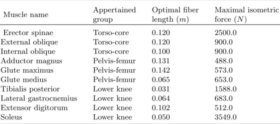
In our study, 10 muscles are selected from the three muscle groups to conduct muscle fatigue analysis. The basic characteristics of these muscles are listed in Table 3.
Table 1. Basic characteristics of the analyzed muscles.
Muscle name Appertained group
Optimal fiber length (m)
Maximal isometric force (N )
Erector spinae Torso-core 0.120 2500.0 External oblique Torso-core 0.120 900.0 Internal oblique Torso-core 0.100 900.0 Adductor magnus Pelvis-femur 0.131 488.0 Glute maximus Pelvis-femur 0.142 573.0 Glute medius Pelvis-femur 0.065 653.0 Tibialis posterior Lower knee 0.031 1588.0 Lateral gastrocnemius Lower knee 0.064 683.0 Extensor digitorum Lower knee 0.102 512.0 Soleus Lower knee 0.050 3549.0
4
Results of simulation
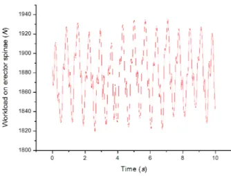
During the arbitrary running, workloads on muscles vary from moment to mo-ment. A typical muscle workload change is shown in Figure 4.
Fig. 3. Workload on erector spinae muscle during 10s arbitrary running at 3.96 m/s.
4.1 General muscle force decline
In order to investigate the fatigue process, the motion data is duplicated to 10 min. During the running process, muscle force capabilities decline with time. After input the fatigability (k) of the subject, the detail information about force capability changes is accessible. A general view of force capability changes of the selected muscles are shown in Figure 4. Here the fatigability is set to 1.0 min−1. Generally, the muscles’ capabilities reduce to 60% to 70% of their maximum after running for 10 min.
4.2 Comparison among different muscles
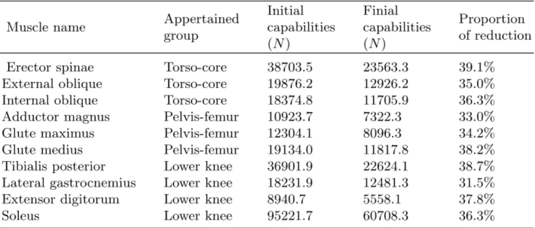
The initial and ending force capabilities of the selected muscles are shown in Table 2.
The proportion of force capability reduction is between 30% to 40% for each of the ten muscles. Erector spinae loses the maximal proportion of force. As far as the selected muscles, torso-core muscle group fatigues no less than the pelvis-femur or the lower knee group (average fatigue level: (36.8 ± 2.1)%, (35.1 ± 2.7)%, (36.1 ± 3.2)%, respectively).
Fig. 4. Force capability lines of ten muscles during 10 min running. k = 1.0 min−1. Force of each muscle is normalized by its maximum.
Table 2. General information of muscle force capabilities
Muscle name Appertained group Initial capabilities (N ) Finial capabilities (N ) Proportion of reduction Erector spinae Torso-core 38703.5 23563.3 39.1% External oblique Torso-core 19876.2 12926.2 35.0% Internal oblique Torso-core 18374.8 11705.9 36.3% Adductor magnus Pelvis-femur 10923.7 7322.3 33.0% Glute maximus Pelvis-femur 12304.1 8096.3 34.2% Glute medius Pelvis-femur 19134.0 11817.8 38.2% Tibialis posterior Lower knee 36901.9 22624.1 38.7% Lateral gastrocnemius Lower knee 18231.9 12481.3 31.5% Extensor digitorum Lower knee 8940.7 5558.1 37.8% Soleus Lower knee 95221.7 60708.3 36.3%
4.3 Influence of fatigability index k
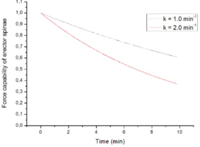
Fatigability is a subject-specific parameter that might also varies between muscle groups. As mentioned in Chapter 2.2, the determined value of k varies from 0.87 min−1to 2.15 min−1for general muscle groups. In the current study, we examine the influence of fatigability by comparing the fatigue level when k = 1.0 min−1
with that when k = 2.0 min−1. A typical comparison is shown in Figure 5. Table 3 manifests the fatigue level comparisons of all the ten muscles. Gen-erally, the muscle reduces to 40% to 50% of its initial maximal capability. The relative sort of muscles’ fatigue level remains unchanged.
Fig. 5. Force capability lines of erector spinae with different preset k values. Force is normalized by the maximum.
Table 3. Comparison of muscle force capabilities between different fatigabilities
Muscle name Appertained group Proportion of capability loss k = 1.0 min−1 k = 2.0 min−1 Erector spinae Torso-core 39.1% 62.9% External oblique Torso-core 35.0% 57.7% Internal oblique Torso-core 36.3% 59.4% Adductor magnus Pelvis-femur 33.0% 55.1% Glute maximus Pelvis-femur 34.2% 56.7% Glute medius Pelvis-femur 38.2% 61.9% Tibialis posterior Lower knee 38.7% 62.4% Lateral gastrocnemius Lower knee 31.5% 53.1% Extensor digitorum Lower knee 37.8% 61.4% Soleus Lower knee 36.3% 59.4%
5
Discussions
According to the simulation data, after 10 minutes’ running at the speed of 3.96 m/s, a healthy male subject is likely to lose 30% to 40% of his maximal muscle capability. The requirement of the running task at the current posture is about 5% of his maximal muscle capability. If the task continues, there would be a time in future when the subject’s muscle capability reduces to near to the required workload. Muscles will enter into a risk zone [23, 22]. Damage will
occur to muscles, which increases the risk of MSDs. The subject would change his posture unconsciously [24]. It is important to predict the exhausting time in the early process of work design.
In Hammer’s research [21], the quadriceps (pelvis-femur muscle group) and plantar flexors (lower knee muscle group) are the major contributors to accel-eration of the body mass center during running, compared with the torso-core muscle group (erector spinae and iliopsoas). While in the current study, erector spinae, who loses 39.2% of its maximal capability, is found to be more fatigue-exposed than the others. Also, torso-core muscle group fatigues no less than the other two muscle groups. This phenomenon indicates that torso-core muscles un-dertake other supportive functions than contributing to body accelerating, such as counterbalancing the vertical angular momentum of the legs.
A subject with a higher fatigability value (k = 2.0 min−1) losses about 20% of his maximal capability more than the subject with a lower fatigability value (k = 1.0 min−1). The influence of fatigability is evident. It is essential to study the fatigability among different human groups.
It should be noticed that the current study considers no effect of fatigue recovery. Future study should integrate the muscle recovery for more accurate calculation.
6
Conclusions and implications
In this study, a plug-in to OpenSim is written on the base of the Force-load mus-cle fatigue model and musmus-cle force generation dynamics to obtain the concrete information about how force-output capability of each muscle declines along time.
Simulation on a three-dimensional, 29 degree-of-freedom human model shows that the force-output capability of ten selected muscles reduced to 60% - 70% after 10 minutes’ running at the speed of 3.96 m/s, with a fatigability value of 1.0 min−1. Torso-core muscle group, which has been found to contribute less to the body’s acceleration in previous research, shows no less proportion of force loss than the other two groups. The difference in fatigue level caused by the change of fatigability is evident, which emphasizes the necessity of the study and determination of fatigability among different human groups.
This work offers a virtual work design platform that helps to predict muscle fatigue and thereby to control the MSDs risks at the early stage of work design. In future works, the Motion Capture System of the ´Ecole Centrale de Nantes will be used to acquire data for industrial tasks. A force platform will be used to validate the muscle properties during experiments. The study of the fatigability k as a function of the people will also be addressed in these new experiments.
7
Acknowledgments
This work was supported by the National Natural Science Foundation of China under Grant numbers 71471095, by Chinese State Scholarship Fund, and by
IN-TERWEAVE Project (Erasmus Mundus Partnership Asia-Europe) under Grants number IW14AC0456 and IW14AC0148.
References
1. B Bigland-Ritchie, CL Rice, SJ Garland, and ML Walsh. Task-dependent factors in fatigue of human voluntary contractions. In Fatigue, pages 361–380. Springer, 1995.
2. Don B Chaffin, Gunnar Andersson, Bernard J Martin, et al. Occupational biome-chanics. Wiley New York, 1999.
3. Eurogip. D´eclaration des maladies professionnelles : probl´ematique et bonnes pra-tiques dans cinq pays europ´eens Allemagne - Danemark - Espagne - France - Italie. Rapport denquˆete, 44 p. www.eurogip.f r/produits − inf ormation/publications − d−eurogip/3906−declaration−des−mpproblematique−et−bonnes−pratiques− dans − cinq − pays − europeens, 2015
4. Thomas J Armstrong, Peter Buckle, Lawrence J Fine, Mats Hagberg, Bengt Jons-son, Asa Kilbom, Ilkka AA Kuorinka, Barbara A Silverstein, Gisela Sjogaard, and Eira RA Viikari-Juntura. A conceptual model for work-related neck and upper-limb musculoskeletal disorders. Scandinavian Journal of Work, Environment & Health, pages 73–84, 1993.
5. Jack, February 2017, retrieved from http : //www.plm.automation.siemens.com /en us/products/tecnomatix/manuf acturing−simulation/human−ergonomics /index.shtml
6. Delmia, February 2017, retrieved from http : //www.3ds.com/products − services/delmia
7. Robert Feyen, Yili Liu, Don Chaffin et al. New software tools improve work-place design. Ergonomics in design: The Quarterly of Human Factors Applications, 7(2):24–30, 1999.
8. Michael Damsgaard, John Rasmussen, Søren Tørholm Christensen, Egidijus Surma, and Mark De Zee. Analysis of musculoskeletal systems in the anybody modeling system. Simulation Modelling Practice and Theory, 14(8):1100–1111, 2006.
9. Scott L Delp, Frank C Anderson, Allison S Arnold, Peter Loan, Ayman Habib, Chand T John, Eran Guendelman, and Darryl G Thelen. Opensim: open-source software to create and analyze dynamic simulations of movement. IEEE transac-tions on biomedical engineering, 54(11):1940–1950, 2007.
10. Don B Chaffin. Digital human modeling for workspace design. Reviews of Human Factors and Ergonomics, 4(1):41–74, 2008.
11. Liang Ma, Damien Chablat, Fouad Bennis, and Wei Zhang. A new simple dy-namic muscle fatigue model and its validation. International Journal of Industrial Ergonomics, 39(1):211–220, 2009.
12. Liang Ma, Damien Chablat, Fouad Bennis, Wei Zhang, Bo Hu, and Fran¸cois Guil-laume. A novel approach for determining fatigue resistances of different muscle groups in static cases. International Journal of Industrial Ergonomics, 41(1):10– 18, 2011.
13. Sophie Sakka, Damien Chablat, Ruina Ma, Fouad Bennis. Predictive model of the human muscle fatigue: application to repetitive push-pull tasks with light external load, International Journal of Human Factors Modelling and Simulation, 5(1):81– 97, 2015.
14. Liang Ma and Jing Chang. Measurement of subject-specific local muscle fatigabil-ity. Advances in Physical Ergonomics and Human Factors: Part II., 15:215–220, 2014.
15. Yoon T., Doyel R., Widule C., Hunter S K. Sex differences with aging in the fatigability of dynamic contractions, Experimental gerontology,70:1-10, 2015. 16. Kent-Braun J A., Ng A V., Doyle J W., Towse T F. Human skeletal muscle
responses vary with age and gender during fatigue due to incremental isometric exercise, Journal of Applied Physiology,93(5): 1813-1823, 2002.
17. Liang Ma, Wei Zhang, Bo Hu, Damien Chablat, Fouad Bennis, and Fran¸cois Guil-laume. Determination of subject-specific muscle fatigue rates under static fatiguing operations. Ergonomics, 56(12):1889–1900, 2013.
18. Felix E Zajac. Muscle and tendon properties models scaling and application to biomechanics and motor. Critical reviews in biomedical engineering, 17(4):359– 411, 1989.
19. Jack M Winters. An improved muscle-reflex actuator for use in large-scale neuro-musculoskeletal models. Annals of biomedical engineering, 23(4):359–374, 1995. 20. Darryl G Thelen et al. Adjustment of muscle mechanics model parameters to
simulate dynamic contractions in older adults. Transactions-American Society Of Mechanical Engineers Journal Of Biomechanical Engineering, 125(1):70–77, 2003. 21. Samuel R Hamner, Ajay Seth, and Scott L Delp. Muscle contributions to propulsion
and support during running. Journal of biomechanics, 43(14):2709–2716, 2010. 22. Liang Ma, Wei Zhang, Damien Chablat, Fouad Bennis, and Fran¸cois Guillaume.
Multi-objective optimisation method for posture prediction and analysis with con-sideration of fatigue effect and its application case. Computers & Industrial Engi-neering, 57(4):1235–1246, 2009.
23. Ruina Ma, Damien Chablat, Fouad Bennis, and Liang Ma. Human muscle fatigue model in dynamic motions. In Latest Advances in Robot Kinematics, pages 349– 356. Springer, 2012.
24. Jason R Fuller, Karen V Lomond, Joyce Fung, and Julie N Cˆot´e. Posture-movement changes following repetitive motion-induced shoulder muscle fatigue. Journal of Electromyography and Kinesiology, 19(6):1043–1052, 2009.
![Fig. 1. Muscle force generation dynamics. Adapted from Zajac (1989) [18].](https://thumb-eu.123doks.com/thumbv2/123doknet/11547554.296287/5.918.210.699.745.827/fig-muscle-force-generation-dynamics-adapted-zajac.webp)