i
My admirations and
endless thanks …
to my foster father
during these past 4
years;
once …
le Pr Giurfa
once …
el señor Giurfa
and once …
simply Martin.
ii
Infinite thanks to countless, but mentionable persons I encountered, for which they contributed in different ways to the finalisation of this dissertation.
My family who shaped me and let me savour Europe, even though it is still not easy for them. Hiromu “Ron” Tanimoto who showed me the stepping stone and encouraged me to take the leap of faith to France.
Prof. Ricarda Scheiner and Prof. Jeremy Niven who reviewed the dissertation in relatively short time and kindly managed to.
Prof. Claudio Lazzari and Jean-Marc who kindly became members of the jury to examine the dissertation as well.
Rodrigo “Muchacho” and Nobuhiro “Nobu” who were my two dear mentors at the beginning of my adventure into the bee world, in good times and in bad, and keep being my mentors in many aspects.
Julie, without her I would have stayed clueless in the laboratory and homeless in France. Edith, without her results my project would not have been present.
Gabriela for her being so welcoming and motherly in and outside the laboratory.
Theo who has been a helpful lab-mate in my early days and become cachaça-mate in later days.
Margaux and Elodie for having been so patient dealing with me and co-authoring my publications.
Isabelle for her advices and supports, particularly remarkable that one time when we had to race to “préfecture de Haute-Garonne” minutes before they closed.
Ayse for being my heroine when things did seem to end badly. Lucie for her efforts in selecting the right foragers.
Je remercie Christine pour ses aides administratives et particulièrement pour avoir réussi à commander la salle de l’UPS qui a le nom le plus cool et très français (ou toulousain).
Maud and Patrick for their technical support.
Gérard pour m’avoir toujours invité de venir faire du ski ensemble qu’on n’a jamais concrétisé.
Postdoc Power Rangers who show me that justice shall prevail; Alfonso, Dañao, Fabien, Sara (whose laughter always brightens up dark corridors) and Sepideh (who was for me identical to Sara on our first encounter).
Constance, Jessica et Sophia pour avoir traduit et corrigé des parts français dans ma thèse, entre autres.
I also thank other actual and previous PAMI members Alexei “Alexov”, Amelie, Aurore, Damien, Cathy, Celeste, Cindy, Clement (✝), Florencia, Florian, Guillaume, Jonas, Lilha Margot, Marie, Marion, Maru, Mathieu, Mehdi, Monique, Pierre, PYM, Thierry and Yannice
iii
Elodie avec sa coupe rasta for letting me know the beauty of nature in France as well as in New Zealand.
Prof. Alison Mercer who hosted me during my stay in New Zealand with outmost hospitality and her spotless team - “Jamie” McQuillan, who passionately taught me gently to stay clean and left, David and Charles.
Prof. Buchner for the α-SynORF antibody which accompanied me during my whole doctoral study. Prof. Karen Mesce for her brain samples. Marie-Laure and Jacques from ITAV for their excellent assistance in confocal microscopy. Anja, Sebastian and Igor “Big Guy” for their helps in developing my technique immunocytochemistry, which made me go this far. Prof. Randolf Menzel, der mich herzlich in seinem Labor empfing und Geheimtipps bezueglich der Elektrophysiologie gab, und Gisela, die mir leidenschaftlich intrazelluläre Ableitung beibrachte. Genauso warmherzig haben mich die anderen Kollegen in Berlin wie Prof. Bjoern Brembs, Sathiskumar “Sati”, Adriana, Andreas, Christine “Chrissi”, and Julien empfangen. Und selbstverständlich meine Freunde in München, im Guten wie im Schlechten: Arnab, Basti “Brenz“, Basti “Swob“, Erika, Johanna, Kristina, Natalie and “Olly” Hawlitschek.
My indonesian friends who keep me checked and sane: Anastasia, “Agus” Karen, Indra “Buabeh”, Kurniawan “Kuri”, Neki “QQ Djavor” and Prudence “Prudi”.
Sophie “Sophsky” and Ana “Muchacha” for connecting me with parts of the world.
Paul for his generous donation of his bottles of scottish whiskeys and south french wines in the infamous fortress Hamer.
La famille Le Liboux, la famille Sirac and la famille Taurand for having me as their oldest son in the family.
My friends from the indonesian student association: Afif, Adrian, Agung, Aziz, Ananda, Adhitya “Saiko”, Bulan, Deni, Devi, Ester, Fito Beng, Ivan, Nadya, Kus, “Nemi” Sinaga, Rama, Reggie, Ruby “Liu Liu”, Surya, Tya, Yoga and Zulkifli “Kipli”.
Disclaimer: Statements above are only the tip of the Eisberg. They don’t represent your true infinite good deeds. Bless you all, I feel overwhelmed having you around me, but we can’t afford getting the acknowledgement longer than the content of the dissertation per se, can we?
iv
TABLE OF CONTENTS
Acknowledgement ... i
Table of contents ... iv
Introduction ... 1
1. Learning and Memory: Basic Definition ... 3
1.1. Classical Conditioning: Principles and Definitions ... 4
1.2. Classical Conditioning in Invertebrates ... 5
2. The Honey Bee as a Model for the Study of Learning and Memory ... 7
2.1. Sucrose Responsiveness in Honey Bees ... 8
2.2. The Appetitive Olfactory Conditioning of PER in the Honey Bee ... 9
2.3. The Architecture of the Honey Bee Brain ... 10
2.4. The Neural Circuits Underlying Appetitive Olfactory Learning in the Honey Bee . 16 2.4.1. Appetitive US (sucrose) Pathway Underlying Olfactory PER Conditioning . 16 2.4.2. Olfactory CS Pathway Underlying Olfactory PER Conditioning …... 18
2.5. Appetitive Memories Induced by Olfactory PER Conditioning ... 20
3. The Advent of a New Conditioning Protocol for Harnessed Bees: the Aversive Olfactory Conditioning for the Sting Extinction Reflex ... 22
3.1. Aversive US Pathway Underlying Olfactory SER Conditioning ... 26
3.2. Olfactory US Pathway Underlying Olfactory SER Conditioning ... 28
3.3. Aversive Memories Induced by Olfactory SER Conditioning ... 29
3.4. Aversive Shock Responsiveness and Colony Organization ... 31
3.5. Shock Sensitivity and Olfactory SER Conditioning ... 34
3.6. Shock Sensitivity, Olfactory SER Conditioning and Caste Specialization within the Hive ... 35
v
Part 1: Pharmacological modulation of aversive responsiveness in honey bees ... 41
Part 2: A tyrosine-hydroxylase characterization of dopaminergic neurons in the honey bee brain ... 85
Part 3: Aversive learning increases the expression of dopamine-receptor genes in specific cell populations of the honey bee brain ... 135
Discussion ... 171
1. Different Functionality of Dopaminergic Neurons (neuropharmacological study – responsiveness to electric shock, an aversive stimulus) ... 174
1.1. Dopaminergic Modulation of Aversive Learning and Memory ... 174
1.2. Dopaminergic Modulation of Aversive Responsiveness ... 175
1.3. Different Classes of Dopaminergic Neurons ... 176
2. Neuroanatomy of the Dopaminergic Network in the Bee Brain ... 178
2.1. Dopaminergic Processes in the MB ... 179
2.2. Dopaminergic Neurons as Modulators of Behaviour ... 181
3. Learning-Dependent Modulation of DA Receptor Expression in the MBs ... 182
3.1. A Molecular Mechanism Accounting for the Rescorla-Wagner Model? ... 185
4. General Conclusions ... 188
References ... 189
1
INTRODUCTION
INTRODUCTION
INTRODUCTION
3
INTRODUCTION
1. Learning and Memory: Basic Definitions
In the course of life, animals acquire experiences, which are an indispensable part of their survival. These experiences increase an animal's knowledge about its environment and allow producing adaptive behavior upon future encounters with the events that generated such experiences. Learning and memory are the two basic capacities that articulate this scenario: Learning is any relatively permanent change in response that occurs as a result of experience acquired individually (Bitterman et al., 1979). Such acquisition may lead, depending on factors such as the number of experiences gathered, their spacing in time, etc, to the formation of memory, which is defined as the capacity to encode, store and retrieve the acquired information in the nervous system (Ebbinghaus, 1913). Different types of memories are established by variations in experimental procedures (e.g. repetition during training, massed vs. spaced trials, etc.), which are distinguishable in terms of their duration (i.e. stability), contents and resistance to extinction (Ebbinghaus, 1885). In this sense, a basic distinction acknowledges that memory is organized in at least two forms: a transient and unstable short-term memory (STM), and a robust, long-lasting long-short-term memory (LTM). Both memory types exhibit distinct temporal courses and underlying molecular processes (Kandel, 2001).
Associative learning is defined as the capacity to learn the predictive links existing between related events in an animal's environment. It allows to extract the logical structure of the world and thus to reduce uncertainties of future events (Pearce, 1987; Rescorla, 1988). Two main paradigms of associative learning are usually distinguished. One is the paradigm of classical (Pavlovian) conditioning (Pavlov, 1927) and the other is the paradigm of operant or instrumental conditioning (Skinner, 1938). While the former relies on learning stimulus – stimulus associations (Pavlov, 1927), the latter relies on acquiring associations linking actions and specific outcomes (reinforcements) of these actions (Skinner, 1938). In classical conditioning the action of the animal is irrelevant for the contingency between stimuli to be acquired; in operant conditioning, on the contrary, it is the action of the animal that determines the occurrence of reinforcement. A considerable amount of literature has been produced on these two learning forms, which have been studied in invertebrates and
Introduction
4
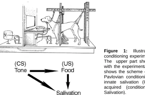
Figure 1: Illustration of the classical conditioning experiment conducted by Pavlov. The upper part shows the harnessing setup with the experimental subject. The lower part shows the scheme of associations underlying Pavlovian conditioning and leading either to innate salivation (US -> Salivation) or to acquired (conditioned) salivation (CS -> Salivation).
vertebrates, including humans. In the following, I will focus exclusively on classical conditioning as it constituted the main experimental framework for most of my works.
1.1. Classical Conditioning: Principles and Definitions
This conditioning form was first discovered by Ivan Pavlov. Pavlov was a Russian physiologist, who at the beginning was not interested by learning but by the physiology of digestion, which he studied using dogs as experimental animals. During his studies, he discovered by chance that dogs began salivating when his assistant came into their sight, and before they received their food. Since the salivary response is an innate response, Pavlov suggested that the salivation in the presence of the assistant represented an acquired
expectation of the food. To test this hypothesis, Pavlov made his subject, a harnessed dog (Fig. 1), to associate the food, evoking the reflex of salivation, with a neutral stimulus unable to evoke it, which was the sound of a metronome. The saliva was collected through a tube and the experimenter could observe and record the salivary response from behind the screen.
Pavlov paired the presentation of both stimuli, sound and food, in such a way that the sound of the metronome always anticipated the food (forward pairing) in order to create the food expectation. After experiencing the sound-food pairings several times, the dogs learned to salivate to the sound of the metronome itself, thus showing the acquisition of a novel response as a consequence of the training. Pavlov then termed the different components of his protocol as: 1) Unconditioned Stimulus (US), the biologically relevant stimulus eliciting an
5
response elicited by the US (here salivation to the food); 3) Conditioned Stimulus (CS), the stimulus that is originally neutral but that acquires the capacity to evoke a learned response (here the sound of the metronome); 4) Conditioned Response (CR), the learned response to the CS (here salivation to the metronome sound).
Thus in his work on classical conditioning, Pavlov concluded that the basis of classical, Pavlovian conditioning resides in the fact that subjects learn to associate an originally neutral stimulus (conditioned stimulus – CS) with a biologically significant stimulus (unconditioned stimulus – US) that elicits a reflexive response (unconditioned response) (Pavlov, 1927). Originally, the CS does not elicit a behavioural response but once an association between the US and the CS is established, the response to the CS will resemble the response to the US.
Different variants of the basic Pavlovian scheme can be conceived. In its basic form (see above), the animal learns a simple link between a CS and a US (CS+, with + indicating the presence of the US), a situation termed absolute Pavlovian conditioning. In differential
Pavlovian conditioning, the animal learns that one CS (CS1) is reinforced, while another CS
(CS2) is non-reinforced (CS1+ vs. CS2-). In the former case, an animal has to learn to respond
to CS+ alone, which is unambiguously associated with reinforcement; in the latter, it has to learn to respond to CS1+ and not to CS2- because both are unambiguously associated with
reinforcement and with the absence of it, respectively.
1.2. Classical Conditioning in Invertebrates
Decades of research have established invertebrates as standard models for the study of classical conditioning. This is because invertebrates learn simple associations and have a relatively simple nervous system that allows associative phenomena to be traced to the cellular and molecular levels in different kinds of laboratory preparations (Giurfa, 2013).
Aversive classical conditioning, for example, has been studied in the mollusc Aplysia. In this sea slug, skin sensory neurons make direct synapses onto motor neurons that control the defensive gill withdrawal reflex; upon a light touch to the naïve animal’s siphon, these
Introduction
6
sensorimotor synapses fail to transmit and the gills are not withdrawn. If, however, the touch is repeatedly paired with a noxious stimulus, such as an electric shock, that does elicit gill withdrawal, the light touch alone eventually comes to elicit a gill withdrawal, whereas, before such training, it did not (Kandel et al. 1979; Walters et al. 1979; Carew et al. 1981; Hawkins et al. 1983; Abrams and Kandel 1988; Hawkins et al. 1989; Byrne et al. 1990; Hawkins et al. 1998).
Classical conditioning has also been intensively studied in the fruit fly Drosophila
melanogaster. Flies can easily be trained to associate an odor (the CS) with an aversive
electric shock (the US). The typical procedure consists in training groups of flies alternatively presented with two different odours, one paired with an electric shock (CS1+), and another
non-paired with the shock (CS2-) (Tully and Quinn, 1985). Retention is measured in a T-maze
where conditioned flies must choose between the CS1+ and the CS2-. Note, however, that
despite its recurrent description as a Pavlovian protocol, the procedure employed involves operant components as the flies freely move within the maze and their actions determine therefore whether a shock will be experienced or not. Although flies certainly associate the odours as CS with the shock or absence of shock as US, the protocol does not exclude the occurrence of operant learning.
On the contrary, true Pavlovian learning can be studied in another preparation conceived for the honeybee Apis mellifera, an insect that has emerged as a powerful model for the study of learning and memory (Giurfa, 2003, 2007; Hammer, 1993, 1997; Menzel, 1999, 2001). Similarly to the reflexive salivation of dogs, an appetitive reflex, the proboscis
extension response (henceforth PER), was known to occur in the bee (Frings, 1944; Frings
and Frings, 1949), and other insects (Minnich, 1921, 1926) upon stimulation of gustatory organs, e.g. the antennae, tarsi or mouth parts, with sugar solution. In the fifties, a Japanese researcher, Matsutaro Kuwabara, realized that this appetitive response could be conditioned in harnessed bees using visual stimuli as CS and sucrose solution delivered to the tarsi as US (Kuwabara, 1957). However, his work did not reach broad impact probably due to the fact that a critical procedural aspect to follow for this protocol to work was the necessity to cut the antennae of the bees prior to conditioning. Indeed, Kuwabara mentioned that bees in his protocol started extending the proboscis to the sucrose solution before it reached the antennae or mouth parts. This was an undesirable effect as the US was supposed to elicit PER only upon contact. He speculated that this effect was due to the presence of hygroreceptors on the
7
spoon. Therefore, he decided to cut the bees’ antennae to avoid this problem, and to elicit the response by stimulating the tarsi with sucrose solution. Depriving the bees of their antennae is not necessarily an ideal handling of the experimental subjects. A damaged animal will probably be less responsive than an intact animal. The low acquisition rates observed in antennae-deprived bees despite long conditioning procedures (Hori et al., 2006; Hori et al., 2007) may be related to this fact. Later, as described below, an olfactory version of this protocol was developed, which allowed to study Pavlovian olfactory learning to an unprecedented level of detail (Bitterman et al., 1983; Takeda, 1961) (see below). This achievement contributed, in part, to the success of the honeybee as one of the most popular models in the neurosciences of learning and memory.
2. The Honey Bee as a Model for the Study of Learning and Memory
The study on honey bee behaviour was pioneered by Karl von Frisch (1886 – 1982). He became famous for the discovery of the honey bee dance, a communication behaviour where a successful forager transmits information to other foragers within the hive about the distance and direction of a profitable food source (von Frisch, 1967). In a natural context and despite their small size, honey bees exhibit an extremely rich behavioural repertoire. At the social level, honey bees exhibit reproductive division of labour (with sterile and reproductive castes), generational overlap and cooperative brood care (Wilson, 1971). During their lifetime, exhibit 'caste polyethism' a term used to indicate that individuals go through different caste stages and perform different tasks at different ages (Robinson and Page, 1989; Wilson, 1971). A newly emerged bee perform the most basic task which is cell cleaning. It progresses with being a nurse where the individual takes care of the queen and feeds larvae. Afterwards the individual starts being a guard gathering first experiences with the outer world. Usually around the 2nd week of life, the bee becomes a forager. During this phase, the bee will start experiencing outer environment and actively uses its cognitive abilities to navigate between the hive and the food sources and to learn and memorise various food-related stimuli. To this end, it exhibits of a rich sensory perception and developed motor performances.
Indeed, bees see the world in colour (Menzel and Backhaus, 1991), perceive shapes and patterns (Giurfa and Lehrer, 2001; Srinivasan, 1994) and resolve movements with a high
Introduction
8
temporal resolution (Srinivasan et al., 1999). Their olfactory sense is able to distinguish a large range of odours (Guerrieri et al., 2005) and the mechanosensory perception is also extremely rich due to thousands of hair cells all around the body and proprioreceptors inside the body (Dacher et al., 2005; Erber et al., 1998; Scheiner et al., 2005).
More importantly, the learning capacities of honey bees, which set the basis for their floral constancy, i.e. the fact that bees remain truthful to the same flower species as long as it offer profitable nectar or pollen reward, are amenable to the laboratory for detailed study. After almost a century of honey bee research, different protocols have been established to assess the bees' learning and memory capabilities (Giurfa, 2007). Here we will focus on one of these protocols, the olfactory conditioning of the PER (Bitterman et al., 1983; Takeda, 1961).
Yet, before presenting the current knowledge on honey bee learning and memory gained through the olfactory conditioning of PER, I will focus on innate US responsiveness and show how the PER preparation allowed characterizing in detail different aspects of how bees respond to the sucrose reward, and how this analysis provided important insights into the organization of labor within the colony and the individual learning itself.
2.1. Sucrose Responsiveness in Honey Bees
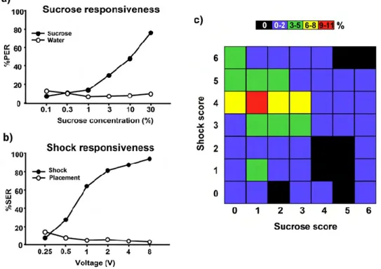
Long before research on PER conditioning started, it was well known that PER could be elicited by stimulating gustatory organs like the antennae, tarsi or mouth parts with sugar solution. The PER had thus been detected in bees (Frings, 1944; Frings and Frings, 1949), flies (Minnich, 1926) and butterflies (Minnich, 1921) among others. Much later, the spontaneous PER upon sucrose stimulation was used to quantify the appetitive responsiveness of bees (i.e. their tendency to respond to sucrose of different qualities) without involving any learned component (Page et al., 1998). This is done by presenting restrained bees with increasing sucrose concentrations delivered to the antennae and determining either the lowest concentration from which the bee starts responding, discriminating it from water (sucrose threshold), or the number of sucrose concentrations to which it responds (sucrose score).
This quantification allowed differentiating nectar- and pollen foragers in terms of their different responsiveness to sucrose (Page and Erber, 2002; Pankiw and Page, 1999; Scheiner
9
foragers, which exhibit lower thresholds and thus higher responsiveness (Page et al., 1998). Although this difference may appear counterintuitive at first sight, the currently accepted explanation is that nectar foragers are more selective when collecting nectar, and thus only respond to the highest sucrose concentrations, which provide the highest energy gain to the individual and the colony. Sucrose responsiveness thresholds have been shown to vary with multiple factors such as age, caste, sex, (Amdam et al., 2006; Page et al., 1998; Pankiw and Page, 1999; Scheiner et al., 2001a, b), foraging experience, genotype, feeding status (Pankiw and Page, 2001), and season (Scheiner et al., 2003), among others.
Later works showed that pollen foragers, being more responsive to sucrose, perform better than nectar foragers during the appetitive PER conditioning (Pankiw and Page, 1999; Scheiner et al., 1999; Scheiner et al., 2001a). This result reflects the fact that the subjective value of sucrose reward, which can be estimated via PER responsiveness, may vary between bees. Thus, the same sucrose concentration may be efficient to support conditioning in some bees while it may not be rewarding enough for other bees which will learn less efficiently when provided with this concentration as US (Scheiner et al., 2005). Summa summarum, bees showing higher sucrose responsiveness perform better during the associative appetitive PER conditioning. In other words, sucrose responsiveness allows us to dissect the effects of genotype and division of labour on associative appetitive learning.
As an important neuronal signalling component, and in facilitating or depressing behavioural responsiveness, three biogenic amines have been examined for their role in the modulation of sucrose responsiveness. These are octopamine, tyramine and dopamine (Scheiner et al., 2002). Modulation was tested by injecting these substances into the bees and determining if and how sucrose responsiveness changed. Octopamine and tyramine increased sucrose responsiveness whereas dopamine decreased it. The decrease induced by dopamine was also induced by a dopamine receptor agonist ADTN (Scheiner et al., 2002).
2.2. The Appetitive Olfactory Conditioning of PER in the Honey Bee
As mentioned above, besides providing insights into sucrose responsiveness PER was used as reflex to be conditioned in a Pavlovian protocol first established by Takeda (1961) who was inspired by the early work of Kuwabara (1957; see above). Kuwabara had reported that PER
Introduction 10 B A odour sucrose odour can be conditioned using colours as CS. Takeda decided to replace colours by odours and set the basis for the olfactory conditioning of PER.
In this protocol, each bee is restrained in an individual harness so that it can only freely move its antennae and mouth-parts (mandibles and proboscis) (Fig. 2).
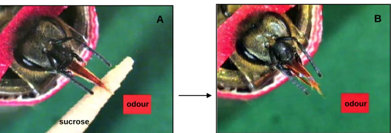
Figure 3: Appetitive olfactory conditioning of proboscis extension reflex (SER). A) A harnessed bee is
trained with paired presentations of an odorant (CS) blown to the antennae and the sugar reward (US) delivered on a toothpick, following an absolute – conditioning design (a single odorant reinforced). B) After a successful conditioning, the bee extends its proboscis to the odour CS alone, which has been learned as predictor of the sucrose US.
The antennae are the bees’ main chemosensory organs. When the antennae of a hungry bee are touched with sucrose solution, the animal reflexively extends its proboscis to reach out to and suck the sucrose (PER). Thus, the sucrose solution inducing the PER is the appetitive US. In the case of classical conditioning, a bee learns to associate the US with a neutral odorant which is blown to the antennae. The odorant does not release such a reflex in naive animals and serves as the CS. The coupling of the CS and the US in a forward-pairing manner results in acquisition of the conditioned odorant (Fig. 2).
The protocol has emerged through the years as a unique tool to access the neural and molecular bases of Pavlovian learning and memory in honey bees as it allowed dissecting the neural circuits underlying CS (odour) and US (sucrose) processing (Giurfa and Sandoz, 2012). Before describing these circuits it is important to provide a general overview of the honey bee brain.
2.3. The Architecture of the Honey Bee Brain
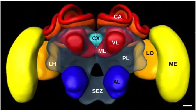
The brain of a honey bee (Fig. 3) has a volume of approximately 1 mm3 and contains around 960.000 neurons (Witthöft, 1967). In general, the honey bee brain is divided into the
11
SPZ comprises several main regions or neuropiles, such as the antennal lobes (AL), the optic lobes (OL), the mushroom bodies (MB), a vast region surrounding the MB generally termed as the protocerebral lobes (PL), a small lateral protrusion of the PL, known as the lateral horn (LH), and the central complex (CX). ALs are the primary olfactory neuropiles and receive direct inputs mainly from chemoreceptors located on the antennae. The OLs are visual processing neuropiles receiving information from the photoreceptors. The MBs occupy approximately 30% of the brain and are higher-order integration centres exhibiting segregated multimodal input and integrated multimodal output; these structures have been historically associated with the presence of memory traces, in particular long-term ones (Menzel, 1999; Menzel and Giurfa, 2001). The PL exhibits also a multimodal organization, with visual and olfactory afferences arriving at different regions of this structure. The CX seems to be involved in different forms of visual processing, from patter recognition (Liu et al., 2006), to polarized light processing (Heinze et al., 2009; Heinze and Homberg, 2007; Homberg et al., 2011) and visual-course setting (Neuser et al., 2008; Strauss, 2002), among others.
Figure 3: 3D-reconstruction of a honey bee brain. This frontal view depicts a honey bee brain with its
neuropiles. AL: antennal lobe; CA: calyx of the mushroom body (MB); VL: vertical lobe; ML: medial lobe; CX: central complex; LO: lobula of the optic lobe (OL); ME: medulla; PL: protocerebral lobe; LH: lateral horn; SEZ: subesophageal zone. Scale = 100 µm; adapted from Brandt et al. 2005.
AL SEZ ME LO VL ML CX CA PL LH
Introduction
12
The AL receives input from 60,000 chemosensory receptor neurons located in specialized cuticular structures (sensilla) on the antennae (Esslen and Kaissling, 1976; Menzel and Muller, 1996). The AL consists of approximately 160 globular structures termed glomeruli, whose number corresponds to that of the olfactory receptor genes recently indentified on the honey bee genome (Robertson and Wanner, 2006). Neurons with the same receptor class project to the same glomerulus. Each glomerulus is a localized site of synaptic interactions between different cell populations: the afferences from olfactory receptors, the lateral connections between glomeruli provided by local inhibitory (GABAergic, histaminergic) interneurons (LIN) and the efferent projection neurons (PN), which connect the AL with the MB, several regions in the PL and the LH. PNs run in three distinct main fibre tracks, namely the antenna-cerebral tracts, currently termed as medial, lateral and mediolateral antennal lobe tract (m-ALT, l-ALT, and ml-ALT) (Abel et al., 2001; Kirschner et al., 2006).
The OL is formed by three succeeding neuropiles: the lamina (LA), the medulla (ME) and the lobula (LO) (Kien and Menzel, 1977b). These are the main centres of visual processing in the bee brain receiving and processing information from the photoreceptors located within ommatidia in the compound eyes.
The lamina is the first visual neuropile, in which the axons of the photoreceptors connect to first order processing interneurons, the lamina monopolar cells (LMC). The LA is made of thousands of optical cartridges, each receiving an axon bundle (containing the nine photoreceptor cell axons) from the overlying ommatidium, as well as the axons of four different types of monopolar cells. Additionally, tangential, centrifugal and horizontal fibers can be found within each cartridge. The spatial arrangement of photoreceptor axons and LMCs within a cartridge remains constant throughout the lamina, thus retaining the retinotopic organization.
The outer chiasm (Ribi, 1974) forms the connection between the LA and the second visual neuropile, the ME. Fibres coming from the anterior part of the LA project to the posterior ME while posterior fibres from the LA project to the anterior ME. The retinotopic organization is retained but it is reversed in the medulla. The medulla is also built of distinct cartridges, receiving the axons of long visual fibres (UV receptors) and from the four LMC of the corresponding cartridge in the LA. Processing of spectral information in the distal part of
13
opponency (Kien and Menzel, 1977a), which sets the basis for their colour vision capacities. The direct connection between the medulla and the third optic ganglion, the lobula (LO), is formed by monopolar cells from the ME, which constitute the inner chiasm. In much the same way as the outer chiasm, neuronal fibres cross the horizontal plane, so that fibres coming from the posterior part of the medulla project to the anterior part of the LO, and vice versa. Therefore, the LO presents the same retinotopic arrangement of visual cartridges as the LA. Neurons responding to directional movement, but also double spectral-opponency neurons, seem to be present in the lobula (Hertel and Maronde, 1987). These neurons project either to the MBs on both brain sides (lobula tract), to the Anterior Optic Tuberculum (AOTU), or build the great commissure (GC) connecting both eyes. Recently, our group has been able to achieve the first optophysiological recordings of neural activity at the neuronal-ensemble level in the visual circuits of the honeybee (Mota et al., 2013; Mota et al., 2011b). It was shown that in the Anterior Optic Tuberculum retinotopy is ventrally-dorsally reversed and, that color processing is compartmentalized in this structure in a way that may refer to a chromatic compass for sky-light-based navigation (Mota et al., 2013).
Different tracts leave the optic lobes to reach the MBs. The most important tract connecting the two optic lobes to the MBs is the anterior superior optic tract (a.s.o.t.) which contains about 300 neurons from the dorso-medial part of the lamina and projecting to the collar and the basal ring of the calices on both brain sides. The anterior inferior optic tract (a.i.o.t.) coming from the ventral part of the medulla joins the a.s.o.t. and projects to the collar and basal rings on both sides. Lastly, the lobula tract (lot) connects each LO with the same regions in the MBs. Interestingly, in both collar and basal ring of the calyces, a strict segregation of information coming from the ME and the LO is observed (Ehmer and Gronenberg, 2002). Moreover, within the collar, projections from the dorsal and ventral parts of the medulla are also segregated. Furthermore, extrinsic lobula neurons also connect with the optic lobe of the contralateral side (Hertel et al., 1987).
The honeybee MB is a higher order multisensory processing/integration centre, which consists of ~160,000 densely packed neurons, the Kenyon cells (KC) (Mobbs, 1982, 1984). Their dendrites form the input sites, calyces (CA), which receive afferences from different sensory pathways (olfactory, visual, gustatory, mechanosensory) and brain regions (AL, OL, SEG) (Strausfeld, 2002) and which project to the output regions of the MBs, the α- and
β-Introduction
14
lobes (Mobbs, 1982), currently identified as vertical (VL) and medial lobes (ML) (Strausfeld, 2002). Furthermore, the KCs are morphologically differentiated into subpopulations: inner compact cells (ICC), non-compact cells (NCC) and outer compact cells (OCC) (Farris et al., 1999). The cell bodies of the ICC are around 4–5 µm and tightly packed into a cone-shaped region at the centre of each calyx. NCC have larger cell bodies (6– 7 μm) and fill up the remaining space within each calyx up to the dorsal border. Dendrites of NCC are detected in the lip and collar regions of the calyces. The OCC are around 6-7 µm as well and their appearance is similar to that of the ICC. They are located lining the outsides of the calyces. Dendrites of OCC can be found in the collar region (Mobbs, 1982). The calyx is segregated into several subregions. Each subregion receives afferents from sensory organs: the lip region receives olfactory afferents; the collar region (col) visual afferents; and the basal ring (br) mixed afferents, in which its inner half is innervated by olfactory afferents and outer half by visual afferents (Ehmer and Gronenberg, 2002; Gronenberg, 2001; Kirschner et al., 2006). The segregation among these subregions continues within the VL layers (Strausfeld, 2002). From both VL and ML, the information is further relayed to other brain regions via the so-called MB extrinsic neurons (MBEN) (Rybak and Menzel, 1993).
Recurrent inhibitory feedback to the MBs is provided by the A3-v cluster of the protocerebral calycal tract (PCT), which connects the major output regions of the MB, the α- and β-lobes and pedunculus, with its major sensory input site, the calyces (Rybak and Menzel, 1993). The second PCT cluster (A3-d) provides local feedback presumably onto MBENs to premotor neurons (Okada et al., 2007). Inhibitory modulation by these GABAergic feedback neurons can be crucial to solve higher-order learning problems (Devaud et al., 2007).
The PL represents a large mass between the AL and the OL (Brandt et al., 2005). Not much is known about the morphological and functional segregations of the PL in honey bee. A prominent structure has been however thoroughly studied. It is an anterior part of the PL, located dorsal to the AL, ventrolateral to the α-lobe of the MB and known as the anterior optic tubercle (AOTU). It is a spherical neuropile that receives input from OL (Mota et al., 2011a). That PL receives information from the visual sensory organ, appears to be conserved across insect models. Prior works in Drosophila, the blowfly and the bumble bee show that this structure is segregated into smaller distinctive neuropiles. These are characterised by the
15
Paulk et al., 2008; Strausfeld and Okamura, 2007) and are termed 'optic glomeruli'.
The LH is a prominent neuropile that is a small lateral protrusion of the protocerebral lobe (Rybak, 1994), compromising at least 4 sub-compartments (Kirschner et al., 2006). It is innervated by various projection neurons (PNs) leaving the AL via different antenno-cerebral tracts (ALTs) (Abel et al., 2001; Kirschner et al., 2006; Muller et al., 2002). Through these various ACTs the LH is connected to larger parts of protocerebral areas and the MBs (Müller et al. 2002; Kirschner et al. 2006). A recent work in the LH of honey bee revealed the principles of olfactory coding occurring at the input of the LH which share similarities with those occurring in the AL (Roussel et al., 2014). This work shows that the LH exhibits a specific coding pattern in response to odorants, thus supporting previous evidences in the locust suggesting that the LH may be involved in simple coding of olfactory stimulus, bilateral olfactory integration and multimodal integration (Gupta and Stopfer, 2012).
The CX is another higher order structure formed by a group of interconnected neuropiles located centrally in the protocerebrum (Homberg, 1985; Strauss, 2002). In the honey bee, it comprises the central body (CB), segregated as an upper (CBU) and a lower (CBL) division, the protocerebral bridge (PB), an arched neuropile located dorsally to the CBU, and by two globular structures located posterior to the PB called noduli (NO). Relatively little is known about the neuroanatomy and physiology of the CX in the honey bee except that its interneurons respond to visual stimuli (Homberg, 1985; Milde, 1988). In other insects, the CX participates in polarization vision, visual information processing and memory storage, spatial orientation and locomotion control (for review see Boyan and Reichert, 2011; Pfeiffer and Homberg, 2014) so that similar roles could be ascribed to the CX of the honey bee.
The SEZ is a fusion of the mandibular, maxillary, and labial neuromeres (Rehder,
1988). This region processes the gustatory, olfactory and mechanosensory input from the proboscis. It seems to be particularly important for gustatory coding (Rehder, 1988; Sanchez et al., 2007). From there, projections are sent to motor neurons controlling the muscles of the mouthparts, thereby mediating the proboscis extension reflex (PER). This region is important for associative appetitive learning as it contains the cell body of an important modulatory neuron involved in olfactory appetitive conditioning, the VUMmx1, which substitutes for sucrose in appetitive olfactory conditioning (Hammer, 1993) (see below).
Introduction
16
2.4. The Neural Circuits Underlying Appetitive Olfactory Learning in the
Honey Bee
Having described the main structures of the bee brain and their known functionalities, it is now possible to go back to the appetitive olfactory conditioning of PER (see above and Fig. 2) and trace the US (sucrose) and CS (olfactory) components to the neural level.
2.4.1. Appetitive US (sucrose) Pathway Underlying Olfactory PER
Conditioning
In the honey bee, the US (sucrose) processing pathway starts at the level of the gustatory receptors which are localized within gustatory sensilla localized on the antennae, tarsi and mouth parts (de Brito Sanchez, 2011; de Brito Sanchez et al., 2007). Sucrose receptor neurons from the mouthparts converge to the SEZ and somehow (the synaptic regions are unknown) connect with a neuron called VUMmx1 (abbreviation for “ventral unpaired median neuron of the maxillary neuromere 1”) (Fig. 4). It responds with long-lasting spike activity to sucrose solution delivered both at the antennae and the proboscis (Hammer, 1993). The dendrites of VUMmx1 arborise symmetrically in the brain and converge with the olfactory pathway at three sites: the ALs, the calyces of the MBs, and the LH, which are key processing stages of olfactory information (CS pathway, see above) in the bee brain.
A B m-ALT l-ALT AL LH m-ALT
17
adapted from Hammer 1993. A: VUMmx1 neuron sends its projection to the antennal lobes (AL), lateral horns (LH) and calyces (lip and basal ring, bR) of the mushroom bodies (MBs) and conveys the appetitive reinforcement signal. B: Olfactory pathway in honey bee showing the afferences from olfactory sensory neurons (oSN) to the glomeruli (G) of the AL. Olfactory information processed in the AL is then relayed by ALTs (l- and m-ALT are depicted) to the MBs which consist of Kenyon cells (KC). The output region of the MBs, the α- and β- lobes is also shown. Scale = 100 µm.
Behavioural learning of an olfactory stimulus can be induced by substituting the sucrose reward in PER conditioning by an artificial depolarisation of VUMmx1 immediately
after olfactory stimulation (forward pairing) (Hammer, 1993). If depolarization precedes olfactory stimulation (backward pairing), no learning is observed. The same forward-backward effect is seen when sucrose is used as reward under similar experimental conditions. In all cases the bees’ response was quantified in terms of the number of spikes of M17 (Rehder, 1987), a muscle controlling the movement of the proboscis. The results thus show that VUMmx1 constitutes the neural correlate of the US in associative olfactory learning.
Classical conditioning relies on the fact that a CS acquires the capacity of replacing the US as it becomes a reliable predictor of reinforcement. This was evident in recordings of VUMmx1 activity after olfactory conditioning (Hammer, 1993). After training a bee to discriminate a rewarded (CS1+) from a non-rewarded odorant (CS2-), it was found that
VUMmx1 fired to the CS1+ and not to the CS2- (Hammer, 1993). Thus, CS1+, the odorant that
reliably predicted the US, acquired the capacity of activating VUMmx1. At the same time, VUMmx1 continued to respond to the US when it was presented unexpectedly, i.e. not preceded by a predictive odorant, but it diminishes its responses to predictable sucrose (Menzel and Giurfa, 2001). Thus, the VUMmx1 neuron has the characteristic properties of a system that provides information on reinforcement-prediction error that is critical to associative learning (Schultz and Dickinson, 2000).
A fundamental characteristic of the VUMmx1 neuron is that it belongs to a group of octopamine-immunoreactive neurons (Kreissl et al., 1994). Activity of VUMmx1 corresponds therefore to the release of the biogenic amine octopamine (OA) by this neuron on its target structures. Thus, in appetitive PER conditioning, octopamine mediates the reinforcing properties of sucrose reward in the bee brain (Hammer, 1993; Hammer and Menzel, 1998; Farooqui et al., 2003). OA was shown to be necessary and sufficient to substitute for the sucrose reward (Hammer and Menzel, 1998) by pairing an odorant with injections of OA as a substitute for sucrose into the MBs or ALs (but not the LH) lobe. This experiment produced a lasting, learning-dependent enhancement of proboscis extension (Hammer and Menzel, 1998). AL
LH
Introduction
18
Several octopamine receptors have been characterised in honey bees (Balfanz et al., 2014; Grohmann et al., 2003; Hauser et al., 2006). However, silencing the expression of only one octopamine receptor, AmOA1, in the honey bee antennal lobe using double-stranded RNA was sufficient to impair appetitive olfactory learning (Farooqui et al., 2003). In a recent pharmacological study (Behrends and Scheiner, 2012), it was shown that activation of this receptor by administration of octopamine leads to a modulation of sucrose sensitivity, in this case a sensitivity increase in newly emerged bees, which are in principle insensitive.
It is notable that reinforcement signalling in the bee brain does not seem to follow the same logic as in the fruit fly brain. Recently, an interconnection between octopaminergic and dopaminergic pathways was discovered in Drosophila, which plays a crucial role in appetitive olfactory conditioning (Burke et al., 2012; Liu et al., 2012). Specifically, a subset of dopaminergic neurons was found, which possess octopaminergic receptors allowing them to receive signals from peripheral octopaminergic neurons signalling the presence of sucrose. These dopaminergic neurons convey the sucrose-reward signal to the mushroom bodies. Their afferences are spatially segregated from those of other subsets of dopaminergic neurons which convey punishment signals to the mushroom bodies (Burke et al., 2012; Liu et al., 2012).
2.4.2. Olfactory CS Pathway Underlying Olfactory PER Conditioning
The olfactory CS processing starts with the olfactory detection of odour molecules. It starts at the level of the antennae, where olfactory receptor neurons are located within olfactory sensilla (sensilla placodea; Esslen and Kaissling, 1976). Sensory neurons endowed with molecular olfactory receptors convey information about odorants to the antennal lobe. Information is processed further in the AL before being conveyed through the PNs to higher order centres such as the LH and the MB (see above and Giurfa and Sandoz, 2012). In the MB, the olfactory input areas are the calyces, specifically the lip and the basal ring subregions. The convergence of these different regions of the olfactory pathway with VUMmx1 arborisations representing the US pathway is particularly remarkable as it provides the structural basis for CS–US associations.
Neural activity at the different stages of the CS processing pathway has been measured using various recording techniques including electrophysiological and opto-physiological means (Abel et al., 2001; Denker et al., 2010; Joerges et al., 1997; Kirschner et
19
neuropiles, olfactory processing has been intensively studied using the calcium imaging technique. These studies have established that odorants are encoded as odor-specific spatiotemporal patterns of glomerular activity at the level of the AL (Carcaud et al., 2012; Joerges et al., 1997; Sachse et al., 1999). By performing calcium imaging experiments shortly (i.e. ca. 15 min) after PER conditioning, it was found that olfactory differential conditioning (CS1+ vs. CS2-) induces an increase in the intensity of the glomerular activation pattern for
the rewarded odorant CS1. No change was recorded in the pattern of the non-rewarded
odorant CS2 (Faber et al., 1999). In addition, a decorrelation of the patterns of odors CS1 and
CS2 was found, suggesting that their discriminability was improved (Faber et al., 1999). This
conclusion was recently confirmed and extended by Rath and co-workers (Rath et al., 2011) who also employed calcium imaging to measure antennal lobe activity two to five hours after differential conditioning. They found that the response patterns to CS1 and CS2 became more
different in bees that learned to discriminate between the two odorants, but not in bees that did not successfully discriminate between them.
Calcium imaging has been also applied in the case of studies performed at the level of the MBs. Recordings of Kenyon cells showed that the combinatorial olfactory code at this level is sparse and temporally sharpened as a consequence of pre- and postsynaptic processing within the mushroom body microcircuits (Szyszka et al., 2005), and due to the probable action of inhibitory recurrent neurons A3-v mentioned above. These responses can be modified by associative learning as shown by PER conditioning studies coupled to calcium imaging recordings. While repeated stimulation with an odour leads to a non-associative decrease in the response strength of Kenyon cells, the pairing of an odour with sucrose induces an associative prolongation of cell responses. After conditioning, Kenyon-cell responses to a rewarded odour (CS1+) recover from the decrease induced by repetition,
while the responses to a non-rewarded odour (CS2−) decrease further. The spatiotemporal
pattern of activated Kenyon cells changes for both odours when compared with the response before conditioning but the change is stronger for the CS2− (Szyszka et al., 2008).
Finally, at the level of the LH, calcium-imaging recording discovered an odour- and pheromone-specific coding. Odour-similarity relationships are mostly conserved between the AL and the LH (Roussel et al., 2014). Since this discovery is very recent, no study has
Introduction
20
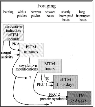
Figure 5: Summary diagram of the memory
phases of the honey bee and their known molecular substrates. A relationship between the temporal organization of these memories and the natural foraging cycles is also shown; adapted from Menzel 1999.
analyzed until now whether the olfactory code existing at the level of the AL is subjected to modifications that are induced by associative olfactory learning.
Taken together, these studies provide an overview about odour processing at several stages along the CS pathway and about the modifications of neural activity induced by olfactory learning in these stages.
2.5. Appetitive Memories Induced by Olfactory PER Conditioning
Appetitive olfactory memories are acquired through PER conditioning and can be retrieved minutes, hours or days later, depending on different experimental factors (Menzel, 2001; Menzel et al., 2001). Among these factors, one can cite the kind of CS, the intensity of the US (i.e. the amount and/or quality of sucrose solution received during conditioning), the number of conditioning trials and the intertrial interval (Menzel et al., 2001).
Different memory phases (short-term, early and late; medium-term, early and late and long-term, early and late) have been characterized accurately in terms of their dynamics and molecular substrates (Menzel, 1999) (Fig. 5). One pairing of an odorant with sucrose (i.e. one conditioning trial) leads to short-term (STM: in the range of sec to min), mid-term (MTM: in the range of hours) an early long-term memory (e-LTM) that can be retrieved 24–48h after conditioning. This e-LTM depends on translation but not on gene transcription and is not,
21
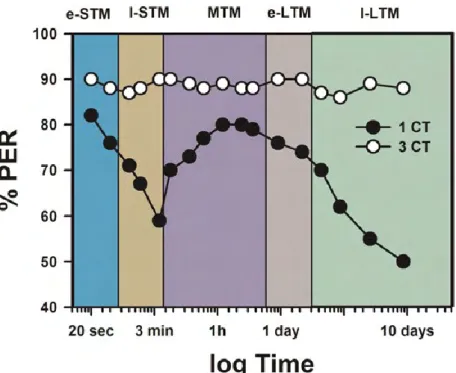
Figure 6: Olfactory memory phases in honeybees. Retention performance (measured as PER
percentage: % PER) depending on trial number and the respective underlying memory phases (after Menzel 2001). The memory retention of a single CS-US association (1 CT; black dots) is compared with three (or more) conditioning trials (3 CT; white dots) with 10-min inter-trial intervals. 1 CT allows good retention performance for up until 1 d. 3 CT induce high performance for several days with two forms of long-term memory, one that depends on translation of already-present mRNA (early-LTM: e-LTM), the other critically depending on de novo gene transcription (late-LTM: l-LTM, starting at 3 d); adapted from Giurfa & Sandoz 2012.
trials, however, induce not only STM, MTM and e-LTM but also a stable late long-term memory (l-LTM) that can be retrieved 72h or more after conditioning (Fig. 6). Unlike e-LTM, l-LTM requires gene transcription and can therefore be inhibited by actinomycin D (Eisenhardt, 2006; Giurfa and Sandoz, 2012; Menzel, 1999, 2001; Schwärzel and Müller, 2006).
Trial spacing is a critical factor for the induction of different types of memory.
Generally, massed trials (i.e. trials succeeding each other in a fast sequence) lead to less stable memories compared to spaced trials (i.e. trials separated in time) which lead to stabilized memories. Longer intertrial intervals lead to better acquisition and retention (Menzel et al., 2001).
The formation of stable l-LTM induces structural modifications in the honey bee brain in accordance with the fact that memories may reside in novel, stabilized connections within olfactory networks that require protein synthesis. These modifications have been found in the AL and in the calyces of MBs (Hourcade et al., 2010; Hourcade et al., 2009).
Introduction
22
In the AL, learning-dependent structural modifications concern the volume of the AL glomeruli. An association with a volume increase in a subset of glomeruli is observable three days after an successful appetitive olfactory conditioning (Hourcade et al., 2009). This increase is odour-specific but does not reflect enhanced activity in the glomeruli encoding the learned odorant as a different pattern of glomerular variation is seen in any case. Rather it may reflect the modulatory effect of VUMmx1 on the inhibitory network of the AL.
In the calyces of the MBs, the neuronal modifications are detectable at the level of the microglomeruli that are present in the calyces. These microglomeruli constitute the interaction site between presynaptic afferent neurons (e.g. PNS coming from the ALs in the case of olfactory information) and the postsynaptic Kenyon cells which make the MBs. Hourcade et al. have reported that the density of olfactory but not visual microglomeruli increases after a successful olfactory l-LTM formation and that this increase is abolished by a protein synthesis inhibitor (Hourcade et al., 2010).
3. The Advent of a New Conditioning Protocol for Harnessed Bees: the
Aversive Olfactory Conditioning of the Sting Extension Reflex
The previous sections highlighted the fact that for approximately a century, research on honey bee learning and memory has focused almost exclusively on appetitive learning, exploiting the fact that bees can learn about a variety of sensory stimuli or to perform certain behaviours if these are rewarded with sucrose solution, the equivalent of nectar collected in flowers. We have seen that this natural foraging scenario is amenable to the laboratory through the olfactory conditioning of PER and how, during the last 50 years, immense progresses have been made in deciphering the neural and molecular bases of appetitive learning and memory using bees as a model.
On the contrary, not much was known about the capacity of honey bees to learn aversive events in their environment. In the fruit fly Drosophila melanogaster, the other insect that has emerged as a powerful model for the study of learning and memory, aversive learning has been the dominant framework (Busto et al., 2010; Davis, 2005; Heisenberg, 2003; Keene and Waddell, 2007; Margulies et al., 2005). In the fruit fly, olfactory aversive conditioning consists in training groups of flies in a T-maze which allows alternated
23
1
another (CS2-) non-paired with the shock (Tully and Quinn, 1985). Retention is tested
afterwards in a dual-choice situation as flies have to choose between the CS+ and the CS- without aversive reinforcement. Successful learning and retention result in CS+ avoidance. This behavioural protocol has allowed the dissection of aversive learning at the cellular and molecular level and identifying the cellular location of different aversive memory traces (Busto et al., 2010; Davis, 2005; Heisenberg, 2003; Keene and Waddell, 2007; Margulies et al., 2005).
Due to obvious differences in behavioural and motivational contexts, in addition to the impossibility to equate US nature and strength, it has been difficult to compare appetitive and aversive learning in bees and flies, respectively, despite their fundamental contribution to understanding learning and memory at multiple levels. As a consequence, the question of whether the mechanisms underlying learning and memory in these two insect models are general or rather specific has remained elusive. Yet, in the last five years, a new conditioning protocol has been established in honey bees, which was conceived to fill this gap (Vergoz et al., 2007a). This protocol is the aversive conditioning of the sting extension response (SER) which is a defensive response to potentially noxious stimuli (Breed et al., 2004). This unconditioned response can be elicited by means of electric-shock delivery to a harnessed bee (Núñez et al., 1997). As no appetitive responses are involved in this experimental context, true punishment (aversive) learning could be studied for the first time in harnessed honey bees.
Inspired by the work of Núñez and coworkers, who used the SER to study the presence of an opioid-like system in honey bees (Núñez et al., 1997), and by the well-established protocol of olfactory PER conditioning (Bitterman et al., 1983; Giurfa and Sandoz, 2012; Takeda, 1961), the protocol of olfactory conditioning of SER was successfully established by Vergoz et al. (Vergoz et al., 2007a). In this protocol, forward-pairing of an odour with an electric shock results in bees learning this contingency and therefore extending their sting in response to the previously punished odour (Vergoz et al., 2007a)(Fig. 7).
Introduction
24
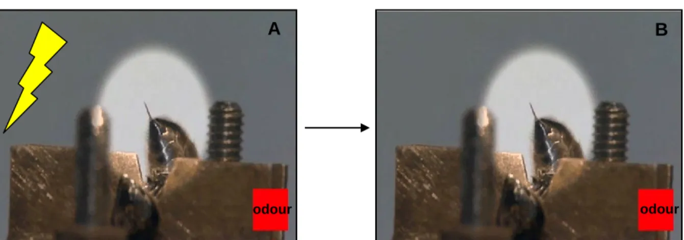
Figure 7: Aversive olfactory conditioning of sting extension reflex (SER). A: A harnessed bee is
trained explicitly paired presentations of an odorant (CS) and the electric shock (US) following an absolute – conditioning design (a single odorant reinforced). B: After a successful conditioning the bee would extend its sting in the presence of the CS alone.
To this end, bees are fixed individually on a metallic holder so that they build a bridge between two brass plates through which a 2 sec mild electric shock (7.5 V) is delivered by a stimulator (60 Hz - AC current) (Fig. 7A). Bees treated in this way extend their sting reflexively in response to the electric shock (Burrell and Smith, 1994; Núñez et al., 1997) (Fig. 7B). Bees of a ‘paired group’ are trained with explicitly paired presentations of an odour (the CS) and the electric shock (the US) following an absolute-conditioning design (a single odorant reinforced). As a control for this kind of conditioning, an ‘explicitly unpaired group’ of bees is presented with unpaired presentations of odour and shock. Figure 8A shows that bees from the paired group learn the odour – shock association and increase conditioned SER to the punished odour during trials. In contrast, bees in the explicitly unpaired group show no significant change in responsiveness to the odour during trials. Thus, the increase of SER observed in the paired group is due to associative learning and not to the simple experience with the odour and the shock. One hour after conditioning, bees of the paired group still remember the conditioned odour while bees of the unpaired do not respond to the odour (Fig. 8A, black and white bars). Therefore, an aversive memory retrievable 1 h after learning is established in the paired but not in the explicitly unpaired group (Vergoz et al., 2007a).
odour odour
25
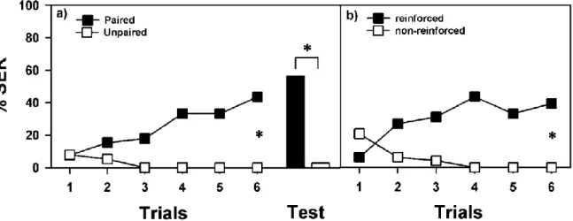
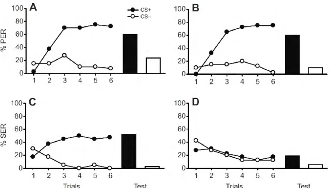
Figure 8: Associative olfactory conditioning of the sting extension reflex (SER) in honeybees. A:
Responses (SER) of bees trained with an odorant explicitly paired with an electric shock (black squares) and with odorant and unpaired electric shock (white squares) during 6 trials. Only the bees in the paired group learned the association and extended their sting as a response to the odorant. One hour after conditioning an olfactory aversive memory was present in the paired (black bar), but not in the unpaired, group (white bar). B: Responses (SER) of bees (n = 48) trained to discriminate an odorant reinforced with an electric shock (black squares) and a non-reinforced odorant (white squares) during 12 trials (6 reinforced and 6 non-reinforced). Bees learned to discriminate between odorants as a result of conditioning; adapted from Vergoz et al. 2007
Moreover, in a differential-conditioning design (Fig. 8B) in which each bee acts as its own control, bees learn to extend their sting to an odour paired with an electric shock and not to respond to another non-reinforced odour. Bees are conditioned during 6 reinforced and 6 non-reinforced trials, presented in a pseudo-random sequence. The resulting learning curves (Fig. 1d) show that bees learn to discriminate between odours as a result of conditioning. Thus, olfactory conditioning of SER is truly associative and does not rely on the simple exposure to the training stimuli, independently of their outcome (Vergoz et al., 2007a).
The use of the term 'aversive' is fully appropriate in the case of this protocol because it was shown that after aversive differential SER conditioning using one odour punished with shock (CS1+) and the other not (CS2-), bees released individually in a mini Y-maze under red
light (i.e. in the dark for bees), which presented the two odours used previously, CS1 and CS2,
avoided significantly the odorant which was coupled with the shock (CS1+). The aversive
nature of SER conditioning in honey bees is clearly emphasised by this result (Carcaud et al., 2009).
Introduction
26
3.1. Aversive US Pathway Underlying Olfactory SER Conditioning
As mentioned above, in appetitive PER conditioning, octopamine mediates the reinforcing properties of sucrose reward in the bee brain (Farooqui et al., 2003; Hammer, 1993; Hammer and Menzel, 1998). In the fruit fly, dopamine was found to mediate the aversive properties of the electric-shock reinforcement used in olfactory conditioning (Aso et al., 2012; Aso et al., 2010; Claridge-Chang et al., 2009; Schwaerzel et al., 2003). In order to establish whether dopaminergic signalling is also crucial for aversive US signalling in bees, neuropharmacological experiments were first performed in order to block this signalling and determine whether olfactory SER conditioning was possible (Vergoz et al., 2007a). Separate groups of bees were injected into the brain through the medium ocellus 30 min before differential conditioning with Ringer solution (control), mianserine or epinastine (octopaminergic blockers) or fluphenazine or flupentixol (dopaminergic blockers).
Ringer-injected bees learned to discriminate the punished from the non-punished odor (Fig. 9A). One hour later, they remembered the aversive association and extended their sting in response to the previously punished odorant. Octopaminergic antagonists (mianserine or epinastine) did not affect performance at any of the concentrations used in these experiments. Figure 9B shows that mianserine-injected bees learned to discriminate the two odorants and responded with SER only to the odorant paired with the electric shock. Retention tests also showed significant discrimination. Thus, octopaminergic antagonists did not impair aversive olfactory learning in honey bees. Dopaminergic antagonists (fluphenazine and flupentixol) had, on the contrary, a dramatic effect on aversive olfactory learning. Flupentixol-injected bees did not learn to discriminate between odorants. Consequently, they did not show discrimination in the tests performed one hour later (Fig. 9C). Fluphenazine had a similar effect although with less effectiveness. These results showed therefore that dopamine-, but not octopamine signalling, is necessary for aversive olfactory learning in honey bees (Vergoz et al., 2007a).
27
conditioning in honey bee. Responses (SER) of bees trained to discriminate an odorant reinforced with an electric shock and a non-reinforced odorant during 12 acquisition trials (six reinforced and six non-reinforced). A retention test was conducted 1h after the last acquisition trial. SER responses are shown for (A) control bees injected with Ringer solution into the brain, (B) bees injected with the octopaminergic antagonist mianserine into the brain and (C) bees injected with the dopaminergic antagonist flupentixol into the brain. Ringer solution- and mianserine-injected bees learned to discriminate the reinforced from the non-reinforced odorant and remembered the difference 1h later. Flupentixol-injected bees did not learn to discriminate the reinforced from the non-reinforced odorant, nor did they respond appropriately in the retention tests. These results show that dopamine but not octopamine receptors are required for aversive olfactory learning in honey bees; adapted from Tedjakumala and Giurfa, 2013
These results prompt a precise neuroanatomical characterization of dopaminergic neurons in the honey bee brain. This characterization is necessary because immunocytochemistry studies using an antiserum against dopamine were performed 25 years ago (Schäfer and Rehder, 1989) but the technique used to stain candidate dopaminergic neurons does not allow to differentiate whether labelled neurons were neurons producing dopamine (true dopaminergic neurons) or neurons incorporating dopamine.
Dopamine-like immunoreactive neurons were identified in most parts of the brain and in the suboesophageal ganglion (Schäfer and Rehder, 1989). Only the optic lobes were devoid of staining. Approximately 330 dopamine-immunoreactive cell bodies were found in each brain hemisphere plus the corresponding suboesophageal hemi ganglion. Most of the stained cell bodies were situated within three clusters: two (C1 & C2) below the α-lobe of the mushroom body, in the inferior medial protocerebrum, and one rather below the lateral calyx (C3). Other stained cell bodies lied dispersed or in small groups around the protocerebral bridge, below the optic tubercles, proximal to the inferior rim of the lobula, and in the lateral and inferior somatal rind of the suboesophageal ganglion (SEG). Due to limitations of the staining technique, not all of the dendritic arborizations and axons of these neurons could be visualized so that where and how dopaminergic circuits contact the olfactory pathway remains to be determined (Schäfer and Rehder, 1989). This information is crucial to study where the association between the odor CS and the electric shock US takes place.
Besides, a dissection of the contribution of the three dopaminergic receptors identified in the honey bee, AmDOP1 (Blenau et al., 1998; Mustard et al., 2003), AmDOP2 (Humphries et al., 2003; Mustard et al., 2003) and AmDOP3 (Beggs et al., 2005), to US signalling in aversive learning is necessary. AmDOP1 and AmDOP3 have been related to the vertebrate D1-like (up-regulates AMP) and D2-like family of dopamine receptors (down-regulates c-AMP), respectively (Beggs et al., 2005; Blenau et al., 1998) while AmDOP2 appears to be