Cloning and expression analysis of an inducible HSP70 gene from
tilapia ¢sh
Alfredo Molina
a, Fre¨de¨ric Biemar
a, Ferenc Mu«ller
a;b;1;*, Arati Iyengar
a;b, Patrick Prunet
a;c,
Norman Maclean
a;b, Joseph A. Martial
a;b, Marc Muller
a;baLaboratoire de Biologie Mole¨culaire et Ge¨nie Ge¨ne¨tique, Universite¨ de Lie©ge, Institut de Chimie B6, B-4000 Sart-Tilman, Belgium bDepartment of Biology, University of Southampton, Bassett Crescent East, Southampton SO16 7PX, UK
cStation INRA/SCRIBE, Campus de Beaulieu, 35042 Rennes Cedex, France
Received 13 March 2000; received in revised form 18 April 2000 Edited by Ned Mantei
Abstract We isolated and characterized the tilapia
(Oreochro-mis mossambicus) HSP70 gene, highly homologous to other
HSP70 genes. A dramatic increase of tilapia HSP70 mRNA
levels was observed after heat shock of whole animals in all
organs tested. Reporter constructs were tested for transient
expression in carp cells and in microinjected zebrafish embryos.
The entire isolated regulatory region (3
3851/+157) was able to
mediate heat shock inducible expression of the reporter gene,
with no preference for a particular tissue. Our studies represent
the first transcriptional analysis of a HSP70 promoter from fish,
revealing a powerful tool to direct controlled, tissue-independent
gene expression in fish.
z 2000 Federation of European Biochemical Societies.
Key words: HSP70; Tilapia; Heat shock; Expression;
Zebra¢sh
1. Introduction
The ¢rst description in Drosophila of a subset of cellular
proteins which are induced upon heat shock (HSP proteins)
[1] has triggered a remarkable amount of research on these
proteins in various organisms and on their function in stress
tolerance. This heat shock response is found universally from
bacteria to human and the HSP genes are among the most
conserved genes during evolution.
The most important and most studied heat shock proteins
form the HSP70 gene family. They play essential roles in
protein metabolism under normal and stress conditions,
in-cluding de novo protein folding, membrane translocation,
degradation of misfolded proteins and other regulatory
pro-cesses. Their expression is regulated by environmental and
physiological stress and non-stressful conditions such as cell
growth, development and pathophysiological states [2]. While
their binding to an unfolded polypeptide chain results in the
stabilization of the unfolded state, their controlled release may
allow progression along the folding pathway [3]. Owing to
their functions, HSP70s have been included in the large family
of chaperones.
The regulation of expression of HSP70 genes occurs mainly
at the transcriptional level. Analysis of the Drosophila HSP70
gene and comparison of di¡erent heat shock regulatory
re-gions led to the identi¢cation of a palindromic `heat shock
element' (HSE), CnnGAAnnTTCnnG [4^6]. More recent
re-sults suggested the view that HSEs are composed of
contigu-ous arrays of a variable number of the highly conserved
se-quence nGAAn arranged in alternating orientation [7^9].
These elements bind the trans-acting heat shock factors
(HSF) which are normally present in the cytoplasm in a
monomeric form (with no DNA-binding activity) and which,
upon heat stress, form trimers and migrate to the nucleus to
bind HSEs with high a¤nity [10^15].
In ¢sh, several cDNAs encoding HSP70 from rainbow trout
[16], medaka [17] or zebra¢sh [18,19] have been described and
a heat-shock-induced increase of mRNA levels was shown.
Genomic sequences of a HSP70 gene family have been
re-ported in the teleost Fugu rubripes [20]. However, no
informa-tion concerning the sequences involved in transcripinforma-tional
reg-ulation of ¢sh HSP70 genes was available until now.
Here, we report the isolation and characterization of the
tilapia (Oreochromis mossambicus) HSP70 gene including
about 1 kb of regulatory sequences. We show that the tilapia
HSP70 promoter is able to confer heat shock regulation to a
reporter gene both in ¢sh cells and in microinjected zebra¢sh
embryos. This promoter thus represents a powerful tool to
drive ubiquitous and controlled expression of a gene of
inter-est in ¢sh.
2. Materials and methods
2.1. OligonucleotidesSynthetic oligonucleotides were obtained from Eurogentec (Seraing, Belgium): library screening PCR probe: forward (H70pf1) TATGTGGCYTTCACHGAYAC-3P and reverse (H70pr1) 5P-TGAGDCKYTTGGCRTCMAAV-3P; semi-quantitative RT-PCR: forward (H70pf2) 5P-TCTGCAGCTAAAGGTGTAGC-3P and re-verse (H70pr2) 5P-TTGAAGGGCCAGTGCTTCATG-3P forward (L18pf1) 5P-TATGTGGCYTTCACHGAYAC-3P and reverse (L18pr1) 5P-TTGGTCCTGCTCATGAACAG-3P; transcription start site: (fH70pr3) 5P-CTACACCTTTAGCTGCAGAC-3P; reporter gene construction: NcoI forward (pH70f1) 5P-CGCCATGGCTG-TCTTCTAGAAAATTCAG-3P and EcoRI reverse (pH70r1) 5P-CG-GAATTCTTTGACTTCGTTTCAAAAAGAGG-3P.
2.2. Probe isolation
2.2.1. Library screening. Degenerate primers (H70pf1 and H70pr1) were designed by comparing several HSP70 sequences and chosen in the most conserved region. After PCR ampli¢cation the 100 bp PCR product was directly cloned into the pCR1II vector (TA-Cloning kit, Invitrogen) and sequenced.
*Corresponding author. Fax: (32)-4-3662968. E-mail: m.muller@ulg.ac.be
1 Present address: IGBMC, 67404 Illkirch Cedex, Strasbourg,
2.3. Library screen
Using the tilapia HSP70 100 bp PCR probe described above, 2Ul06
plaques (recombinant V-GEM11) of a tilapia genomic library [21] were screened. The probe was labeled by random priming (T7QuickPrime1
kit, Pharmacia Biotech) using [K32P]dCTP (3000 Ci/mmol,
Amer-sham). Hybridization and washing were carried out under stringent conditions. After four additional screening cycles, puri¢ed positive clones were ampli¢ed [22] and the phage DNA was puri¢ed (Lambda midi kit, Qiagen, Hilden, Germany). Subcloning was performed into pBluescript II KS+ (Stratagene). Restriction mapping and Southern blotting were performed using standard procedures [23].
2.4. Cap site determination by primer extension
Total RNA from liver was puri¢ed using TRIzol reagent (Gib-coBRL) and 1 Wg was used to perform primer extension experiments using the GeneAmp, Thermostable rTth Reverse Transcriptase RNA PCR kit (Perkin Elmer). After 10 cycles of annealing (48³C, 10 min), extension (70³C, 5 min) and denaturation (95³C, 1 min) using the 5P £uorescein labeled fH70pr3 primer, the reaction was treated as is described in [24] and loaded on an automated sequencer (A.L.F., Pharmacia). The data were analyzed using the ALF manager 3.02 software.
2.5. Semi-quantitative RT-PCR
From a group of adult mature female tilapia Oreochromis niloticus (mean weight: 121 þ 26 g, n = 6) reared in freshwater at 27³C, half were transferred at 37³C for 1 h, whereas a control group was kept at 27³C. All ¢sh were kept at 27³C for an additional 2 h and tissues were sampled. Fish were anaesthetized; organs were dissected and immediately frozen in liquid nitrogen. Poly-A RNA was isolated
from these tissues using the QuickPrep1 Micro mRNA Puri¢cation kit (Amersham/Pharmacia Biotech). RT-PCR experiments were per-formed using the Titan1 One Tube RT-PCR kit from Boehringer Mannheim. A 283 bp HSP70 fragment was ampli¢ed using the H70pf2 and H70pr2 primers. As a control, a 192 bp fragment of the constitutively expressed L18 ribosomal protein gene was ampli¢ed (GenBank number: AF240375) using the L18 primers L18pf1 and L18pr1. Five Wl aliquots of the PCR reaction were collected after 18, 20, 22, 24, 26, 28 and 30 cycles to determine the linear range of the reaction, in two independent experiments. Twenty-six cycles were chosen and the HSP70 and L18 reactions were analyzed in the same slot on a 2% agarose gel.
2.6. Reporter gene construction
The tiHSP70-0.3LacZ expression vector was constructed as follows: a 360 bp PCR fragment, including the appropriate restriction sites at the 5P-end of the primers pH70f1 and pH70r1, was ampli¢ed from the pB3-tihsp subclone, and introduced into the expression vector pGCV-Lac-Z (positive control) [25], from which the SV40 promoter had previously been deleted by double digestion with Sapl and NcoI fol-lowed by religation.
The tiHSP70-1.0LacZ expression vector was obtained by inserting the 700 bp XbaI fragment from the pB3-tihsp into the XbaI site of the tiHSP70-0.3LacZ construct.
The linear fragments containing the promoter, reporter gene and poly-A signal unit for microinjection into zebra¢sh embryos were excised with NcoI and BamHI and puri¢ed using the Easy-Pure1 kit (BI0zym bv, the Netherlands). The negative, promoterless control was obtained by excision with the same enzymes directly from the pGCV-Lac-Z vector.
Fig. 1. The nucleotide sequence of the tiHSP70 gene and the deduced amino acid sequence in the coding region are shown. Upper case letters are used for the coding sequence. The transcription start site is indicated by +1. The putative TATA-box and cis-acting elements are boxed and the three putative HSE elements are in bold. Isolated nGAAn or nTTCn sequence units are underlined. The sequence of the PCR probe used to screen the library is underlined (positions +299/+398). The oligonucleotide used to determine the transcription start site is shown in italics. The complete isolated sequence including the coding region and the 3P-end of the gene has been deposited in the EMBL databank (ac-cession number AJ001312).
2.7. Cell culture and transfection experiments
Epithelioma papulosum cyprini (EPC) cells, derived from carp epi-dermal herpes virus-induced hyperplasia lesions, [26] were grown in BHK-21 medium supplemented with 10% fetal bovine serum and 1% penicillin^streptomycin at 24³C in 5% C02. Twenty pmol of puri¢ed
plasmid DNA was transfected in 800 Wl of resuspended cells (1.5U106
cells/ml of medium) by electroporation in 4 mm cuvettes using a single pulse (250 V, 1500 WF) delivered by a Easyject apparatus (Eurogentec, Seraing, Belgium). Transfected cells were immediately transferred to four culture dishes (55 mm). After 48 h, the cells were harvested by scraping, washed two times in PBS and resuspended in lysis bu¡er [27]. Protein concentration and L-galactosidase activity were deter-mined according to [23].
Heat shocks were performed by transferring the plates previously sealed with para¢lm in a thermoregulated bath at the appropriate temperature. After the treatment, the cells were returned for 2 h at 24³C and the LacZ activity was determined.
2.8. Microinjection procedure
Fish care and embryo rearing was performed as described by [28]. Eggs were collected and microinjected (300 pl, 5O ng/Wl) at the one cell stage, targeting to the cytoplasm/yolk boundary of the zygote. Following microinjection, the embryos were replaced at 28.5³C in small tanks containing 500 ml of Holtfreter's solution to allow correct development. Heat shock treatment was carried out on 1 day old embryos at 40³C for 15 min, followed by a recovery for 2 h at 28.5³C. LacZ expression was determined as previously described [29].
3. Results
3.1. Genomic library screening
A 100 bp probe speci¢c for the tilapia HSP70 coding
se-quence was obtained by performing a PCR on tilapia DNA
using the primers H70pf1 and H70pr1 (Fig. 1). By screening
2.0U10
6V-phage plaques from a tilapia genomic library, 104
positive clones were obtained. Eight of them were selected and
puri¢ed by three additional screenings. Restriction analysis
revealed the presence of three di¡erent groups of clones.
One of them, V-11tiHSP, containing approximately 15 kb of
insert, was chosen for restriction mapping and Southern blot
analysis (data not shown). A 3 kb SacI fragment, shown to
hybridize with the HSP70 probe, was subcloned (pB3-tihsp)
and subdivided further into three fragments of approximately
1 kb by PstI digestion, which were again subcloned
(pB3A-tihsp, pB3Btihsp and pB3C-tihsp) and completely sequenced
on both strands. An oligonucleotide hybridizing to the 3P-end
of the pB3-tihsp insert was used to sequence the 3P-£anking
region using the V-11tiHSP DNA. The isolated promoter,
5P-untranslated and coding sequence up to +469 is shown in Fig. 1.
3.2. Sequence analysis of the tilapia HSP70 gene
Computer analysis of the isolated sequence reveals a long
open reading frame encoding a 640-amino acid protein
corre-sponding to a HSP70 family protein (Fig. 1). Amino acid
sequence comparison con¢rmed the high degree of
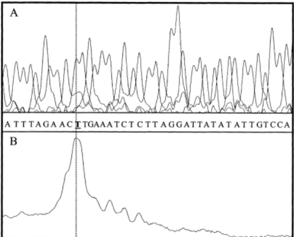
conserva-Fig. 2. Transcription start site. Primer extension was performed us-ing adult tilapia liver RNA. Comparison of the primer extension product with the products of a sequencing reaction on the pB3-tihsp is shown.
Fig. 3. Expression analysis in di¡erent organs. RT-PCR ampli¢ca-tion products for a tiHSP70 fragment (283 bp) and the constitutive control L18 fragment (192 bp) from RNA Poly-A from control
(3) and heat shock treated (+) ¢sh are shown. The complete experi-ment was performed twice with identical results.
Fig. 4. Transient expression of tiHSP70 promoter/LacZ in cultured cells. A: Schematic representation of the reporter constructs used. TiHSP70-1.0Lac-Z contains the complete isolated tiHSP70 control region (3851/+157) fused upstream from the ATG codon of the LacZ gene. TiHSP70-0.3LacZ contains the control region starting just upstream from HSE3 (3182/+157). B: EPC cells transfected with either reporter construct or the pGCV-LacZ (transfection posi-tive control) were subjected or not to a heat shock for 60 min at 37³C. The cells were left 2 h at 24³C for recovery, harvested and lacZ activity was measured. L-Galactosidase activity was normalized to the protein concentration and fold induction relative to the non-treated cells is shown. C: EPC cells transfected with the tiHSP70-1.0LacZ with or without heat shock for 60 min at 28, 32, 37 and 42³C. Triplicate experiments were performed three times, the data are represented as mean þ S.D.
tion of HSP70 in various species: 83, 84, 87 and 91% identity
with human [30], rat [31], Xenopus laevis [32] and trout [16]
sequences, respectively. The fact that the genomic sequence
contains no intron is consistent with what is observed in
in-ducible HSP70 genes from other species, further supporting
the conclusion that the isolated sequence corresponds to an
inducible tilapia HSP70 gene (tiHSP70).
The complete coding region of the tiHSP70 gene and about
1 kb of regulatory sequence, including the 5PUTR region, was
isolated. To determine the transcription initiation (CAP) site,
a primer extension experiment was performed using a
£uores-cent oligonucleotide located in the tiHSP70-coding region (see
Fig. 1). One clear signal was obtained which, by comparing
the migration of the primer extension product with that of the
products from a sequencing reaction performed on the
pB3-tihsp using the same primer (Fig. 2), allowed to de¢ne the
start site at 172 bp upstream from the ATG.
Several putative cis-acting sequences were found upstream
from the CAP site (Fig. 1). In addition to a TATA box at
338, two GC boxes were detected at position 3444 and
3708, a CAAT box at 3272 and an inverted CAAT box at
3831 were found. Three elements corresponding to the HSE
consensus were found, respectively, at positions 3815, 3500
and 3179. Several additional nGAAn elements were detected
in both orientations. Moreover, HSE2 and HSE3 form the
5P-part of a 32 bp direct repeat.
3.3. TiHSP70 gene expression by semi-quantitative RT-PCR
Two adult tilapias were heat shocked at 37³C for 1 h and
left to recover for 2 h at normal temperature (27³C). Poly-A
RNA was extracted from di¡erent organs of these ¢sh and of
two untreated ¢sh. TiHSP70 mRNA levels were assessed in
each organ by RT-PCR, using expression of the L18
riboso-mal protein as invariant control (Fig. 3). After heat shock,
tiHSP70 RNA was readily detected in each organ, while no
signal was observed in the control ¢sh, indicating that the
tiHSP70 gene is highly inducible. Induction levels seem to
be roughly the same in the di¡erent organs tested.
3.4. Transfection experiments
Transient expression studies were performed in carp EPC
cells to evaluate the transcriptional regulation of the tiHSP70
promoter by thermal stress. To that purpose, two constructs
containing either the whole available regulatory region
(tiHSP70-1.0LacZ) or starting just upstream from HSE3
(tiHSP70-0.3LacZ) were tested (Fig. 4A). Heat shock for 1 h
at 37³C resulted in a clear, 16-fold stimulation of
L-galac-tosidase expression only for the tiHSP70-1.0LacZ construct,
while the tiHSP70-0.3LacZ or the control pGCV-LacZ
con-structs were una¡ected (Fig. 4B). Di¡erent temperature
treat-ments were tested using the inducible tiHSP70-1.0LacZ
con-struct (Fig. 4C). A slight induction was observed after l h at
32³C. A heat shock at 37³C resulted in a two-fold stimulation
Fig. 5. Transient expression of tilapia HSP70 promoter-LacZ constructs in 1 day old zebra¢sh embryos. Zebra¢sh embryos were microinjected at the one cell stage with the tiHSP70-1.0LacZ; tiHSP70-0.3LacZ or pGCV-LacZ linearized constructs. One day old embryos were subjected to a heat shock at 40³C for 15 min, allowed to recover for 2 h at 28.5³C and stained with X-gal. Untreated controls were stained in parallel. In-jection of supercoiled tiHSP70-1.0LacZ led to a stronger expression compared to the linearized fragment, however, survival rates were much lower (data not shown). Cumulative patterns of LacZ expression were obtained by superimposing the expression patterns of all the individual embryos within a same batch.after 15 min which increased up to 16-fold after 1 h. Higher
temperatures (42³C) led to a much faster response (six-fold
after 15 min) which did not increase after longer treatment. In
this case, detachment of the cells from the plate indicated
cellular death.
3.5. Transient expression in zebra¢sh embryos
The transcriptional activity of the tiHSP70 promoter was
also tested in whole animals by microinjection into zebra¢sh
eggs. When the tiHSP70-0.3LacZ construct was injected, no
expression of lacZ was observed even when the embryos were
subjected to heat shock (Fig. 5) (a total of 81 individuals were
analyzed). In contrast, injection of tiHSP70-1.0LacZ resulted
in a strong expression only after heat shock (0/82 positive
individuals without heat shock versus 37/83 after heat shock).
As expected, no LacZ expression was observed with the
pro-moterless construct (not shown) while the positive control
(pGCVLacZ) directed constitutive expression, both with
(9/29 positives) or without heat treatment (6/29 positives).
By superimposing the di¡erent locations of expression from
all the individual embryos within a same batch, a cumulative
pattern of expression was obtained. The results (Fig. 5) show
that the heat shock-induced expression directed by the
tiHSP70 promoter displays no preference for any particular
tissue, the pattern is comparable to that obtained with the
SV40 promoter containing pGCVlacZ.
4. Discussion
We report the cloning, sequencing and expression analysis
of the tiHSP70 gene. Furthermore, the ¢rst analysis of a ¢sh
HSP70 promoter and its regulation by stress is presented.
Analysis of the deduced amino acid sequence revealed a
remarkable homology to HSP70 genes from other species,
as much as 91% similarity with trout HSP70. Perfectly
con-served regions correspond to the functional domains involved
in ATP and peptide binding. In particular, amino acids
di-rectly involved in interaction with HSP40 and in ATPase
ac-tivity [33] are identical to those in other species. Although the
C-terminal end appears to be less conserved, the last 8 amino
acids, GPTIEEVD, are again identical in all the species. This
feature was previously shown to be characteristic of
nuclear-cytosolic HSP70s [34].
Another striking feature of the tiHSP70 gene is the absence
of introns. An uninterrupted open reading frame of 1920 bp
encoding the entire protein was found. Such a gene structure
is characteristic of inducible HSP genes, in contrast to the
constitutively expressed HSP (cognate heat shock HSC)
genes, and of other stress induced genes such as glutathione
S-transferase D [35]. Intron splicing is normally required for
translocation of most mRNAs from the nucleus to the
cyto-plasm, however an inhibition of RNA-splicing by stress was
shown [36^38]. It was suggested that the absence of introns in
stress-induced genes allows to compensate for this inhibition
thereby enabling preferential expression of these proteins
dur-ing cellular stress, the nuclear export signal bedur-ing probably
provided by the mRNA secondary or tertiary structure [39].
Semi-quantitative RT-PCR experiments demonstrate the
presence of tiHSP70 mRNA in various tissues only after
heat shock. Taken together, these observations strongly
sug-gest that the isolated sequence corresponds to a heat
induc-ible, nuclear-cytosolic tiHSP70 gene.
Analysis of the 5P regulatory region showed no evident
homologies to other HSP70 promoters or 5P leader sequences.
However, basal cis-acting elements (TATA, CAAT and
GCGGG boxes) and putative HSEs are detected, although
at di¡erent positions and in di¡erent arrangements. In the
HSP70 promoters from other species, a CAAT box localized
near the TATA box, around 370 [40^42], was shown to be
required for full-stress induction [43^45]. The role of the two
CAAT boxes in the tiHSP70 promoter, respectively at 3831
and 3272, in transcriptional regulation is at present unclear.
The function of the tiHSP70 promoter was tested in vivo by
transient expression in cell culture and microinjected zebra¢sh
embryos. As the 5P leader sequence was shown to mediate
preferential translation of HSP70 mRNAs in several species
[46^48], this region was included in our reporter constructs
although no obvious sequence conservation was found. Only
the longest construct containing the complete isolated
regula-tory region (tiHSP70-1.0LacZ) was able to mediate high
tran-sient expression and only after heat shock both in transfected
cells and in developing embryos. These results suggest that the
most proximal putative HSE3 is not su¤cient to confer heat
inducibility, in contrast to what was observed in HSP gene
promoters from other species. In addition, as the shorter
con-struct (tiHSP70-0.3LacZ) still contains the 5P leader sequence,
we can rule out that the response observed with
tiHSP70-1.0LacZ is due to enhanced translation or stabilization of
preexisting mRNAs. Thus, the region between 3851 and
3182 in the tiHSP70 promoter mediates transcriptional
stim-ulation upon heat shock; further experiments will show which
of the HSE1, HSE2, CAAT or GC-boxes are precisely
in-volved.
Transient expression in EPC cells revealed a time- and
`dose'-dependent expression of the reporter gene, with the
highest stimulation at 37³C for 1 h. This response behavior
is similar to that observed in other cold-blooded organisms
[49,50].
Even if the transient expression of the tiHSP70-1.0Lac-Z
transgene after heat shock is mosaic, the cumulative
expres-sion patterns of all injected embryos demonstrate no
prefer-ence for a particular tissue. This observation is in agreement
with the expected ubiquitous expression of HSP70 proteins
and with our results showing a very homogeneous expression
in all the tissues from heat shocked tilapia (Fig. 3). In
con-clusion, the tiHSP70 promoter is probably able to confer heat
shock response to a reporter gene in the entire animal.
The introduction of novel genes into animals follows two
major objectives: (1) gene regulation, expression and
develop-mental studies in vivo and (2) the production of commercially
valuable transgenic species bene¢ting from the acquisition of
desirable traits [51]. In this respect, the availability of
induc-ible promoters able to control the expression of a particular
gene by a simple stimulus would be very useful. Cellular
stress, in particular heat shock, is an easy and non-toxic
way to treat an animal. Moreover, the mechanisms mediating
stress response are highly conserved. The results reported here
demonstrate that the tilapia HSP70 promoter represents a
potentially powerful tool to drive the expression of a
trans-gene of interest in ¢sh under controlled conditions.
Acknowledgements: We are grateful to S.E. Wendelaar Bonga (Nijme-gen, The Netherlands) for providing ¢sh material. We would like to thank R. Carpeaux for excellent technical assistance. This work was
supported by the `Re¨gion Wallone' (ULg 1815); the `Services Fe¨de¨r-aux des A¡aires Scienti¢ques, Techniques et Culturelles' (PAI P4/30 and `Actions de Recherche Concerte¨es': 95/00-193); the Fonds Na-tional de la Recherche Scienti¢que (FNRS) (33.4537.93 and 39.4569.95) and the EU (No. BIO4-CT97-0554). M.M. is a `Cher-cheur quali¢e¨' at the FNRS. A.M. held fellowships from `CGRI' and `ULg patrimoine'.
References
[1] Ritossa, F. (1962) Experientia 18, 571^573. [2] Morimoto, R.I. (1998) Genes Dev. 12, 3788^3796. [3] Hartl, U.F. (1996) Nature 381, 571^580.
[4] Holmgren, R., Corces, V., Marimoto, R., Blackman, R. and Meselson, R. (1981) Proc. Natl. Acad. Sci. USA 78, 3775^ 3778.
[5] Pelham, H.R. (1982) Cell 30, 517^528.
[6] Bienz, M. and Pelham, H.R. (1987) Adv. Genet. 24, 31^72. [7] Xiao, H. and Lis, J.T. (1988) Science 239, 1139^1142.
[8] Amin, J., Ananthan, J. and Voellmy, R. (1988) Mol. Cell. Biol. 8, 3761^3769.
[9] Fernandes, M., Xiao, H. and Lis, J.T. (1994) Nucleic Acids Res. 22, 167^173.
[10] Rabindran, S.K., Haroun, R.I., Clos, J., Wisniewski, J. and Wu, C. (1993) Science 259, 230^234.
[11] Morimoto, R.I. (1993) Science 259, 1409^1410.
[12] Baler, R., Dahl, G. and Voellmy, R. (1993) Mol. Cell. Biol. 13, 2486^2496.
[13] Sarge, K.D., Murphy, S.P. and Morimoto, R.I. (1993) Mol. Cell. Biol. 13, 1392^1407.
[14] Sistonen, L., Sarge, K.D. and Morimoto, R.I. (1994) Mol. Cell. Biol. 14, 2087^2099.
[15] Nakai, A., Kawazoe, Y., Tanabe, M., Nagata, K. and Morimo-to, R.I. (1995) Mol. Cell. Biol. 15, 5168^5178.
[16] Kothary, R.K., Jones, D. and Candido, E.P. (1984) Mol. Cell. Biol. 4, 1785^1791.
[17] Arai, A., Naruse, K., Mitani, H. and Shima, A. (1995) Jpn. J. Genet. 70, 423^433.
[18] Lele, Z., Engel, S. and Krone, P.H. (1997) Dev. Genet. 21, 123^ 133.
[19] Santacruz, H., Vriz, S. and Angelier, N. (1997) Dev. Genet. 21, 223^233.
[20] Lim, E.H. and Brener, S. (1999) Cell Mol. Life Sci. 668^678. [21] Swennen, D., Poncelet, A.C., Sekkali, B., Rentier-Delrue, F.,
Martial, J.A. and Belayew, A. (1992) DNA 11, 673^684. [22] Grossberger, D. (1987) Nucleic Acids Res. 15, 6737.
[23] Sambrook, J., Fritsh, E.F., and Maniatis, T. (1989) Molecular Cloning: A laboratory Manual, 2nd edn., Cold Spring Harbor Laboratory Press, Cold Spring Harbor, NY.
[24] Myo«ha«nen, S. and Wahlfors, J. (1993) Biotechniques 14, 16^17.
[25] Poncelet, D.A., Bellefroid, E.J., Bastiaens, P.V., Demoitie¨, M.A., Marine, J.C., Pendeville, H., Alami, Y., Devos, N., Lecocq, P., Ogawa, T., Muller, M. and Martial, J.A. (1998) DNA Cell Biol. 17, 931^942.
[26] Fijan, N., Sulimanavoic, D., Bearzotti, M., Muzinic, M.D., Zwi-lenberg, L.O., Chilmonzyk, S., Vautherot, J.F. and de Kinkelin, P. (1983) Ann. Virol. 134, 207^220.
[27] Brasier, A.R. and Tate, J.E. (1989) Biotechniques 7, 1116^1122. [28] Wester¢eld, M. (1993) The zebra¢sh Book. A guide for the labo-ratory use of zebra¢sh (Danio rerio), University of Oregon Press, Eugene, OR.
[29] Mu«ller, F., Lele, Z., Va¨radi, L., Menczel, L. and Orba¨n, L. (1993) FEBS Lett. 324, 27^32.
[30] Hunt, C. and Morimoto, R.I. (1985) Proc. Natl. Acad. Sci. USA 82, 6455^6859.
[31] Mestril, R., Chi, S.H., Sayen, M.R. and Dillmann, W.H. (1994) Biochem. J. 15, 561^569.
[32] Bienz, M. (1984) EMBO J. 3, 2477^2483.
[33] Gassler, C.S., Buchberger, A., Laufen, T., Mayer, M.P., Schroder, H., Valencia, A. and Bukau, B. (1998) Proc. Natl. Acad. Sci. USA 95, 15229^15234.
[34] Boorstein, W.R., Ziegelho¡er, T. and Craig, E.A. (1994) J. Mol. Evol. 38, 1^17.
[35] Toung, Y.P., Hsieh, T.S. and Tu, C.P. (1993) J. Biol. Chem. 268, 9737^9746.
[36] Yost, H.J. and Lindquist, S. (1986) Cell 25, 185^193.
[37] Yost, H.J. and Lindquist, S. (1991) Mol. Cell. Biol. 11, 1062^ 1068.
[38] Bond, U. (1988) EMBO J. 7, 3509^3518.
[39] Huang, Y., Wimler, K.M. and Carmichael, G.G. (1999) EMBO J. 18, 1642^1652.
[40] Wu, C. (1984) Nature 309, 229^234.
[41] Wu, B.J., Kingston, R.E. and Morimoto, R.I. (1986) Proc. Natl. Acad. Sci. USA 83, 629^633.
[42] Greene, J.M., Larin, Z., Taylor, I.C., Prentice, H., Gwinn, K.A. and Kingston, R.E. (1987) Mol. Cell. Biol. 7, 3646^3655. [43] Lum, L.S., Hsu, S., Vaewhongs, M. and Wu, B. (1992) Mol. Cell.
Biol. 12, 2599^2605.
[44] Ovsenek, N. and Heikkila, J.J. (1992) Biochem. Cell. Biol. 70, 339^342.
[45] Ago¡, S.N. and Wu, B. (1994) Oncogene 9, 3707^3711. [46] McGarry, T.J. and Lindquist, S. (1985) Cell 42, 903^911. [47] Klemenz, R., Hultmark, D. and Gehring, W.J. (1985) EMBO J.
4, 2053^2060.
[48] Hess, M.A. and Duncan, R.F. (1996) Nucleic Acids Res. 24, 2441^2449.
[49] Krone, P.H. and Heikkila, J.J. (1988) Development 103, 59^67. [50] Misra, S., Zafarullah, M., Price-Haughey, J. and Gedamu, L.
(1989) Biochim. Biophys. Acta 1007, 325^333.
[51] Iyengar, A., Mu«ller, F. and Maclean, N. (1996) Transgenic Res. 5, 147^166.