UNIVERSITÉ DE MONTRÉAL
ELECTRO-MECHANICAL MANIPULATION OF MAMMALIAN CELLS
IN SUSPENSION
LUKE MACQUEEN
DÉPARTMENT DE GÉNIE PHYSIQUE ÉCOLE POLYTECHNIQUE DE MONTRÉAL
THÈSE PRÉSENTÉE EN VUE DE L‟OBTENTION DU DIPLÔME DE PHILOSOPHIAE DOCTOR
(GÉNIE PHYSIQUE) DÉCEMBRE 2010
UNIVERSITÉ DE MONTRÉAL
ÉCOLE POLYTECHNIQUE DE MONTRÉAL
Cette thèse intitulée:
ELECTRO-MECHANICAL MANIPULATION OF MAMMALIAN CELLS IN SUSPENSION
présentée par: MACQUEEN Luke
en vue de l‟obtention du diplôme de: Philosophiae Doctor a été dûment acceptée par le jury d‟examen constitué de :
Mme. BOUDOUX Caroline, Ph. D., présidente
M. WERTHEIMER Michael, R., D. Sc. A., membre et directeur de recherche M. BUSCHMANN Michael, Ph.D., membre et codirecteur de recherche M. GERVAIS Thomas, Ph. D., membre
DEDICATION
I would first like to thank Professor Michael Wertheimer, my Ph.D. supervisor, for his excellent guidance and encouragement throughout my time in the M.Sc. A. and Ph.D. programs at the École Polytechnique. Dr. Wertheimer introduced me to the exciting interdisciplinary research topic of biological cell- “electro-manipulation”, and he initiated meetings with Professor Michael Buschmann, thus forming the nucleus of my Ph. D program.
I also thank Professor Michael Buschmann, my Ph.D. co-supervisor, who welcomed me into his tissue-engineering laboratories, where I performed the majority of the experiments reported in this thesis. In addition to productive days in the labs, I will always remember with fondness the famous summer BBQs.
Professors Wertheimer and Buschmann have been ideal Ph. D. co-supervisors, and I have benefited greatly from their generosity and expertise.
Next, I would like to thank Dr. Mohamed Latreche, Dr. Grzegorz Czeremuszkin, and Dr. Gilles Dennler, for their help getting me started in the plasma laboratories and Mr. Yves Drolet for his outstanding technical support in the plasma laboratories.
I thank the staff at the microfabrication laboratories (LMF), in particular Dr. Souleymane Bah for training me within the LMF, often spending many hours in the process. With this training, I was able to fabricate devices, which continue to work reliably several years later.
I thank Dr. Marc Thibault for his help with the confocal imaging system and other related equipment, and for interesting scientific conversations and collaborations.
I thank Professor Olivier Guenat and Dr. Thomas Gervais for their interest in my research and for their encouragement.
I have also benefitted greatly from many other people at the École Polytechnique de Montréal during my M.Sc. and Ph.D. programs, and I am very grateful to them all.
RÉSUMÉ
Le but principal de cette thèse était de décrire le développement d'une plate-forme microfabriquée et son application pour la caractérisation mécanique des cellules mammifères vivantes. La technique emploie les forces de polarisation électriques pour emprisonner et étirer des cellules dans des champs électriques non-uniformes et variables dans le temps. Ce travail a été motivé par la sous-utilisation apparente des champs électriques pour la caractérisation mécanique des cellules; les méthodes décrites ici ont permis la caractérisation mécanique de cellules mammifères diverses et précédemment non étudiées.
La caractérisation mécanique de cellules vivantes est habituellement réalisée en sondant des structures locales près de la surface des cellules. Très peu de ces techniques appliquent des contraintes uniaxiales aux cellules entières et la comparaison des données de diverses techniques est donc difficile. Par contre, des champs électriques peuvent être employés pour exercer ces forces uniaxiales sur les cellules suspendues. La plupart des cellules mammifères adoptent une géométrie quasi-sphérique lorsque suspendues dans un milieu (aqueux) liquide; ceci simplifie à la fois la manipulation des cellules ainsi que l'interprétation des données mécaniques. Les champs électriques exercent des forces sans contact mécanique significatif entre les cellules et les structures du dispositif et peuvent donc être décrits comme un « rayon tracteur », qui peut déplacer, emprisonner, ou déformer électriquement les objets polarisables telles les cellules biologiques. En plus, les électrodes nécessaires sont facilement incorporées dans les dispositifs microfabriqués, ce qui suggère que les techniques basées sur des champs électriques seront de plus en plus utilisées.
La caractérisation mécanique des cellules par « électro-déformation » (ED) se place dans le contexte plus grand de l‟électro-manipulation de cellules. Afin de mieux comprendre le comportement des cellules dans les champs électriques, nous avons commencé nos études en utilisant la diélectrophorèse (DEP) pour placer des monocytes humains (U937) dans un champ électrique non-uniforme, avant d‟effectuer l'électroporation (EP) permettant la livraison de transgènes. Un ensemble d'électrodes planaires inertes microfabriquées sur un substrat de verre a été utilisé pour la DEP et l‟EP. Les propriétés diélectriques des cellules ont été estimées et la modélisation (par éléments finis) des champs électriques nous à permis de prévoir le positionnement des cellules. Le point à partir du quel les impulsions électriques ont augmenté la perméabilité des membranes cellulaires aux molécules fluorescentes et aux plasmides d'ADN
d'impulsion, l‟EP était soit irréversible (ayant pour résultat la cytolyse), réversible (menant à la livraison de gènes), ou non discernable, selon la position des cellules. Nos résultats démontrent clairement que l‟EP des cellules dans un champ électrique non-uniforme peut être commandée par DEP.
Les mêmes microélectrodes planaires utilisées pour DEP et EP ont alors été employées pour mesurer les propriétés mécaniques de différents types de cellules mammifères en suspension, en déformant des cellules individuelles dans les champs électriques non-uniformes et variables dans le temps. Les contraintes électriques produits par ces microélectrodes planaires ont été employées pour emprisonner et étirer les cellules, alors que l‟ED des cellules était observée et photo-documentée par microscopie optique. Deux types de cellule distincts ont été comparés après des données convenables à un modèle de contrainte à trois-paramètres (SLS) et à un de deux paramètres (PL). Les cellules de type « Chinese hamster ovary » (CHO) étaient approximativement deux fois plus rigides que les promonocytes humains (U937); les CHOs avaient un comportement élastique avec le rétablissement de la forme initiale, alors que les U937 témoignent à la déformation plastique.
Nous avons alors ensuite exécuté des expériences d‟électrodéformation avec deux types de cellules additionnelles (L929 et HEK293) où la microscopie confocal a été employée pour la visualisation et l‟analyse semi-quantitative de la structure du cytosquelette (CSK) des cellules. Nous avons traité les cellules U937 avec de la latrunculin-A (Lat-A) ou de l‟acrylamide (ACR) pour évaluer respectivement leurs effets sur les microfilaments (MF) et les filaments intermédiaires (IF) du CSK. Nous avons démontré que les propriétés viscoélastiques des cellules individuellement déformées sont dépendentes de l'épaisseur de l'actine corticale (AC) et ont été significativement affectées par des traitements de Lat-A. Les cellules U937 et HEK293 possèdent de minces AC et sont plus facilement déformées que les cellules CHO et L929, qui étaient plus rigides et possèdent des couches plus épaisses d‟AC.
Les résultats présentés dans cette thèse démontrent que les champs électriques produit par des électrodes microfabriquées permettent la caractérisation mécanique de plusieurs types de cellules mammifères, et atteignent donc l'objectif principal de ce travail, du développement des microdispositifs pour la caractérisation mécanique de cellules individuelles.
ABSTRACT
The purpose of this study has been to describe the development and demonstration of a microfabricated platform for mechanical characterization of individual living mammalian cells in suspension. The technique uses electrical polarization forces to trap and stretch cells in time-varying, non-uniform fringing electric fields. This work was motivated by the apparent under-utilization of electrical stresses for the mechanical characterization of live cells, and the methods described here permitted mechanical characterization of diverse (previously uncharacterized) mammalian cell-types.
Mechanical characterization of cells is usually achieved by probing local structures near the cell-surface, and very few techniques apply uniaxial stresses to whole individual cells. Most mammalian cells adopt a relatively simple (spherical) geometry when they are suspended in a liquid (aqueous) medium. This simplifies cell-manipulation and permits relatively straight-forward interpretation of mechanical data. Mammalian cells are increasingly being used outside of their natural environments, for example within microfluidic devices, which requires precise cell-manipulation protocols. Present miniaturization trends within experimental biotechnology are producing new tools for the precise manipulation of individual living cells, and electric fields feature prominently within this context. Electric fields exert forces on cells without the requirement of mechanical contact between cells and device structures and can therefore be described as “tractor beams”, which can move, trap, or deform electrically polarisable objects such as biological cells.
Mechanical characterization of cells by electro-deformation (ED) will be described within the larger context of cell electro-manipulations. To better understand the behaviour of cells in electric fields, we used dielectrophoresis (DEP) to position human monocytes (U937) within a non-uniform electric field prior to electro-poration (EP) for gene delivery. DEP positioning and EP pulsing were both accomplished using a common set of inert planar electrodes, micro-fabricated on a glass substrate. A single-shell model of the cell‟s dielectric properties and finite-element modeling of the electric field distribution permitted us to predict the major features of experimentally observed cell positioning. The extent to which electric pulses increased the permeability of the cell-membranes to florescent molecules and to pEGFP-Luc DNA plasmids were found to depend on prior positioning. For a given set of pulse parameters, EP was either irreversible (resulting in cytolysis), reversible (leading to gene delivery), or not detectable,
dependent EP of cells in a non-uniform electric field can be controlled by DEP.
The same planar microelectrodes used for DEP and EP were then used to measure mechanical properties of individual mammalian cells in suspension by deforming the cells in time-varying, non-uniform electric fields. Electrical stresses generated by the planar microelectrodes were used to trap and stretch cells, while (ED) was observed using optical microscopy. Two distinct cell-types were compared after fitting strain data with a three-parameter “standard linear solid” (SLS) model of viscoelasticity, and with a two-parameter power-law (PL) method. Chinese hamster ovary (CHO) cells were found to be approximately twice as stiff as U937 human promonocytes, and CHO cells displayed an elastic behaviour with full recovery of initial shape, while U937 strain data bore witness to plastic deformation.
We then extended these measurements to include two additional cell-types (L929 and HEK293); confocal immuno-fluorescent microscopy was used for visualization and semi-quantitative analysis of the cell-cytoskeleton (CSK) for all cell-types. We treated U937 cells with microfilament (MF)- and intermediate-filament (IF)- disrupting drugs, latrunculin-A (Lat-A), and acrylamide (ACR), respectively, to assess their effects on the CSK and on the mechanical properties of that cell-type. The measured viscoelastic properties of individually deformed cells depended on cortical actin (CA) thickness and were significantly affected by Lat-A treatments. U937 and HEK293 cells had thin CA and were more easily deformed than CHO and L929 cells, which were stiffer and had thicker CA layers.
The results presented in this thesis demonstrate that electrical stresses generated by micro-fabricated electrodes permit mechanical characterization of distinct mammalian cell-types, and therefore accomplish the main objective of this work.
CONDENSÉ EN FRANÇAIS
Les propriétés mécaniques du tissu vivant, y compris celles des cellules individuelles, ont des rôles essentiels dans les phénomènes biologiques. L'étude des propriétés mécaniques des cellules est devenue un sous-champ important de la biologie cellulaire. Par contre, leurs petites tailles et leur sensibilité aux contraintes mécaniques rendent les cellules difficiles à manœuvrer et leurs propriétés mécaniques sont donc difficiles à mesurer. Dans cette thèse, on présente un examen des méthodes utilisées pour la caractérisation mécanique des cellules, et on démontre le besoin de nouvelles méthodes qui peuvent déformer des cellules sans contact mécanique significatif entre les cellules et les structures du dispositif. L'utilisation des champs électriques pour l‟ "électro-déformation" (ED) de cellules est présentée comme un candidat potentiel pour la caractérisation mécanique des cellules. La méthode ED se fait en utilisant des électrodes microfabriquées, ce qui est de plus en plus favorisé pour les mesures biomécaniques sur de petits échantillons tels les cellules. Les avantages de l'ED comparée aux autres techniques qui sont également employées pour la caractérisation mécanique de cellules en suspension, sont les suivants: (i) L'ED élimine le besoin de pièces mobiles ou de microbilles dans la suspension de cellules; (ii) le contact mécanique entre les cellules et les structures de dispositif est réduit au minimum; (iii) les électrodes planaires pour l'ED sont facilement microfabriquées en parallèle, ce qui permet des mesures simultanées sur plusieurs cellules, et intégration simple dans des biopuces; (iv) les tensions électriques de valeurs programmables permettent l'étude des propriétés mécaniques sur plusieurs échelles de temps.
Récemment, des dispositifs microfabriqués telles les micropuces ont été utilisés pour analyser et manipuler des spécimens biologiques de plus en plus complexes pour des applications en sciences de la vie comme l‟analyse génétique et le clonage. Les avantages d'utiliser des micropuces pour la bio-analyse incluent l'utilisation de plus petits échantillons et la possibilité d'exécuter beaucoup d'expériences simultanées et commandées par ordinateur. Bien qu‟un progrès significatif ait été accompli, les plates-formes pour étudier la mechanobiologie de cellules vivantes sont à leur premier pas et la manipulation efficace des cellules biologiques demeure un défi important. L'objectif principal de cette thèse était donc de démontrer la caractérisation mécanique des cellules mammifères vivantes en utilisant des champs électriques. Pour atteindre cet objectif, des électrodes planaires ont été conçues pour produire des champs électriques
non-l'observation et l‟aquisition des images de déformation par microscopie optique.
La thèse commence (Chapitre 1) avec une revue des techniques employées pour étudier les propriétés mécaniques des cellules. Nous prêtons une attention particulière aux cellules en suspension et aux méthodes exigeant un contact mécanique minimal avec les cellules. Les méthodes actuellement utilisées pour mesurer les propriétés biomécaniques des cellules suspendues appliquent généralement des forces magnétiques, optiques, ou électriques pour manœuvrer et déformer les cellules. Les méthodes magnétiques et optiques exigent typiquement un attachement de particules métalliques ou autre aux cellules (dans les cellules ou sur leurs surfaces) afin de faire la couplage des forces. Les forces électriques agissent quant à elles directement sur la cellule et sont donc idéales pour l'application des forces sans l'addition de particules ou d'autres matériaux à la suspension. Bien que les forces électriques aient été employées précédemment par d'autres pour déformer des cellules, la plupart des études décrivent un type spécifique de cellules comme les érythrocytes (globules rouges) et la majorité des cellules mammifères n'ont pas été étudiés. Notre but devient alors plus clair et la question à savoir si les champs électriques provenant des microélectrodes sont suffisamment grands pour déformer plusieurs types de cellules mammifères autres que les globules rouges devient encore plus pertinente.
La deuxième partie de la thèse (Chapitre 2) présente notre démarche menant à la fabrication des outils nécessaires à l‟ED de cellules suivie de nos résultats validant l‟applicabilité de notre méthode d‟ED pour la caractérisation mécanique de cellules. Ce chapitre constitue en fait une synthèse des trois articles en Annexes I-III déjà publiés. Pour faciliter les expériences d‟ED pendant l'observation par microscopie optique, des microélectrodes ont été fabriquées sur des lames de microscope en verre standard. Pour augmenter la compatibilité de ces substrats avec les procédures photolithographiques, des couches minces à base de silicium ont été déposées sur les substrats avant la fabrication d'électrodes. Ensuite, des électrodes en platine ont été évaporées sous vide sur lesquelles des couches minces électriquement isolantes ont été déposées afin de permettre leur utilisation dans les suspensions (aqueuses) liquides. Plusieurs géométries d'électrode ont été considérées et la géométrie idéale finalement choisie pour le piégeage et déformation de cellules, décrite dans cette thèse, est composée d'une rangée d'électrodes coniques de « piégeage » opposées à une contre-électrode centrale. Le champ électrique non-uniforme, E,
produit par ces électrodes, a été simulé utilisant les méthodes d‟éléments finis (FEM). Des cellules individuelles emprisonnées à chaque extrémité des électrodes sont électriquement polarisées avec des valeurs d‟E plus élevées près des pôles, comparées aux valeurs inférieures près de l'équateur. Dans le cas (non-uniforme) actuel, E est plus élevé près du pôle qui fait face au bout de l'électrode de « piégeage » et les cellules sont donc emprisonnées dans cette région: Une fois piégées, les cellules sont déformées en augmentent E. Les champs électriques agissent donc en tant que « rayon tracteur » et sont capables de déplacer, d'emprisonner, et d'étirer les cellules biologiques, sans chauffer de manière significative la suspension ou endommager autrement les cellules.
Afin de réaliser le piégeage fiable des cellules, nous avons dû mesurer leurs propriétés diélectriques. Ceci a été réalisé en mesurant le mouvement des cellules sous l'influence d'un E non-uniforme et variable dans le temps (sinusoïdal). Ce phénomène s‟appelle "diélectrophorèse" (DEP) a et été mesuré en fonction de plusieurs conditions expérimentales. Nous avons donc mesuré le spectre-DEP des cellules et, en comparant ensuite les données mesurées aux modèles théoriques de la structure cellulaire, le positionnement prévisible des cellules était possible. Ces expériences ont établi la gamme des conditions expérimentales qui sont employées par la suite pour l'ED de cellules.
Afin d'établir les conditions expérimentales préservant la viabilité cellulaire nous avons réalisé des essais de viabilité pendant les expériences de DEP. Nous avons aussi étudié les effets des champs électriques pulsés sur la viabilité de cellules. Les champs électriques pulsés augmentent la perméabilité de la membrane cellulaire (CM) par un phénomène d'électroporation (EP) et les effets d‟EP sur la viabilité de cellules ont aussi été examinés sous plusieurs conditions expérimentales. Au cours de ces expériences, l‟EP a été utilisée dans la transfection de gènes pour la modification génétique des monocytes (U937) humains et nous avons combiné des expériences de DEP et d‟EP pour des applications originales. Le placement de cellules par DEP avant d‟effectuer l‟EP a démontré que les modifications génétiques peuvent se faire selon un protocole sélectif au type cellulaire. Ainsi, il semble possible de viser spécifiquement un type cellulaire parmi une population hétérogène pour une application donnée comme pour la modification génétique ou la destruction.
Apres la présentation des résultats de DEP et EP, la caractérisation mécanique des cellules par ED en utilisant les mêmes microélectrodes planaires est présentée. Nous avons constaté que
l‟ED de divers type de cellules mammifères. Nous présentons nos premièrs résultats ED avec deux types de cellules au Chapitre 2, et d'autres résultats sont présentés au Chapitre 3 où quatre types de cellules ont été employées et où leur structure a étés analysée par microscopie confocal. Les propriétés mécaniques des U937s et des cellules épithéliales de type « Chinese hamster ovary » (CHO) ont d‟abord été comparées. On a ensuite paramétrisé les propriétés mécaniques de ces deux types de cellules avec deux modèles simples qui sont fréquemment employés dans la littérature. Ces mesures forment notre introduction à l‟ED comme technique de caractérisation mécanique de cellules mammifères.
Pour démontrer plus en profondeur l'utilité de l'ED, on présente (au Chapitre 3) la caractérisation structurale et mécanique de quatre types de cellules différents. Il est connu que les cellules biologiques vivantes s'adaptent à leur environnement en procédant à des ajustements structuraux parfois significatifs. Les géométries adaptatives des cellules sont permis en grande partie par la flexibilité d'organisation du cytosquelette (CSK), qui est composé principalement de 3 réseaux moléculaires distincts: les microfilaments d‟actine (MF), les filaments intermédiaires (IF), et les microtubules (MT).
La membrane cellulaire (CM) peut être considérée comme la « peau » des cellules; très mince (épaisseur ~10 nanomètres) avec une contribution négligeable à la rigidité globale de la cellule par rapport au CSK. En conditions isotoniques ou les cellules n‟est pas gonflée la CM n‟est pas étirée au maximum et permet donc les contraintes en tension. Donc, la CM limite les valeurs maximales de contrainte d‟une cellule mais, pour de petites contraintes, les MFs du CSK déterminent la plupart de la rigidité mesurée. Situé dans le cortex intracellulaire, une zone étroite juste sous la CM, la plupart des filaments d'actine sont arrangés dans un maillage qui exclut la plupart des organelles du cytoplasme. Les MFs sont associés à l'interaction mécanique des cellules avec leurs environnements externes, et ils ont des rôles importants dans la propagation, la division et la motilité cellulaire. L'ensemble et l'organisation de ces réseaux moléculaires sont réglés par des facteurs génétiques, épigénétiques, et environnementaux ; les morphologies de cellules sont donc diverses et dynamiques.
Afin d'interpréter correctement des mesures mécaniques, il est utile de considérer les cas où la géométrie cellulaire est simple; c.à.d. quasi-sphérique, comme en suspension. Les propriétés mécaniques des cellules en suspension influencent de nombreuses fonctions biologiques dans
plusieurs contextes in vivo et ex vivo: Par exemple, les cellules du système circulatoire ont été intensivement étudiées d'une perspective mécanique et il est connu que leur rigidité accrue limite leur passage à travers de petits capillaires, limitant leur migration trans-endothélial. Les mesures de la rigidité cellulaire ont donc été essentielles pour déterminer les effets biomécaniques de diverses drogues et de traitements concernant des cellules dans la circulation. La géométrie sphérique des cellules suspendues a également comme conséquence la modélisation plus simple, et permet l'analyse claire des structures sous-cellulaires telles que les MFs. Pour les cellules en suspension, la plupart des MFs forment une coquille d'actine corticale (CA), dont l'épaisseur dépend du type cellulaire. Nous avons mesuré l'épaisseur du CA dans chacun des quatre types afin d'étudier son influence sur les propriétés mécaniques mesurées. L'influence d‟agents qui perturbent le CSK a également été examinée, tels la latrunculin-A (Lat-A) et l‟acrylamide (ACR), pour perturber les MFs et IFs, respectivement. En comparant les propriétés mécaniques mesurées des cellules traitées ou non, nous pouvons déduire l'influence de ces divers éléments structuraux du CSK.
Des quatre types de cellules examinées ici, les fibroblastes L929 étaient les plus rigides, suivis de CHO, HEK293, et U937, respectivement. Ces différences entre les propriétés mécaniques mesurées ont été attribuées à leur CSK différent et nous avons observé un rapport inverse entre l‟épaisseur du CA et la déformation maximale des cellules avec les L929 ayant avec le CA le plus épais, suivi de CHO, HEK293, et finalement d'U937. L'épaisseur du CA des types de cellules examinées ici varie considérablement. Par exemple, pour les cellules L929, l'épaisseur du CA était de ~ 25% du rayon de cellules, tandis que pour les cellules U937 c'était de ~ 10 %. Les U937 sont des monocytes humains dérivés du lymphome histiocytic et sont cultivés en suspension; ils proviennent donc du système circulatoire et le fait qu‟ils soient moins rigides par rapport aux autres types mentionnés ici est donc logique. Les cellules HEK293s ont également été facilement déformées avec un CA mince. Bien que les HEK293s aient été dérivées d'une source humaine embryonnaire de rein, elles ont été récemment liées aux cellules neurales, et leur lignée vraie n'est pas connue. Nous concluons donc que l‟épaisseur du CA est une cause déterminante des propriétés mécaniques au niveau de la cellule entière et que ces propriétés peuvent être mesurées par ED.
En perturbant le CSK par des traitements de Lat-A ou ACR nous pouvions réduire la rigidité des cellules. Le CA a été distinctement perturbé par Lat-A mais les effets d‟ACR étaient
nous pourrions donc commander l'élasticité des cellules par le choix appropriés des temps de traitement. A partir des traitements de Lat-A, nous avons constaté que la rigidité des cellules pouvait être réduite par un facteur de ~ 2 avant que la viabilité des cellules ne soit affectée. En utilisant de plus longs temps de traitement, l'élasticité des cellules a été encore réduite et les cellules ne sont pas revenues à leurs formes sphériques originales après l'étirement, indicatif d‟un effet sur la santé cellulaire. Les traitements de Lat-A ont eu comme conséquence un CA plus mince et ont mené aux trous dans le CA, ce qui était clairement mis en évidence par les observations au microscope confocal. Les traitements avec ACR à basses concentrations ont quant à eux affecté les réseaux IF, mais de longs temps de traitement étaient exigés pour causer des changements mesurables de l'élasticité, ayant comme conséquence la rupture concomitante des IFs et des MFs. Le rôle de l'ACR est donc moins clair que le Lat-A car il n‟est pas spécifique à un seul type de réseau du cytosquelette aux concentrations utilisées ici.
Nous avons également noté plusieurs résultats inattendus de ces traitements qui ont facilité la manipulation des structures sous-cellulaires. Ces résultats additionnels incluent l'électro-manipulation du noyau et de l'extrusion de matériaux du cytosol et sont présentés dans les sections finales du Chapitre 3. Ces résultats inattendus suivent donc les résultats principaux d'ED et sont présentés afin de donner au lecteur un sens du potentiel pour les futurs dispositifs en lesquels la chirurgie unicellulaire est exécutée utilisant des techniques semblables à ceux présentées ici. Par exemple, l'utilisation des champs électriques pour la fusion des cellules multiples est bien établie et employée couramment pour la production et le clonage d'hybridome. Notre démonstration que ces méthodes peuvent être accomplies avec les microélectrodes planaires est originale et importante pour le développement de futurs dispositifs.
Pour récapituler, nos résultats permettent d‟affirmer que notre objectif principal a été atteint en démontrant clairement l'applicabilité de l‟ED pour la caractérisation mécanique de diverses cellules mammifères (précédemment non caractérisées) en suspension. Une large variété de manipulations cellulaires et sous-cellulaires ont été effectuées: DEP, EP, et ED pour le positionnement de cellules, la modification génétique, et la déformation, respectivement, ont été tous accomplis utilisant les mêmes microélectrodes planaires. Bien que ces techniques aient été précédemment rapportées par d'autres, on a présenté des applications originales de chaque technique et les expériences d'ED présentées ici sont les premières qui ont comparé
systématiquement la biomécanique des cellules mammifères de divers types en fonction de leur structure cytosquelettique.
Les électrodes planaires décrites sont aisément incorporées dans les plates-formes de microtechnologie, et la technique du « rayon tracteur » peut déplacer ou déformer les objets polarisables électriquement tels que les cellules biologiques, tout en réduisant au minimum le contact mécanique entre ces objets et les structures du dispositif. Ceci réduit le collage de cellules et permet des analyses en continu, auxquelles différentes cellules individuelles sont déplacées avec précision entre les stations expérimentales dans le dispositif. En suivant les méthodes décrites ici pour la microfabrication à basse température (dépôts de couches assistés par plasma), nous avons fabriqué des dispositifs d'ED sur le verre ou les substrats en plastique transparent (de polymère), et nous nous attendons donc à de futures réalisations diverses basées sur les méthodes décrites dans cette thèse.
DEDICATION ... iii
ACKNOWLEDGMENTS ... iv
RESUME ... v
ABSTRACT ... vii
CONDENSÉ EN FRANÇAIS ... ix
TABLE OF CONTENTS ... xvi
LIST OF TABLES ... xviii
LIST OF FIGURES ... xix
LIST OF SYMBOLS AND ABREVIATIONS ... xxi
LIST OF APPENDICES ... xxiv
INTRODUCTION ... 1
CHAPTER 1. MECHANICAL PROPERTIES OF MAMMALIAN CELLS ... 5
1.1 Introduction to mammalian cells ... 5
1.1.1 Structural properties ... 5
1.1.2 Mammalian cells in suspension ... 7
1.1.3 Measured mechanical properties ... 8
1.2 Microtechnology platforms for life-science applications ... 11
1.3 Electro-manipulation of cells ... 12
1.3.1 Dielectrophoresis ... 12
1.3.2 Electroporation ... 14
1.3.3 Combined dielectrophoresis and electroporation ... 15
1.3.4 Electrodeformation ... 16
CHAPTER 2. METHODS AND RESULTS ... 18
2.1 Methods ... 18
2.1.1 Theory ... 18
2.1.1.1 Calculation of applied electrical stresses...19
2.1.1.2 Viscoelastic properties (SLS model)...25
2.1.2 Finite element methods ... 27 2.1.3 Microfabrication of electrodes ... 29 2.1.4 Cell culture ... 31 2.1.5 Cell-viability tests ... 32 2.1.6 Cell-suspension medium ... 32 2.1.7 Electro-manipulation of cells ... 33 2.1.7.1 Dielectrophoresis ... 33 2.1.7.1 Electroporation ... 36 2.1.7.1 Electrodeformation ... 36
2.1.8 Disruption of the U937-cytoskeleton ... 38
2.1.9 Confocal microscopy ... 38
2.2 Results ... 41
2.2.1 Dielectrophoresis ... 41
2.2.2 Electroporation ... 44
2.2.3 Electrodeformation ... 48
CHAPTER 3. SUPPLEMENTARY RESULTS ... 51
3.1 Dielectrophoresis in gels ... 51
3.2 Electrofusion ... 51
3.3 Electrodeformation ... 53
3.3.1 Electrodeformation of several mammalian cell-types ... 53
3.3.2 Cortical actin thickness ... 55
3.3.3 Modification of the U937 cytoskeleton ... 58
3.4 Electro-manipulation of sub-cellular structures ... 62
CHAPTER 4. GENERAL DISCUSSION ... 64
CONCLUSION ... 67
BIBLIOGRAPHY ... 68
Table 1.1 Methods used to characterize the passive mechanical properties of cells...9
Table 2.1 Mechanical properties of cells derived using the SLS model...50
Table 2.2 Model parameters for the “power-law solid” (PL)...50
Table 3.1 Viscoelastic (SLS) parameters of several cell-types...54
Table 3.2 Cortical thickness and maximal strain-values...57
Table 4.1 Physical and viscoelastic (SLS) properties of several cell-types...65
LIST OF FIGURES
Figure 1.1 Structural features of mammalian cells...6
Figure 1.2 Methods used to measure mechanical properties of cells...9
Figure 1.3 Depiction of the typical length-scales and elastic modulus values for several materials...11
Figure 1.4 Increased use of dielectrophoresis (DEP) in the last decade...14
Figure 1.5 Several effects of pulsed electric fields applied to cells...14
Figure 1.6 Electro-deformation of an erythrocyte (red-blood-cell, RBC)...16
Figure 2.1 Simplified dielectric model of a suspended mammalian cell...20
Figure 2.2 The real part of the (dipole) polarization factor...21
Figure 2.3 Frequency-dependent plots of the complex polarization factor...22
Figure 2.4 Simplified model of cell-deformation...24
Figure 2.5 Finite-element simulations of E...27
Figure 2.6 Finite-element simulations of E, magnified view...28
Figure 2.7 Ti/Pt electrodes deposited on a glass microscope slide...30
Figure 2.8 Microscopy system used for ED...34
Figure 2.9 Cell-viability during DEP...35
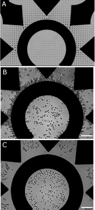
Figure 2.10 Trapping and stretching of U937 cells...37
Figure 2.11 Fluorescence intensity of actin labeled with Alexa-488-phalloidin...39
Figure 2.12 Positioning of U-937 monocytes by DEP...42
Figure 2.13 The crossover frequency, f0, versus conductivity of the extracellular medium, for U-937 monocytes...43
Figure 2.14 Position-dependent EP of U-937 monocytes. ...45
Figure 2.15 Calculated position-dependence of EP of U-937 monocytes, for different values of U...46
Figure 2.16 Position-dependent transfection of U937 cells...47
Figure 2.17 CHO stretching and recovery...48
Figure 2.18 U937 stretching and recovery...49
Figure 2.19 Strain and relaxation data for U937and CHO cells, fit using the “standard linear solid” (SLS) or power-law (PL) models...50
Figure 3.3 Time-dependent strain-data, γ(t), of four cell-types...54
Figure 3.4 Cortical actin in four differing cell-types...57
Figure 3.5 Linear correlation between maximum strain and cortical actin thickness...58
Figure 3.6 Time-dependent effects of 10 μM latrunculin-A treatment in U937 cells...59
Figure 3.7 Time-dependent effects of 10 mM acrylamide treatment in U937 cells...60
Figure 3.8 Strain data for U937 cells following treatment with 10 μM latrunculin-A or 10 mM acrylamide...61
Figure 3.9 Electromanipulation of a cell-nucleus...62
Figure 3.10 Electromanipulation of blebs and nanoparticles...63
Figure 3.11 Electromanipulation of cell-ghosts...63
Figure 4.1 Planar electrode array micro-fabricated on a transparent insulating (flexible) polymer substrate...65
LIST OF SYMBOLS AND ABBREVIATIONS
U electric potential V
E electric field strength V/m
Erms electric fields strength (root-mean-squared) V/m
f frequency Hz
f0 crossover frequency (DEP) Hz
J material compliance 1/Pa
C cell compliance factor m2/V2
k elastic constant Pa
E0 initial elastic modulus Pa
ER relaxed elastic modulus Pa
G0 initial shear modulus Pa
GR relaxed shear modulus Pa
a position of maximum pixel intensity (m)
A power-law prefactor s-α/Pa
rc cell radius (m)
d distance (minimal gap between electrodes) (m)
dCM cell-membrane thickness (m)
dexp cortical actin thickness (m)
d% cortical actin thickness (percent of cell-radius) (dimensionless)
F force N
Ia pixel intensity (dimensionless)
Imax maximum pixel intensity (dimensionless)
kout exponential decay-factor outside the cell (1/m) kin exponential decay-factor inside the cell (1/m)
K Clausius-Mosotti factor dimensionless
L length m
L0 initial length m
Lx length of ellipsoid major-axis m
rc cell radius m
α power-law exponent dimensionless
γ strain dimensionless
γx strain of ellipsoid major-axis dimensionless γy strain of ellipsoid minor-axis dimensionless
ε dielectric permittivity F/m
η viscosity Pa·s
ν Poisson‟s ratio dimensionless
σ electric conductivity S/m
τ time constant (or “relaxation time”) s
ω angular frequency rad/s
ς stress Pa
ACR Acrylamide
AFM Atomic force microscopy
CA Cortical actin
CM Cell-membrane
CSK Cell-cytoskeleton
DEP Dielectrophoresis
nDEP Negative dielectrophoresis pDEP Positive dielectrophoresis
DS Dielectric spectroscopy ED Electro-deformation EF Electro-fusion EP Electro-poration ER Electro-rotation IF Intermediate filament Lat-A Latrunculin-A
MEMS Micro-electro-mechanical systems
MF Microfilament
MFS Minimum feature size
MPA Micropipette aspiration
MSC Mesenchymal stem cell
MT Microtubule
MTC Magnetic twisting cytometry
OT Optical trapping
PECVD Plasma-enhanced chemical vapour deposition
APPENDIX I. PECVD of nanocrystalline Si layers on high-Tg polymer substrates ...74 APPENDIX II. Gene delivery by electroporation after dielectrophoretic
positioning of cells in a non-uniform electric field...86 APPENDIX III. Mechanical properties of mammalian cells in suspension
INTRODUCTION
The mechanical properties of living tissue, including individual cells, play a key role in the understanding of biological phenomena. Therefore, the study of cell-mechanics has become an important sub-field of cell biology. In recent years, microtechnology platforms have been used to analyse and manipulate increasingly complex biological specimens for life-science applications. Although significant progress has been made towards the goal of manipulating individual biological cells, many basic questions remain unanswered and improved technology-platforms are required to achieve reliable manipulation and characterization of individual mammalian cells. Structural and mechanical properties are among the measurable characteristics, which are fundamental to the forms and functions of cells, and have therefore been studied using a large variety of techniques. However, few of these techniques are readily microfabricated and very few can handle individual cells without requiring significant mechanical contact between cells and device structures. Non-contact methods for mechanical manipulation of cells use either magnetic, optical, or electrical forces, but the former two typically require the attachment of beads to the cells, in order to provide adequate force-coupling. Electrical forces act directly on the cell without requiring the addition of beads or other materials to the cell suspension, and are therefore more suitable for non-contact cell manipulations. Electric fields act as “tractor beams” and are capable of moving, trapping, and stretching biological cells, without significantly heating the cell-suspension or otherwise damaging the cells. It therefore seems likely that electric fields are underutilized for the study of cell mechanics.
The first part (Chapter 1) of this thesis introduces the basic structural components of mammalian cells, which determine the cell‟s mechanical properties. A review of experimental techniques that have been used to measure the cell‟s mechanical properties is then provided. This literature review includes recently developed miniaturized systems, and highlights the need for new methods to characterize cells in suspension. The use of applied electric fields for various cell-manipulations will be reviewed, and their potential use for mechanical characterization of cells in suspension will be presented. The second part (Chapter 2) summarizes three published articles, which are included in Appendices I-III. Chapter 2 is divided into two parts: A methods section (2.1) and a results section (2.2). The experimental methodology includes theory, modelling, micro-fabrication, and cell-manipulation protocols. Simplified models of the cell were
the fringing electric field generated by the electrodes was simulated using finite element methods (FEM). I used standard microfabrication procedures to produce planar (Ti/Pt) electrodes on glass or plastic substrates. Lastly, I describe the use of this microdevice for cell manipulations, during observation by optical microscopy. Chapter 3 contains supplementary results, which have not yet been published.
The main objective of this work was the development and demonstration of a microtechnology platform for the mechanical characterization of individual mammalian cells in suspension. Although electrical forces have been used previously by others to deform cells, most reports describe erythrocytes (red blood-cells) and the majority of mammalian cell-types have not been studied. We hypothesized that cell-deformation in strong electric fields can be used to characterize the mechanical properties of a wide variety of previously uncharacterized mammalian cells. I therefore designed novel planar electrode structures for generating spatially non-uniform fringing electric fields, and used them to trap and deform several (previously uncharacterized) mammalian cell-types in suspension, during observation by optical microscopy. The planar electrodes are readily incorporated within array-based microtechnology platforms, and the “tractor beam” technique can displace or deform electrically polarisable objects such as biological cells, with minimal mechanical contact between these objects and the device structures. This reduces cell-sticking and permits assembly-line assays, in which individual cells are precisely moved between experimental stations within the device.
To accomplish the main objective of individual cell deformation, the following three specific challenges were addressed: (i) low-temperature deposition of thin film “barrier” materials to protect electrodes within liquid (aqueous) solutions; (ii) design and testing of electrode geometries, which permit the capture and deformation of individual cells; and (iii) dielectric characterization of cells to increase the reliability and versatility of cell-manipulation using electric fields.
To facilitate ED experiments during observation by optical microscopy, electrodes were microfabricated using standard glass microscope slides as substrates. Electrode structures were then patterned using photolithography and Pt/Ti electrodes were evaporated under vacuum. Electrically insulating thin films were also deposited over the electrodes in order to permit their
use within liquid (aqueous) suspensions. To ensure that these thin film fabrication procedures were compatible with emerging soft-lithographic procedures, I developed methods for silicon-based thin film deposition at low temperatures (T < 150 °C), using plasma-enhanced chemical vapour deposition (PECVD).
Several electrode geometries were considered for the purposes of this work, and the ideal geometry for cell- trapping and –deformation is described; it consisted of an array of tapered “cell-trapping” electrodes, which are opposed by a central counter electrode. The non-uniform E, which is generated by these electrodes, was simulated using finite-element-methods (FEM). Individual cells, which are trapped at each electrode tip, become electrically polarized, with higher E-values near the cell-poles compared with lower E-values near the equator. In the present (non-uniform) case, E is highest near the cell-pole that faces the electrode tip and cells are therefore trapped in this region. I defined positions within 50 μm of the electrode edges as strong-E regions, and otherwise as weak-strong-E. The values of strong-E averaged over the strong and weak regions differed by a factor of approximately three. For example, when the applied voltage was U = 10 V, the maximum value of E = U/l, where l is the inter-electrode distance, was 200 kV m-1. The E values averaged over the strong- and weak- E regions were ~180 kV m-1 and ~60 kV m-1, respectively. These values are consistent with previous reports of dielectrophoresis (DEP) and electro-deformation (ED). DEP results were used to validate cell-positioning capabilities and to determine proper operating conditions for ED.
Cells were patterned according to the electric field distribution, E, which was predicted by FEM simulations. Cells were moved either towards or away from the strong-E regions, depending on experimental conditions. Cell-positioning by DEP prior to the delivery of electric pulses for EP, demonstrated position-dependent EP of the cell-membrane. These results showed that the outcome of EP could be controlled by DEP and, suggested that cell-type dependence of these results was possible. Thus, it seems possible that cells of a particular type, which form part of a heterogeneous cell-suspension, may be specifically targeted for genetic modification or destruction.
Mechanical characterisation of cells by ED is then presented: We found that planar microelectrodes did indeed generate (non-uniform) E of sufficient magnitude to induce substantial deformation of individual (L929, CHO, HEK293, and U937) cells in suspension.
frequently encountered in the literature. In the three-parameter “standard linear solid” (SLS) model, a single relaxation process characterizes the cell‟s strain relaxation, whereas, in the two-parameter power-law (PL) model, a continuum of relaxation processes is assumed. The main objective of this thesis was therefore achieved and cell-deformations, observed by optical microscopy, enabled the fitting of measured strain data with well-known mechanical models of cell behaviour. These results constitute the first systematic study of ED with several different mammalian cell-types.
The most important contribution of this work is the experimental development and demonstration of microfabricated ED platforms. Some advantages of ED over other techniques used for mechanical characterization of individual cells in suspension are the following: (i) ED obviates the need for moving parts or for micro-beads in the cell suspension; (ii) mechanical contact between cells and device structures is minimized; (iii) planar electrodes for ED are easily micro-fabricated as arrays, which enables simultaneous measurements on several cells, and simple integration within biochips; and (iv) programmable ranges of U values enable the study of mechanical properties over multiple timescales.
CHAPTER 1. MECHANICAL PROPERTIES OF MAMMALIAN CELLS
This chapter introduces the basic structural components of mammalian cells, which determine the cell‟s mechanical properties; it also reviews experimental techniques, which have been used to measure the mechanical properties of cells and cell-structures. Emphasis is placed on suspended cells; following a brief introduction to microtechnology platforms for cell-based analysis and modification, cell-manipulation using electric fields is reviewed. The chapter concludes with an explanation of the motivation and the objectives of this thesis.
1.1 Introduction to mammalian cells
Biological cells are the smallest functionally autonomous units of living organisms, and are therefore referred to as the “building blocks” of life. Cells are defined structurally by a distinct boundary, the cell-membrane (CM), which separates the interior (the cytosol) from the external environment. Inside the cell is a complex dynamic arrangement of organelles and interacting molecular networks, which give life to the cell.
1.1.1 Structural properties
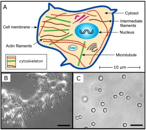
Living biological cells adapt to their environments continuously, often undergoing drastic structural adjustments. Adaptive cell-geometries are permitted by the organizational flexibility of the cytoskeleton (CSK), which consists of several distinct but interacting molecular networks (simplified in Fig. 1.1): These include actin-based microfilaments (MF), intermediate filaments (IF), and microtubules (MT) [1]. The assembly and organization of these CSK networks are regulated by genetic, epigenetic, and environmental factors, and cell morphologies are therefore diverse and dynamic [2]. In order to reliably interpret mechanical measurements performed on individual (whole) cells, it is therefore necessary to establish conditions in which the cell‟s major structural features can be regulated and their roles in determining the measured mechanical properties established [3].
Figure 1.1 Structural features of mammalian cells. (A) Simplified model of a cell with
major features labelled; adapted from [3]; (B) HEK293 cells in monolayer culture; scale bar = 30 μm (C) HEK293 cells in suspension; scale bar = 30 μm.
The CM can be thought of as the cell‟s “skin”; it is very thin (dCM ~10 nm, [2]), and its contribution to the overall stiffness of the cell is small in comparison to the CSK [4]. In isotonic conditions, the CM is ruffled and therefore not maximally extended; for example, in hypotonic media, the expansion of chondrocytes to twice their isotonic volumes was observed [5]. The CM therefore limits maximal strain values of cells but, for small strains, the MF CSK mostly determines measured stiffness [4].
Located mostly in the cortex, a narrow zone just beneath the CM, most actin filaments are arranged into a meshwork that excludes most organelles from the cortical cytoplasm [6]. MFs are therefore closely associated with the mechanical interaction of cells with their external environments, and they have important roles in cell spreading and motility. Dramatic demonstrations of MF remodelling in response to mechanical cues has been achieved using micro-patterned substrates to define novel cell-geometries [7].
The mechanical microenvironment seen by cells in vivo depends on the type of tissue in which they are located. Solid tissues exhibit a range of stiffness values, as measured by the elastic modulus, E0 (kPa) [8]: Brain (~1), muscle (~10), collagenous bone (~50). The microenvironment‟s stiffness has a strong impact on cell-phenotypes and is known to determine lineage specification of multipotent cells: For example, polyacrylamide gels of varying stiffnesses were found to be either neurogenic (0.1 – 1 kPa), myogenic (8 – 17 kPa), or osteogenic (25 – 40 kPa), when used as substrates for mesenchymal stem cell (MSC) differentiation and proliferation [8]. Measured mechanical properties of individual cells are therefore used to establish potential relationships between the stiffness of cells, their microenvironments, and their composite tissues.
1.1.2 Mammalian cells in suspension
The mechanical properties of mammalian cells in suspension are important determinants of biological functionality in several in vivo and ex vivo contexts. Cells of the circulatory system, for example, have been extensively studied from a mechanical perspective, and increased stiffness of diseased erythrocytes (red blood-cells, RBC) and leukocytes is known to restrict their flow through small capillaries [9-12]. Increased stiffness of both lymphoid and myeloid leukemia cells has been shown to result from some chemotherapy treatments [10], and cell-stiffness has been used as marker for various cancers [3].Measurements of stiffness have therefore been essential for determining the biomechanical effects of various drugs and treatments relevant to cells in circulation.
Mammalian cells are increasingly being used for the production of recombinant proteins and related products in large-scale bioreactors [13], and suspension culture permits mammalian cells to grow in bioreactors by methods similar to those used in microbial systems that enable scale-up [14]. The range of available culture conditions is, however,
17]. To reduce the harmful effects of mechanical stresses, shear-stress modifiers are often added to cell suspensions, although some of these additives can enter through the cell-membrane, with unknown consequences to the health and function of the cultured cells [19].
Methods to quantify mechanical properties of suspended cells can therefore be used to improve the design of new cell-types, bioreactors, and micro-fluidic devices, by predicting the cellular elastic and viscoelastic responses to various forces.
In contrast with the diversity of adherent cell geometries, mammalian cells often assume a roughly spherical geometry when they are suspended in a liquid (aqueous) medium. With the notable exception of RBCs [20], cell-types for which spherical geometries have been observed in suspension include the following: Fibroblasts [4], articular chondrocytes [5], adipose derived adult stem (ADAS) cells [22], bone marrow-derived adult mesenchymal stem cells (MSCs) [22], and others examined in later sections (Chapters 2 and 3) of this thesis. Thus, a rounded (suspended) geometry is common to many cell-types from diverse mammalian tissue sources. In suspended cells, cortical actin (CA) forms a semi-rigid shell, which is clearly visible using fluorescence microscopy [5]. Although interior structural elements such as the cell-nucleus contribute to the cell's exact shape via their loose coupling to the cortex, the CA (and its thickness) mainly determines the cell's structural response [4].
1.1.3 Measured mechanical properties
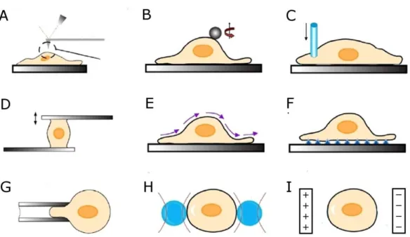
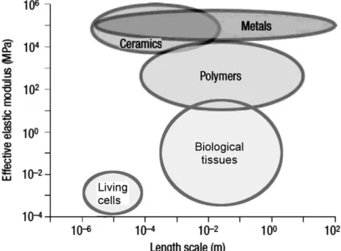
The mechanical properties of cells have been studied in detail using a large variety of experimental techniques (Fig. 1.2), which generally involve the application of external mechanical stresses, followed by observation of the cell‟s structural responses. These techniques are classified according to the several criteria (Table 1.1), including the amplitude and distribution of stresses applied to the cell. The majority of these techniques probe small regions on or within the cells and therefore measure local mechanical properties. At these sub-cellular length-scales, mechanical properties are heterogeneous and differing measurement protocols have therefore resulted in a large range of reported cell-elasticity values (Fig. 1.3: 100 Pa < E0 < 10,000 Pa) [3].
Figure 1.2 Methods used to measure mechanical properties of cells. (A) atomic force
microscopy; (B) magnetic twisting cytometry; (C) cyto-indentation; (D) micro-plate stretching; (E) fluid flow rheometry; (F) substrate deformation; (G) micro-pipette aspiration; (H) optical trapping; (I) electro-deformation. Adapted from [3].
Table 1.1 Methods used to characterize the passive mechanical properties of cells
Method Force range (nN) Cell morphology* Probe area§ References
AFM 0.01 – 100 Spread Local [22]
MTC 0.01 – 100 Spread Local [33]
Indentation 0.01 – 100 Spread Local [40]
MPA 0.01 – 100 Rounded Local [9]
OT 0.01 – 1 Rounded Local [3]
ED 0.01 – 10 Rounded Global Present work
*
Cell morphologies typically used; exceptions are discussed in the main text.
§ Probe area: “Global” if the whole cell is stretched without requiring mechanical contact between the testing device and a localized region of the cell.
Very few techniques are available for the mechanical characterization of whole suspended cells. Although traditional techniques such as AFM have been adapted for use with suspended cells [22], they are nevertheless limited to probing local structures. This limitation is partially overcome by the following four techniques: (i) micropipette aspiration (MPA: Fig. 1.2 G) [9];
H), [21, 34, 35]; and (iv) electro-deformation (ED: Fig. 1.2 I) [36-39]. In the next three paragraphs (following Fig. 1.2 and Table 1.1), I will discuss these four techniques (MPA, MTC, OT, and ED) separately.
Measurement of the apparent membrane tension of sea urchin eggs (rc ~ 50 μm), by partial aspiration of cells into narrow (~ 10 μm) pipette capillaries, was reported in 1954 [23]. This technique was soon named “micropipette aspiration”, and was applied to RBCs [24-26] and other cells of the circulatory system such as granulocytes [29].MPA has also been applied to adherent cell-types, such as articular chondrocytes [5], which are not traditionally cultured in suspension. Only a portion of the cell is aspirated during MPA experiments, and pipettes possessing several differing diameters are often used to characterize cells. Furthermore, significant mechanical contact between cells and pipettes complicates the interpretation of mechanical deformation data.
Magnetic fields have been used to study cell rheology since the 1940s [30, 31]. Magnetic fields exert forces on small particles, which are either injected into the cell [30], or attached to the cell-surface [33]. Magnetic-twisting-cytometry (MTC) therefore probes local structures and different transmembrane receptors were found to differ in their ability to mediate force transfer across the cell surface [32].
Radiation pressure resulting from differing optical properties (refractive indices) of suspension media and suspended bodies has been used to manipulate a wide variety of particles and objects by “optical trapping” (OT) [34, 35, 41]. Optical radiation can, however, easily damage cells [42], and stresses are therefore usually coupled indirectly, for example by attaching beads to the cells (Fig 1.2 H).
Electrical stresses and electro-deformation (ED) result from differing dielectric properties of suspension media and suspended bodies. Like OT, ED is generally applicable to suspended bodies such as drops of liquid [43] or membrane-bound vesicles [44]. Although ED of biological cells has been sporadically reported since the 1970s, including ED of amoebae [45], murine Sp2 myeloma cells [46], and non-mammalian protoplasts [39], RBCs remain the only cell-type to have been studied in detail, by multiple independent researchers [36-38, 47, 48]. ED will be discussed in greater detail (in Sect. 1.3.4), after microtechnology platforms and electro-manipulation techniques have been introduced.
Figure 1.3 Depiction of the typical length-scales and elastic modulus values for several materials. Adapted from [49].
1.2 Microtechnology platforms for life-science applications
As discussed in the previous section of this thesis, the diameter of a typical suspended mammalian cell is on the order of ~ 10 μm. Modern semiconductor manufacturing processes are capable of producing electronic devices with minimum feature sizes (MFS) well below 100 nm [50]. Many semiconductor foundries are finding new opportunities in the life-sciences, where MFS-requirements are less stringent than they are for most electronic applications. Life-science applications do, however, present new challenges associated with the handling of complex biological objects within aqueous environments [51]. The need to fabricate on-chip channels and chambers to handle aqueous suspensions has resulted in the emerging fields of soft-lithography [52],and microfluidics [53].
(MEMS) technologies, which have been developed for a wide variety of applications [54]. Integration of MEMS-based micromanipulation tools within microfluidic platforms permits precise manipulation of individual biological cells [51, 55].
As tools for bio-analysis continue to be miniaturized, with the goal of integrating multiple laboratory functions within small, electronically controlled devices, these “Labs On Chips” (LOCs) will replace traditional laboratory assays with ever-decreasing on-chip footprints [56].Commercial LOC platforms for the analysis of molecules, such as DNA or proteins, are available, and several LOC platforms have been recently developed to handle cells [57, 58] and tissue constructs [59, 60]. The potential advantages of increased parallelism, smaller sample sizes, and computer-controlled automation of experiments motivates our search for effective micromanipulation techniques, which can be included in LOC platforms. Efforts made in this thesis to develop low-temperature, plasma-based fabrication methods for use with typical soft-lithography materials are represented by our first published article (Appendix I).
1.3 Electro-manipulation of cells
From early dielectric spectroscopy (DS) experiments, the dielectric properties of individual cells were originally inferred by varying their concentrations in suspension and by measuring the dielectric properties of the suspensions [61, 62]. Subsequent observations revealed diverse types of interactions between electrically polarised cells in an externally applied electric field, E: In a spatially non-uniform E, displacement of cells toward either stronger- or weaker- E-regions was observed to depend on the cells‟ dielectric properties [63], and application of pulsed E disrupted cell-structures such as the CM [64]. In combination, pulsed and oscillating E have been used to fuse two or more cells into new hybrid cell-types [65]. Transient cell-deformation occurs in sufficiently strong pulsed- [38] or oscillating- [36] E.
1.3.1
Dielectrophoresis
Biological cells of various types can be distinguished from one-another and displaced within a liquid medium using dielectrophoresis (DEP) [63, 66, 67]. In a spatially non-uniform
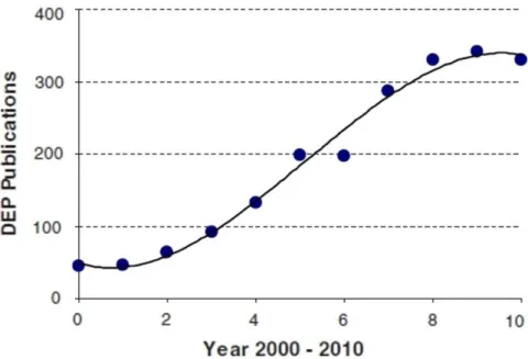
E, the differential electric polarizability of cells and their suspending medium produces the DEP force, which can be either attractive (towards the strong-E regions) or repulsive (towards the weak-E regions), depending on experimental conditions. Attractive or repulsive DEP forces are usually referred to as “positive” (pDEP) or “negative” (nDEP), respectively. Measurement of the DEP force as a function of experimental variables produces DEP “spectra” which are characteristic of each cell-type; DEP has therefore been used to identify and select rare cells from heterogeneous populations [68, 69]. DEP forces are significantly larger than other forces arising from gravity (sedimentation) or fluid flow under normal conditions [70, 72], and DEP has therefore been widely used to trap and hold cells in precise locations [71, 72]. In microfluidic devices, DEP has been used to transport and position cells with sufficient precision to enable single-cell manipulation [73]. Increased use of microtechnology platforms for life-science applications, which demand efficient methods to manipulate living cells on an individual basis, has therefore resulted in a growing interest in DEP (Fig. 1.4) [74].
Figure 1.4 Increased use of dielectrophoresis (DEP) in the last decade. The number of
DEP publications is plotted against time (in years), over the last decade (years 2000-2010). Reproduced from [74].
1.3.2
Electro-poration
Electro-poration or electro-permeabilization (EP) results from the application of an intense electric field to bring about structural changes of the cell membrane that increase its permeability. It is well known that irreversible EP leads to cytolysis [75], while reversible EP can be used to transfer molecules such as DNA into the cells while maintaining high rates of cell survival [64]. Generally, pulsed electric fields are used and the extent of EP is determined by parameters such as the strength, duration, and repetition rate of the electric pulses. Critical values of the electric field strength, which determine whether cell-membrane EP is reversible or irreversible, are specific to each cell-type and are usually determined by performing experiments at different E-values [76, 77].
Figure 1.5 Several effects of pulsed electric fields applied to cells. Increased
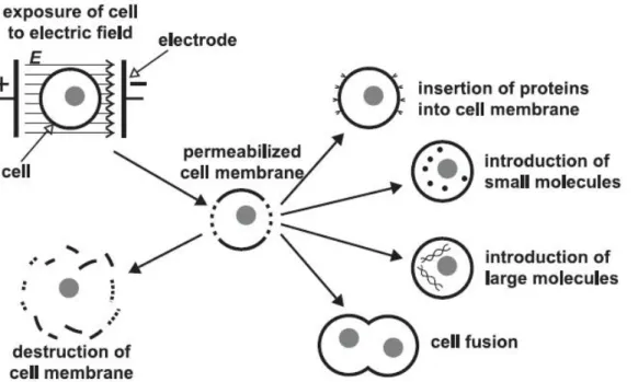
permeability of the CM, which results from the application of a pulsed electric field, has been used to insert molecules into the cell, to fuse two or more cells together, or to destroy cells; image is from [77].
Traditionally, in vitro EP has been accomplished using electrodes with mm spacing, and the position of individual cells within the electrode chamber did not need to be considered [78]. In contrast, micro-fabricated devices for EP accommodate relatively small numbers of cells and their smaller (sub-mm) electrode dimensions require consideration of spatial non-uniformities in E and of cell-positioning with respect to the electrodes [79-83]. Cell-positioning by DEP is known to complement EP experiments. For example, the alignment of cells by DEP after EP has been used for cell-cell fusion [65], and EP of DEP-trapped cells increased the sensitivity of impedance-based cell-detection [84]. In a spatially non-uniform E, the dependence of EP on the field‟s amplitude results in position-dependent EP, and therefore leads to regions within the chamber where either reversible or irreversible EP may prevail [85, 86].
1.3.3
Combined dielectrophoresis (DEP) and electro-poration (EP)
The combined use of DEP and EP is dramatically demonstrated by “electro-fusion” (EF) of cells: In this procedure, DEP is used to align cells in pearl-chain formations and subsequent EP results in fusion of two or more cells [65]. DEP has also been used to trap cells prior to impedance measurements and cell-destruction by EP [84]. The cell-type selectivity of DEP permits ED-based genetic modifications or cell-destruction to be performed with cell-type selectivity. Having judged this topic worthy of further research, and in order to demonstrate predictable cell-positioning by DEP, we explored the extent to which reversible or irreversible EP of cells could be controlled by DEP. This combined DEP/EP work is represented by our second published article (Appendix II).Figure 1.6 Electro-deformation of an erythrocyte (red-blood-cell, RBC). (A) schematic
view of a cell (RBC) in an inhomogeneous electric field, E (solid lines); the dotted-line represents the unstained RBC-shape, prior to application of E; (B)transient elongation of an RBC in response to a step-wise increase in the amplitude of E; adapted from [47].
1.3.4
Electro-deformation (ED) of cells
Detailed studies of ED were first reported by Engelhardt et al., using RBCs: [36, 47, 87]. In their system, cells were trapped at the edges of “razor”-shaped electrodes by DEP, permitting improved optical observation when compared with circular wires (Fig. 1.6 A). The measured time-dependent strain and relaxation properties of RBCs (Fig. 1.6 B), permitted mechanical characterisation of these cells using simplified mechanical models, which were previously used to describe materials such as polymers [88]. They developed several differing experimental protocols, in which the patterns of applied electrical stresses were varied in amplitude and time using programmable waveforms, thereby demonstrating the versatility of ED.
Work with RBCs was extended by Krueger and Thom (1997) to subzero temperatures (-15°C < T < 25°C): They discovered that RBCs could be stretched to nearly twice their original lengths, even at temperatures below 0ºC without destruction. This work has been continued recently by some of the same authors [48]. ED has also been reported, on several occasions, by U. Zimmermann and co-workers, among their very extensive