HAL Id: hal-00023081
https://hal.archives-ouvertes.fr/hal-00023081
Submitted on 20 Apr 2006HAL is a multi-disciplinary open access archive for the deposit and dissemination of sci-entific research documents, whether they are pub-lished or not. The documents may come from teaching and research institutions in France or abroad, or from public or private research centers.
L’archive ouverte pluridisciplinaire HAL, est destinée au dépôt et à la diffusion de documents scientifiques de niveau recherche, publiés ou non, émanant des établissements d’enseignement et de recherche français ou étrangers, des laboratoires publics ou privés.
The actin cytoskeleton is involved in signalling
protoplast embryogenesis induced by agarose embedding
Christian Brière, Henri Barthou, Alain Jauneau, Gilbert Alibert, Michel
Petitprez
To cite this version:
Christian Brière, Henri Barthou, Alain Jauneau, Gilbert Alibert, Michel Petitprez. The actin cy-toskeleton is involved in signalling protoplast embryogenesis induced by agarose embedding. Physi-ologia Plantarum, Wiley, 2004, 122, pp.115-122. �10.1111/j.1399-3054.2004.00374.x�. �hal-00023081�
The actin cytoskeleton is involved in signalling protoplast
embryogenesis induced by agarose embedding
etitpreza , avenue de 1326 Castanet Tolosan Cedex, France
ge, Auzeville losan, France
cIFR FR40, Pôle de Biotechnologie Végétale, 18 chemin de Borde Rouge, Auzeville F31326
in protoplasts al of the cell reorganized Treatment of contacts with ion and cable ork and the was lost. Furthermore, embryoid formation induced by agarose embedding
h latrunculin B, known work is involved in the signalling process that leads to polarity acquisition and embryoid determination in agarose-embedded protoplasts.
Keywords: actin, adhesion, cytoskeleton, protoplast, sunflower, Helianthus annuus
Christian Brièrea,b, Henri Barthoua, Alain Jauneauc, Gilbert Aliberta and Michel P a Laboratoire de Biotechnologie et Amélioration des Plantes, INP-ENSAT l'Agrobiopole, Auzeville, F3
bUMR CNRS 5546, Pôle de Biotechnologie Végétale, 18 chemin de Borde Rou F31326 Castanet-To
Castanet-tolosan, France
Abstract:
The organization of actin microfilaments was studied by immunofluorescence isolated from sunflower hypocotyls and cultured in an agarose matrix. Remov wall completely disrupted the actin cytoskeleton, which became progressively into cortical microfilament arrays and actin cables during protoplast culture. protoplasts with Arg-Gly-Asp motif-containing peptides, to inhibit putative cell the agarose matrix, strongly affected this repair process: microfilament elongat formation were inhibited and the connectivity between the cortical netw perinuclear basket
was reduced. Similar effects were observed with a short treatment wit to disrupt actin microfilaments. These results indicate that the actin net
Introduction
In Helianthus annuus (Chanabé et al. 1991; Krasnyanski and Menczel 1993; 1994) as in Arabidopsis thaliana (O'Neill and Mathias 1993) inclusion of pro solid matrix induces changes in their division mode and development pathways. protoplasts cultured in liquid medium divide symmetrically and form loose colonies, whereas protoplasts embedded in agarose divide mostly asymmetricall to compact embryo-like structures (Petitprez et al. 1995). It has been shown that matrix stabilizes the microtubule cytoskeleton (Caumont et al. 1997), ii) embry
Alibert et al. toplasts in a In sunflower, ly associated y, giving rise i) the agarose oid formation u et al. 1999). the linkage plant cells, existence of such components (Pont
that integrin-atembe et al. tif, known to tin) in animal nts plant cell et al. 1998), mbrane-wall ll interactions sh and Heath llular matrix ton.
s studies have shown the role of the microtubule cytoskeleton in adhesion-signal transduction leading to a shift in protoplast developmental
embryoid differentiation (Caumont et al. 1997; Barthou et al. 1999). We ment of actin in this signal transduction pathway. We show that ning peptides
e cultured as ere used for n L4 medium
e acetic acid, 4.4 µM N6-benzyl aminopurin and 0.45 µM 2,4-dichlorophenoxy acetic acid (TLD medium). Protoplasts were then embedded in 0.5% (w/v) low-melting-point agarose (Sea plaque, FMC Bioproducts, Rockland, ME, USA) and solidified into 250 µL droplets which were submerged in TLD medium according to Chanabé et al. (1991). Cultures were maintained at 25°C in the dark. The plating efficiency (= percent of divided protoplasts) was assessed at day 7. At day 10, cultures were transferred to the light (25µE m-2 s-1, 16h illumination) and the number of microcolonies and embryoids counted at day 21. Embryoid formation was expressed as the is mediated by contact between the agarose matrix and the cell surface (Bartho
These data strongly suggest the existence of trans-membrane proteins ensuring between the extracellular matrix and the cortical cytoskeleton.
In animal cells, the existence of such proteins is well known, the most prominent and best-studied matrix receptors belonging to the integrin family (Hynes 1992). In
although clear evidence is still lacking concerning the
Lezica et al. 1993; Reuzeau and Pont-Lezica 1995), several studies have shown like proteins are present in Arabidopsis, Allium and Chara (Gens et al. 1996; K 1997) but whether they actually are integrins remains to be proved.
Numerous studies have focused on the role of the Arg-Gly-Asp (RGD) mo mediate the interaction between integrin and its ligands (vitronectin or fibronec cells (Haas and Plow 1994). A synthetic peptide bearing the RGD motif preve growth (Schindler et al. 1989), protoplast adhesion (Zhu et al. 1993; Canut embryoid differentiation in sunflower protoplasts (Barthou et al. 1999), and me adhesion in Pelvetia (Henry et al. 1996). RGD dependent plasmalemma-cell wa have also been implicated in plant defence response (Kiba et al. 1998; Meller 2001). Taken together, these data strongly suggest that membrane-extrace binding sites forward extracellular signals into the cell possibly via the cytoskele In sunflower protoplasts, previou
program resulting in report here on the involve
disrupting actin microfilament arrays by actin drugs or by RGD motif contai prevents or inhibits embryoid development.
Material and Methods
Protoplast isolation and culture
Seeds of Helianthus annuus L. var. Emil (Pioneer Hibred International) wer described by Chanabé et al. (1989). Hypocotyls of seven-day-old seedlings w isolation of protoplasts which were cultured at a final density of 5x10⁴ mL-1 i (Lénée and Chupeau 1986) containing 14.6 mM sucrose, 16 µM 1-naphthalen
ratio between the number of embryoids and the total number of microcolonies (embryoid + loose colonies).
garose layers ated with Poly-L-Lysine (Sigma), submerged in TLD ed in the same conditions.
Calbiochem GDSP (final concentration 40µM) was added to the ter protoplast embedding. Controls were performed by adding
k solution in Meudon, France). 3-day old protoplasts, cultured in liquid er before or TLD and the 0.03% (w/v) razine-N,N’-N’-tetraacetic 1989). Fixed n to increase ere treated 3 insed twice in (w/v) normal 5°C with in (Amersham, UK) diluted 250 fold in TBS-Mg. They Mg, once in 0.1% (w/v) bovine serum albumin dissolved in
TBS-nate (FITC)-1/50 in in antifading rd). scope (Leica, counted, and loose colony or embryoid) were scored to determine the plating efficiency.
Actin pattern: immunolabelled protoplasts were examined with a Leitz epifluorescence microscope equipped with excitation and barrier filters BP450-490/BP520-525. Photographs were taken with a Wild MPS51S camera and a Fujichrome 400 film for colour slides. Protoplasts were scored according to the characteristics of the actin pattern: type of filaments (only dots or patches, short filaments, or presence of long filaments), type of organization (parallel or random) of actin arrays (when present), and presence of actin wires.
For immunofluorescence studies protoplasts were embedded in 0.5% (w/v) a spread onto coverslips previously co
medium, and cultur
Effect of RGD peptides
Hexapeptides GRGDSP and GDGRSP (5mM stock solution in water) were from (Meudon, France). Hexapeptide GR
culture medium immediately af
the GDGRSP peptide in the same conditions.
Effect of cytoskeleton drugs
Phalloidin (10mM stock solution in ethanol) and Latrunculin B (1mM stoc DMSO) were from Calbiochem (
TLD medium, were incubated in the presence of these drugs for 6h, eith immediately after protoplast inclusion in agarose. They were then rinsed in culture continued in standard conditions.
Indirect immunofluorescence
Protoplasts on coverslips were fixed for 1 h in 2% (w/v) paraformaldehyde and glutaraldehyde in microtubule stabilizing buffer (MSB) containing 50 mM pipe bis (2-ethanesulfonic acid), 5 mM ethylene glycol-bis (β-aminoethylether) N, acid (EGTA), 5 mM MgSO4, 0.08% Nonidet P40, pH 6.9 (Traas et al. protoplasts were treated with Triton X-100 (0.25%, v/v, in MSB) for 30 mi membrane permeability. To reduce aldehyde-induced fluorescence, samples w times 10min with 10mM NaBH4 in 10mM phosphate buffer saline, pH 8, r TBS-Mg (10mM Tris buffer saline, 30mM MgCl2, pH 7.6) and treated with 5% goat serum in the same buffer for 30 min. Protoplasts were incubated overnight at 2 N350 mouse monoclonal anti F-Act
were rinsed twice in
Mg (TBS-BSA) and further incubated for 2h with fluorescein isothiocya conjugated sheep anti-mouse immunoglobulin antibody (Amersham), diluted BSA. After one wash in the same buffer, coverslips were mounted on slides medium Citifluor (Link analytical, Oxfo
Microscopy observation
Plating efficiency: protoplasts in culture were observed on an inverted micro Heidelberg, Germany). For each plate a minimum number of 100 objects was the type of cell (divided or undivided), or the type of microcolony (
Quantitative image analysis of actin labelling
Due to the fixation process, many protoplasts did not keep a spherical shap flattened. This had the advantage of allowing the observation of large
e, but were parts of the ts on which nation and a quisition and 10 nm) were 450-490 nm tal condition o Plus image d subtraction, adjusted to ers were then ilter to further software. For ere recorded. hole number tion process of protoplasts may lead to underestimate the maximum length of filaments, due to possible folding or breakdown of long filaments, and thus to underestimate
usions drawn erent culture
ivision frequency (%) = 100 x 21, embryoid quency (%) = 100 x number of embryoids / number of (embryoids + loose colonies). The values are the means of 3 to 5 experiments in which 700 to 2100 protoplasts and 50 to 385 embryoids were respectively observed at day 7 and day 21. Percent data were normalized by cytoskeleton network without changing the focus. These flattened protoplas
measurements of filament length was possible were thus used for quantitative imaging. A DMIRBE inverted microscope (Leica) equipped for epifluorescence illumi Color Coolview camera (Photonic Science, UK) were used for image ac digitisation. Fluorescence images (512x512 pixels, 8 bits depth, pixel size 3 recorded with a PL APO 40X oil immersion objective (NA 1.25) and with a BP excitation filter and an LP 515 nm emission filter (Leica). For each experimen digital images of 50 protoplasts were recorded and analysed with Image-Pr processing software (Media Cybernetics, Maryland, USA). After backgroun each image was segmented by binarization, the threshold being manually eliminate the background noise outside the protoplast. Two morphological filt applied to the resulting binary image: a thinning filter followed by a dilatation f reduce the noise. The lengths of segmented objects were then assessed by the every cell, the number of segments, the maximum length and the total length w For each condition, the figures represent the average of all these values for the w of protoplasts measured .
The fixa
differences between culture conditions. But this will have no effect on concl from statistical tests showing significant differences of length between diff conditions.
Statistics
The division frequency at day 7 was calculated as follows: d
% sin
Arc
number of divided protoplasts / number of observed protoplasts. At day fre
using transformation before comparison with Student’s t-test.
ol and treated protoplasts were compared with the χ2
escence from labelling was observed and sorted into different categories (Fig. 1). Freshly isolated protoplasts (i.e. observed about 1 hour after inclusion in agarose) showed no filamentous actin but only punctuated labelling all over the cell surface and perinuclear labelling (Fig. 1A1). Rapidly, within the first day of culture, short microfilaments (MFs) formed and extended through the whole protoplast cortex (Fig 1B1): after 6h, 32% of the protoplasts showed a punctate pattern, 53% showed short MFs, whereas only 15% had long MFs (Fig. 1A2-C2). The proportion of protoplasts with long MFs (Fig. 1C1) increased progressively during the culture, exceeding 60% after 48 hours. In a few protoplasts these long MFs appeared to form a parallel array Distributions of actin patterns in contr
test.
Results
Actin network dynamics in control protoplasts
The dynamics of the actin network in protoplasts was followed by immunofluor day 0 to day 6 in culture. For each protoplast the overall organization of actin
(Fig. 1D1). This parallel organization was observed in an increasing number o with time but never concerned more than 20% of the protoplasts (Fig. 1D2 cortical MF network, the perinuclear basket of MFs appeared to be progressively the cortical network by cytoplasmic actin wires (Fig.1E1) which started to appe
f protoplasts ). Beside this connected to ar during the toplasts (Fig. ce images of he number of network in a of nucleation rotoplasts the , up to 296 at ime, the maximum length of microfilaments increased from 20µm to 70µm (Fig2B) and the total length of the actin network increased from 200µm to 500µm and microfilament
zation
ing the RGD usion or after ays of culture. The GDGRSP peptide was used in control experiments. At day 21 . 3C, a strong t cultures. A and 96 hours ed very short ic actin wires (Fig. 4C). No For a better filaments, we in structures: t to only slightly l cultures. On rrays of microfilaments, which appeared as early as the first day of ig. 5B). This ction of the assembly of : at 120h, as onnecting the (Fig.2, black bars). It can be observed that for the three parameters assessed (number of actin segments, max. length of actin filaments and total length of the actin array) after one day of culture the values of the assay were lower than in the control and the difference increased with time. This was particularly true for the length of the longest filament and for the total length of the actin network in 4 and 5-day-old protoplasts: they were significantly lower in RGD-treated protoplasts than in the control. Although the decrease of the network length may be partially explained by a decrease of the number of filaments, it is clear that there is a significant first 3 days of culture and were observed in more than 80% of 5-day-old pro
1E2).
In order to quantify the appearance of the cortical actin network, fluorescen protoplasts were analysed numerically and three parameters were assessed: t filaments, the maximum length of a filament, and the total length of the protoplast (Fig. 2, empty bars). The number of filaments depends on the number sites whereas their length reflects the elongation activity. In freshly isolated p number of actin segments was low (108 at 6h) and increased gradually with time 144h (Fig2A). At the same t
(Fig.2C). These data reveal an intense activity of actin nucleation elongation in cultured protoplasts.
RGD peptide inhibits embryoid formation and affects actin microfilament organi
To inhibit cell contacts with the agarose matrix, the hexapeptide GRGDSP, bear motif, was added to the culture medium either immediately after protoplast incl 1 to 4 d
microcolonies (Fig. 3A) and embryoids (Fig. 3B) were counted. As shown in Fig inhibition of embryoid formation was observed in RGD-treated protoplas maximal effect was obtained when GRGDSP peptides were added between 48 of culture.
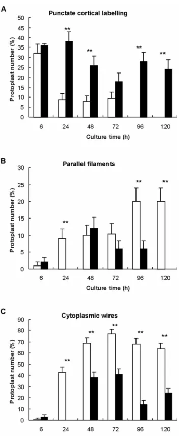
Protoplasts cultured for 24 h in the presence of the GRGDSP peptide display cortical microfilaments often restricted to actin dots (Fig. 4A), the cytoplasm were often broken (Fig.4B) leaving the nucleus disconnected from the cortex such effect was observed in protoplasts treated with the GDGRSP peptide. understanding of the effect of the RGD peptide on the organization of micro assessed the proportions of protoplasts showing one of the three following act
cortical actin dots, parallel cortical microfilaments, and cytoplasmic actin wires. In contras control experiments, the proportion of protoplasts showing actin dots decreased
in RGD-treated cultures (Fig.5A), reaching 25% at 120h against 0% in contro the contrary, parallel a
culture in controls, were not observed before 48h in RGD-treated protoplasts (F delay in the cortical array organization was correlated with a marked redu percentage of protoplasts exhibiting such a parallel cortical array. Finally cytoplasmic wires was also strongly inhibited by RGD peptides (Fig. 5C) compared to the control, less than half of the protoplasts possessed actin wires c nucleus to the cortical array.
reduction of microfilament length. These results strongly suggest that RGD peptides mainly f the matrix-alloidin – or r et al. 1999). to ensure that ore, previous cell wall has immediately embrane and were used to trations. Then one sample was immediately fixed and
in standard d after 7 days o the control. esenting long oids were not tration tested, on process. of increasing on of cortical both as short ot stained by toplasts were sion rate was ced by about of this effect nt alone was dilution (1:5000) corresponding to the concentration of solvent in 200nM proportion of control cultures (25.2 ± 1.8 %). Thus, the inhibition of embryoid differentiation observed in latrunculin B treated cultures
explained by a solvent effect but is really due to an effect of the drug.
Similar results were observed when the drugs were applied for 6h immediately before e time of
In sunflower protoplasts embedded in agarose the first division is asymmetric and leads to embryoid development, whereas in liquid medium the first division is symmetric, leading to the formation of loose microcolonies (Petitprez et al. 1995). An early marker of protoplast polarization is the migration of the nucleus from the cell centre towards the periphery of the protoplast. In Pelvetia and Micrasteria, actin microfilaments (MFs) and microtubules (MTs) have been associated with the migration of the nucleus towards the cortex (Meindl et al. 1994). In Fucus, it was suggested that actin-driven targeted secretions of sulphated compounds could fix the cell division axis (Brownlee and Bouget 1998). Moreover, in this inhibit the actin microfilament elongation process.
Involvement of the actin cytoskeleton in embryoid formation
In order to study the involvement of actin microfilaments in the transduction o adhesion signal, protoplasts were treated with drugs known to stabilize – ph depolymerise – latrunculin B – actin microfilaments (Gibbon et al. 1999; Mathu In these experiments 3-day-old protoplasts cultured in liquid medium were used before the treatment they had recovered a complete actin network. Furtherm work (Caumont et al. 1997) has shown that, on average, less than 40% of the been rebuild in 3-day-old protoplasts. Protoplasts were embedded in agarose and incubated with the drugs for 6h in order to allow connections between the cell m the extracellular matrix to establish. Increasing concentrations of the drugs determine the optimum concen
immunolabelled with anti-actin, another sample was rinsed and cultured conditions. The percentages of cell division and of embryoids were then assesse and three weeks of culture, respectively.
Phalloidin did not modify the microfilament network up to 1µM as compared t At 10µM, a marked stabilizing effect was observed, nearly all protoplasts pr microfilaments and actin wires. The division rate and the proportion of embry modified by the treatment (Table 1). It clearly appears that whatever the concen including the 10µM stabilizing dose, phalloidin did not alter the protoplast divisi Latrunculin B is a very potent actin-depolymerizing drug. We tested the effect concentrations of latrunculin B, ranging from 10nM to 50µM, on the organizati actin MFs. At 10nM, cortical MFs were short, at 200nM cortical actin appeared MFs and dots, at 1µM there were only cortical dots and many protoplasts were n anti-actin antibody, and at 50µM there were no visible MFs and most of the pro unlabelled (Fig. 1F). Between 10nM and 200nM, no modification of the divi observed (Table 1). In contrast, embryoid differentiation was significantly redu 30%, but no clear dose-response relationship was found between the intensity and the tested concentrations. In control experiments with DMSO, the solve added at a final
latrunculin B treatments. No significant difference was observed between the embryoids in DMSO treated cultures (24.3 ±1.7 %) and in
cannot be
inclusion in agarose.
Taken together these results indicate that modification of the actin cytoskeleton at th protoplast inclusion contributes to determine embryoid differentiation.
latter model, the fixation of the first division axis requires linkage of MFs to the the participation of putative transmembrane factors (Quatrano et al. 1991). In tob wall matrix is necessary for the positioning of the nucleus (Katsuta and Shibao these results implicate the actin network in the cell fate: its connections, on one
cell wall with acco, the cell ka 1988). All side with the be crucial to of the actin reported in ctin network en previously arpova et al. ls (Seagull et phases were ucleation and of filaments ablishment of cytoplasmic s tracks for intracellular he cortex and ver the exact ed in agarose plast cultures also inhibited ot observed. already been ct similar to were able to al. 1998). In D treatment with the cell n reported to be 1997) and of , even if such arthou et al. fied. To date, (Wasteneys plant cells. a continuum e perinuclear microtubule establishment of this continuum (before cell wall synthesis) appears to be essential for cell fate determination. In liquid media, where extracellular contacts are lacking, embryoids cannot develop. It is thus likely that agarose embedding of protoplasts allows the formation of transmembrane adhesion complexes that anchor the protoplast to this extracellular matrix and stabilize the cytoskeleton network. This would help to determine and/or fix the polarity of the cell leading to asymmetric division and embryoid development. In this report, we show that not only microtubules but also the actin cytoskeleton are involved in this signalling process. extracellular matrix and on the other side with the peri-nuclear basket appear to
the initiation of cell polarity.
In our model, freshly prepared protoplasts exhibited complete disorganization network resulting from the digestion of the cell wall matrix as previously
Nicotiana (Rutten and Derksen 1990). Rapidly, the reconstruction of the a
occurred and showed different patterns (as illustrated figure 1) which have be described in a wide variety of eukaryotic organisms (Clayton and Lloyd 1985; K 1998) and correspond to interphasic MF networks already reported in plant cel al. 1987; Traas et al. 1987). During the rebuilding of the actin network, two main distinguished: i) an increase of actin dots, which likely corresponds to active n assembly of monomers, and ii) an increase of maximum and total lengths resulting from the elongation of actin microfilaments. We also observed the est connectivity between the cortical actin network and the perinuclear basket by actin wires. It has been suggested that these cables could act a
trafficking (Stamnes 2002) along which material would be transported between t the nucleus and so could participate in the cell polarity (Reddy 2001). Whate mechanisms involved in the actin network reconstruction, protoplasts embedd are further able to divide in an asymmetric manner and to give rise to embryoids. The application of latrunculin B or RGD peptides to agarose-embedded proto reduced the formation of embryoids. Cable formation and MF elongation were and connectivity between the cortical network and the perinuclear basket was n The modification of cell fate through the application of RGD peptides has reported in maize where calli became more compact and displayed an effe plasmolysis (Labouré et al. 1999). In Arabidopsis thaliana cells, RGD peptides bind purified plasma membranes and to inhibit cell-cell adhesion (Canut et sunflower protoplast, plasmolysis inhibited embryoid formation, as did RG (Barthou et al. 1999) suggesting that the association of the plasma membrane wall was involved in cell development. In plant cells, this association has bee indispensable to maintain the stationary organisation of the MFs (Ryu et al. cortical microtubules (Akachi et al. 1990; Akachi and Shibaoka 1991). However adhesion sites have been demonstrated in our model (Caumont et al. 1997; B 1999), the protein components of adhesion complexes remain to be clearly identi only spectrin-like, integrin-like and vinculin-like epitopes have been evidenced and Galway 2003), but none of these proteins have been clearly characterized in Overall, these results strongly suggest that in agarose-embedded protoplasts, occurs between the agarose extracellular matrix, the plasma membrane and th basket via the actin MF network. Previous results have shown that the cytoskeleton is also involved in this continuum (Barthou et al. 1999). The early
The nature of the adhesion complexes and their impact on microtubule and actin organization remain to be determined.
We thank Philippe Anson and Marie-Josée Tavella for their technical support in plant culture and protoplast preparation.
les by the cell ll in cultured tobacco cells. Planta 182: 363-369
ssociation of l Sci 98: 169-anabe C, Burrus M (1994) Sunflower tissue and cell cultures and their
, Alibert G (1999) RGD-mediated membrane-r pmembrane-rotoplasts. m zygote to N, Ferrara P, Pont-Lezica R psis thaliana in C, Souvré microtubule ypocotyl protoplasts. Physiol Plant 99: 129-134
nt of colony Studies on plant regeneration from tematic plant p Cell Res 156: 231-238
ualization by oning microscopy of integrin and associated proteins at the cell cts on pollen Haas TA, Plow EF (1994) Integrin-ligand interactions: a year in review. Curr Opin Cell Biol 6: 656-662
Henry CA, Jordan JR, Kropf DL (1996) Localized membrane-wall adhesions in Pelvetia zygotes. Protoplasma 190: 39-52
Hynes RO (1992) Integrins: versalility, modulation, and signaling in cell adhesion. Cell 69: 11-25
Karpova TS, Mcnally JG, Moltz SL, Cooper JA (1998) Assembly and function of the actin cytoskeleton of yeast: Relationships between cables and patches. J Cell Biol 142: 1501-1517
Acknowledgements
References
Akashi T, Kawasaki S, Shibaoka H (1990) Stabilization of cortical microtubu wa
Akashi T, Shibaoka H (1991) Involvement of transmembrane proteins in the a cortical microtubules with the plasma membrane in tobacco BY-2 cells. J Cel 174
Alibert G, Aslane-Ch
use in biotechnology. Plant Physiol Bioch 32: 31-44 Barthou H, Petitprez M, Brière C, Souvré A
matrix adhesion triggers agarose-induced embryoid formation in sunflowe Protoplasma 206: 143-151
Brownlee C, Bouget F, Y, (1998) Polarity determination in Fucus: Fro multicellular embryo. Semin Cell Dev Biol 9: 179-185
Canut H, Carrasco A, Galaud JP, Cassan C, Bouyssou H, Vita
(1998) High-affinity RGD-binding sites at the plasma membrane of Arabido links the cell wall. Plant J 16: 63-71
Caumont C, Petitprez M, Woynaroski S, Barthou H, Brière C, Kallerhoff J, Bor A, Alibert G (1997) Agarose embedding modifies cell-wall regeneration and organization in sunflower h
Chanabé C, Burrus M, Alibert G (1989) Factors affecting the improveme formation from sunflower protoplasts. Plant Sci 64: 125-132
Chanabé C, Burrus M, Bidney D, Alibert G (1991)
protoplasts in the genus Helianthus. Plant Cell Rep 9: 635-638
Clayton L, Lloyd C (1985) Actin organization during the cell cycle in meris cells. Ex
Gens JS, Reuzeau C, Doolittle KW, McNally JG, Pickard BG (1996) Covis computational optical-secti
membrane of living onion protoplasts. Protoplasma 194: 215-230
Gibbon BC, Kovar DR, Staiger CJ (1999) Latrunculin B has different effe germination and tube growth. Plant Cell 11: 2349-2363
Katembe WJ, Swatzell LJ, Makaroff CA, Kiss JZ (1997) Immunolocalization of integrin-like leton and the cell wall in nuclear
) Interaction lant defense eration from Rep 12: 260-263 wth of maize 8 nthus annuus Plant Sci 43: d to elaborate psis thaliana.
ssociated with the migrating lls. J Cell Sci or expression Arabidopsis enesis. J Exp Bot 44: 1579-1585
cterisation of ue origin. Plant Cell kers in onion
cus zygotes: : 97-178 nt-Lezica RF (1995) Comparing plant and animal extracellular
matrix-ents in regenerating and
471-479
licated in the ation of the actin cytoskeleton in mesophyll cells of Vallisneria. Plant Cell lant cell wall es for growth. J Cell Biol 108: 1955-1965.
Seagull R, Falconer M, Weerdenburg C (1987) Microfilaments: dynamic arrays in higher plant cells. J Cell Biol 104: 995-1004
Stamnes M (2002) Regulating the actin cytoskeleton during vesicular transport. Curr Opin Cell Biol 14: 428-433
Traas JA, Doonan JH, Rawlins DJ, Shaw PJ, Watts J, Lloyd CW (1987) An actin network is present in the cytoplasm throughout the cell cycle of carrot cells and associates with the dividing nucleus. J Cell Biol 105: 387-395
proteins in Arabidopsis and Chara. Physiol Plant 99: 7-14 Katsuta J, Shibaoka H (1988) The roles of the cytoske
positioning in tobacco BY-Z cells. Plant Cell Physiol 29(3): 403-413
Kiba A, Sugimoto M, Toyoda K, Ichinose Y, Yamada T, Shiraishi T (1998 between cell wall and plasma membrane via RGD motif is implicated in p responses. Plant Cell Physiol 39: 1245-1249
Krasnyanski S, Menczel L (1993) Somatic embryogenesis and plant regen hypocotyl protoplasts of sunflower (Helianthus annuus L.). Plant Cell
Labouré AM, Faik A, Mandaron P, Falconet D (1999) RGD-dependent gro calluses and immunodetection of an integrin-like protein. FEBS Lett 442: 123-12 Lénée P, Chupeau Y (1986) Isolation and culture of sunflower protoplasts (Helia L.): factors influencing the viability of cell colonies derived from protoplasts. 69-75
Mathur J, Spielhofer P, Kost B, Chua N (1999) The actin cytoskeleton is require and maintain spatial patterning during trichome cell morphogenesis in Arabido Development 126: 5559-5568
Meindl U, Zhang D, Hepler PK (1994) Actin microfilaments are a
nucleus and the cell cortex in the green alga Micrasterias. Studies on living ce 107: 1929-1934
Mellersh D, Heath M (2001) Plasma membrane-cell wall adhesion is required f of plant defence responses during fungal penetration. Plant Cell 13: 413-424. O'Neill CM, Mathias RJ (1993) Regeneration of plants from protoplasts of
thaliana L. cv. Columbia (C24), via direct embryog
Petitprez M, Brière C, Borin C, Kallerhoff J, Souvré A, Alibert G (1995) Chara protoplasts from hypocotyls of Helianthus annuus in relation to their tiss
Tiss Org Cult 41: 33-40
Pont Lezica RF, Mac Nally JG, Pickard BG (1993) Wall-to-membrane lin epidermis: some hypotheses. Plant Cell Environ 16: 117-123
Quatrano RS, Brian L, Aldridge J, Schultz T (1991) Polar axis fixation in Fu components of the cytoskeleton and extracellular matrix. Development Supp. 1: 11-16 Reddy A (2001) Molecular motors and their function in plants. Int Rev Cytol 204 Reuzeau C, Po
cytoskeleton connections - are they alike? Protoplasma 186: 113-121 Rutten TLM, Derksen J (1990) Organization of actin filam
outgrowing subprotoplasts from pollen tube of Nicotiana tabacum L. Planta 180: Ryu J, Mizuno K, Takagi S, Nagai R (1997) Extracellular components imp stationary organiz
Physiol 38: 420-432
Schindler M, Meiners S, Cheresh D (1989) RGD-dependent linkage between p and plasma membrane: consequenc
Traas JA, Burgain S, Dumas de Vaulx R (1989) The organization of the cytosk meiosis in eggplant (Solanum melongena (L.))
eleton during : microtubules and F-actin are both necessary nd form: an ita NC (1993) Enrichment of vitronectin- and fibronectin-like proteins in NaCl-adapted plant cells and evidence for their involvement in plasma membrane - cell wall adhesion. Plant J 3: 637-646
formation in trunculin B embedding and were next cultured in standard conditions. Division rate was assessed at day 7; proportion of embryoids was assessed at day 21. Values are expressed as percentages of control expe an values ± standard-errors from three measurements (control = 100).
Phalloidin Latrunculin 0.1µM 1µM 10µM 10nM 50nM 200nM for coordinated meiotic division. J Cell Sci 92: 541-550
Wasteneys G, Galway M (2003) Remodelling the cytoskeleton for growth a overview with some new views. Ann Rev Plant Biol 54: 691-722
Zhu J-K, Shi J, Singh U, Wyatt SE, Bressan RA, Hasegawa PM, Carp
Table 1: Effect of phalloidin and latrunculin B on the division rate and embryoid
protoplast culture. 3-day-old protoplasts were treated for 6 h by phalloidin or la immediately after agarose
riments. Me
Cell divisions 109.0 ± 6 112.0 ± 3 107 ± 3 109 ± 1 109 ± 2 99 ± 2 Embryoids 111 ± 7 75 ± 10 63 ± 2 67 ± 6
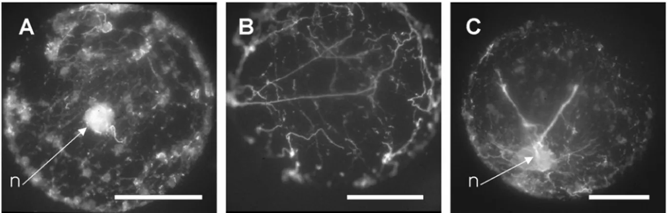
Fig.1: Actin patterns observed in sunflower protoplasts (A1 to E1) and frequency variations of these patterns (A2 to E2) during culture. (A) punctate dotted pattern of actin labelling, (B) short microfilaments, (C) random network of long microfilaments, (D) parallel network of actin microfilaments, and (E) actin wires connecting the nucleus to the cortex. (F) Absence of actin labelling in a protoplast treated with 50µM latrunculin B. n = nucleus, bar = 10µm. Frequencies are based on the observation and scoring of 100 protoplasts, at each time.
Fig.2: Variations of actin microfilament length during control (empty bars) and RGD-treated (black bars) protoplast culture. (A) Number of objects (dots or filaments) / protoplast, (B) maximum length, and (C) total length of actin filaments per protoplast. Mean ± S.E.M. from 13 (control) and 15 (RGD treatment) protoplasts. Treatments were compared with t-test: * P=0.05, ** P = 0.01. Control = protoplasts incubated with GDGRSP peptide.
Fig. 3: Loose colony (A) and embryoid (B) observed in a 3-week-old culture embedded in agarose. Bar = 10µm (C) Effect of RGD peptides on embryo
of protoplasts id formation: 100µM GRGDSP peptide was added to the culture medium either immediately after protoplast isolation and inclusion, or after different times of culture. Proportion of embryoids (= number of embryoid / total number of microcolonies) was assessed in 3-week-old cultures. Control = protoplasts grown without addition of RGD peptides. The effect of the treatment was significant whatever the treatment time.
Fig. 4: Effect of RGD peptides on actin organization in 1-day-old protoplasts. Protoplasts incubated with the GRGDSP hexapeptide, showing (A) a disrupted cortical actin network, (B) and (C) broken actin wires. n = nucleus, bar = 10µm
Fig. 5: Quantitative analysis of actin organization in RGD-treated protoplasts according to culture time. Treated protoplasts were incubated with the GRGDSP hexapeptide. Control protoplasts were incubated with the GDGRSP hexapeptide. (A) cortical actin dots, (B) cortical parallel microfilaments, (C) cytoplasmic wires. Black bars = RGD-treated, Empty bars = control. Treatments were compared with χ2 test : * P=0.05, ** P = 0.01