HAL Id: hal-01206863
https://hal.archives-ouvertes.fr/hal-01206863
Submitted on 1 Oct 2015
HAL is a multi-disciplinary open access
archive for the deposit and dissemination of
sci-entific research documents, whether they are
pub-lished or not. The documents may come from
teaching and research institutions in France or
abroad, or from public or private research centers.
L’archive ouverte pluridisciplinaire HAL, est
destinée au dépôt et à la diffusion de documents
scientifiques de niveau recherche, publiés ou non,
émanant des établissements d’enseignement et de
recherche français ou étrangers, des laboratoires
publics ou privés.
ACCURATE AND ROBUST SHAPE DESCRIPTORS
FOR THE IDENTIFICATION OF RIB CAGE
STRUCTURES IN CT-IMAGES WITH RANDOM
FORESTS
Mariem Gargouri, Julien Tierny, Erwan Jolivet, Philippe Petit, Elsa Angelini
To cite this version:
Mariem Gargouri, Julien Tierny, Erwan Jolivet, Philippe Petit, Elsa Angelini. ACCURATE AND
ROBUST SHAPE DESCRIPTORS FOR THE IDENTIFICATION OF RIB CAGE STRUCTURES
IN CT-IMAGES WITH RANDOM FORESTS . IEEE International Symposium on BIOMEDICAL
IMAGING, Apr 2013, San Francisco, United States. �hal-01206863�
ACCURATE AND ROBUST SHAPE DESCRIPTORS FOR THE IDENTIFICATION OF RIB
CAGE STRUCTURES IN CT-IMAGES WITH RANDOM FORESTS
Mariem Gargouri
?†Julien Tierny
?Erwan Jolivet
‡Philippe Petit
†Elsa D. Angelini
?? Institut Mines Telecom - CNRS LTCI - Telecom ParisTech, Paris, FRANCE
† LAB-PSA Peugeot Citroen-Renault, FRANCE
‡ CEESAR - European centre of studies and risk analysis, FRANCE
ABSTRACT
This paper presents a new automatic technique for the seg-mentation of the rib cage on CT images. Motivated by a usage scenario in the context of large, heterogeneous databases of CT-images, we introduce two shape descriptors to be used in conjunction with a Random Forests (RF) classifier. These de-scriptors were specifically designed to address the challenges of rib identification under various acquisition conditions af-fecting subject’s orientation and image quality. Extensive ex-periments demonstrate the superiority of our proposed shape descriptors in nominal configurations. Robustness with re-spect to subject’s orientation variation and additive noise is also demonstrated, with an improvement of classification per-formance of up to 25%, comparing to intensity-based descrip-tors, without neither pre-registration nor pre-smoothing.
Index Terms— CT images, bone segmentation, thorax, random forests, shape descriptors.
1. INTRODUCTION
There is an increasing interest in image-based anatomical models, exploiting diverse 3D volumes of medical images from several modalities. In this work, we are interested in human skeleton modeling based on CT images. In the context of our work, numerical models of human skeletons are used by car manufacturers who dedicate important research efforts in developing numerical simulations of crash tests to improve the safety of their new vehicles. These numerical simula-tions enable to study the mechanical reacsimula-tions of specific bones during diverse types of car crashes. Until now, nu-merical models of human skeletons have been built based on CT images of individual subjects, which had to be manually segmented.
However, a large database of numerical models derived from a diverse population of subjects (categorized by gender, age and morphology) is required to build up a sufficient and reliable knowledge of fracture risks at the level of a popula-tion. In this context, it is planned to use in the near future a large, heterogeneous, database of thousands of CT-images of diverse subjects. Constructing such a database requires to use
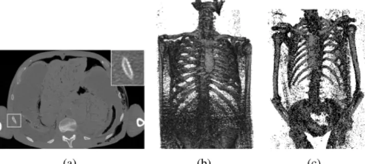
(a) (b) (c)
Fig. 1.Illustrations of the challenges in rib identification on a heterogeneous database of CT images: (a) Axial slice of the torso showing very thin ribs’ cortical cross-sections (see the inset zoom) with a typical thickness of 1.5 mm; (b) High noise level in some acquisitions; (c) Variable subject’s posture.
automated segmentation tools to extract detailed bone struc-tures. In this context, we focus in this paper on the rib cage segmentation, required to study car crash-induced thoracic in-juries, which are life threatening and remain among occupant safety priorities for car manufacturers.
Regarding bone segmentation on CT images, a variety of approaches have been derived to extract large bones such as the tibia or the femur [1, 2, 3] and small bones such as ver-tebrae [4, 5]. However, ribs turn out to be particularly chal-lenging for existing automatic segmentation techniques. In-deed, the thickness of the cortical bone is of the same order of magnitude as the voxel resolution used for patient imag-ing. Therefore, the intensity of cortical bone voxels can dras-tically decrease and become difficult to distinguish from the surrounding soft tissues (see Fig. 1). Based on this observa-tion, we describe in this paper an approach for the identifica-tion of rib cage structures that leverages the expertise of users through a learning procedure. In particular, Random-Forests (RF) [6] have shown to be a very successful learning-based approach [7], especially with rather simple shape descriptors, for the segmentation of several types of bone structures on CT images [8, 9]. In addition, in the context of a large het-erogeneous database of CT images, the classification proce-dure should be robust to variability in acquisition settings (and therefore noise level) and to the possible changes in subject postures (see Fig. 1). In this paper, we propose to use two new shape descriptors in a RF classifier that enable a reliable
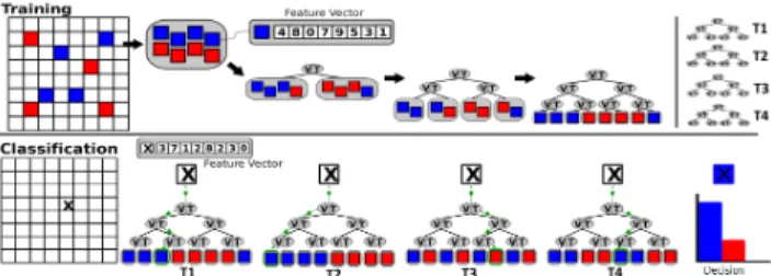
Fig. 2. Illustration of the Random Forests classifier structure. During the training (top), a random subset of labeled samples is used to build a binary decision tree. For each node, random variables of the sample’s feature vector (top) are evaluated and the pair Variable-Threshold which best splits the subset is associated to the node. The process is repeated for a large number of trees (top right). During the classification (bottom), the sample to classify is sent to each decision tree and the class of the sample is determined by the combination of the predictions of the trees of the forest.
and robust identification of the rib cage structures. Extensive experiments demonstrate the superior accuracy in classifica-tion performance of our proposed descriptors (in comparison to intensity-based descriptors used in [8]) as well as signif-icant classification performance improvements (up to 25%) on data-sets subject to posture changes and random noise ad-dition. The remainder of the paper is organized as follows: Sec. 2 describes our overall segmentation framework, Sec. 3 our proposed shape descriptors, and Sec. 4 presents our ex-perimental results.
2. RIB CAGE LABELING WITH RANDOM FORESTS
Random Forests (RF) [6] is a classifier that combines multi-ple randomized decision trees. Based on a simmulti-ple algorithm, it yields very fast and accurate supervised classifications es-pecially for large-dimension feature spaces [7]. RF classi-fiers can also be generalized easily to multi-class classifica-tion tasks.
In our work, each input CT volume, representing a human torso (from the pelvis to the neck), was encoded on a voxel grid G of size ∼1500 × 512 × 512 (voxel resolution: 0.75 × 0.97 × 0.97 mm) where each voxel v = (x, y, z) is associated with an intensity value i(v). To speed up both the training and the classification procedures, the set of bone voxels B to label is pre-identified as the set of voxels whose intensity is higher than a given threshold iB(typically, iB= 400HU ):
B = {v ∈ G / i(v) ≥ iB} (1)
Fig. 1 and 4 illustrate the ensemble of bone structures to label on three different CT scans.
2.1. RF training procedure
The input to the RF training procedure is a set of bone voxels manually pre-labelled by an expert. Each bone voxel v ∈ B is associated with a feature vector f (v) ∈ Rn (in our case, a set of n real values) and a class c(v) ∈ {0, 1} (respectively, non-rib voxelsand rib voxels). The feature vector can be com-posed of various measurements and we further discuss some examples in Sec. 3. As illustrated in Fig. 2, the training
procedure builds a set of binary decision trees, called a for-est F = {T0, T1, . . . TNT−1} (where NT is the number of
trees in the forest). Each tree T ∈ F is built independently in the following manner, given a random subset R of B (of size NR). Each node of T is first associated with a set of NV
pairs of Variable-Threshold. The variables are picked ran-domly among the components of the feature space (between 0 and n) and their corresponding thresholds are also picked randomly within the range of values of each feature. Each pair of Variable-Threshold yields a binary classification of R into R0 and R1 that is evaluated by its gain G on the Gini
index: G = G(R) − G(R0) × |R0| |R| − G(R1) × |R1| |R| (2) and where the Gini index G (a standard purity measure in clas-sification) is given by:
G(R) = 2 ×|{v ∈ R / c(v) = 0}| |R| ×
|{v ∈ R / c(v) = 1}| |R| (3) The pair of Variable-Threshold which maximizes G is then stored in the node and the sets R0 and R1 are provided as
inputs to the creation of two children-nodes. The rest of T is constructed recursively with this algorithm until the tree reaches a pre-set maximal depth Dmax or until G(R) = 0.
Finally, each leaf of T is labelled with the class represented in majority by its associated voxels (see Fig. 2).
2.2. RF classification procedure
As illustrated in Fig. 2, the classification of a new voxel v is straightforward and fast. Each decision tree T is traversed by v from its root by routing v to the appropriate child-node if the node’s variable in f (v) is lower or greater than the node’s threshold. The leaf node in which v stops its traversal will then yield a prediction on c(v). Finally, the predictions of all the trees are combined to assign the final value for c(v), via majority voting.
3. ROBUST SHAPE DESCRIPTORS FOR RF CLASSIFICATION
In this section, we present three shape descriptors that we pro-pose to use in the RF classifier for the automated segmenta-tion of the rib cage. In particular, we introduce two new shape descriptors which aim at providing more accurate and robust classifications than intensity-based features.
3.1. Intensity-based features
For our experiments, we evaluated an intensity-based descrip-tor, called baseline descripdescrip-tor, which was introduced in pre-vious work [8] and is computed as follows: For each voxel v ∈ B to train or classify, a set of 60 boxes of random size (with a length between 3 and 21 voxels along each dimen-sion), and centered at random locations within the neighbor-hood of v (within 20 voxels) is generated. A feature vector fbaseline ∈ R60is then computed with the average intensity
Fig. 3. Illustration of the intensity and shape descriptors around a pixel p marked with a cross. From left to right: baseline (intensity-based) descriptor with box neighbor-hoods, axial inertia and rotation invariant shape context descriptors, both with spherical concentric neighborhoods.
the randomization regarding the position and size of the boxes only occurs once, during the initialization of the RF training. The same box parameters are employed for the classification task.
This baseline descriptor, due to its simplicity, is well suited for the labeling of homogeneous structures. However, in the context of rib identification, as illustrated in Fig. 1, the average intensity of voxels exhibits some heterogeneity along the rib, and the intensity of the surrounding soft tis-sues is quite homogeneous, which injects some ambiguity in the classifier. In addition, although, in theory, average intensities should be robust to noise, this baseline descriptor is not rotation invariant, which prevents it from yielding ac-curate classification in case of posture change. We propose to overcome these difficulties with our new shape features introduced in the following sub-sections.
3.2. Axial-inertia descriptors
In order to circumvent the ambiguities related to homogeneity measures within and around the ribs, we focus our new fea-tures on the description of the local geometry of the bone vox-els B. In particular, we propose to evaluate the local curva-ture of the set of bone voxels in small neighborhoods around the voxels of interest. Given a voxel p, a neighborhood re-gion Spis defined within a sphere of radius r and centered in
p. We then compute on the set of neighborhood bone voxels Bp = {v ∈ BT Sp}, its barycenter bpand its inertia matrix
Ip, defined as: Ip= A −F −E −F B −D −E −D C (4) with: A = X v∈Bp (y(v) − y(b))2+ (z(v) − z(b))2 B = X v∈Bp (x(v) − x(b))2+ (z(v) − z(b))2 C = X v∈Bp (x(v) − x(b))2+ (y(v) − y(b))2 D = X v∈Bp (y(v) − y(b)) × (z(v) − z(b)) E = X v∈Bp (x(v) − x(b)) × (z(v) − z(b)) F = X v∈Bp (y(v) − y(b)) × (x(v) − x(b))
where (x(v), y(v), z(v)) are the XYZ coordinates of a voxel v.
The eigen vectors of Ip correspond to the principal
di-rections of the bone voxels SB, and the corresponding eigen
values quantify the moments of inertia of the bone voxels Bp
along these principal directions (i.e. their deviation from the direction of the corresponding eigen vector). Let λmaxp and λmin
p be the maximal and minimal eigen values of Ip The
proposed feature vector, which characterizes the axial iner-tia, is denoted fAI(p) ∈ R42, and composed of the pairs
(λmax p ,
λmax p
λmin
p ) for 21 spheres with a radius r progressively
increasing from 10 to 30 voxels. The greater λmaxp , the more
aligned the bone voxels are. As illustrated in Fig. 3, this de-scriptor measures the deviation of the bone voxels from their local principal direction, within neighborhood of increasing scales. Note that, by design, this descriptor is rotation invari-ant.
3.3. Rotation invariant shape context (RISC) descriptors The second proposed descriptor is inspired from the shape-context descriptor [10], which is popular in computer vision. To obtain a rotation-invariant version of this descriptor (and avoid registration ambiguities), As illustrated in Fig. 3, we compute, for a given voxel p, a set of 56 bands between spheres centered in p and of radius values randomly sampled between 2 and 35 voxels. Then for each band, the num-ber of bone voxels is calculated, yielding a feature vector fRISC(p) ∈ R56. This descriptor is rotation-invariant and
depicts the local volume occupied by bone voxels in spherical bands of increasing radius around the voxel of interest.
4. EXPERIMENTS AND RESULTS
We experimented RF classification exploiting each of the three descriptors, for the task of rib labeling on thoracic CT scans. For each descriptor, we trained the RF classifier on a set of 437,000 randomly chosen bone samples (45% rib bones, 55% non-rib bones) (taken from one thoracic CT scan of an average size old male (above 60 years old)) and tested it with 3,938 randomly chosen bone samples (48% rib bones, 52% non-rib bones) from a different subject with similar size and age. We set the parameters of the RF to the following values: NV = 7, NT = 500, Dmax = 10 and NR is a
random value (between 10 and |B|). The training procedure, for each descriptor, on a desktop computer with an i7 CPU (2.93GHz) and 8Gb of RAM, with an implementation com-bining MatLab and C++ codes (using OpenCV), took about 3 hours while the entire classification task took on average 0.5 seconds. A visual illustration of the labeling quality is provided in Fig. 4 for one thorax.
Table 1 reports the classification performance with each descriptor. The best results were obtained for our RISC de-scriptor, improving by 4% the baseline performance.
Table 1. Performance of RF classification for rib bone extraction us-ing three different descriptors: baseline, axial inertia (AI) and the rotation-invariant shape-context (RISC).
Baseline AI RISC Correct classification 92.12% 90.32% 96.19% False positives 0.45% 7.54% 0.83%
Table 2. Performance of RF classification for rib bone extraction on the same CT scan as in Table 1, but rotated by 20◦.
Baseline AI RISC Correct classification 81.28% 87.22% 97.33% False positives 3.02% 11.7% 1.01%
Table 3. Performance of RF classification for rib bone extraction on the same CT scan as in Table 1, corrupted with additive random noise.
Baseline AI RISC 1% noise Correct classification 91.92% 87.51% 96.92% False positives 2.18% 10.99% 0.81% 2% noise Correct classification 72.09% 76.38% 97.48% False positives 24.42% 22.65% 0.68%
As discussed in the introduction, homogenous acquisition settings cannot be assumed when processing large databases of CT-images. In particular, subjects can have different pos-tures and CT scanners can generate images with varying noise characteristics that can be hard to predict beforehand and not represented during training. Addressing such issues with pre-smoothing of the images might be highly unreliable, based on our experience. Classification performance was further tested on the same CT scan thorax and points to label as in Table 1 but after some modification: (1) rotation of the volume by 20◦ around the direction orthogonal to the coronal plane to simu-late posture change, with results in Table 2. We observed that both AI and RISC descriptors provided significant classifica-tion improvements (up to 16%) over the baseline descriptor performance; (2) addition of a white Gaussian random noise (which amplitude was set as a fraction of the whole inten-sity range, at 1% and 2%), with results in Table 3. In that case, the RISC descriptor outperformed the baseline and AI descriptors by up to 25%. Note that various combinations of these three descriptors were also experimented but with no significant improvement over the RISC descriptor alone. La-beling of a full thoracic CT scan using such combination is illustrated in Fig. 4
5. DISCUSSION AND CONCLUSION
In this paper, we presented a new approach for the automatic segmentation of rib bones in CT-images which leverages the expertise of users through a learning procedure. We intro-duced two shape descriptors to be used in conjunction with a Random Forests classifier. Experiments demonstrated the su-periority of rotation-invariant shape context descriptors over
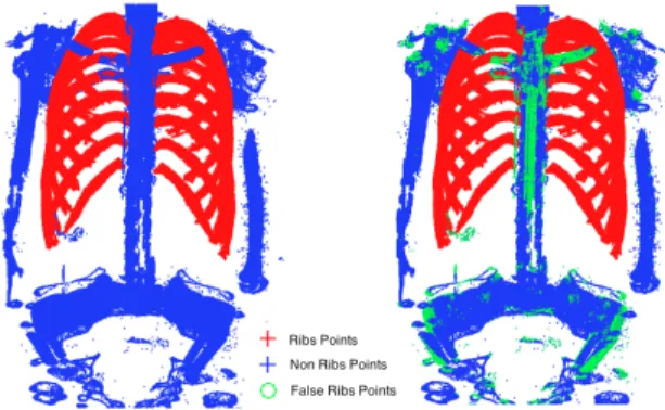
Fig. 4. Rib bone labeling on a full thoracic CT scan: (left) manual expert segmen-tation, (right) RF classification combining baseline and RISC descriptors. (Blue: true non-rib points, Red: true rib points, Green: false rib points).
nominal intensity-based descriptors, especially under varia-tions of subject’s posture and noise level (with improvements over existing descriptors of 4%, 16% and 25% respectively). Such descriptors seem therefore well suited for the automated classification of rib bones on heterogeneous databases of CT images, without requiring pre-registration nor smoothing.
In future work, we will extend our descriptors for multi-class RF multi-classification to individually segment each rib bone, and to distinguish between cortical and spongious bone com-ponents.
6. REFERENCES
[1] X.M. Pardo, M.J. Carreira, A. Mosquera, and D. Cabello, “A snake for CT image segmentation integrating region and edge information,” Image and Vision Computing, vol. 19, pp. 461–475, 2001.
[2] Y. Kang, K. Engelke, and W.A. Kalender, “A new accurate and pre-cise 3-D segmentation method for skeletal structures in volumetric CT data,” IEEE Transactions on Medical Imaging, vol. 22, pp. 586–598, 2003.
[3] M. Krcah, G. Szekely, and R. Blanc, “Fully automatic and fast segmen-tation of the femur bone from 3D-CT images with no shape prior,” in IEEE ISBI, 2011, pp. 2087–2090.
[4] T. Klinder, M.E. Ostermann, A. Franz, R. Kneser, and C. Lorenz, “Au-tomated model-based vertebra detection, identification and segmenta-tion in CT images,” Medical Image Analysis, vol. 13, pp. 471–482, 2009.
[5] J. Ma, L. Lu, Y. Zhan, X. Zhou, M. Salganicoff, and A. Krishnan, “Hi-erarchical segmentation and identification of thoracic vertebra using learning-based edge detection and coarse-to-fine deformable model,” in MICCAI, 2010, pp. 19–27.
[6] L. Breiman, “Random forests,” Machine Learning, vol. 45, pp. 5–32, 2001, 10.1023/A:1010933404324.
[7] Rich. Caruana, Nikos. Karampatziakis, and Ainur. Yessenalina, “An empirical evaluation of supervised learning in high dimensions,” Pro-ceedings of the 25th International Conference on Machine Learning (2008), pp. 96–103, 2008.
[8] K. Rajpoot, A. Noble, V. Grau, C. Szmigielski, and H. Becher, “Ran-dom forest classification for automatic delineation of myocardium in real-time 3D echocardiography,” in Functional Imaging and Modeling of the Heart, 2009, pp. 447–456.
[9] A. Criminisi, J. Shotton, D. Robertson, and E. Konukoglu, “Regression forests for efficient anatomy detection and localization in CT studies,” in MICCAI Medical Computer Vision Workshop, 2010, pp. 106–117. [10] G. Mori, S. Belongie, and J. Malik, “Shape contexts enable efficient