Ghada Ghazal1, Jules Gagnon1, Pierre-Étienne Jacques2, Josette-Renée Landry2, François Roberts2,3 and Sherif Abou Elela1*.
1 RNA Group, *Département de microbiologie et d’infectiologie, Faculté de médecine et des sciences de la santé, Université de Sherbrooke, Sherbrooke, Québec, Canada, J1H 5N4. 2 Institut de recherches cliniques de Montréal, 110 Avenue des Pins Ouest, Montréal, Québec, Canada, H2W 1R7.
3 Département de Médecine, Faculté de Médecine, Université de Montréal.
*Corresponding author Sherif Abou Elela, Ph. D.
Département de microbiologie et d’infectiologie, Faculté de médecine et des sciences de la santé, Université de Sherbrooke,
3001 12e Ave Nord,
Sherbrooke, Québec, Canada J1H 5N4
Tel: (819) 564-5275 Fax: (819) 564-5392
E-mail address: Sherif.Abou.Elela@USherbrooke.ca
Character count: 45 735
Running title: Rnt1p dependent transcription termination Subject category: Posttranscriptional Regulation
Summary
Transcription termination of messenger RNA (mRNA) is normally achieved by polyadenylation followed by Rat1p dependent 5’-3’ exoribonuleolytic degradation of the downstream transcript. Here we show that the yeast orthologue of the dsRNA-specific ribonuclease III (Rnt1p) may trigger Rat1p dependent termination of RNA transcripts that fail to terminate near polyadenylation signals. Rnt1p cleavage sites were found downstream of several genes and the deletion of RNT1 resulted in transcription read-through. Inactivation of Rat1p impaired Rnt1p dependent termination and resulted in the accumulation of 3’ end cleavage products. These results support a new model for transcription termination in which co-transcriptional cleavage by Rnt1p provides access for exoribonucleases in the absence of polyadenylation signals.
Introduction
Transcription termination plays an important role in determining the fate and function of the RNA. For example, formation of polyadenylated RNA could signal protein translation, while aberrant termination may trigger RNA degradation (Zhao et al., 1999). There are currently two models for transcription termination in eukaryotes; the first is called the “torpedo” model, which is the predominant mode of termination in protein coding genes (Luo et al., 2006; Tollervey, 2004), and the other is the “allosteric” model, which appears to be favoured in genes producing short non-coding RNA (Kim et al., 2006; Vasiljeva et al., 2008). In the case of “torpedo” termination the polyadenylation signal that is often found near the end of protein coding genes triggers an endonucleolytic RNA cleavage generating an entry site for the 5’-3’ exoribonuclease Rat1p that in turn destabilizes the RNAP II elongation complex (Kim et al., 2004). On the other hand, the “allosteric” mode of termination does not require cleavage or the presence of a polyadenylation signal but depends on the binding of a termination complex in close proximity to the promoter (Carroll et al., 2004; Vasiljeva et al., 2008).
In yeast, mutations that change the 3’ end sequence of mRNAs or inactivate the exoribonuclease Rat1p result in transcription read-through that often terminates before the promoter of the downstream genes (Kim et al., 2004; Luo et al., 2006). Indeed, the intergenic regions in yeast are normally littered with non-canonical polyadenylation sites that become active upon the disruption of the primary site of transcription termination (Grec et al., 2000; Milligan et al., 2005). Therefore, simple defects in a canonical termination site will not automatically lead to the production of polycistronic RNA transcripts. Consistently, most of the confirmed RNAP II-transcribed polycistronic transcripts in yeast are processed by the yeast dsRNA specific RNase III (Rnt1p) and does not require polyadenylation signals for
termination (Ghazal et al., 2005). Rnt1p cleaves specific RNA stems terminating with NGNN or AAGU tetraloops (Ghazal and Elela, 2006) found near pre-rRNA (Abou Elela et al., 1996; Kufel et al., 1999), snRNAs (Abou Elela and Ares, 1998), snoRNAs (Ghazal et al., 2005) or occasionally within mRNA coding sequence (Ge et al., 2005; Larose et al., 2007). Recently, Rnt1p was also shown to promote termination of RNAP I by giving access to Rat1p in a mechanism analogous to that of the polyadenylation-dependent “torpedo” mode of termination (El Hage et al., 2008; Kawauchi et al., 2008). However, the impact of Rnt1p on RNAP II transcription remains unexplored.
Since Rnt1p processes clusters of RNAP II transcribed snoRNAs in yeast (Ghazal et al., 2005) we explored the possibility that the enzyme also influences the expression of neighbouring protein coding genes. Accordingly, we have searched for clusters of open reading frames (ORF) separated by a canonical Rnt1p cleavage signal and identified several conserved regions with the potential to produce dicistronic transcripts. One of these transcripts was expressed in vivo in an Rnt1p dependent manner. In depth mutational analysis and chromatin immunoprecipitation of this transcription unit indicated, that Rnt1p is required for terminating mRNA transcripts that fail to terminate near polyadenylation signals. Genome-wide search for Rnt1p dependent transcription termination sites identified additional genes that require Rnt1p for alternative transcription termination. Together, the results presented here reveal a new mechanism for gene regulation in which Rnt1p triggers mRNA degradation by inducing polyadenylation independent “torpedo” like transcription termination.
Results
Rnt1p represses the expression of dicistronic mRNA in yeast
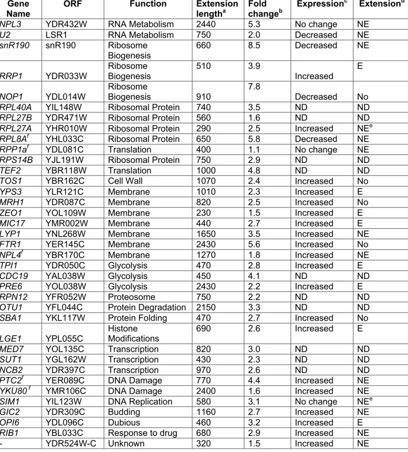
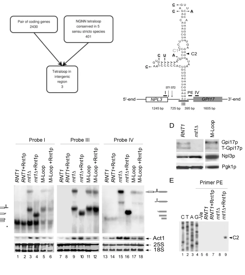
Rnt1p cleaves RNA stems terminating with NGNN tetraloops to initiate the processing of polycistronic snoRNA (Ghazal et al., 2005). Accordingly, we reasoned that the presence of Rnt1p cleavage signal within a cluster of genes could be indicative of dicistronic mRNA expression. To examine this possibility, we searched a group of 5 sensu stricto Saccharomyces species (Herrero, 2005; Liti et al., 2006) for the presence of conserved NGNN stem-loops located between gene-pairs transcribed in the same direction (Figure 1A). Three conserved tetraloops were found but only one in a dicistronic transcript containing the sequence of the NPL3 and GPI17 genes was detected in the absence of Rnt1p (Figure 1B). Using gene specific probes, we monitored the expression of NPL3 and GPI17 in the presence (RNT1) or the absence (rnt1∆) of RNT1. As shown in figure 1C, the probe specific to the NPL3 coding sequence (Probe I) detected the mature Npl3 mRNA (Russell and Tollervey, 1995; Russell and Tollervey, 1992) in RNA extracted from wild type cells (Lane 1). In contrast, rnt1∆ RNA (Lane 3) exhibited two additional large RNA species. Hybridizing RNT1 RNA with GPI17 specific probes (Probe IV) highlighted a band (Lane 13) corresponding to the predicted size of the mature GPI17 mRNA (Zhu et al., 2005). Surprisingly, the expression of the mature GPI17 mRNA was reduced in rnt1∆ RNA (Lane 15) and a large transcript migrating with the same speed as that observed with the NPL3 specific probe (Lane 15) was detected. This indicates that the deletion of RNT1 inhibits the expression of GPI17 and leads to the accumulation of a large transcript containing both Npl3 and Gpi17 sequences. This was further confirmed by a probe (Probe III) hybridizing to the intergenic region (Lane 9).
Consistently, western blot using antibodies against Npl3p and Gpi17p (Figure 1D) revealed that while RNT1 deletion does not affect Npl3 mRNA translation it inhibits the production of Gpi17p. Since GPI17 is essential (Zhu et al., 2005), we presume that a small amount of proteins, below the detection level of the western blot, is expressed in rnt1∆ cells. In any case, the results clearly show that Rnt1p is required for the normal expression of Gpi17p.
Direct cleavage of the extended Npl3-Gpi17 mRNA by Rnt1p was confirmed in vitro using recombinant Rnt1p. Total RNA extracted from RNT1 or rnt1∆ cells was incubated with recombinant Rnt1p and the impact on Npl3 and Gpi17 mRNA was monitored by Northern blotting (Figure 1C). As expected, Rnt1p did not affect the mature Npl3 (Lanes 2) or Gpi17 (Lanes 14) mRNAs. On the other hand, Rnt1p converted the large extended RNA transcripts observed in rnt1∆ cells to smaller fragments (Lanes 4, 10, and 16) corresponding to the predicted cleavage products (Figure 1B). The exact location of Rnt1p cleavage was determined by reverse transcription using a primer (PE) complementary to the sequence downstream of the loop predicted to bind Rnt1p (Figure 1B). Again, no cleavage product was detected in wild type RNA (Figure 1E lanes 6 and 7), while a band corresponding to a cleavage 16 nucleotides downstream of the conserved NGNN tetraloop (C2) was observed in rnt1∆ RNA in the presence (Lane 9) and not in the absence (Lane 8) of recombinant Rnt1p. To directly determine the impact of Rnt1p cleavage on the expression of NPL3-GPI17, we mutated Rnt1p cleavage signal and monitored the effect on mRNA synthesis. Six point-mutations were introduced in the two stem-loop structures (Ghazal and Elela, 2006) predicted to be cleaved by Rnt1p (Figure 1B) and the impact was monitored by Northern blot. A large Npl3- Gpi17 transcript similar to that detected in rnt1∆ RNA (Lanes, 3, 9 and 15) was observed in cells harbouring the stem-loop mutations (M-Loop) (Lanes 5, 11, and 17). The extended RNA produced from the gene carrying mutations in the loops was not cleaved by
recombinant Rnt1p in vitro confirming that the cleavage site was indeed disrupted (Lanes 6, 12, and 18). However, the disruption of the cleavage signal reduced the expression of GPI17 to a lesser extend than RNT1 deletion (Figure 1C). Indeed, western blot analysis indicated that the stem loop mutation does not inhibit the expression of Gpi17p (Figure 1D). Therefore, while direct cleavage by Rnt1p is required for inhibiting the accumulation of the Npl3-Gpi17 transcript the presence of Rnt1p itself may play additional role in modulating the expression of GPI17.
NPL3 termination-sequence induces the accumulation of Rnt1p dependent
read-through transcripts
To determine the elements regulating the expression of GPI17, we deleted the predicted promoter regions of either the NPL3 or GPI17 gene (Figure 2A) and monitored the impact on RNA expression. The deletions were achieved by inserting a URA3 gene fused to ADH1 termination sequence (Akada et al., 2006; Noble and Johnson, 2005) upstream of the translation start codon of the chromosomal copies of either NPL3 or GPI17. Deletion of the NPL3 promoter (npl3pr∆) blocked the expression of Npl3 mRNA (Figure 3B) in both the presence (Lane 4) and the absence of RNT1 (Lane 5). The deletion of the NPL3 promoter increased the expression of Gpi17 mRNA in rnt1∆ cells (Lane 14) but not in RNT1 cells (Lane 13). Notably, deletion of the NPL3 promoter abolished the expression of the read-through Npl3-Gpi17 RNA even in the absence of RNT1. This indicates that the NPL3 promoter is required for the expression of the Npl3-Gpi17 RNA but not for the synthesis of normal Gpi17 mRNA. In addition, these results indicate that Rnt1p is not required for transcription initiated from the GPI17 promoter. Changes in the GPI17 expression upon the deletion of Rnt1p and NPL3 promoter may stem from the general effect of RNT1 deletion on stress and membrane
related proteins (Ge et al., 2005; Lee et al., 2005; Tremblay, 2002). Deletion of the GPI17 promoter (gpi17pr∆) abolished the expression of Gpi17 mRNA (Lane 17) without affecting the expression of Npl3 mRNA in wild type cells (Lane 8). Replacement of GPI17 promoter by URA3 gene reduced the length of the Npl3 read-through transcripts accumulating in rnt1∆ cells (Lane 9). These new extended transcripts did not hybridize to probes specific to GPI17 (Lane 18) indicating that NPL3 transcription terminated upstream of the URA3 gene (Figure 2A). We conclude that transcription read-through of NPL3 is dependent at least in part on the sequence near the gene 3’ end.
The transcriptional read-through of NPL3 may be influenced by the promoter or termination sequence. To differentiate between these two possibilities, we first replaced the NPL3 promoter with that of ACT1 and monitored the impact on RNA expression (Figure 2A and B). As expected, transcripts produced from the ACT1 promoter (PACT1-NPL3) were slightly larger
than those driven from the endogenous NPL3 promoter due to changes in the transcription start site, irrespective of RNT1 expression (Lanes 6 and 7). On the other hand, PACT1-NPL3
did not significantly change the expression level of either mature Npl3 (Lane 6) or Gpi17 mRNA (Lane 15) when expressed in wild type cells. Changing NPL3 promoter in rnt1∆ cells reduced transcriptional read-through (Lane 7 and 16) and permitted the expression of mature Gpi17 mRNA (Lane 16). This suggests that either ACT1 promoter enhances termination near the canonical NPL3 termination site or that the changes in the site of transcription initiation influence the termination efficiency.
To evaluate the impact of NPL3 termination on the accumulation of the extended Npl3-Gpi17 transcripts, we replaced the sequence between the translation stop codon of NPL3 and the polyadenylation signal with the ADH1 termination signal (Figure 2A). The mutations were
introduced either in RNT1 cells or in cells carrying mutations in Rnt1p cleavage signals. As shown in figure 2C, the introduction of the ADH1 terminator abolished the expression of read-through transcripts regardless of Rnt1p cleavage (Lanes 4, 5, 9 and 10). This demonstrates that the production of extended Npl3-Gpi17 RNA is largely due to the leaky termination of the NPL3 gene. Therefore, Rnt1p seems to function as a fail-safe terminator of NPL3.
Rat1p is required for transcription termination downstream of Rnt1p cleavage site
To determine the fate of the RNA cleaved by Rnt1p, we examined the impact of known exoribonucleases on the expression of NPL3 and GPI17 transcripts. As shown in figure 3, deletion of the nuclear 3’-5’ exoribonuclease Rrp6p induced the expression of 3’ extended Npl3 RNA that migrates with the same speed as Rnt1p cleavage products (lanes 4, 13, 22, and 31). This suggests that Rnt1p 5’ end cleavage products are degraded by Rrp6p under normal growth conditions. Surprisingly, inactivation of a temperature-sensitive allele of the 5’-3’ nuclear exonuclease Rat1p (rat1-1) reduced the expression of both mature Npl3 and Gpi17 mRNAs (Lanes 6, 15, 24 and 33). The general reduction in mRNA could be explained by poor termination and subsequent degradation of the aberrant RNA since Rat1p was previously shown to be required for the termination of polyadenylated mRNA (Kim et al., 2004). Indeed, Rat1p may affect the 3’ end formation by influencing the recruitment of 3’ end processing factor and the choice of polyadenylation site (Luo et al., 2006). At permissive temperature the deletion of the cytoplasmic 5’-3’ exoribonuclease XRN1 in rat1-1 cells resulted in little perturbation of the Npl3 mRNA, while causing an accumulation of a 5’ end-extended Gpi17 RNA species consistent with the 3’ end cleavage product of Rnt1p (Lanes 7, 16, 25, and 34). At restrictive temperature, the rat1-1 xrn1∆ and rat1-1 cells exhibited the same profile of NPL3 expression (Lanes 6, 8, 15, and 17). However, rat1-1 xrn1∆ RNA exhibited a new transcript
corresponding to the size of the stem-loop structure cleaved by Rnt1p (Lane 26). In addition, other bands corresponding to Rnt1p 3’ end cleavage products containing the GPI17 sequence were detected (Lane 35). Deletion of the nonsense mediated decay exoribonuclease Upf1p had little effect on RNA expression (Lanes 9, 18, 27, 36). This result indicates that Rnt1p cleavage leads to the degradation of the 3’ end cleavage product by Rat1p in the nucleus or, surviving this, by Xrn1p in the cytoplasm. We conclude that Rnt1p cleavage generates an entry site for the 5’-3’ exoribonuclease Rat1p, which causes “torpedo” like transcription termination.
To link Rnt1p directly to transcription termination, the pattern of RNAP II association with the NPL3 and GPI17 genes was examined by chromatin immunoprecipitation (ChIP) using antibodies against the RPB1 subunit (Malagon et al., 2006) in the presence or the absence of Rnt1p. As expected, in RNT1 cells (Figure 3B) the RNAP II co-immunoprecipitated with DNA fragments corresponding to the promoter (A) and coding sequence of NPL3 (B and C) but not with known untranscribed regions of chromosome V (Ctl). DNA corresponding to the intergenic region between NPL3 and GPI17 (D, E and F, white columns) co-immunoprecipitated as or more efficiently than the DNA corresponding to the coding region or the sequence downstream of GPI17 (G, H and I, white columns). Strikingly, the deletion of RNT1 significantly increased the association of RNAP II with the intergenic region between NPL3 and GPI17 (D, E, and F, grey columns) and the GPI17 ORF (G, H and I, grey columns). This suggests that Rnt1p is required for the efficient termination of NPL3.
In order to understand how Rnt1p influences transcription termination, we immunoprecipitated Rnt1p and monitored its association with the actively transcribed NPL3 and GPI17 genes (Figure 3B, black columns). Interestingly, Rnt1p co-precipitated with fragments corresponding
to NPL3 promoter region (A), NPL3 coding sequence (B and C), and the NPL3 transcription termination site (D). Weak associations with the intergenic region (E and F) and the 5’ end (G) of GPI17 were also detected. Rnt1p did not co-precipitate with GPI17 3’ end fragments (H and I). The strongest association was found between Rnt1p and fragment immediately adjacent to NPL3 3’ end. Inactivation of a temperature sensitive allele of RNAP II also inhibited the association of Rnt1p with all DNA fragments (data not shown). These data indicate that Rnt1p associates with actively transcribed DNA and is required for transcription termination downstream of NPL3 3’ end.
RNT1 deletion perturbs transcription termination of several RNAP II transcribed genes
To examine the possibility that Rnt1p mediated the transcription termination of genes other than NPL3 we initiated a new search in silico looking for all conserved Rnt1p cleavage signals downstream of known polyadenylation sites. As indicated in Table 1, five stem-loop structures other than that near NPL3 3’ end were found with score above 0.8, which was previously established as reasonable cut-off (Ghazal et al., 2005). Three of the newly identified genes were either not expressed under vegetative growth or the identified Rnt1p stem-loop overlapped with the sequence of downstream tRNA (data not shown). Interestingly two additional genes known to code for RNA binding proteins (NAB2 and RPL8A) were identified. Northern blot analysis of the mRNA produced by these two genes indicated that RNT1 deletion causes transcriptional read-through downstream of the canonical termination site (Table 1 and Figure 4A and B). In addition, deletion of RNT1 increased the amount of the RNA generated from the NAB2 (Figure 4A lanes 1 and 2) and RPL8A genes (Figure 4B lane 1 and 2). This increase in expression could be due either to an increase in transcription rate as evident in the case of RPL8A (Figure 4D), increased RNA stability in the absence of RNT1
or combination of both factors. Deletion of the exoribonuclease XRN1 and the inactivation of the RAT1 resulted in the accumulation of an extended Rat1 RNA and fragments corresponding to the sequence downstream of Rnt1p cleavage site located near the 3’ end of Rpl8A and Nab2 (Figure 4A and B lane 9). Chromatin immunoprecipitation indicated that Rnt1p interact with the transcriptional units of NAB2 and RPL8A and confirmed transcription read-through of these genes upon the deletion of RNT1 (Figure 4C and D). These data further confirm the role of Rnt1p in suppressing the accumulation of transcription read-through products by generating an entry site for the exoribonculeases Rat1p and Xrn1p.
In order to examine the global impact of Rnt1p on transcription termination we analyzed the overall pattern of RNAP II occupancy in the presence and the absence of RNT1. RNAP II specific chromatin immunoprecipitation was performed as described above from RNT1 or rnt1∆ cells and the extracted DNA hybridized to a DNA microarray containing an average of 4 probes per kilobase across the whole yeast genome. Extended association of RNAP II at the 3’ end of 39 genes that could not be attributed to overlapping or neighbouring genes were identified (Table 2). The length of these extensions varied from 230 to 2440 nts with an average extension length of 815 nts. As expected, the transcription read-through near the NPL3 and RPL8A genes was identified. However, the NAB2 gene was not detected because it is located next to a very highly transcribed gene that generates an RNAP II signal overlapping with the intergenic region located downstream from the NAB2 gene. The accuracy of the systematic analysis of RNAP II read-through was tested by quantitative PCR on 10 genes identified by ChIP-chip. In all cases, the increased association of RNAP II was confirmed by quantitative PCR (data not shown). Interestingly, the ChIP-chip approach identified a transcriptional read-through in two non-coding RNA (U2 snRNA (Abou Elela and Ares, 1998) and snR190 (Chanfreau et al., 1998)) that were shown to be processed by Rnt1p.
In both cases the deletion of RNT1 leads to the transcription of extended RNA species (Abou Elela and Ares, 1998; Chanfreau et al., 1998). To link the RNAP II profile to RNA expression we examined the RNA transcripts produced by 29 genes displaying transcriptional read-through by ChIP-chip. Northern blot analysis indicated that 24 of those 29 genes indeed produce a larger RNA species in rnt1∆ cells (Table 2). Interestingly, most of these genes (18/24) were also overexpressed in the mutant cells. In about a third of the cases (8/24) some transcriptional read-through could be detected in wild type cells, suggesting that some RNAP II molecules can escape from Rnt1p surveillance. Only four of the genes we have tested were downregulated in rnt1∆ cells, while three showed no difference in expression. Most of these genes (6/7), however, produced extended transcript in rnt1∆ cells, suggesting that the termination function of Rnt1p is not linked to its role in the regulation of transcription level. As shown in figure 5, three of Rnt1p dependent transcriptional read-through resulted in the accumulation of discitronic transcripts that include the sequence of two neighbouring genes while, in other cases, the extension terminated in the intergenic region. Together these data suggest that Rnt1p impact on transcription is not limited to NPL3-GPI17 cluster and may extend to genes with different functions.
Discussion
In this study, we have shown that Rnt1p cleavage signal may function as polyadenylation independent fail-safe terminator. Deletion of the dsRNA specific ribonuclease Rnt1p induced the expression of a long read-through transcript containing the sequence of the NPL3 and GPI17 genes (Figure 1). In vitro, recombinant Rnt1p cleaved the extended RNA species at the predicted NGNN stem-loop structure in the absence of any other factors (Figure 1). In vivo Rnt1p was found associated with actively transcribed NPL3 and its deletion resulted in transcriptional read-through interfering with the transcription of the downstream gene coding for Gpi17p (Figure 3). Rnt1p dependent transcriptional read-through was also detected in several genes with a variety of functions indicating that the impact of Rnt1p on transcription is not limited to a single gene (Table 2). Together the results reveal a new mode of gene regulation were polyadenylation independent transcription termination triggers degradation of nascent RNA transcripts.
Npl3p is an RNA-binding protein implicated in the export of mRNA and it functions as an antagonist of transcription termination (Burkard and Butler, 2000; Krebber et al., 1999; Lund et al., 2008). In addition, it was recently proposed that phosphorylated Npl3p inhibits efficient recognition of the canonical polyadenylation signal of its own transcript (Lund et al., 2008). However, the mechanism by which Npl3p influences mRNA processing remained unclear. Npl3p binds its actively transcribed gene and overexpressing Npl3p causes transcription read-through that is normally inhibited by Rnt1p (data not shown). The endonucleolytic cleavage prevents read-through and preserves the transcriptional activity of the downstream genes. Indeed, the biological advantage conferred by this mechanism is evident from the conservation of the Rnt1p cleavage signal in five closely related Saccharomyces species
(Figure 1A). This phenomenon is not an isolated event since we have found other RNA binding proteins (Table 2 and Figure 4) that could benefit from Rnt1p triggered termination. For examples, Nab2p (Roth et al., 2005) and Rpl8ap (Cusick, 1994) are known to bind RNA and Nab2 was shown to be autoregulated via the induction of transcription read-through (Roth et al., 2005).
The two current models of RNAP II transcription termination (torpedo and allosteric) do not explain how the transcription of long non-polyadenylated RNA terminates. The “torpedo” model requires cleavage near the polyadenylation signals and the “allosteric” model functions only with short non-coding RNA (Kim et al., 2006; Luo et al., 2006). We propose a modified “torpedo” mode of termination (Figure 6) in which Rnt1p circumvent the need for polyadenylation signals by generating an entry site for the Rat1p exonuclease leading to termination of transcription. In this model, polyadenylation is not required and therefore long RNA can be produced without being obligatorily transported and translated. Recruitment of Rnt1p to the termination site is likely to be signalled by phosphorylation dependent interaction of the RNA II CTD. Interestingly, Rnt1p was shown to interact with the RNAP II CTD in a two hybrid system when the phosphorylation site required for either polyadenylation dependent termination (Serine 2) or that required for Nrd1p complex dependent non-polyadenylated RNA termination (Serine 5) is mutated (Ghazal and Abou Elela unpublished data). This suggests that indeed Rnt1p represent transcription termination alternative in situations where neither conventional “torpedo” nor “allosteric” modes of termination are possible (e. g. long non-polyadenylated RNA). Indeed, it was recently shown mechanistically in a model system that Rnt1p elicit RNAP II termination by a torpedo mechanism (see Rondon et al., this issue). Rnt1p cleaves the 3’ end of the non-polyadenylated U2 snRNA and in the absence of Rnt1p a longer polyadenylated transcript is produced (Abou Elela and Ares, 1998). Indeed, deletion of
Rnt1p leads to transcriptional read-through in the U2 gene (Table 2). This mode of transcription termination is not unique to RNAP II. Rnt1p cleavage at the 25S pre-rRNA gives access to Rat1p allowing it to terminate transcription by RNAP I in a “torpedo” like fashion (El Hage et al., 2008).
The impact of Rnt1p on transcription is not limited to transcription termination. In many cases we have observed an overall increase in RNAP II occupancy associated with an increase in gene expression in the absence of RNT1 (Table 2). The effect of Rnt1p on the level of transcription and termination are not necessarily linked. In the case of NPL3, the disruption of Rnt1p cleavage site lead to transcriptional read-through but the level of expression is lower than that observed upon the deletion of RNT1 (Figure 1C). On the other hand, increased gene expression in rnt1∆ do not necessarily lead to transcription read-through (Larose et al., 2007). Indeed, genome-wide analysis of gene expression in the absence of Rnt1p identified many RNA transcripts that are over-expressed upon the deletion of RNT1 and the vast majority did not exhibit changes in the site of transcription termination (Ge et al., 2005). Therefore, in certain cases the recruitment of Rnt1p to the active transcription complex may directly modulate transcription independent of the cleavage at the 3’ end of the nascent RNA.
Discovering that Rnt1p cleavage induces Rat1p dependent transcription termination mandates re-examination of Rnt1p function in RNA processing. It is currently accepted that Rnt1p processes most non-coding RNA in yeast including pre-rRNA, snRNA and snoRNA (Abou Elela and Ares, 1998; Abou Elela et al., 1996; Ghazal et al., 2005). However, it is not clear why this processing step is necessary and why in certain cases the lack of this processing leads to the generation of polyadenylated RNA (Abou Elela and Ares, 1998). The results presented here suggest that in many cases Rnt1p cleavage is not introduced as an
obligatory processing step but rather as a transcription terminator required in order to avoid the polyadenylation of aberrant RNA. This interpretation is also compatible with the role of Rnt1p in mRNA degradation (Ge et al., 2005; Larose et al., 2007). In this case, Rnt1p will not simply degrade the newly transcribed mRNA but will also terminate transcription. This indeed explains why an enzyme localized in the nucleus plays a role in the regulation of mostly cytoplasmic RNA species like mRNA. However, the discovery that Rnt1p cleavage elicits transcription termination raises questions about the mechanism of polycistronic snoRNA processing. In this scenario, Rnt1p cleaves between snoRNAs that are transcribed as a single transcript leading to the maturation of these different RNAs (Ghazal et al., 2005). Therefore, if Rnt1p cleavage leads to transcription termination, the downstream snoRNA will not be produced. This apparent paradox could be explained by the presence of specific sequence elements or transcription factors that specifically prevent the 5’ end generated by Rnt1p cleavage of these gene clusters from being digested by Rat1p. Case by case studies of Rnt1p cleavage and its link to termination will reveal the existence of these elements. Meanwhile, the data presented here reveal a new model of polyadenylation independent transcription termination and provide a mechanism by which transcription termination may regulate gene expression.
Experimental Procedures Strains and plasmids
Yeast strains were grown and manipulated using standard procedures (Guthrie and Fink, 1991). Yeast strains used in this study are listed in supplemental Table 1. For details, see Supplemental material file 1.
Search for Rnt1p cleavage signals
All uninterrupted pairs of ORFs transcribed in the same orientation were identified in the April 9th 2008 version of Saccharomyces Genome Database (SGD). Independently, all conserved NGNN-capped stem-loops in five sensu stricto Saccharomyces species (S. cerevisiae, S. paradoxus, S. mikatae, S. kudriavzevii and S. bayanus) were identified using the genome multiple alignment from UCSC (http://hgdownload.cse.ucsc.edu/goldenPath/
sacCer1/bigZips/multizYeast.zip). To assess conservation, three criteria where considered: the conserved G in position two of the tetraloop, the capacity of the two closing base pairs to form canonical Watson-Crick base pairs and the formation of 23 nt NGNN-capped stem-loop as predicted by Vienna RNA 1.6.5. When these three criteria were validated at the same position in the alignment for the five species, the stem-loop was considered as conserved. We found that three of these conserved NGNN-capped stem-loops are located in the intergenic region between consecutive coding transcripts.
RNA analysis
RNA extractions, Northern hybridization and primer extension were performed as previously described(Ghazal et al., 2005). Primer extension was performed using (20 ng/µl) of reverse primer CAAATTCTTTGAAATTAGCCTGACCCAAAC, and 10 µg of RNA. The primers used to
generate the randomly labeled probes (Perbal, 1988) used for Northern blots are listed in Supplemental Table 2. Cleavage of total RNA was conducted as previously described (Ghazal et al., 2005) using 50 µg of total RNA and 8 pmol of purified Rnt1p (Lamontagne and Abou Elela, 2001). Standard 1.2 % agarose or 6% polyacrylamide gels were used to separate low and high molecular weight RNAs respectively.
Western Blotting
Total protein extracts and western blot analysis were performed as described before (Catala et al., 2008). Cells were grown in 50 ml culture. Proteins (10-20 µg) were loaded on 12% SDS gel, transferred and incubated with 1:3000 dilution of antibodies against Npl3p (Russell and Tollervey, 1992) and 1:500 Gpi17p (Zhu et al., 2005). Anti-rabbit HRP was used as secondary antibody at a dilution of 1:80 000. Pgk1p was detected using anti-mouse HRP antibody as a secondary at a dilution of 1:16000 (Sigma-Aldrich Canada Ltd., Oakville, Ont).
Chromatin immunoprecipitation and microarray analysis
Chromatin extracts were prepared as described (Strahl-Bolsinger et al., 1997; Taggart et al., 2002). Immunoprecipitations were performed with monoclonal anti-Rpb1 8WG16 (Covance, Berkeley, CA) and polyclonal anti-Rnt1p (Lamontagne et al., 2000) as described earlier (Catala et al., 2008). The method used for quantitative PCR amplification is outlined in supplemental material. ChIP material was labeled and hybridized on DNA microarrays (Agilent Technologies) containing 44,290 Tm-adjusted 60-mer probes covering the entire yeast genome for an average density of one probe every 287 bp (±100 bp) as described before (Rufiange et al., 2007). The data were normalized and replicates were combined using a weighted average method as described previously (Rufiange et al., 2007). The combined datasets are available supplemental file 2. Comparing RNAP II density beyond the 3’ end of ORFs identified genes with termination defects in rnt1∆ cells. In addition, those exhibiting
previously noted changes in expression after the deletion of RNT1 were closely inspected to ensure that no obvious candidates are missed through the automated selection process.
Accession Numbers
The ChIP-chip data in this paper have been deposited in NCBIs Gene Expression Omnibus (GEO) (http://www.ncbi.nlm.nih.gov/geo/) and are accessible through GEO series accession number GSExxxx.
Acknowledgements
We thank Momchil Vodenicharov for help with chromatin immunoprecipitation and Julie Parenteau with yeast genetics. We also thank Jean-François Lucier for the design of PCR primers. We are indebted for Mark Schmitt, Julie Parenteau, Mathieu Catala and Bruno Lamontagne for critical reading of the manuscript. This work was supported by a grant from the Canadian Institute of Health Research. S. A. is a Chercheur Boursier National of the Fonds de la Recherche en Santé du Québec. F.R. holds a New Investigator Award from the Canadian Institute of Health Research. P-É.J. holds a post-doctoral award from the IRCM training program in cancer research funded by the CIHR. J.-R.L is a research fellow of the Terry Fox Foundation through an award from the National Cancer Institute of Canada.
References:
Abou Elela, S., and Ares, M., Jr. (1998). Depletion of yeast RNase III blocks correct U2 3' end formation and results in polyadenylated but functional U2 snRNA. EMBO Journal 17, 3738-3746. Abou Elela, S., Igel, H., and Ares, M., Jr. (1996). RNase III cleaves eukaryotic preribosomal RNA at a U3 snoRNP-dependent site. Cell 85, 115-124.
Akada, R., Kitagawa, T., Kaneko, S., Toyonaga, D., Ito, S., Kakihara, Y., Hoshida, H., Morimura, S., Kondo, A., and Kida, K. (2006). PCR-mediated seamless gene deletion and marker recycling in Saccharomyces cerevisiae. Yeast 23, 399-405.
Blancafort, P., Ferbeyre, G., Sariol, C., and Cedergren, R. (1997). PolI-driven integrative expression vectors for yeast. J Biotechnol 56, 41-47.
Burkard, K.T., and Butler, J.S. (2000). A nuclear 3'-5' exonuclease involved in mRNA degradation interacts with Poly(A) polymerase and the hnRNA protein Npl3p. Mol Cell Biol 20, 604-616.
Carroll, K.L., Pradhan, D.A., Granek, J.A., Clarke, N.D., and Corden, J.L. (2004). Identification of cis elements directing termination of yeast nonpolyadenylated snoRNA transcripts. Mol Cell Biol 24, 6241-6252.
Catala, M., Tremblay, M., Samson, E., Conconi, A., and Abou Elela, S. (2008). Deletion of Rnt1p alters the proportion of open versus closed rRNA gene repeats in yeast. Mol Cell Biol 28, 619-629. Chanfreau, G., Rotondo, G., Legrain, P., and Jacquier, A. (1998). Processing of a dicistronic small nucleolar RNA precursor by the RNA endonuclease Rnt1. Embo J 17, 3726-3737.
Chinnusamy, V., Gong, Z., and Zhu, J.K. (2008). Nuclear RNA export and its importance in abiotic stress responses of plants. Curr Top Microbiol Immunol 326, 235-255.
Cusick, M.E. (1994). Purification and identification of two major single-stranded binding proteins of yeast Saccharomyces cerevisiae as ribosomal protein L4 and histone H2B. Biochim Biophys Acta 1217, 31-40.
El Hage, A., Koper, M., Kufel, J., and Tollervey, D. (2008). Efficient termination of transcription by RNA polymerase I requires the 5' exonuclease Rat1 in yeast. Genes Dev 22, 1069-1081.
Ge, D., Lamontagne, B., and Abou Elela, S. (2005). RNase III-Mediated Silencing of a Glucose-Dependent Repressor in Yeast. Curr Biol 15, 140-145.
Ghazal, G., and Elela, S.A. (2006). Characterization of the reactivity determinants of a novel hairpin substrate of yeast RNase III. J. Mol. Biol. 363, 332-344.
Ghazal, G., Ge, D., Gervais-Bird, J., Gagnon, J., and Abou Elela, S. (2005). Genome-wide prediction and analysis of yeast RNase III-dependent snoRNA processing signals. Mol Cell Biol 25, 2981-2994. Grec, S., Wang, Y., Le Guen, L., Negrouk, V., and Boutry, M. (2000). Cryptic polyadenylation sites within the coding sequence of three yeast genes expressed in tobacco. Gene 242, 87-95.
Guthrie, C., and Fink, G.R. (1991). Guide to Yeast Genetics and Molecular Biology (San Diego, CA: Academic Press).
Herrero, E. (2005). Evolutionary relationships between Saccharomyces cerevisiae and other fungal species as determined from genome comparisons. Rev Iberoam Micol 22, 217-222.
Kawauchi, J., Mischo, H., Braglia, P., Rondon, A., and Proudfoot, N.J. (2008). Budding yeast RNA polymerases I and II employ parallel mechanisms of transcriptional termination. Genes Dev 22, 1082-1092.
Kim, M., Krogan, N.J., Vasiljeva, L., Rando, O.J., Nedea, E., Greenblatt, J.F., and Buratowski, S. (2004). The yeast Rat1 exonuclease promotes transcription termination by RNA polymerase II. Nature 432, 517-522.
Kim, M., Vasiljeva, L., Rando, O.J., Zhelkovsky, A., Moore, C., and Buratowski, S. (2006). Distinct pathways for snoRNA and mRNA termination. Mol Cell 24, 723-734.
Krebber, H., Taura, T., Lee, M.S., and Silver, P.A. (1999). Uncoupling of the hnRNP Npl3p from mRNAs during the stress-induced block in mRNA export. Genes Dev 13, 1994-2004.
Kufel, J., Dichtl, B., and Tollervey, D. (1999). Yeast Rnt1p is required for cleavage of the pre-ribosomal RNA in the 3' ETS but not the 5' ETS. RNA 5, 909-917.
Lamontagne, B., and Abou Elela, S. (2001). Purification and characterization of Saccharomyces cerevisiae Rnt1p nuclease, Vol 342 (San diego, CA: Academic Press).
Lamontagne, B., Tremblay, A., and Abou Elela, S. (2000). The N-terminal domain that distinguishes yeast from bacterial RNase III contains a dimerization signal required for efficient double-stranded RNA cleavage. Mol Cell Biol 20, 1104-1115.
Larose, S., Laterreur, N., Ghazal, G., Gagnon, J., Wellinger, R.J., and Elela, S.A. (2007). RNase III-dependent regulation of yeast telomerase. J Biol Chem 282, 4373-4381.
Lee, A., Henras, A.K., and Chanfreau, G. (2005). Multiple RNA surveillance pathways limit aberrant expression of iron uptake mRNAs and prevent iron toxicity in S. cerevisiae. Mol Cell 19, 39-51.
Liti, G., Barton, D.B., and Louis, E.J. (2006). Sequence diversity, reproductive isolation and species concepts in Saccharomyces. Genetics 174, 839-850.
Lund, M.K., Kress, T.L., and Guthrie, C. (2008). Autoregulation of Npl3, a yeast SR protein, requires a novel downstream region and serine phosphorylation. Mol Cell Biol 28, 3873-3881.
Luo, W., Johnson, A.W., and Bentley, D.L. (2006). The role of Rat1 in coupling mRNA 3'-end processing to transcription termination: implications for a unified allosteric-torpedo model. Genes Dev 20, 954-965.
Malagon, F., Kireeva, M.L., Shafer, B.K., Lubkowska, L., Kashlev, M., and Strathern, J.N. (2006). Mutations in the Saccharomyces cerevisiae RPB1 gene conferring hypersensitivity to 6-azauracil. Genetics 172, 2201-2209.
Milligan, L., Torchet, C., Allmang, C., Shipman, T., and Tollervey, D. (2005). A nuclear surveillance pathway for mRNAs with defective polyadenylation. Mol Cell Biol 25, 9996-10004.
Noble, S.M., and Johnson, A.D. (2005). Strains and strategies for large-scale gene deletion studies of the diploid human fungal pathogen Candida albicans. Eukaryot Cell 4, 298-309.
Perbal, B. (1988). A Practical Guide to Molecular Cloning, Second edn (New York: John Wiley and Sons, Inc.).
Roth, K.M., Wolf, M.K., Rossi, M., and Butler, J.S. (2005). The nuclear exosome contributes to autogenous control of NAB2 mRNA levels. Mol Cell Biol 25, 1577-1585.
Rufiange, A., Jacques, P.E., Bhat, W., Robert, F., and Nourani, A. (2007). Genome-wide replication-independent histone H3 exchange occurs predominantly at promoters and implicates H3 K56 acetylation and Asf1. Mol Cell 27, 393-405.
Russell, I., and Tollervey, D. (1995). Yeast Nop3p has structural and functional similarities to mammalian pre- mRNA binding proteins. Eur J Cell Biol 66, 293-301.
Russell, I.D., and Tollervey, D. (1992). NOP3 is an essential yeast protein which is required for pre-rRNA processing. J Cell Biol 119, 737-747.
Steinmetz, E.J., Warren, C.L., Kuehner, J.N., Panbehi, B., Ansari, A.Z., and Brow, D.A. (2006). Genome-wide distribution of yeast RNA polymerase II and its control by Sen1 helicase. Mol Cell 24, 735-746.
Strahl-Bolsinger, S., Hecht, A., Luo, K., and Grunstein, M. (1997). SIR2 and SIR4 interactions differ in core and extended telomeric heterochromatin in yeast. Genes Dev. 11, 83-93.
Taggart, A.K., Teng, S.C., and Zakian, V.A. (2002). Est1p as a cell cycle-regulated activator of telomere-bound telomerase. Science 297, 1023-1026.
Tollervey, D. (2004). Molecular biology: termination by torpedo. Nature 432, 456-457.
Tremblay, A. (2002). Etude de la fonction de la RNase III eucaryote et identification de ses partenaires cellulaires dans un criblage double-hybrides. In Department of Microbiology (Sherbrooke, Université de Sherbrooke).
Vasiljeva, L., Kim, M., Mutschler, H., Buratowski, S., and Meinhart, A. (2008). The Nrd1-Nab3-Sen1 termination complex interacts with the Ser5-phosphorylated RNA polymerase II C-terminal domain. Nat Struct Mol Biol 15, 795-804.
Zhao, J., Hyman, L., and Moore, C. (1999). Formation of mRNA 3' ends in eukaryotes: mechanism, regulation, and interrelationships with other steps in mRNA synthesis. Microbiol Mol Biol Rev 63, 405-445.
Zhu, Y., Fraering, P., Vionnet, C., and Conzelmann, A. (2005). Gpi17p does not stably interact with other subunits of glycosylphosphatidylinositol transamidase in Saccharomyces cerevisiae. Biochim Biophys Acta 1735, 79-88.
Table 1. List of Rnt1p cleavage sites near sites of transcription termination
Gene Function Loop Scorea Effect of RNT1 Deletionb RPL8A LSU r-protein 0.961 3’ end extension
NPL3 Poly A binding protein 0.955 3’ end extension
LOH1 Unknown 0.920 NE
NAB2 Poly A binding protein 0.878 3’ end extension FAB1 Membrane kinase 0.876 NE
BNI5 Septin organization 0.850 NE
a Loop score was determined as described earlier (Ghazal et al., 2005). The higher the score the more likely that it is cleaved by Rnt1p. b NE indicates no evidence to support 3’ end extension.
Table 2: Genome-wide screen for RNT1 dependent transcription termination Gene
Name ORF Function Extension lengtha Fold changeb Expression
c Extensiond
NPL3 YDR432W RNA Metabolism 2440 5.3 No change NE
U2 LSR1 RNA Metabolism 750 2.0 Decreased NE
snR190 snR190 Ribosome Biogenesis 660 8.5 Decreased NE RRP1 YDR033W Ribosome Biogenesis 510 3.9 Increased E NOP1 YDL014W Ribosome Biogenesis 910 7.8 Decreased No
RPL40A YIL148W Ribosomal Protein 740 3.5 ND ND
RPL27B YDR471W Ribosomal Protein 560 1.6 ND ND
RPL27A YHR010W Ribosomal Protein 290 2.5 Increased NEe RPL8Af YHL033C Ribosomal Protein 650 5.8 Decreased NE
RPP1af YDL081C Translation 400 1.1 No change NE
RPS14B YJL191W Ribosomal Protein 750 2.9 ND ND
TEF2 YBR118W Translation 1000 4.8 ND ND
TOS1 YBR162C Cell Wall 1070 2.4 Increased No
YPS3 YLR121C Membrane 1010 2.3 Increased E
MRH1 YDR087C Membrane 820 2.5 Increased No
ZEO1 YOL109W Membrane 230 1.5 Increased E
MIC17 YMR002W Membrane 440 2.7 Increased E
LYP1 YNL268W Membrane 1650 3.5 Increased NE
FTR1 YER145C Membrane 2430 5.6 Increased No
NPL4f YBR170C Membrane 1270 1.8 Increased NE
TPI1 YDR050C Glycolysis 470 2.8 Increased E
CDC19 YAL038W Glycolysis 450 4.1 ND ND
PRE6 YOL038W Glycolysis 2430 2.2 Increased E
RPN12 YFR052W Proteosome 750 2.2 ND ND
OTU1 YFL044C Protein Degradation 2150 3.3 ND ND
SBA1 YKL117W Protein Folding 470 2.7 Increased No LGE1 YPL055C
Histone
Modifications 690 2.6 Increased E
MED7 YOL135C Transcription 820 3.0 ND ND
SUT1 YGL162W Transcription 430 2.3 ND ND
NCB2 YDR397C Transcription 970 2.6 ND ND
PTC2f YER089C DNA Damage 770 4.4 Increased NE
YKU80 f YMR106C DNA Damage 2400 1.6 Increased NE SIM1 YIL123W DNA Replication 580 3.1 No change NEe
GIC2 YDR309C Budding 1160 2.7 Increased NE
OPI6 YDL096C Dubious 460 3.2 Increased E
RIB1 YBL033C Response to drug 680 2.9 Increased NE
a The length of the extension (in nucleotides) predicted by the RNAP II ChIP-chip upon the deletion of RNT1. b The rnt1∆ / RNT1 fold change of RNAP II occupancy in the identified extension adjusted by the difference of RNAP on the complete gene. c-d Variation in the level of expression of mature RNA fragment c or size d as detected by Northern blot (ND, not determined; E, RNA extension detected in wild type strains that is increased in the absence of RNT1; NE, new extension detected only in the absence of RNT1 and; No, no extension). e Extension due to intron retention. f Manually selected genes. The changes in the expression of U2 and snR190 in the absence of RNT1 were previously reported (Abou Elela and Ares, 1998; Chanfreau et al., 1998).
Figure legends
Figure 1. Identification of Rnt1p dependent dicistronic RNA. (A) In silico search of conserved gene clusters separated by NGNN stem-loop structures using Saccharomyces cerevisiae Genome Database (SGD) annotations. (B) Illustration of NPL3-GPI17 gene cluster showing the size of each gene fragment at bottom. Positions of Northern blot probes (I and IV) and reverse transcription primers are shown on top. C1 and C2 indicate the position of the predicted cleavage sites. Black arrowhead indicates cleavage observed in vitro using total RNA. Grey arrowhead indicates cleavage observed with a 5’ end labeled model substrate in vitro (data not shown). Point mutations disrupting Rnt1p cleavage site are shown in bold. ST1 and ST2 indicate the position of previously reported polyadenylation sites near the NPL3 3’ end (Chinnusamy et al., 2008; Steinmetz et al., 2006). (C) Rnt1p is required for the cleavage of the extended Npl3-Gpi17 RNA in vivo and in vitro. RNA was extracted from wild type (RNT1), rnt1∆, and from cells carrying mutations in Rnt1p cleavage site (M-Loop) and incubated either alone or in the presence of recombinant Rnt1p. Schematics of the different RNA transcripts are indicated beside each gel. Open and grey boxes represent NPL3 and GPI7 ORFs respectively. (D) Western blot analysis of the Npl3p and Gpi17p. Proteins were extracted from RNT1, rnt1∆ and M-Loop cells separated on 12% SDS gel and visualized using antibodies specific to Gpi17p, Npl3p or the control Pgk1p. Note that Gpi17p exists in two forms, a full-length membrane-bound version (Gpi17p) and a truncated free form (T-Gpi17p) (Zhu et al., 2005). (E) Rnt1p cleaves Npl3-Gpi17 extended RNA in vitro. Reverse transcription using a primer downstream of the predicted cleavage site was performed using RNA extracted from wild type or rnt1∆ cells incubated with or without recombinant Rnt1p. Sequencing of DNA corresponding to the same region is indicated on the left as a marker. The position of the cleavage (C2) is indicated on the right.
Figure 2. Identification of cis-acting elements controlling the expression of the Npl3-Gpi17 RNA. (A) Schematics representation of the different mutations introduced in the promoter and termination regions of NPL3 and GPI17. The promoter of NPL3 was either replaced by ACT1 promoter (ACT1P) or deleted by inserting a URA3 gene linked to a strong ADH1 terminator (URA3-AT). The 3’ end sequence containing the two reported NPL3 polyadenylation signals (Steinmetz et al., 2006) was replaced by a strong ADH1 terminator (ADH1T) (Blancafort et al., 1997). The promoter region of GPI17 was replaced by URA3-AT. All replacements and deletions were carried in the chromosomal copies of the genes and the names of the resulting yeast strains are shown on top. (B) NPL3 promoter is not required for the production of mature Gpi17 mRNA. Northern blot analysis was performed using RNA extracted from cells carrying the different mutations and visualized by probes specific to either NPL3 (I) or Gpi17 (IV) mRNA. The stained rRNA is shown as a loading control. (C) The NPL3 3’ end is required for transcriptional read-through. Northern blot analysis was performed using RNA extracted from the different mutations as described in B.
Figure 3. Rnt1p cleavage triggers transcription termination. (A) Degradation of Rnt1p cleavage product by 5’-3’ exoribonuclease is required for the expression of GPI17. RNA was extracted from cells lacking the 3’-5’ nuclear exoribonuclease Rrp6p (rrp6∆), cells expressing a temperature sensitive allele of the 5’-3’ nuclear exoribonuclease RAT1 grown at permissive (rat1-1 26°C) or restrictive conditions (rat1-1 37°C), rat1-1 cells lacking 5’-3’ cytoplasmic exoribonuclease XRN1 grown at the permissive (rat1-1 xrn1∆ 26°C) or restrictive (rat1-1 xrn1∆ 37°C) temperature, and cells lacking the non-sense mediated decay ribonuclease
Upf1p (upf1∆). The different RNAs were visualized by probes complementary to different regions of the NPL3-GPI17 cluster. The rRNA is included as loading control. (B) Rnt1p enhances the transcription termination of NPL3. Chromatin Immunoprecipitations were performed using antibodies against the RNAP II protein subunit Rpb1p in the presence (white column) or the absence (grey column) of RNT1. The association of Rnt1p with transcribed genes was examined using antibodies against Rnt1p (black column). The precipitated DNA was amplified by real-time PCR using primers specific to different regions within the NPL3-GPI17 clusters (indicated on the top). A total of two biological and three technical replicates were used to calculate the relative levels of DNA precipitated and the average values are indicated. A primer-pair amplifying a known untranscribed region of chromosome V was used as negative control (Ctl). Standard deviations between replicate experiments was ± 0.05.
Figure 4. Rnt1p cleavage signal identifies sites of alternative transcription termination in genes coding for RNA binding proteins. Extended Nab2 (A) and Rpl8A (B) RNA are cleaved by Rnt1p in vivo and in vitro. RNA was extracted from RNT1, rnt1∆, rrp6∆, xrn1∆ cells or cells expressing a temperature sensitive allele of RAT1 grown at permissive (rat1-1 26°C) or restrictive conditions (rat1-1 37°C) or rat1-1 cells lacking 5’-3’ cytoplasmic exoribonuclease XRN1 grown at the permissive (rat1-1 xrn1∆ 26°C) or restrictive (rat1-1 xrn1∆ 37°C) temperature. In vitro cleavage assay of RNA extracted from rnt1∆ cells was carried by incubation with recombinant Rnt1p (rnt1∆ + Rnt1p). RNAs were visualized by probes complementary to either a sequence near NAB2 3’ end (V) or unique sequence that is found in the 3’UTR of RPL8A and not the RPL8B isoform (VI). The rRNA is included as loading control. The asterisk indicates 3’ end RNA degradation products that were observed occasionally. The 3’ end cleavage project of NAB2 generated by Rnt1p in vitro was too faint
to detect in the exposure shown. NAB2 (C) and RPL8A (D) Chromatin immunoprecipitations were performed using antibodies against the RNAP II protein subunit Rpb1p in the presence (white column) or absence (grey column) of RNT1 or using antibodies against Rnt1p (Black column) in wild type cells as described in Figure 3B. Immunoprecipitations of RPL8A chromatin was performed either in wild type strain (data not shown) or strains lacking the RPL8B to avoid cross-amplification of homologous sequence (D). The data were obtained and calculated as mentioned in Figure 3B.
Figure 5. RNAP II chromatin immunoprecipitation in the absence of RNT1 identifies genes with alternative transcription termination. Northern blot analysis was carried out as described in Figure 1. The probe position relative to the gene structure is shown on top. The nature of the different transcripts is schematically represented on the side. The asterisk indicate cross hybridization with rRNA. In all cases extensions were not detected in cells expressing RNT1 even after prolonged exposure.
Figure 6. Model describing the impact of Rnt1p on transcription termination and mRNA stability. Under normal conditions, (RNT1) transcription of RNA binding proteins (RBP) genes like NPL3, NAB2, or RPL8A is autoregulated. When the amount of RBPs is low (On condition), transcription terminates at the canonical site via Rat1p dependent “torpedo” mechanism leading to the production of mature RNA and protein synthesis. When the RBPs accumulate in the cell (Off condition), they bind near the termination site of their gene leading to transcription read-through up to Rnt1p cleavage signal downstream. Cotranscriptional cleavage by Rnt1p gives access to Rat1p leading to “torpedo” like termination. However, in this case the resulting RNA is rapidly degraded by the exoribonuclease Rrp6p, Rat1p and Xrn1p. In the absence of RNT1, transcription continues to the polyadenylation signal of the
downstream gene or until it meats a cryptic polyadenylation site in the intergenic region. In both cases, the polyadenylated RNA is transported and translated disrupting the auto-regulatory circuit of the RBP.
Figure 1 Ghazal et al., 2009 Npl3p Gpi17p Pgk1p T-Gpi17p C2
A
B
C
D
G-C A-U G-C A-U U-A A-U U.G C-G U-A U-A C-G U-A C-G U-A U.G G-C U.G U.G A-U U-A U-A C-G U-A A-U U-A A-U A G U U C A NPL3 GPI17 1245 bp 725 bp 395 bp 1605 bp 5'-end 3'-end C G C A A A C A U A U U C G A A A GU C2 C1 ST2 ST1 I IV III C C A C C U A Act1 25S 18S 1 2 3 4 5 6 7 8 9 10 11 12 13 14 15 16 17 18 PEFigure 2 Probe I Probe IV Probe I Probe IV 18S 25S URA3-AT
A
B
C
NPL3 GPI17 1245 bp 725 bp 395 bp 1605 bp 5'-end 3'-end I IV npl3pr∆ URA3-AT gpi17pr∆ ADH1T ST2 TADH1-NPL3 18S 25S ACTP PACT1-Npl3 1 2 3 4 5 6 7 8 9 10 11 12 13 14 15 16 17 18 * 1 2 3 4 5 6 7 8 9 10 ST1Probe I Probe II Probe III NPL3 GPI17 Probe IV 18S 25S 1 2 3 4 5 6 7 8 9 10 11 1213 1415161718 19 20 21 22 23 24 25 26 27 28 29 30 31 3233 3435 36 Figure 3 A B C D E F G H I NPL3 GPI17 0.05 0.10 0.20 0.25 0.30 0.35 0.40 0.15 0.00 Ctl A B C D E F G H I Position of Amplified Fragments
A B C D NAB2 RPL8A 18S 25S RPL8A C A B D E F A B C D E NAB2 0.00 0.05 0.10 0.20 0.25 0.30 0.15 Ctl A B C D E F Position of Amplified Fragments
1 2 3 4 5 6 7 8
VI
9
1 2 3 4 5 6 7 8 9
Ctl A B C D E Position of Amplified Fragments
0.00 0.05 0.10 0.20 0.25 0.30 0.15 V Figure 4 25S 18S * *
*
SSE2 NPL4 VIII VII DOT6 PTC2 X IX
*
*
XIII RPP1a RIB1 PGM2 YKU80 XII XI XIV Figure 5 Ghazal et al., 2009A
B
C
D
E
1 2 3 4 1 2 3 4 1 2 3 4 1 2 1 2 25S 18S 25S 18S 25S 18S 25S 18SP RBP Poly A PII Figure 6 Ghazal et al., 2009 Canonical termination RBP RBP P RBP RBP PII Rnt1p Rbp mRNA AAAA
Translation Rat1p Rat1p
Rrp6p
Polyadenylation independent termination
RNA decay Feed back P RBP Poly A PII Canonical termination RBP RBP P RBP RBP PII Rbp mRNA AAAA Translation Rat1p Feed back
Random termination or pausing
AAAA
rnt1∆
On Condition Off Condition
RBP Translation