Université de Montréal
A Systematic In Vitro Metabolism Study of Opioids using Rat Liver S9
Fractions and Mass Spectrometry Revealed CYP2D Metabolism is
Impaired with age
par
SABRIN FUAD SALMIN
Département de biomédecine vétérinaire
Faculté de médecine vétérinaire
Mémoire présenté en vue de l'obtention du grade de maître ès sciences (M.Sc.) en
sciences vétérinaires option pharmacologie
Juillet 2016
i
Résumé
La codéine et l'oxycodone sont des opioïdes utilisés pour soulager la douleur. Le succès du traitement dépend du métabolisme par les enzymes CYP450. Selon les médicaments utilisés, l’altération de métabolisme peut entraîner de graves conséquences, y compris des modifications dans leurs profils d'efficacité, de sécurité et de toxicité. L'objectif de cette étude consistait à évaluer par HPLC-MS / MS (trappe ionique linéaire) l'effet de l'âge sur le métabolisme de l'enzyme CYP2D chez les rats. La codéine et l'oxycodone ainsi que leurs métabolites spécifiques, morphine (CYP2D), norcodeine (CYP3A), oxymorphone (CYP2D) et noroxycodone (CYP3A), ont été quantifiés en utilisant la méthode de dilution isotopique. Les fractions S9 de foie de rats (Sprague-Dawley) de sexe masculin âgés de 3, 6, 12 et 18 mois ont été préparées et les paramètres de Michaelis-Menten ont été déterminés pour les principales voies métaboliques. La vitesse maximale de la réaction enzymatique (Vmax) a montré une saturation rapide des sites actifs de CYP2D dans la fraction s9 de foie chez les rats âgés de 18 mois. Les valeurs de Vmax observées pour la codéine chez les rats âgés de 3, 6 et 12 mois étaient respectivement de 0,327 ± 0,027, 0,355 ± 0,021 et 0,273 ± 0,028. Concernant l'oxycodone, les valeurs étaient de 0,690 ± 0,096, 0,499 ± 0,087, 0,153 ± 0,01 respectivement. Cependant, la valeur de Vmax chez les rats âgés de 18 mois était de 0,092 ± 0,003 pour la codéine et de 0,153 ± 0,01 pour l’oxycodone. Vraisemblablement, la valeur de Vmax affecte de manière significative (P ≤ 0,05) la clairance enzymatique de ces médicaments (CLuint = Vmax / Km). Les résultats de cette étude suggèrent que l’enzyme CYP2D subit un changement conformationnel avec l'âge, particulièrement chez les rats âgés de 18 mois. Ce changement conformationnel conduit à une diminution significative du taux de formation des métabolites opioïdes actifs, ce qui entraine une diminution de la clairance enzymatique de ces médicaments. En conclusion, cette étude suggère une détérioration des CYP2D et CYP3A chez les rats vieillissants, ce qui affecte le métabolisme des médicaments.
Mots-clés: Opioïdes, vieillissement, métabolisme des médicaments, CYP450, CYP2D, CYP3A, spectrométrie de masse.
ii
Abstract
Codeine and oxycodone are opioids used in the treatment of pain. The outcome of the treatment is ultimately related to their metabolism by CYP450 enzymes. Depending on the drugs used, alterations in the metabolism of drugs by CYP450 enzymes can lead to severe consequences including alterations in their efficacy, safety and toxicity profiles. The objective of this study was to assess the effect of age on CYP2D enzyme metabolism in rats using HPLC-MS/MS (Linear Ion Trap). Codeine and oxycodone along with specific metabolites morphine (CYP2D), norcodeine (CYP3A), and oxymorphone (CYP2D), noroxycodone (CYP3A) were quantified using isotopic dilution method. Liver S9 fractions from 3, 6, 12, and 18 month-old male Sprague Dawley rats were prepared and Michaelis-Menten parameters were determined for their primary metabolic pathways. The derived maximum enzyme velocity (Vmax) suggested a rapid saturation
of the CYP2D active sites in the liver S9 fractions of 18 month-old rats. The observed Vmax
values for codeine in 3, 6 and 12 month-old rats respectively were 0.327 ± 0.027, 0.355 ± 0.021 and 0.273 ± 0.028 and for oxycodone, the values were 0.690 ± 0.096, 0.499 ± 0.087, 0.153 ± 0.01. For 18 month-old rats, Vmax value was 0.092 ± 0.003 for codeine compared to 0.153 ± 0.01
for oxycodone. The Vmax value affected significantly (P ≤ 0.05) the enzyme-mediated clearance
of these drugs (CLuint = Vmax / Km). The results of this study suggest that CYP2D enzyme
undergoes a conformational change with age, particularly in 18 month-old rats considered to be geriatric in age, thus leading to a significant decrease in the rate of the formation of active opioid metabolites and a decrease in enzymatic clearance of these drugs. This study suggests that there is an impairment of CYP2D and CYP3A in aging rats, which affects their drug metabolism.
iii
Table of index
Résumé ... i Abstract ... ii Table of index ... iii Tables List ... v List of figures ... vi Abbreviations list ... viii Acknowledgment ... x Introduction ... 1 I LITERATURE REVIEW ... 3 I.1 Historical View of Opioids ... 3 I.2 Opioids ... 5 I.3 Opioid receptors and their mechanism of action ... 5 I.3.1 Endogenous opioids ... 11 I.4 Pharmacological drugs targeting opioids receptors ... 14 I.4.1 Morphine ... 14 I.4.2 Codeine ... 14 I.4.3 Oxycodone ... 15 I.4.4 Naloxone ... 15 I.4.5 Naltrexone ... 16 I.4.6 Tramadol ... 16 I.4.7 Fentanyl ... 17 I.5 Codeine ... 18 I.5.1 Physico-chemical properties of codeine ... 18 I.5.2 Metabolism of codeine ... 18 I.5.3 Effects of codeine: ... 20 I.5.4 Excretion ... 20 I.5.5 Pharmacokinetics ... 21 I.5.6 Pharmacogenetics ... 22 I.5.7 Effect of age ... 23 I.6 Oxycodone ... 26 I.6.1 Physico-chemical properties of oxycodone ... 26 I.6.2 Metabolism of oxycodone ... 26 I.6.3 Effects of oxycodone ... 28 I.6.4 Oxycodone excretion ... 29 I.6.5 Oxycodone pharmacokinetics ... 29 I.6.6 Effect of age ... 31 I.7 Physiological and pharmacokinetic effects of aging ... 32 I.7.1 Absorption ... 34 I.7.2 Distribution ... 34 I.7.3 Metabolism ... 35 I.7.4 Elimination ... 36 I.8 Age-related changes on pharmacodynamics of drugs ... 36 I.9 Effects on polymorphism and aging ... 37 I.10 In vitro metabolism models ... 38 I.10.1 Hepatocytes ... 39iv I.10.2 Liver slices ... 39 I.10.3 Perfused liver ... 39 I.10.4 Cytosol ... 40 I.10.5 Cell lines ... 40 I.10.6 Transgenic cell lines ... 40 I.10.7 Microsomes ... 40 I.10.8 Liver S9 fractions ... 41 I.10.9 Supersomes ... 41 I.11 Substrate depletion model ... 42 I.12 Animal Cytochrome P450 ... 44 I.13 Common probe substrates ... 46 II Hypothesis and objectives ... 48 III Article ... 49 IV Discussion ... 78 V Conclusion ... 84 VI Bibliographic References ... 86 VII Annex ... xi
v
Tables List
Table 1. Summary of the affinity of endogenous opioids to specific receptors.………..13
Table 2: Summary of opioid receptors, their tissue location and the response to their activation resulting from the binding of endogenous opioids onto the receptors………..13
Table 3: Affinity of some of the commonly used opioids along with their metabolic pathway and the effect produced……….17
Table 4: Codeine and some of its effects on important organs in the body………..20
Table 5: Pharmacokinetic data of codeine collected from different publications show a wide variability in the values reported………...……21
Table 6: Recommended dosing of codeine by CYP2D6 phenotypes………...……22
Table 7: Oxycodone and some of its effects on important organs in the body….………....28
Table 8: Pharmacokinetics of oxycodone in humans...30
Table 9: Recommended dosing of oxycodone by CYP2D6 phenotypes………...……...32
Table 10: CYP enzymes of the major drug-metabolizing CYP family in humans, rat, mouse, dog and monkey. The isoforms in red represent the rat analogs to the human isoforms……….45
vi
List of figures
Figure 1: Sites of action of opioid analgesics. The grey pathway shows the sites of action on the pain transmission pathway from periphery to central nervous system. The red pathway shows the actions on pain modulating neurons in the mid brain and medulla. 1- Opioids can act directly on peripheral tissue. 2- Inhibition of pain by opioids can be done at the spinal cord. 3- Some opioids act directly on the brain to relieve pain………...……….7
Figure 2: Activation cycle of G-proteins by G-protein-coupled receptors. 1-An agonist binds to the G-coupled protein receptor. 2- Agonist activates the G-coupled protein receptor. 3- Conformational change 4- Due to the conformational change the attachment of guanosine triphosphate (GTP) to the α-subunit takes place 5- α-subunit with GTP dissociates from the β-γ stable dimer. Both the α-subunit and β-γ stable dimer modulate several cellular signaling pathways. 6- Once energy is used the α-subunit with GDP reunites with the β-γ stable dimer until it is activated again………...………..…...8
Figure 3: One mechanism of opioid action. When an opioid binds to an opioid receptor in the membrane of a neuron, calcium channels close, blocking positively charged calcium ions from entering the cell. In addition, cAMP levels decrease and potassium channels open, allowing positive potassium ions to exit the cell. These events hyperpolarize the cell, increasing the charge difference between the cell’s interior and the extracellular environment and making the neuron less likely to fire an action potential………...………..…...9
Figure 4: Pre- and post-synaptic effects of MOR agonists on synaptic transmission. Opioids specifically depress neurotransmitter release from nociceptive Aδ-fiber and C-fiber via presynaptic inhibition mediated by an inhibition of N-type, and to a lesser extent of P/Q-type voltage-dependent Ca2+-channels. Opioids regulate neuronal excitability and transduce receptor activation to downstream signal transduction pathways via the post synaptic inhibition……….10
vii
Figure 6: Drug-metabolizing enzymes involved in the metabolism of codeine. Codeine is bioactivated by CYP2D6 to morphine and demethylated by CYP3A4 to norcodeine, its inactive
metabolite………...………19
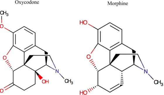
Figure 7: The chemical structures of oxycodone and morphine………...………26 Figure 8: Drug-metabolizing enzymes involved in the metabolism of oxycodone. The major metabolic pathway of oxycodone is the formation of noroxycodone by CYP3A4 enzymes. Noroxycodone is further metabolized to noroxymorphone by CYP2D6 enzymes. The minor metabolic pathways of oxycodone result in formation of oxymorphone by CYP2D6 enzymes, and 6-ketoreduction of oxycodone to α-/β-oxycodol. Oxymorphone is further metabolized to noroxymorphone by CYP3A4 enzymes……….…………..…27
Figure 9: The content of CYP2D and CYP3A, after bottom-up proteomics, showed no statistical difference between age groups when stastically compared using ANOVA and Tukey’s multiple
viii
Abbreviations list
AUC Area under the curve
cAMP Cyclic adenosine monophosphate
CNS Central nervous system
C-6-G Codeine-6-glucuronide
CL Clearance
Cluint Enzyme-mediated clearance
CYP450 cytochrome P450
DOR δ-opioid receptor
EM Extensive metabolizer
ER Endoplasmic reticulum
FDA Food and drug administration GABA γ-amino butyric acid
GDP Guanosine diphosphate
GIT Gastrointestinal tract
Gly Glycine
GTP Guanosine triphosphate
HPLC High performance liquid chromatography
IM- Intermediate Metabolizer
i.m. Intramuscular
i.v. Intravenous
Km Michaelis-Menten constant
Leu Leucine
KOR k-opioid receptor MOR µ-opioid receptor
MS Mass spectrometry
M-3-G Morphine-3-glucuronide
M-6-G Morphine-6-glucuronide
NADPH Nicotinamide adenine dinucleotide phosphate
ix ORL-1 Opioid-like receptor 1
Phe Phenylalanine
PM Poor metabolizer
Tyr Tyrosine
UDPGA Uridine 5'-diphospho-glucuronic acid
UGT Uridine 5'-diphospho-glucuronyltransferase
UM Ultra-rapid metabolizer
x
Acknowledgment
I like would to thank my supervisor Dr Francis Beaudry for giving me the opportunity to pursue a master degree under his supervision. His patience, understanding and perseverance acted as my guiding light. A heartfelt thank you to him for pointing out my weaknesses and helping me to overcome them.
I extend my initial gratitude to my supervisory committee members Younes Chorfi and a later member Dr Kalidou Ndiaye who helped me in my analytical and research thinking and never failed to advise me for my own betterment. A special thanks to Pascal Vaschon and Marie-Chantal Giroux for the work they did in collecting tissue samples for this project and to the Canadian Association for Laboratory Animal Sciences for their funding.
Finally thank you to Professor Amer Silim and his wife Mrs Carol Jones for they are the reason I was able to come and pursue my masters studies at the Université de Montréal.
Last but not least I am grateful to my parents for allowing me to pursue my post graduate studies in a country so far away from home and to my friends and colleagues for the constant support, help and encouragement.
1
Introduction
The recent advancement in the past decade in the medical field has led to an increase in the average life expectancy. In 2013, a survey showed that 14.1% of the US population are above the age of 65 and this is expected to increase to 21.7% by 2040 (Ortman et al., 2014). Elderly patients suffer from pain due to reasons, which include but are not limited to diseases. To improve the quality of life of these patients, they are prescribed medications for pain relief and life style changes that include eating healthy, losing a small amount of weight and increasing physical activity.
Opioids are considered first-line therapy for pain relief. They are widely used for the management of all types of pain ranging from mild pain such as backache to severe pain such as post-surgical pain or cancer pain. The pharmacokinetics and pharmacodynamics of opioids are known to be altered with aging (Mézière et al., 2013; Woodhouse & Wynne, 1988). Extensive research and literature reviews are available on this subject and some of the widely accepted explanations include decline in lean body mass, decrease in hepatic blood flow and disease conditions. In addition, studies suggest a decreased hepatic metabolism and renal clearance of drugs in elderly patients due to physiological aging or the presence of diseases like renal failure, hepatic failure and cardiovascular complications (Mangoni & Jackson, 2004). The reduction in hepatic clearance with age has not been satisfactorily elucidated and remains to some extent controversial.
A large number of drugs are metabolized in the liver by CYP450 enzymes to active or inactive metabolites. The efficacy of the drugs bioactivated by CYP450 enzymes and the clearance of drugs metabolized by these enzymes are affected by any change in the activity of these enzymes. The isoforms of CYP450: CYP3A4, CYP2D6, CYP2C9, CYP2C19, CYP2C8 and CYP2B6 contribute greatly to opioid metabolism in the body. CYP3A4 and CYP2D6 are the primary metabolizers of a large proportion of opioid drugs. Alteration in the functionality of CYP3A4 and CYP2D6 with age leads to a change in the efficacy, safety, toxicity and side effects of these drugs.
2
Codeine and oxycodone are well-known substrates for CYP2D and CYP3A. Both drugs act on the µ-opioid receptor (MOR) in the CNS to cause analgesia. Codeine is metabolized by CYP2D6 to morphine, to which it owes its analgesic effect, and by CYP3A4 to noroxycodone, an inactive metabolite. Oxycodone is metabolized to oxymorphone, an active metabolite and noroxycodone, an inactive metabolite by CYP2D6 and CYP3A enzymes respectively. In rats, it has been shown that CYP2D6 corresponds to CYP2D1 and CYP3A4 corresponds to CYP3A2 in terms of structure and function (Martigoni et al., 2006). Previous studies have shown that CYP3A is impaired in 18-month old rats leading to a decreased enzymatic clearance of ketamine (Santamaria R. et al. 2015).
In general, drugs present in the pharmaceutical markets have not been sufficiently tested in geriatric and pediatric patients due to ethical considerations and regulatory restrictions. An understanding of factors that influence drug metabolism is important for a safe and effective drug use in the elderly. Enzyme modification can have a significant impact on the formation and elimination of active and inactive metabolites thus resulting in a profound effect on the pharmacological and toxicological outcomes.
The aim of this research project was to study the effect of age in rats on the impairment of drug metabolism by CYP2D enzyme which can be due to a decrease in the metabolic function of the enzyme with age hence leading to a decrease in active metabolite formation and accumulation of parent drug in the body. In this study, rat liver S9 fractions from 3, 6, 12 and 18 month-old rats were used to evaluate the effect of aging on metabolic stability and enzyme-mediated clearance by CYP2D and CYP3A, based on Michaelis-Menten approach for specific metabolic pathways. Codeine and oxycodone were used to characterize functional changes since they are well-defined substrates for CYP2D and CYP3A functionality. HPLC-MS method was used to quantify codeine, norcodeine, morphine, oxycodone, oxymorphone and noroxycodone using isotopic dilution strategy in liver S9 fraction suspension.
3
I LITERATURE REVIEW
I.1 Historical View of Opioids
Around 3400 BC the Sumerians on Mesopotamia were among the first people to have cultivated the poppy plant, Papaver somniferum. They called it Hul Gil, the ‘joy plant’. Eventually the plant spread throughout the ancient world to every civilization in Europe and Asia and it was used to treat pain and many other ailments (Smith, 2009).
The poppy plant was cultivated in the ancient civilizations of Persia, Egypt and Mesopotamia. Archaeological evidence and fossilized poppy seeds suggest that Neanderthal man may have used the opium poppy over thirty thousand years ago. Less controversially, the first known written reference to the poppy appears in a Sumerian text dated around 4,000 BC. Papaver
somniferum has long been popular in Europe. Fossil remains of poppy-seed cake and poppy-pods
have been found in Neolithic Swiss lake-dwellings dating from over 4,000 years ago. Poppy images appear in Egyptian pictography and Roman sculpture and opium could readily be bought on the street-markets of Rome. By the eighth century AD, opium use had spread to Arabia, India and China. The Arabs both used opium and organized its trade.
A significant advance in opium-processing occurred in the sixteenth century. In freebase form, the alkaloids found in opium are significantly less soluble in water than in alcohol. Philippus Aureolus Theophrastus Bombastus von Hohenheim (1490-1541), better known as Paracelsus, claimed: "I possess a secret remedy which I call laudanum and which is superior to all other heroic remedies". He concocted laudanum [literally: "something to be praised"] by extracting opium using brandy, thus producing, in effect, tincture of morphine. Laudanum remained largely unknown until the 1660 when English physician Thomas Sydenham compounded a proprietary opium tincture that he also called laudanum, although it differed substantially from laudanum by Paracelsus. By the nineteenth century, vials of laudanum and raw opium were freely available at any English pharmacy or grocery store.
Until the nineteenth century, the only opioids used medicinally or recreationally took the form of crude opium. Opium is a complex chemical cocktail containing sugars, proteins, fats, water, meconic acid, plant wax, latex, gums, ammonia, sulphuric and lactic acids, and numerous
4
alkaloids, most notably morphine (10%-15%), codeine (1%-3%), noscapine (4%-8%), papaverine (1%-3%), and thebaine (1%-2%). All of the latter, apart from thebaine, are used medicinally as analgesics. The opioid analgesics are of inestimable value because they reduce or stop pain without causing a loss of consciousness. They also relieve coughs, spasms, fevers and diarrhea.
Even thebaine, though without analgesic effect, is of immense pharmaceutical value. This is because it can be used to produce semi-synthetic opioid morphine analogues such as oxycodone (Percodan), dihydromorphenone (Dilaudid), hydrocodone (Vicodin) and etorphine (Immobilon). Classes of morphine analogue include diphenylpropylamines (e.g. methadone), 4-phenylpiperidines (e.g. meperidine), morphinans (e.g. levorphanol) and 6,7-benzomorphans (e.g. metazocine). Although seemingly structurally diverse, all these compounds either possess a piperidine ring or contain the critical part of its ring structure (Williams, 2011).
Morphine was first isolated from opium in 1805 by a German pharmacist, Wilhelm Sertürner. who described it as the Principium somniferum. He named it morphium after Morpheus, the Greek god of dreams. Doctors had long hunted for effective ways to administer drugs without ingesting them. Taken orally, opium is liable to cause unpleasant gastric side effects. The development of the hypodermic syringe in the mid-nineteenth century allowed the injection of pure morphine, after which it started being used for minor surgical procedures, for post operative and chronic pain and as an adjunct to general anesthetics. In 1874, English pharmacist C.R. Alder Wright boiled morphine and acetic acid to produce diacetylmorphine. Diacetylmorphine was then synthesized and marketed commercially by the German pharmaceutical company, Bayer and in 1898, Bayer launched heroin the best-selling drug-brand of all time made from morphine (Rassool, 2009).
Codeine, a less powerful drug that is found in opium but can be synthesized, was first isolated in 1830 in France by Jean-Pierre Robiquet, to replace raw opium for medical purposes. In 1874, chemists trying to find a less addictive form of morphine made heroin. But heroin had twice the potency of morphine, and heroin addiction soon became a serious problem. Methadone was first synthesized in 1937 by German scientists Max Bockmühl and Gustav Ehrhart at the IG Farben company. They were searching for a painkiller that would be easier to use during surgery, with less addiction potential than morphine or heroin. New painkillers that came to the market in the
5
USA with approval from the Food and Drug Administration includes Vicodin in 1984, OxyContin in 1995 and Percocet in 1999. These are all synthetic opiates which mimic the body’s own painkillers.
Opioids are considered to be the standard care for the management of acute and chronic pain, but long-term administration of opioids for the treatment of chronic non-cancerous pain is controversial (Rosenblum et al., 2008). The concerns related to the effectiveness, safety and abuse of opioids has led to two different approaches; the first one is a more restrictive perspective, whereas the second one is a greater willingness to endorse this treatment (Smith, 2009). It was only after the publication of reports on the safety and efficacy of opioids prescribed to small numbers of patients with chronic non-malignant pain and the publication of a seminal article entitled “The Tragedy of Needless Pain” that opioid prescription for chronic non-malignant pain began to be practiced.
I.2 Opioids
Opioids refer to compounds that bind to opioid receptors (OR) and exert their pharmacological or physiological effects on the body. An important effect of opioids is the reduction in the perception of pain. The human body has an innate pain relieving system made of endogenous opioids. Like their endogenous counter parts opioid drugs bind to OR to produce analgesia with some undesirable effects (Holden et al., 2005).
I.3 Opioid receptors and their mechanism of action
OR and their respective genes have been characterized at cellular, molecular and pharmacological levels into four different OR systems µ-opioid receptor (MOR), k-opioid receptor (KOR), δ-opioid receptor (DOR), and opioid receptors like-1 (ORL-1) (Al-Hasani & Bruchas, 2011). OLR-1 is genetically closely associated with the previous three receptors, but displays a pharmacological profile that greatly differs from MOR, KOR and DOR as ORL-1 does not bind opioid ligands (Connor & Christie, 1999; Mollereau & Mouledous, 2000). The MOR, KOR, DOR and ORL-1 receptors are encoded by OPRM1, OPRD1, OPRK1 and OPRL1 genes respectively. The OR genes are highly conserved at the sequence encoding the seven transmembrane fragment but they vary at the carboxyl and amino termini. The difference in
6
affinity to opioid ligands and the distinct signaling pathways of the OR is a result of this variation (Levran et al., 2012).
OR are expressed in the central nervous system and in the peripheral organs such as the heart, intestines, kidneys, lungs, liver and reproductive tracts (Abbadie et al., 2004; Beadles-Bohling & Wiren, 2005; Mansour et al., 1995; Villemagne et al., 2002; Vogt et al., 1995; Wittert et al., 1996; Xia & Haddad, 1991, 2001). They are involved in a large number of physiological processes that include growth, reproduction, respiration, immunological response, physiology of the gastrointestinal tract (GIT) and pain signaling in the central and peripheral nervous system. The expression and distribution of OR vary among different organs and different species (Barry & Zuo, 2005).
OR have been identified on cell bodies in the dorsal root ganglion and on central terminals of primary afferent neurons within the dorsal horn of the spinal cord (Stein et al., 1996). They have also been detected on peripheral nervous system on primary afferent neurons in animals (Coggeshall et al., 1997; Hassan et al., 1993; Li et al., 1996; Mousa et al., 2001; Stein et al., 1990; Wenk & Honda, 1999) and in humans (Stein et al., 1996). The binding characteristics of OR on primary afferent neurons were shown to be very similar to those in the brain by binding experiments (Hassan et al., 1993).
In the pain-modulation descending pathway, OR are expressed in the medulla and the periaqueductal gray area. They are also expressed in the limbic, the midbrain and the cortical structures (Al-Hasani & Bruchas, 2011). The activation of OR at these locations directly inhibits neurons, which in turn inhibits spinal cord pain transmission (Ahlbeck, 2011; McNicol et al., 2003).
7
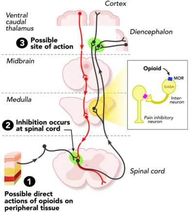
Figure 1: Sites of action of opioid analgesics. The ascending pathway shows the sites of central action on the pain transmission. The descending pathway shows the actions on pain modulating neurons in the mid brain and medulla. 1- Opioids can act directly on peripheral tissue. 2- Inhibition of pain by opioids can takes place at the spinal cord. 3- Some opioids act directly on the brain to relieve pain. Adapted from Al-Hasani & Bruchas, 2011.
OR are G-protein-coupled receptors with seven transmembrane-spanning domains and are present on neuronal cell membranes. G-proteins consist of α, β and γ subunits. The α subunit is coupled with a guanosine diphosphate (GDP). When an agonist binds to an OR, it causes a conformational change leading to the attachment of guanosine triphosphate (GTP) to the α-subunit, which in turn dissociates it from the β-γ stable dimer and the receptor (Fig.2). Both the α-subunit and β-γ stable dimer can modulate several cellular signaling pathways (Childers et al., 1979; Childers & Snyder, 1978; Gether, 2000). These include among others, stimulation or inhibition of adenylate cyclase (Gether, 2000; Minneman & Iversen, 1976), activation of phospholipases and regulation of potassium and calcium channels. This complex along with the β and γ group subsequently act on different intracellular pathways (Childers et al., 1979; Childers & Snyder, 1978) (Fig.2)
8
Figure 2: Activation cycle of G-proteins by G-protein-coupled receptors. 1-An agonist binds to the G-coupled protein receptor. 2- Agonist activates the G-coupled protein receptor. 3- Conformational change takes place. 4- Due to the conformational changes the attachment of GTP to the α-subunit takes place 5- α-subunit with GTP dissociates from the β-γ stable dimer. Both the α-subunit and β-γ stable dimer modulate several cellular signaling pathways. 6- Once energy is used the α-subunit with GDP reunites with the β-γ stable dimer until it is activated again. Adapted from Jähnichen S, 2006.
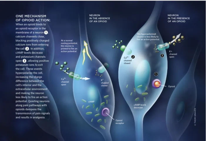
OR have also been shown to promote Ca2+ influx, stimulate phosphoinositide hydrolysis and stimulate adenylyl cyclase (Powell et al., 2002). Figure 3 illustrates one example of the mechanism of opioid action.
9
Figure 3: One example of the mechanism of opioid action. When an opioid binds to an OR in the membrane of a neuron, calcium channels close, blocking positively charged calcium ions from entering the cell. In addition, cAMP levels decrease and potassium channels open, allowing positive potassium ions to exit the cell. These events hyperpolarize the cell, increasing the charge difference between the cell’s interior and the extracellular environment and making the neuron less likely to fire an action potential. Adapted from Grens K, 2011.
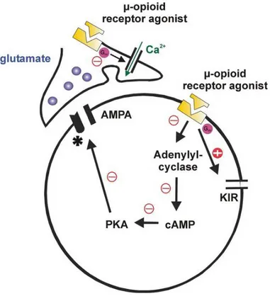
The activation of all three types of OR by an agonist leads to the inhibition of adenylyl cyclase and modulation of membrane Ca2+ and K+ conductance. The increase in the K+ conductance and decrease in Ca2+ conductance by opioids reduces membrane excitability and contributes to the analgesic property of opioids. The inhibition of cAMP by opioids implies a more complex pathway for opioid regulation of cellular mechanism (Gong et al., 1998).
10
Figure 4: Pre- and post-synaptic effects of MOR agonists on synaptic transmission. Opioids specifically depress neurotransmitter release from nociceptive Aδ-fiber and C-fiber via presynaptic inhibition mediated by an inhibition of N-type, and to a lesser extent of P/Q-type voltage-dependent Ca2+-channels. Opioids regulate neuronal excitability and transduce receptor activation to downstream signal transduction pathways via the postsynaptic inhibition.
Some opioids bind presynaptically to MOR on the central terminals of primary afferent nociceptive nerve fibres and others post synaptically on superficial dorsal horn neurons (Fig.4). KORs are located presynaptically on primary afferent neurons in the dorsal horn of the spinal cord where they participate in the inhibition of the release of excitatory neurotransmitter such as substance P, calcitonin gene-related peptide and glutamate. DORs are present on postsynaptic terminals of secondary order neurons and they decrease the excitability caused by the activation of other postsynaptic receptors such as neurokinin-1 receptor and N-methyl-D-aspartate. MORs are located either at presynaptic or postsynaptic terminals, therefore, they can either modulate the release of excitatory neurotransmitters or decrease the excitability of postsynaptic receptors (McDonald & Lambert, 2015). Presynaptic MORs not only inhibit neurotransmitter release of gamma-amino butyric acid (GABA), but they also decrease the inhibition of dopamine pathways causing more dopamine to be released. Exogenous opioids through this mechanism cause inappropriate dopamine release which leads to abnormal synaptic plasticity causing addiction.
11
I.3.1 Endogenous opioids
Endogenous opioids are opioid peptides released by neurons. There are four families of endogenous opioid peptides: endorphins, enkephalins, dynorphins and a recently classified family called endomorphins. All these peptides are derived by the cleavage of larger protein precursors. The precursors prepro-opiomelanocortin, preproenkephalin and preprodynorphin, which are encoded by three corresponding genes code for enkephalins, endorphins and dynorphins respectively (Koneru et al. 2009).
I.3.1.1 Endorphins
Endorphins are endogenous polypeptides that are considered natural painkillers. In vertebrate animals during strenuous exercise, excitement, pain and orgasms, the hypothalamus and pituitary gland produces endorphins, which causes the feeling of well-being and analgesia (Sprouse-Blum
et al., 2010). Endorphins act by binding to MOR, hence causing analgesia and a sense of
euphoria. They also cause the release of many sex hormones. There are four types of endorphins present in the body α-, β-, δ- and γ-endorphins, the most important and powerful endogenous opioid neurotransmitter is the β-endorphins. In the CNS, β-endorphins primarily bind to the presynaptic MOR inhibiting the release of GABA, thus resulting in excess production of dopamine. In the peripheral nervous system, β-endorphins bind to MOR both pre- and post-synaptically and inhibit the release of tachykinin, particularly substance P, a key protein responsible for the transmission of pain (Sprouse-Blum et al., 2010). β-endorphins have the highest affinity for MOR, with medium affinity for DOR and lowest affinity to KOR (Holzer, 2014).
I.3.1.2 Enkephalins
Nociception in the body is partly regulated by pentapeptides called enkephalins. Proenkephalin and prodynorphin are proteolytically cleaved into enkephalin peptides (Orduna & Beaudry, 2016). Enkephalins exist mainly in two forms: met-enkephalin and leu-enkephalin, both of which are found in abundance in the brain and endocrine tissues (Michael Comb et al., 1982). They are also expressed in the gut, where OR agonists interact with pathways of the enteric nervous system that regulate GI motility and secretion (Hughes et al., 1977). They play an
12
important role in the behavior, cardiac function, cellular growth, immunity and ischemic tolerance. Among their functions are pain perceptions, mood and behavior by altering emotional responses as well as acting on cardiovascular and respiratory functions (Przewlocki & Przewlocka, 2001). Enkephalins are distinguished by their C-terminal amino acid sequence, the opioid motif of met-enkephalin consists of Tyr–Gly–Gly–Phe–Met, whereas for leu-enkephalin it consists of Tyr–Gly–Gly–Phe–Leu (M. Comb et al., 1982). Their selectivity for DOR is moderately higher than MOR (Nieto et al., 2005).
I.3.1.3 Dynorphins
Dynorphins are formed by the cleavage of precursor proteins called prodynorphin by protein convertases (Berman et al., 2000; Day et al., 1998). Two types of dynorphins are formed: dynorphin A and dynorphin B. They are a family of endogenous opioid peptides that have potent analgesic effects and have been identified as neuropeptides involved in endogenous pain inhibition (Kuner, 2010; Mika et al., 2011). They are produced in different parts of the brain including midbrain, hippocampus, pons, medulla and spinal cord. They are also responsible for homeostasis, appetite control, pain regulation and body temperature regulation (Koneru et al. 2009). They exert their effects through KOR. The KOR and the endogenous dynorphins are rich in the ventral tegmental area, nucleus accumbens, and prefrontal cortex, brain regions that regulate mood and motivation. Neurochemical and electrophysiological data have shown that KOR activation in these regions decreases dopamine transmission (Shippenberg et al., 2007) that maybe effective in the treatment of depression and drug addiction (Shippenberg, 2009).
I.3.1.4 Endomorphins
Endomorphins have a key role in pain perception, responses related to stress and complex functions such as reward, arousal and vigilance (Fichna et al., 2007). These tetrapeptides differ from the other known endogenous opioid peptides through their amino acid sequence, in which tyrosine residue is followed by glycine. All other endogenous opioids share the Tyr-Gly-Gly-Phe amino acid sequence at the N-terminus. Endomorphins are widely distributed in the CNS and have the highest known affinity and specificity for MOR. There are two types of endomorphins: endomorphin1 and endomorphin2. Both inhibit nociceptive transmission in the spinal cord through MOR (Przewlocki et al., 1999).
13
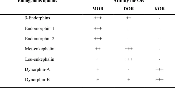
Endogenous opioids Affinity for OR
MOR DOR KOR
β-Endorphins +++ ++ - Endomorphin-1 +++ - - Endomorphin-2 +++ - - Met-enkephalin ++ +++ - Leu-enkephalin + +++ - Dynorphin-A + - +++ Dynorphin-B + + +++
Table 1. Summary of the affinity of endogenous opioids to specific receptors. (Merg et al., 2006; Raynor et al., 1994; Stein et al., 2009)
OR CNS location Response on activation
MOR Brain ( laminae III and IV
of the cortex, thalamus, periqueductal gray), spinal
cord (substantia gelatinosa)
supraspinal analgesia, physical dependence
respiratory depression, miosis, euphoria, reduced GIT motility
DOR Brain (pontine nucleus,
amygdala, olfactory bulbs, deep cortex).
Analgesia may be associated with mood change
KOR Brain (hypothalamus,
peri-aqueductal gray, claustrum), spinal cord
(substantia gelatinosa)
Spinal analgesia, diuresis, dysphoria, sedation, miosis, disrealization, dispersonalization
Table 2. Summary of OR, their tissue location and the response to their activation resulting from the binding of endogenous opioids onto the receptors. Adapted from (Cherny, 1996). OR are also present in the autonomic nervous system, peripheral nerves and GIT, where they can mediate effects on heart rate, nociception and GIT motility.
14
I.4 Pharmacological drugs targeting opioids receptors
I.4.1 Morphine
Morphine is a naturally occurring opiate found in opium plant, Papaver somniferum. It acts on the MOR present in the CNS to relieve pain. It is used to treat moderate to severe pain. Morphine is generally prescribed for inpatients or post-operative patients, it is also given to terminally ill patients. It has high abuse potential. Morphine exerts its effect by binding to MOR, KOR and DOR, but morphine has a higher affinity for MOR. Morphine can be administered orally, intravenously, rectally, subcutaneously, epidurally, by inhalation and by snorting (Martin et al., 2016). The liver metabolizes morphine to morphine-6-glucuronide (M-6-G) and morphine-3-glucuronide (M-3-G) both of which are then excreted through the kidneys. M-6-G is more potent than morphine (Frances et al., 1990) and it is said to have higher affinity for the DOR than the MOR. M-6-G is also suggested to be safer than morphine because it causes lower respiratory depression due to the difference in their affinities towards the OR (Kilpatrick & Smith, 2005). The side effects of morphine include nausea, vomiting, constipation, myosis, sedation, drowsiness, allergy, euphoria, delirium, tolerance and respiratory depression. The adverse effects of morphine on the recovery of locomotor function may be linked to the activation of the KOR (Aceves et al., 2016). Studies performed on knockout mice lacking the MOR, show that mice display no morphine-induced analgesia (Matthes et al., 1996) or respiratory depression (Romberg et al., 2003) with morphine, suggesting that the two effects are closely linked. Morphine is the standard drug against which all other opioid analgesics are compared.
I.4.2 Codeine
Codeine has significantly lower affinity to MOR in comparison to morphine (Knaggs et al., 2004). Codeine is converted to morphine by CYP2D6 in the liver and imparts its analgesic action through morphine (Desmeules et al., 1991; Sindrup et al., 1990). It is also converted by CYP3A4 to norcodeine, which is reported to be an inactive metabolite. It is considered to be the agent of choice for the treatment of mild to moderate pain in both adults and children. Side effects of codeine include dizziness, shortness of breath, nausea and vomiting, which are all due to its
15
opioid effect. Severe adverse reactions to codeine include respiratory depression, circulatory depression, respiratory arrest, shock and cardiac arrest (Barh et al., 2013).
Codeine is used as an antitussive in some cough-suppressing drugs. Cough is normally caused by the activation of cough reflex arc that includes vagal afferent nerves, cough center in the medulla and the efferent nerves. Inhibition at any site of this arc relieves cough. The antitussive effect of codeine is thought to be primarily by activation of the MOR and KOR receptors. Binding studies have demonstrated that codeine is more selective for MOR than KOR or DOR receptor (Kotzer
et al., 2000; Mignat et al., 1995). KOR agonists also have antitussive activity, therefore, both
MOR and KOR are considered as candidates for being the receptors, which contribute to its antitussive activity (Takahama & Shirasaki, 2007). Codeine is one of the well-characterized drugs used for analyzing the function of CYP2D enzyme in the liver. An elaboration on codeine, its pharmacokinetics, pharmacogenomics and pharmacodynamics is discussed in chapter III.
I.4.3 Oxycodone
Oxycodone is a semisynthetic opioid analgesic made from thebaine, a naturally occurring opioid alkaloid in the opium plant (Freund et al., 1999). Oxycodone is prescribed for moderate to severe pain and produces analgesia via the MOR. Oxycodone also has some affinity for a subtype of KOR and little to no affinity for DOR (Lalovic et al., 2006; Nielsen et al., 2007; Virk & Williams, 2008). In vitro and in vivo data suggest differences in the mechanism of action of morphine and oxycodone (Smith, 2008). Pretreatment with norbinaltorphimine, a KOR-selective antagonist blocks antinociception of intracerebroventricular oxycodone, but not morphine and pretreatment with MOR-selective antagonist naloxonazine blocks intracerebroventricular morphine but not oxycodone (Nielsen et al., 2007).
I.4.4 Naloxone
Naloxone is known to block or reverse the effects of opiate medications, which includes extreme drowsiness, respiratory depression, mental depression and loss of consciousness (Hardman JG & Limbird LE, 2006), and as such, is used for the treatment of mild to severe overdose with opioids. Reversal of respiratory depression caused by overdose with partial opiate agonists, may be incomplete or may require higher doses of naloxone. Intravenous naloxone acts within two
16
minutes and its effect lasts for thirty minutes, because of its short time effect compared to longer acting effect of opioids, multiple doses of naloxone is required to reverse the effects of opioids. In opioid abusers, naloxone can precipitate abrupt withdrawal symptoms such as body aches, restlessness, anxiety, tachycardia and seizure. Naloxone has been reported to be a MOR inverse agonist (Sirohi et al., 2009). Published data suggests that naloxone has 9-fold higher affinity for MOR in comparison to KOR and 60-fold greater affinity to DOR (Codd et al., 1995). Naloxone in the absence of opioids has not been documented to have any effect on the body since it is a competitive antagonist of OR.
I.4.5 Naltrexone
Naltrexone and its active metabolite 6ß-naltrexol are antagonists at the MOR, KOR to a lesser extent and DOR to a possibly insignificant extent. It is used in the management of opioid dependence. Despite being approved by the FDA for opioid addiction it is more frequently used for alcoholism. Ultra-low doses of naltrexone influence morphine analgesia and tolerance through spinal cord action. These doses increase acute morphine analgesia, whereas a high dose of naltrexone blocks analgesia. Furthermore ultra-low doses of naltrexone inhibits the development of morphine tolerance and partially restores morphine potency in animals previously showing tolerance (Ling et al., 1986). Recent studies in mice showed that ultra-low doses of systemic naltrexone augment morphine-induced analgesia and inhibit the development of tolerance and/or physical dependence.
I.4.6 Tramadol
Tramadol is a centrally acting opioid analgesic closely related to codeine and morphine. Tramadol consists of two enantiomers, tramadol and O-desmethyltramadol, both of which contribute to analgesia via different mechanisms and both are agonists at MOR. Tramadol also inhibits serotonin and norepinephrine reuptake thus enhancing the inhibitory effects on pain transmission in the spinal cord. Tramadol is metabolized in the liver by CYP2D6 to O-desmethyltramadol, which is an active metabolite. It also gets metabolized by CYP2B6 and CYP3A4 to N-desmethyltramadol, an inactive metabolite. It undergoes Phase II conjugation reaction to form glucuronides and sulphates. Tramadol and its metabolites are mainly excreted by the kidneys. A wide variability in the pharmacokinetic properties of tramadol can be partially attributed to CYP polymorphism. Tramadol is effective in reducing pain caused by trauma, renal
17
or biliary colic, labour and chronic pain of malignant or non-malignant origin especially neuropathic pain. Due to its dual mechanism of action tramadol is useful in opioid-resistant chronic pain.
I.4.7 Fentanyl
It is a semisynthetic opioid that is 50 to 100 times more potent than morphine. It was introduced into clinical practise in the early 1960s. Fentanyl is a MOR agonist. It is used as an adjunct to general anaesthesia and to induce and maintain anaesthesia. The off label indications of fentanyl include its use as an analgesic for severe pain. Fentanyl contains a boxed warning for respiratory depression even at low therapeutic doses, which is caused by its direct action on the brain stem respiratory centers. It also depresses cough reflex by direct action on the cough center in the medulla. It is metabolized in the liver and intestinal mucosa by CYP3A4 isoform to norfentanyl. A study stated that fentanyl could modify the response to noxious pressure in inflamed paws via a peripheral site of action in rats, thus suggesting that it has possible role in the modulation of nociception in inflamed tissues via peripherally located opioid receptors (Stein et al., 1988).
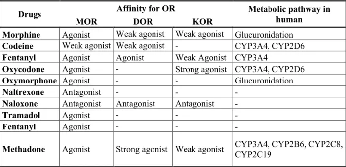
Drugs Affinity for OR Metabolic pathway in human
MOR DOR KOR
Morphine Agonist Weak agonist Weak agonist Glucuronidation
Codeine Weak agonist Weak agonist - CYP3A4, CYP2D6
Fentanyl Agonist Agonist Weak Agonist CYP3A4
Oxycodone Agonist - Strong agonist CYP3A4, CYP2D6
Oxymorphone Agonist - - Glucuronidation
Naltrexone Antagonist - - -
Naloxone Antagonist Antagonist Antagonist -
Tramadol Agonist - - -
Fentanyl Agonist - - -
Methadone Agonist Strong agonist Weak agonist CYP3A4, CYP2B6, CYP2C8, CYP2C19
Table 3: Affinity of some of the commonly used opioids along with their metabolic pathway and the effect produced (Smith & Peppin, 2014).
18
I.5 Codeine
I.5.1 Physico-chemical properties of codeine
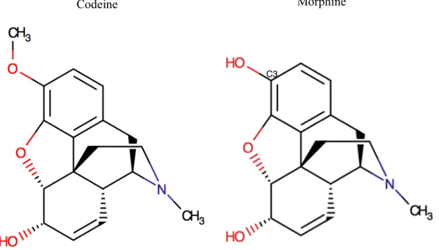
Codeine is chemically known as methyl morphine (C18H21NO3) and has a molecular weight of
299.36424 g/mol. It is obtained either naturally from Papaver somniferum plant or by methylation of morphine.
Figure 5: The molecular structures of codeine and morphine.
The stronger analgesic activity of morphine is attributed to the free hydroxyl group at C3 enabling a high affinity receptor binding whereas a low affinity receptor binding was reported for codeine (Mignat et al., 1995). The methylation of the hydroxyl group of morphine led to a more lipophilic compound that is codeine (Kay et al., 1967).
I.5.2 Metabolism of codeine
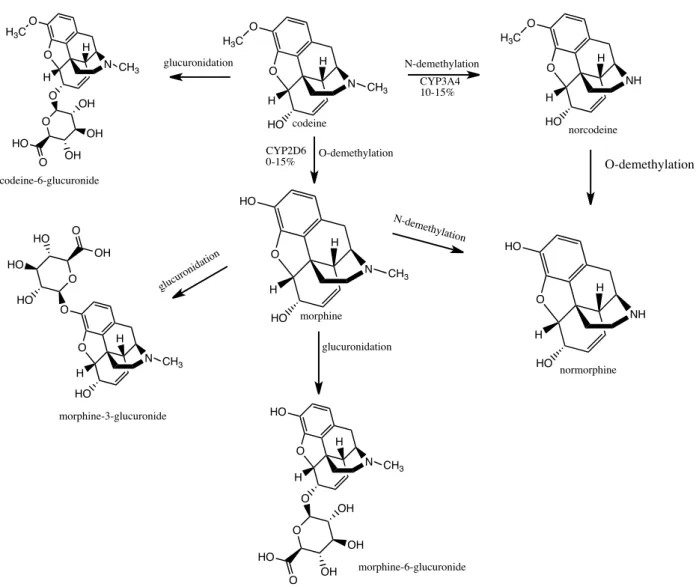
Codeine is metabolized in the liver by CYP2D and CYP3A to form primary metabolites including morphine and norcodeine (Caraco et al., 1996). Figure 6 represents the metabolic pathway of codeine in the liver. Codeine by itself is not an analgesic; it is only through its
19
conversion to morphine by CYP2D in the liver that it expresses its analgesic effect (Desmeules
et al., 1991; Sindrup et al., 1990).
The affinity of codeine to the MOR is very low compared to morphine, which has 200 folds higher affinity for the receptor than codeine (Chen et al., 1991; P. Madadi & Koren, 2008; Yue
et al., 1991). Morphine is one of the most potent opioid drugs present in the market and hence
the analgesic effect felt after taking codeine is, without doubt, due to the morphine generated after its metabolism. Morphine is further converted to M-6-G and M-3-G by glucuronidation. Since codeine is metabolized by CYP2D enzyme into morphine, any changes in the enzymatic activity of CYP2D will lead to a variation in its pharmacological effect, which includes codeine efficacy in terms of pain relief, toxicity due to its accumulation and side effects exacerbation.
Figure 6: Drug-metabolizing enzymes involved in the metabolism of codeine. In human, codeine is bioactivated by CYP2D6 to morphine and demethylated by CYP3A4 to norcodeine an inactive metabolite. Adapted from Stamer et al. 2010.
H N HO O H3C O CH3 H H N HO HO O CH3 H H NH HO O H3C O H H N O O H3C O CH3 H O OH OH OH O HO H N HO O O CH3 H O HO HO HO O OH H N O HO O CH3 H O OH OH OH O HO glucuronidation glucuroni dation N-demethylation O-demethylation N-demethylation H NH HO HO O H CYP3A4 10-15% glucuronidation CYP2D6 0-15% codeine norcodeine normorphine O-demethylation morphine morphine-6-glucuronide morphine-3-glucuronide codeine-6-glucuronide
20
I.5.3 Effects of codeine:
Codeine acts on the CNS by acting as an analgesic. Other effects include anxiolysis, euphoria and feelings of relaxation. A direct effect of codeine on the brain stem respiratory centres can cause respiratory depression. On the vascular system, it is known to produce peripheral vasodilatation that could cause orthostatic hypotension and fainting. Similar to other opioids, it can cause histamine release, which plays a role in opioid-induced hypotension. Histamine release may cause pruritus, flushing, red eyes and sweating. Effects of codeine on the GIT include decrease in gastric, biliary and pancreatic secretions resulting in indigestion. It can also cause a reduction in motility and an increase in tone in the atrum of the stomach and duodenum. In the small intestine, propulsive peristaltic waves in the colon are decreased, while tone is increased which results in constipation. Table 4 is a summary of codeine and some of its effects on important organs in the body.
Codeine
Site Effects
Central nervous system Analgesia, anxiolysis, euphoria, relaxation. Respiratory system Respiratory depression.
Cardiovascular system Orthostatic hypotension
Gastrointestinal tract Decrease gastric, biliary and pancreatic secretions; decrease gastrointestinal tract motility, constipation. Table 4: Codeine and some of its effects on important organs in the body.
I.5.4 Excretion
Codeine and its metabolites are excreted primarily by kidneys in humans, monkeys and dogs (Casarett & Doull, 1977). Urinary excretion is rapid with approximately 67% of total alkaloids being eliminated in the urine within 6 hours after the administration of the drug. Excretion is almost complete by 24 hours, but trace amounts of codeine and its metabolites can still be found in urine for several days. Codeine metabolites were determined by using HPLC in urine of male rats. Unchanged codeine, codeine glucuronide, free morphine and morphine-3-glucuronide were detected in 24 hours urines of the rats. Codeine is metabolized by glucuronidation and by oxidative N- and O-demethylation in rats (Oguri et al., 1990)
21
I.5.5 Pharmacokinetics
Codeine is readily absorbed from the GIT and has a reported oral bioavailability of 80.5% ± 30.9% (Guay et al., 1988). It is rapidly distributed from blood to body tissues, passes the blood brain barrierand is found in fetal tissues and breast milk. Lipophilicity of codeine from reported literature was found to be 0.60 (octanol/pH 7.4) + 0.89 (Moffat et al., 2005; Moffat, 1986). The apparent volume of distribution of codeine has been reported to be approximately 3-6 L/kg indicating extensive distribution of the drug into tissues. The total body clearance, renal clearance and non renal clearance of codeine were reported to be 15.0 ± 3.4 ml/min/kg, 1.71 ± 0.78 ml/min/kg and 13.32 ± 2.83 ml/min/kg (Guay et al., 1988). The reported half-life of codeine in humans is about 2.9 hours.
Table 5 presents data collected from different publications where the pharmacokinetics of codeine was evaluated.
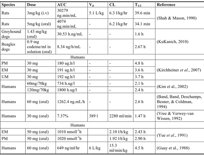
Species Dose AUC Vd CL T1/2 Reference
Rats 3mg/kg (i.v) 30279
ng.min/mL 5.1 L/kg 6.3 l/kg/hr 39.6 min
(Shah & Mason, 1990)
Rats 5mg/kg (oral) 4074 ng.min/mL - 6.2 l/kg/hr 34.1 min
Greyhound dogs 1.43 mg/kg (oral) 30.53 h.ng/mL - - 1.6 h (KuKanich, 2010) Beagles dogs 0.9 mg codeine/ml in solution (oral) 8.34 ng/h/mL - - 2.67 h Humans PM 30 mg 180 ug.h/l - - 4.8 h (Kirchheiner et al., 2007) EM 30 mg 191 ug.h/l - - 3.6 h UM 30 mg 192 ug.h/l - - 3.7 h
Humans 60mg/70kg 734 h.ug/l - - 2.1 h (Kim et al., 2002)
120mg/70kg 1800 h.ug/l - - 2.4 h
Humans 60 mg (oral) 1262.4 ng.mL/h - - 2.6 h
(Band, Band, Deschamps, Besner, & Coldman, 1994)
Humans 30 mg (oral) 7.37% 389 l 2280 ml/min 1.47 h (Vree & Verwey-van
Wissen, 1992)
Humans
EM 50 mg (oral) 1010 nmoll-1h 2.10 l/h/kg 2.43 h
(Yue et al., 1991)
PM 50 mg (oral) 1020 nmoll-1h 1.92 l/h/kg 2.90 h
Humans 60 mg (oral) 649 ng/ml/hr 6 L/kg 15.3 ml/min/kg 4.5 h (Guay et al., 1988)
Table 5: Pharmacokinetic data of codeine collected from different publications show a wide variability in the values reported. The – represents values that were not reported. Abbreviations PM, EM and UM described in legend.
22
I.5.6 Pharmacogenetics
Recent studies showed that the pharmacokinetics of codeine varies between individuals and can only be described via pharmacogenomics of CYP2D6 polymorphism. Humans can be categorized according to CYP2D6 polymorphism into poor metabolizers (PM), intermediate metabolizers (IM), extensive metabolizers (EM), and ultra-rapid metabolizers (UM). CYP2D6 alleles are characterized as normal function, reduced function and non-functional based on the expected activity level of the enzyme for which they encode (Crews et al., 2012). Two non-functional alleles result in PM phenotype, at least one reduced non-functional allele in IM and at least one functional allele in EM, multi copies of a functional allele, due to duplication or multiple duplications of the CYP2D6 gene in UM phenotype (Table 4). In UM and EM a large dose of codeine is metabolized to morphine leading to morphine toxicity that can cause severe respiratory depression and death. In PM codeine is not converted to morphine and hence the affected individuals do not experience any analgesia from codeine and thus continue to experience pain (Table 6).
Table 6: Recommended dosage of codeine based on CYP2D6 phenotypes. Adapted from Crews
et al., 2012. Likely phenotype Genotypes Implications of codeine metabolism Codeine therapy recommendation Ultrarapid metabolizer An individual carrying more than two copies of alleles.
Increased formation of morphine following codeine administration leading to higher risk of toxicity.
Avoid codeine use due to potential toxicity.
Extensive metabolizer
An individual carrying two alleles encoding full function allele together with reduced-function allele.
Normal morphine formation. Use label recommended age or weight-specific dosing.
Intermediate metabolizer
An individual carrying one reduced and one non-functional allele.
Reduced morphine formation.
Use label recommended age or weight-specific dosing. If no response, consider alternative analgesics such as morphine or non-opioid. Poor metabolizer An individual carrying no functional alleles.
Greatly reduced morphine formation following codeine administration leading to insufficient pain relief.
Avoid codeine use due to lack of efficacy.
23
Studies show that morphine AUC after codeine administration varied more than 30-fold between PM and UM groups (Johansson et al., 1991; Mikus et al., 1994; Mortimer et al., 1990; Yue, et
al., 1997). A strong correlation between the number of active CYP2D6 genes and plasma
concentrations as well as urinary recovery ratios of all O-demethylated codeine metabolites was observed. The plasma concentrations and AUCs of morphine between UMs and EMs differed about 1.5-fold with a nearly exact linear gene-dose effect. There was an almost exact linear relationship between the number of active CYP2D6 gene copies and codeine total clearance. The
metabolic ratios calculated as codeine/morphine and
(codeine+Codeine-6-Glucuronide)/(morphine+M-3-G+M-6-G) varied significantly depending on CYP2D6 activity. Cases have been reported of adults with UM phenotype suffering opioid toxicity even with small doses of codeine (Madadi et al., 2009).
In PMs, the ratios were about 10-fold higher compared to EM or UM. A significant trend towards lower ratios with increasing CYP2D6 activity was observed. UM resulted in a 1.5-fold higher morphine production compared to EM. This difference is only moderate but the risk for opioid intoxication might be increased in UMs if other additional factors such as reduction in renal function or inhibition of other enzyme systems occur at the same time (Kirchheiner et al., 2007). When sedation and miosis as opioid effect-related parameters are observed a slight decrease in pupil diameter in all groups was observed but no CYP2D6 genotype-related effects on miosis have been detected. A longer period of miosis was observed in PM though not statistically significant (Kirchheiner et al., 2007). Significant variability in both the pharmacokinetics and pharmacodynamics of codeine has been shown in animal and adult human laboratory experimental studies (Chen et al., 1991; Cleary et al., 1994; Eckhardt et al., 1998; Mikus et al., 1991).
I.5.7 Effect of age
It has been suggested that neonates and infants have a reduced metabolic capacity for codeine as CYP2D6 activity is, at most, less than 1% of the adult values in foetal liver microsomes (Quiding et al., 1992). CYP2D6 expression rapidly reaches adult levels within the first 6 months of age (Stevens et al., 2008). Although another study suggests that enzyme activity may be less than 25% of adult values up to 5 years of age (Tateishi et al., 1997). CYP2D6 genotype seems to
24
be concordant at 2 weeks of age (Madadi et al., 2009). A significant relationship between phenotype and plasma morphine was shown in children (Sindrup & Brøsen, 1995).
The United States Food and Drug Administration (FDA) have recently issued a boxed warning contraindicating the use of codeine after tonsillectomy and/or adenoidectomy in children. Post operatively, children suffering from obstructive sleep apnea passed away after therapeutic doses of codeine were prescribed. The children were later confirmed to be UM of codeine and may have suffered from breathing problems, but it is hard to determine UM in children and hence codeine has been contraindicated (Ciszkowski et al., 2009; Kelly et al., 2012). Three fatal or life threatening cases have been reported in children. The fatal cases had functional gene duplication encoding for CYP2D6 caused a greater production of potent morphine from codeine. Severe respiratory depression in an EM was also reported (Caraco et al., 1996). Health Canada has also reviewed the safety of prescription of codeine in children and it no longer recommends its use in children less than twelve years of age. It can be used to stepdown from stronger opioids in instances when a patient’s pain is decreasing over time (Ginsburg et al., 2009).
In children antitussive dosages of 3-5 mg/kg/d have produced somnolence, ataxia, miosis, vomiting, rash, facial swelling and pruritus. Respiratory depression requiring mechanical ventilation occurred in 3% of children receiving doses greater than 5 mg/kg/d. Hepatic glucuronidation pathway is completely underdeveloped in infants, which places them at particular risk for adverse dose-related effects. In adults a linear relationship has been shown to exist between a codeine dosage in the range of 7.5-60 mg/d and a decrease in chronic cough (Crews et al., 2012).
Available research findings imply that age specific differences in the pharmacokinetics of codeine may be significant. In a comparison of i.m and rectal codeine administration in children aged 3 months to 12 years, peak plasma levels were achieved between 30 and 60 minutes in both groups but rectal bioavailability was found to be lower. The plasma drug concentration data indicates that a rectal dose of codeine of 0.5 mg/kg in children can result in similar or slightly greater plasma concentration of codeine and its metabolites than after 60 mg in adults (Williams
25
Furthermore, nursing mothers are advised not to take codeine. In one case, a 13-day old breast fed infant was reported dead, but the mother was on a less than usual amount of codeine prescribed for episiotomy. She was genetically tested and confirmed to be an UM. The amount of morphine in her breast milk was at a toxic level and hence the baby died of opioid toxicity (Madadi et al., 2007). Codeine is considered comparatively safe in the elderly and is still being prescribed (Chau et al., 2008). A case of an elderly patient complaining of no pain relief after taking therapeutic doses of codeine, patient was confirmed to be a PM of codeine (Susce et al., 2006).
The role of codeine in treatment of the elderly is a subject of debate due to lack of evidence and common adverse effects experienced by them. Assessment, evaluation and treatment of pain can be very challenging due to alterations in the pharmacokinetics of opiates, which occur with normal physiological aging, polypharmacy, multiple comorbidities and the potential of more side effects or treatment failures are other problems that should be taken in consideration. Codeine is considered a safe analgesic however there have been calls to withdraw it from the market. A meta-analysis of opioids found that the modest benefits of codeine were outweighed by adverse effects in the treatment of osteoarthritis of the knee or hip (Iedema J., 2011). A cohort study found that the risk of injury was higher in older people using combination products of codeine than those taking other sedative drugs (Buckeridge et al., 2010).
The variation in pharmacological effect from children dying of morphine toxicity to elder patients experiencing no analgesic effect with increased risk of adverse effects, makes us assume that there is a change in CYP2D6 activity with age. The variation of CYP2D6 metabolic activity with age, may lead to a change in phenotype from UM and EM metabolizers to PM. The patients may hence experience no analgesia and codeine might accumulate in their body systems causing opioid toxicity and lack of pain relief. Overall the efficacy and side effects of codeine in different age groups has not so far been sufficiently investigated.
26
I.6 Oxycodone
I.6.1 Physico-chemical properties of oxycodone
Oxycodone is a semisynthetic opioid derived from the naturally occurring opium alkaloid,
thebaine. Chemically oxycodone is a
4,5-alpha-epoxy-14-hydroxy-3-methoxy-17-methylmorphinon-6-onehydrochloride. The molecular formula of oxycodone is C14H21NO4 and
its molecular weight is 315.36364g/mol.
Figure 7: The chemical structures of oxycodone and morphine.
The structural difference between oxycodone and morphine means that oxycodone is not directly susceptible to phase II metabolism therefore it has greater bioavailability of 60-87% (Ordonez et
al., 2007). Also because of the difference in structure, oxycodone causes less
immunosuppression in comparison to other opioids.
I.6.2 Metabolism of oxycodone
In human, oxycodone is extensively metabolized by CYP2D6 and CYP3A4 in the liver to oxymorphone and noroxycodone respectively (Kalso, 2005). Systemic exposure to oxycodone and metabolically derived noroxycodone was approximately 2.3- and 6.4-fold higher in Dark
27
Agouti rats compared to Sprague-Dawley rats following a subcutaneous administration of single bolus of oxycodone at 2mg/kg whereas the circulating concentrations of metabolically derived oxymorphone remained very low (Huang et al., 2005). Oxymorphone is a more potent opioid receptor agonist than oxycodone, whereas noroxycodone is less potent and has weaker antinociception than the parent drug (Kalso, 2005). Noroxycodone undergoes further oxidation by CYP3A4 to noroxymorphone, which is active at opioid receptors but it, does not seem to cross the blood brain barrier. The glucuronide metabolic pathway for oxycodone has so far not been established. The analgesic effect of oxycodone is due to the parent drug itself but some analgesia is a contribution of one of the active metabolites oxymorphone, which is more potent than oxycodone, but is formed in small quantities.
Figure 8: Drug-metabolizing enzymes involved in the metabolism of oxycodone. The major metabolic pathway of oxycodone is the formation of noroxycodone via CYP3A4 enzymes. Noroxycodone is further metabolised to noroxymorphone via CYP2D6 enzymes. The minor metabolic pathways are formation of oxymorphone via CYP2D6 enzymes, and 6-keto reduction to α-/β-oxycodol. Oxymorphone is further metabolised to noroxymorphone via CYP3A4 enzymes. Adapted from Andreassen et al., 2010
O HO O H OHH N CH3 O O O H OHH N CH3 H3C O O O H OHH NH H3C O O O H OH H NH H O HO HO H OH H N CH3 keto reduction 8% O-demethylation CYP2D6 11% N-demethylation CYP3A4 CYP3A4 47% CYP2D 6 oxycodol oycodone noroxycodone noroxymorphone oxymorphone