Gene expression pattern
Cloning and expression of the TALE superclass homeobox Meis2 gene
during zebrafish embryonic development
Fre´de´ric Biemar
a,b, Nathalie Devos
a, Joseph A. Martial
a, Wolfgang Driever
b, Bernard Peers
a,*
aLaboratoire de Biologie Mole´culaire et de Ge´nie Ge´ne´tique, Institut de Chimie, Baˆtiment B6, Universite´ de Lie`ge, B-4000 Liege (Sart Tilman), Belgium bInstitut fu¨r Biologie I, Abt. Entwicklungsbiologie, Universita¨t Freiburg, Hauptstrasse 1, D-79104 Freiburg, Germany
Received 20 April 2001; received in revised form 3 September 2001; accepted 3 September 2001
Abstract
Meis and Prep/Pknox (MEINOX family) proteins, together with Pbx (PBC family) proteins, belong to the TALE superfamily
character-ized by an atypical homeodomain containing three additional amino acids between helix 1 and helix 2. Members of the MEINOX and PBC
families have been isolated in Caenorhabditis elegans, Drosophila, Xenopus, chick, mouse and human, and play crucial roles in many
aspects of embryogenesis. Here, we report the isolation of meis2 in zebrafish. Expression of meis2 is first detected at the beginning of
gastrulation. Later during embryogenesis, meis2 transcripts are found in distinct domains of the central nervous system with the strongest
expression in the hindbrain. Expression was also detected in the isthmus, along the spinal cord and in the lateral mesoderm. As development
proceeds, meis2 is also expressed in the developing retina, pharyngeal arches, and in the vicinity of the gut tube. q 2001 Elsevier Science
Ireland Ltd. All rights reserved.
Keywords: Homeobox; TALE; Meis; Zebrafish; Development
1. Results and discussion
The Drosophila TALE proteins, Extradenticle (EXD) and
Homothorax (HTH), have been implicated in numerous
important regulatory processes throughout embryogenesis,
including HOM-C dependent regulation of segment identity
(Mann, 1995; Wilson and Desplan, 1995), peripheral
nervous system (PNS) patterning (Kurant et al., 1998),
and establishment of territories in the eye (Pai et al.,
1998; Pichaud and Casares, 2000), wing and leg imaginal
discs (Morata and Sanchez-Herrero, 1999). In addition,
HTH may be involved in determination of antennal identity
(Dong et al., 2000; Yao et al., 1999) and salivary gland
development (Andrew et al., 2000; Henderson and Andrew,
2000). In vertebrates, members of the PBC and MEINOX
families have been shown to play crucial roles in hindbrain
patterning (Ferretti et al., 2000; Maconochie et al., 1997;
Popperl et al., 1995; Salzberg et al., 1999) and limb
outgrowth (Capdevila et al., 1999; Gonzalez-Crespo et al.,
1998; Mercader et al., 1999; Mercader et al., 2000). Yet,
their function in multiple aspects of vertebrate development
is still not completely understood.
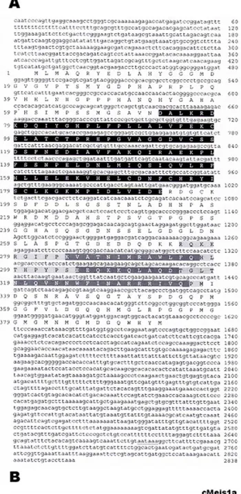
We have isolated meis2, a member of the TALE
super-class of homeodomain proteins. The 2.8 kb meis2 cDNA
contains an open reading frame encoding a protein of 393
amino acids, which exhibits the atypical homeodomain and
the MEINOX domain characteristic of Meis proteins
(Burglin, 1998). Amino acid sequence comparison with
human, mouse, chicken, Xenopus, Drosophila and
Caenor-habditis elegans Meis proteins reveals extremely high
homology within these conserved domains (the MEINOX
and the homeodomain of zebrafish and mouse meis2
proteins are 97.5 and 98.4% identical, respectively).
Phylo-genetic analysis suggests that the zebrafish Meis isolated in
this study is a true ortholog of chick and mouse Meis2
proteins (Fig. 1B), as it is clustered with the cMeis2 and
mMeis2 orthologs.
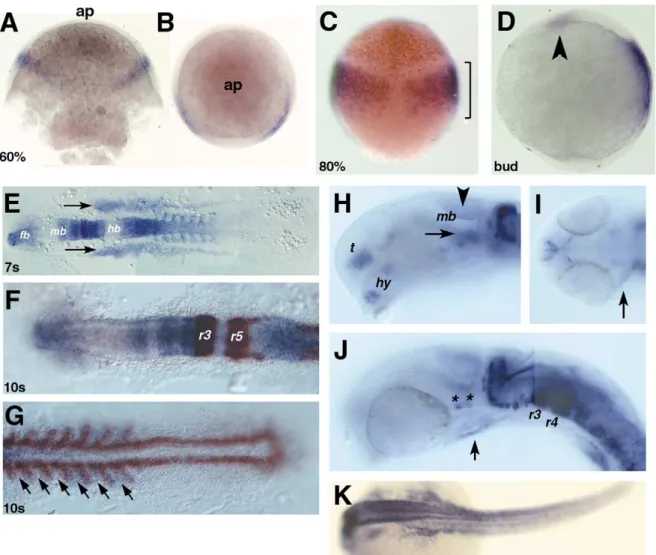
Zebrafish meis2 transcripts are first detected during
gastrulation, at 60% epiboly, in two lateral domains within
the ectoderm (Fig. 2A,B). At 80% epiboly, these expression
domains have enlarged (Fig. 2C), now covering the whole
area of the prospective hindbrain (Woo and Fraser, 1995).
Double labeling with otx2, a marker for the presumptive
forebrain and midbrain (Li et al., 1994) confirms that
meis2 expressing cells are restricted to the presumptive
hindbrain. Between 100% epiboly and late bud stage (Fig.
2D), a second expression domain appears, presumably in the
anterior neural plate (Fig. 2D, arrowhead). During
somito-0925-4773/01/$ - see front matter q 2001 Elsevier Science Ireland Ltd. All rights reserved. PII: S 0 9 2 5 - 4 7 7 3 ( 0 1 ) 0 0 5 5 4 - 8
www.elsevier.com/locate/modo
* Corresponding author. Tel.: 132-4-366-33-74; fax: 132-4-366-29-68. E-mail address: bpeers@ulg.ac.be (B. Peers).
genesis (Fig. 2E–G), expression is detected within the
fore-brain, posterior midbrain and anterior hindbrain
(rhombo-meres, r1 through r3; Fig. 2F). In the spinal cord, strong
expression is seen anteriorly, that gradually weakens
poster-iorly (Fig. 2E,F). meis2 expression is also seen in the somite,
but is absent from the presomitic mesoderm. The alternate
pattern shown by meis2 and myoD expression indicates that
meis2 is expressed in the anterior border of each somite
(Weinberg et al., 1996; Fig. 2G, arrows). meis2 expression
is also found in two lateral mesodermal domains, which
extend anteriorly up to the mid–hindbrain junction (Fig.
2E–F).
At 24 h post-fertilization (hpf), meis2 expression
continues in several distinct domains within the developing
forebrain, including basal ventral telencephalon and
hypothalamus (Fig. 2H). In the midbrain, expression is
seen in both the ventral tectum (Fig. 2H, arrowhead) and
tegmentum (Fig. 2H, arrow). Interestingly, several clusters
of meis2 positive cells are also detected laterally in the
ventral part of the midbrain (Fig. 2J, asterisks), similar to
the expression of hoxa1a (Shih et al., 2001). Groups of cells
at the rostral border, as well as in the caudal dorsal part of
the cerebellum, also express meis2 (Fig. 2H,J). Within the
hindbrain (Fig. 2J), r3 and r4 exhibit the strongest
expres-sion, while other rhombomeres retain patchy expression
ventrally. The nascent heart tube also shows weak levels
of meis2 expression (Fig. 2I,J, arrow). In the trunk, meis2
is also expressed in the hypaxial musculature (Fig. 2K).
meis2 remains strongly expressed in the zebrafish
hind-brain until 77 hpf (Fig. 3A–D). Additional expression is
seen in the retina (Fig. 3E–G). Surprisingly, we did not
observe expression in the pectoral fin buds at any stages
(Fig. 3H–J). We detected meis2 transcripts in pharyngeal
arches at 45 and 55 hpf (Fig. 3K,L), notably in the region
of the jaw in the presumptive mandibular and hyoid
primor-dia (Fig. 3L). We also found meis2 expression in the vicinity
of the gut tube, presumably in the pronephric glomerulus or
an endodermally-derived organ, such as the liver or
pancreas (Fig. 3L).
Thus, although the zebrafish meis2 expression pattern
resembles that of the mouse Meis2 ortholog, especially in
the central nervous system (CNS), our study points out some
striking differences. Firstly, no expression was observed in
the pectoral fin buds, as described for the mouse and chick
(Capdevila et al., 1999; Mercader et al., 1999). Secondly,
the restriction of zebrafish meis2 expression in the anterior
portion of the somites was not observed for mouse Meis2. In
addition, Meis2 is expressed in the segmental plate in the
mouse, but not in zebrafish (Cecconi et al., 1997;
Oulad-Fig. 1. (A) Nucleotide sequence and open reading frame of the zebrafish meis2 gene. The conserved MEINOX domain is highlighted in black. The homeodomain is highlighted in light grey and the three helixes in dark grey. (B) A phylogenetic tree established with TREEVIEW shows the evolution-ary relationships between zebrafish Meis2 and Meis proteins of other species.
Abdelghani et al., 1997). Finally, whereas expression of
Meis1 but not Meis2 was reported in the retina in mice
(Toresson et al., 2000), we do find expression of meis2 in
the zebrafish retina at 36 hpf onwards.
2. Materials and methods
A 246 bp fragment, corresponding to a highly conserved
region of meis genes, was isolated using a degenerate
PCR-based approach (forward primer, 5
0-ATHTTYGARAART-GYGAR-3
0; and reverse primer, 5
0-RTGRCARAARTT-RTCRCA-3
0) from a 15–19 hpf zebrafish cDNA library (a
generous gift from Dr Bruce Appel). The amplified
frag-ment was subsequently used as a probe to obtain a
full-length cDNA from a shield stage library available from
RZPD (http://www.rzpd.de). One positive clone was
isolated, sequenced in both directions and deposited in
GenBank (accession number, AF170065).
Whole-mount in situ hybridizations was performed as
described (Hauptmann and Gerster, 1994).
3. Note added in proof
While this manuscript was in preparation, the cloning of
the same gene and its expression during embryogenesis was
reported by T. Zerucha and V.E. Prince (Zerucha and
Prince, 2001).
Fig. 2. Expression of meis2 in wildtype zebrafish embryos from gastrulation to 24 h post-fertilization (hpf). Dorsal (A) and animal pole (B) views at 60% epiboly. (C) Eighty percent epiboly, dorsal view; and (D), bud stage, lateral view. Arrowhead points at the new expression in the presumptive anterior neural plate. (E) Seven-somite stage, dorsal, flat-mounted view. Arrows point at the bilateral mesodermal domains. (F,G) Ten-somite stage, dorsal, flat-mounted view showing expression of meis2 (in blue), Krox20 (F) and myoD (G) (in brown). Arrows point at the anterior border of each somite. (H–K) expression of meis2 at 24 hpf. (H) Lateral view, anterior to the left, dorsal to the top of the fore- and midbrain regions. Arrowhead points at the ventral tectum, and arrow points at the tegmentum. (I) Dorsal view, anterior to the left of a flat-mounted embryo showing expression in the forming heart tube. (J) Lateral view, anterior to the left, dorsal to the top of the mid- and hindbrain regions. Asterisks show patchy expression in the ventral midbrain. (K) Dorsal view, anterior to the left showing the expression in the hypaxial muscles. fb, forebrain; hb, hindbrain; mb, midbrain; hy, hypothalamus; r(3, 4, 5), rhombomere (3, 4, 5); sc, spinal cord; t, telencephalon.
Acknowledgements
The authors wish to thank Dr Bruce Appel and the RZPD
for providing cDNA libraries, Zoltan Varga for helpful
discussions, Karen Lunde and Marianne Voz for critical
reading of the manuscript. F.B. holds a doctoral fellowship
from the ‘Fonds pour la Formation a` la Recherche dans
l’industrie et dans l’Agriculture (FRIA)’. B.P. is ‘Chercheur
Qualifie´’ from the ‘Fonds National pour la Recherche
Scien-tifique (FNRS)’. This work was supported by a grant from
the EC Fifth Framework (QLRT-1999-00149 to B.P. and
W.D.).
Fig. 3. Expression of meis2 in late embryonic stages at: (A,D,E,H), 36; (K), 45; (B,F,I), 48; (L), 55; and (C,G,J), 77 hpf. (A–C) Dorsal views, anterior to the left showing overall expression in the brain region at: (A), 36; (B), 48; and (C), 77 hpf. Arrowhead points at the expression in the rostral part of the cerebellum. (D) lateral view of the embryo depicted in (A). Arrowheads point at the expression in the rostral part of the cerebellum. (E–G) Dorsal views, anterior to the left showing expression in the retina at: (E), 36 (E, arrows); (F), 48; and (G), 77 hpf. (H–J) Lateral views, anterior to the left of a pectoral fin bud at: (H), 36; (I), 48; and (J), 77 hpf. (K) Sagittal section of the head region at 45 hpf showing overall brain expression. Arrowhead points at expression in the posterior pharyngeal arch. (L) Sagittal section at 55 hpf showing expression in the presumptive mandibular and hyoid primordium (arrowhead) and gut region (arrow).
References
Andrew, D.J., Henderson, K.D., Seshaiah, P., 2000. Salivary gland devel-opment in Drosophila melanogaster. Mech. Dev. 92, 5–17.
Burglin, T.R., 1998. The PBC domain contains a MEINOX domain: coevo-lution of Hox and TALE homeobox genes? Dev. Genes Evol. 208, 113– 116.
Capdevila, J., Tsukui, T., Rodriquez Esteban, C., Zappavigna, V., Izpisua Belmonte, J.C., 1999. Control of vertebrate limb outgrowth by the proximal factor Meis2 and distal antagonism of BMPs by Gremlin. Mol. Cell 4, 839–849.
Cecconi, F., Proetzel, G., Alvarez-Bolado, G., Jay, D., Gruss, P., 1997. Expression of Meis2, a Knotted-related murine homeobox gene, indi-cates a role in the differentiation of the forebrain and the somitic meso-derm. Dev. Dyn. 210, 184–190.
Dong, P.D., Chu, J., Panganiban, G., 2000. Coexpression of the homeobox genes Distal-less and homothorax determines Drosophila antennal identity. Development 127, 209–216.
Ferretti, E., Marshall, H., Popperl, H., Maconochie, M., Krumlauf, R., Blasi, F., 2000. Segmental expression of Hoxb2 in r4 requires two separate sites that integrate cooperative interactions between Prep1, Pbx and Hox proteins. Development 127, 155–166.
Gonzalez-Crespo, S., Abu-Shaar, M., Torres, M., Martinez, A.C., Mann, R.S., Morata, G., 1998. Antagonism between extradenticle function and Hedgehog signalling in the developing limb. Nature 394, 196–200. Hauptmann, G., Gerster, T., 1994. Two-color whole-mount in situ
hybri-dization to vertebrate and Drosophila embryos. Trends Genet. 10, 266. Henderson, K.D., Andrew, D.J., 2000. Regulation and function of Scr, exd,
and hth in the Drosophila salivary gland. Dev. Biol. 217, 362–374. Kurant, E., Pai, C.Y., Sharf, R., Halachmi, N., Sun, Y.H., Salzberg, A.,
1998. Dorsotonals/homothorax, the Drosophila homologue of meis1, interacts with extradenticle in patterning of the embryonic PNS. Devel-opment 125, 1037–1048.
Li, Y., Allende, M.L., Finkelstein, R., Weinberg, E.S., 1994. Expression of two zebrafish orthodenticle-related genes in the embryonic brain. Mech. Dev. 48, 229–244.
Maconochie, M.K., Nonchev, S., Studer, M., Chan, S.K., Popperl, H., Sham, M.H., Mann, R.S., Krumlauf, R., 1997. Cross-regulation in the mouse HoxB complex: the expression of Hoxb2 in rhombomere 4 is regulated by Hoxb1. Genes Dev. 11, 1885–1895.
Mann, R.S., 1995. The specificity of homeotic gene function. Bioessays 17, 855–863.
Mercader, N., Leonardo, E., Azpiazu, N., Serrano, A., Morata, G., Marti-nez, C., Torres, M., 1999. Conserved regulation of proximodistal limb axis development by Meis1/Hth. Nature 402, 425–429.
Mercader, N., Leonardo, E., Piedra, M.E., Martinez, A.C., Ros, M.A., Torres, M., 2000. Opposing RA and FGF signals control proximodistal vertebrate limb development through regulation of meis genes In process citation. Development 127, 3961–3970.
Morata, G., Sanchez-Herrero, E., 1999. Patterning mechanisms in the body trunk and the appendages of Drosophila. Development 126, 2823– 2828.
Oulad-Abdelghani, M., Chazaud, C., Bouillet, P., Sapin, V., Chambon, P., Dolle, P., 1997. Meis2, a novel mouse Pbx-related homeobox gene induced by retinoic acid during differentiation of P19 embryonal carci-noma cells. Dev. Dyn. 210, 173–183.
Pai, C.Y., Kuo, T.S., Jaw, T.J., Kurant, E., Chen, C.T., Bessarab, D.A., Salzberg, A., Sun, Y.H., 1998. The Homothorax homeoprotein activates the nuclear localization of another homeoprotein, extradenticle, and suppresses eye development in Drosophila. Genes Dev. 12, 435–446. Pichaud, F., Casares, F., 2000. Homothorax and iroquois-C genes are
required for the establishment of territories within the developing eye disc In process citation. Mech. Dev. 96, 15–25.
Popperl, H., Bienz, M., Studer, M., Chan, S.K., Aparicio, S., Brenner, S., Mann, R.S., Krumlauf, R., 1995. Segmental expression of Hoxb-1 is controlled by a highly conserved autoregulatory loop dependent upon exd/pbx. Cell 81, 1031–1042.
Salzberg, A., Elias, S., Nachaliel, N., Bonstein, L., Henig, C., Frank, D., 1999. A Meis family protein caudalizes neural cell fates in Xenopus. Mech. Dev. 80, 3–13.
Shih, L., Tsay, H., Lin, S., Hwang, S.L., 2001. Expression of zebrafish Hoxa1a in neuronal cells of the midbrain and anterior hindbrain. Mech. Dev. 101, 279–281.
Toresson, H., Parmar, M., Campbell, K., 2000. Expression of Meis and Pbx genes and their protein products in the developing telencephalon: impli-cations for regional differentiation. Mech. Dev. 94, 183–187. Weinberg, E.S., Allende, M.L., Kelly, C.S., Abdelhamid, A., Murakami, T.,
Andermann, P., Doerre, O.G., Grunwald, D.J., Riggleman, B., 1996. Developmental regulation of zebrafish MyoD in wild-type, no tail and spadetail embryos. Development 122, 271–280.
Wilson, D.S., Desplan, C., 1995. Homeodomain proteins. Cooperating to be different. Curr. Biol. 5, 32–34.
Woo, K., Fraser, S.E., 1995. Order and coherence in the fate map of the zebrafish nervous system. Development 121, 2595–2609.
Yao, L.C., Liaw, G.J., Pai, C.Y., Sun, Y.H., 1999. A common mechanism for antenna-to-Leg transformation in Drosophila: suppression of homo-thorax transcription by four HOM-C genes. Dev. Biol. 211, 268–276. Zerucha, T., Prince, V.E., 2001. Cloning and developmental expression of a