REVIEW
E-learning in cardiovascular imaging: another step
towards a structured educational approach
Bernard Cosyns
1*
, Jose Juan Gomez De Diego
2, Alexandros Stefanidis
3,
Maurizio Galderisi
4, Laura Ernande
5, S. Richard Underwood
6, Chiara Bucciarelli-Ducci
7,
Patrizio Lancellotti
8,9, and Gilbert Habib
10, on behalf of the EACVI education,
web-communication and certification committees
Document reviewers: Thor Edvardsen and Julien Magne
1
Department of Cardiology (CHVZ) and In Vivo Cellular and Molecular Imaging (ICMI) Laboratory, University Hospital of Brussels, Vrij Unviversiteit van Brussel, 101 Laarbeeklaan 1090 Brussels, Belgium;2
Laboratorio de Imagen Cardı´aca, Hospital Clı´nico San Carlos Madrid, Madrid, Spain;3
1st Department of Cardiology, Echocardiography Laboratory, General Hospital of Nikea, Piraeus, Greece;4
Department of Advanced Biomedical Sciences, Federico II University Hospital, Naples, Italy;5
Service des Explorations Fonctionnelles, DHU Ageing-Thorax-Vessel-Blood, Henri Mondor Hospital, Assistance Publique Hoˆpitaux de Paris, and INSERM U955, Team 8, Universite´ Paris-Est Cre´teil (UPEC), Paris, France;6
National Heart and Lung Institute, Imperial College, London SW3 6LY, UK;7
Bristol Heart Institute, NIHR Bristol Cardiovascular Biomedical Research Unit, University of Bristol, Bristol, UK;8
University of Lie`ge Hospital, GIGA Cardiovascular Sciences, Heart Valve Clinic, Department of Cardiology, University Hospital SartTilman, Lie`ge, Belgium;9
GVM Care and Research, Napoli, Italy; and
10
Cardiology Department, Hoˆpital La Timone, Marseille, France
Received 23 January 2015; accepted after revision 25 January 2015; online publish-ahead-of-print 6 March 2015
Introduction
The availability of educational resources in cardiovascular imaging varies between European countries, and access to imaging technol-ogy also differs. A survey of European cardiovascular imaging by the European Association of Cardiovascular Imaging (EACVI) pro-vides a snapshot of imaging practice in 40 countries of the European Society of Cardiology (http://www.escardio.org/communities/EACVI/ imaging-organisations/Documents/eacvi-highlight-book-v5.pdf). Con-sistency of education is important, but it must also take account of personal and national requirements.
Definition of e-learning
The European Commission definition of e-learning is ‘the use of new multimedia technologies and of the Internet to improve the quality of learning by facilitating access to resources and services, as well as remote exchanges and collaboration’. However, the relationship between e-learning and the quality of education is unproved, and an alternative definition is that of LabSET, ‘online learning focuses on the development of skills by the learner and structured by inter-actions with the tutor and peers’. The term ‘online’ refers to a com-puter attached to some form of network, either local (intranet) or global (Internet).
E-learning can be used in a variety of ways, such as to introduce, extend, or complete face-to-face teaching, and it is not limited to dis-tance learning if delivered synchronously in a virtual classroom or
auditorium. It can be delivered by a variety of methods including downloads of audio or video sessions.
Advantages of e-learning
E-learning allows techniques to be used that fit with different learning styles including the use of complex digital content and innovative methods such as meta-cognition and. Formative assessment also helps to introduce complex skills in cardiovascular imaging, and it can improve the quality of the interaction between trainer and trainee. It is more flexible in time and space, which may be particularly relevant for clinicians who often have a heavy workload, and it improves accessibility and cost.
Keys to successful e-learning
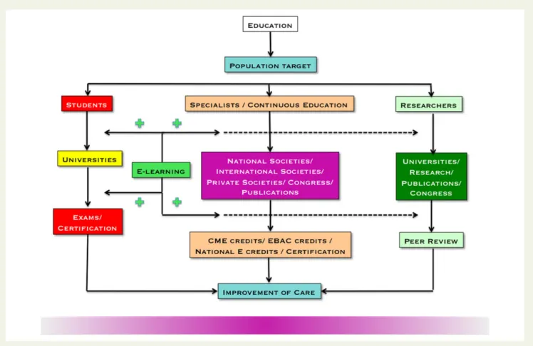
Educational tools and resources should be adapted to the target audience, and they may differ radically depending on whether the aim is for students, specialists, or researchers (Figure1). E-learning can indeed be adapted and similar content can be presented in a variety of ways, including slides, webinars, cases, courses, and key-reference libraries (http://www.escardio.org/communities/EACVI/ education/echo-box/Pages/welcome.aspx).
A common framework for structured e-learning is a fundamental step towards more homogeneous education. All the core syllabi of the EACVI family have been updated in that perspective and published1,2 or are available online (http://www.escardio.org/
*Corresponding author. Tel:+32 2 3872620, E-mail: [email protected]
Published on behalf of the European Society of Cardiology. All rights reserved.&The Author 2015. For permissions please email: [email protected].
European Heart Journal – Cardiovascular Imaging (2015) 16, 463–465 doi:10.1093/ehjci/jev022
by guest on April 24, 2015
communities/EACVI/education/Documents/cardiac-ct-core-syllabus-2014-final.pdf). The combination of e-learning and mentoring is particularly helpful, and this increases compliance by maintaining motivation. Other forms of motivation are self-evaluations and ‘fun’ exercises and collaborative work. For example a certification process is already available for cardiac magnetic resonance (CMR) (http://www.escardio.org/communities/EACVI/accreditation/cmr/ Pages/certification-processes.aspx), and remote mentoring is being considered when local support is limited.
Examples of real cases are important in any clinical specialty, and these are commonly presented in courses or congresses and can be made available to share clinical experience (http://www.escardio. org/communities/EACVI/education/resources-meetings-courses/ Pages/welcome.aspx) and extended to include case reports and image libraries (http://www.escardio.org/EACVI-clinical-case). The sharing of resources by social media has already been adopted by the young community (https://www.linkedin.com/groups/Welcome-to-EACVI-NEW-Club-5153456.S.5814745717431894017) and should be extended to the entire imaging community. Cardiovascular imaging can be complex and is ideally presented in modules such as a basic echocardiography course (http://www.escardio.org/ communities/EACVI/education/Pages/echocardiography-elearning-course.aspx), advanced courses (transoesophageal echocardiog-raphy, stress echocardiography), and similar in nuclear cardiology, cardiac CT and CMR (http://www.escardio.org/communities/EACVI/
club-35/education/Pages/tutorials.aspx). It is important to manage the pace of learning and for the process not to be too ‘linear’. Online reference tools such as glossaries, links, and online help are valuable.
How to use e-learning?
If e-learning is to be effective, it must encourage completion of the task and this requires high-quality material and a combination of personal and group study supported by an experienced trainer. EACVI e-learning material is peer reviewed and of high standard, such as with simultaneous online and printed paper release of rec-ommendation papers in the European Journal of Cardiovascular Imaging.3–7
Limitations of e-learning
E-learning does not replace traditional teaching methods, but it com-pliments them. It is important that it does not engender self-confidence to the extent that traditional and clinical methods are ignored since even with sophisticated clinical simulators there is no substitute for hands on experience. Access to online facilities is still difficult for some people, and language is also a problem unless resources are made available in languages other than English. Figure 1 Potential targets for e-learning in education.
B. Cosyns et al.
464
by guest on April 24, 2015
Conclusion
E-learning can assist education in cardiovascular imaging and can complement traditional education techniques. It has a number of advantages, and it can supplement interaction with peers and with formal trainers. It is flexible in time and space, it can be widely avail-able, and it is relatively cheap. Resources should be adapted to each intended audience and should be supplemented by techniques for both formative and summative assessment. We plan in future to assess the impact of EACVI educational programmes in clinical prac-tice throughout Europe.
Conflict of interest: None declared.
References
1. Cosyns B, Garbi M, Separovic J, Pasquet A, Lancellotti P; Education Committee of the European Association of Cardiovascular Imaging Association (EACVI). Update of the echocardiography core syllabus of the European Association of Cardiovascular Imaging (EACVI). Eur Heart J Cardiovasc Imaging 2013;14:837 – 92.
2. Petersen SE, Almeida AG, Alpendurada F, Boubertakh R, Bucciarelli-Ducci C, Cosyns B et al. Education Committee of European Association of Cardiovascular Imaging Association (EACVI). Update of the European Association of Cardiovascular Imaging (EACVI) Core Syllabus for the European Cardiovascular Magnetic Resonance Certification Exam. Eur Heart J Cardiovasc Imaging 2014;15:728 – 9.
3. Plana JC, Galderisi M, Barac A, Ewer MS, Ky B, Scherrer-Crosbie M et al. Expert con-sensus for multimodality imaging evaluation of adult patients during and after cancer therapy: a report from the American Society of Echocardiography and the European Association of Cardiovascular Imaging. Eur Heart J Cardiovasc Imaging 2014;15: 1063 – 93.
4. Popescu BA, Stefanidis A, Nihoyannopoulos P, Fox KF, Ray S, Cardim N et al. Updated standards and processes for accreditation of echocardiographic laboratories from The European Association of Cardiovascular Imaging. Eur Heart J Cardiovasc Imaging 2014;15:717 – 27.
5. Neskovic AN, Edvardsen T, Galderisi M, Garbi M, Gullace G, Jurcut R et al. Focus cardiac ultrasound: the European Association of Cardiovascular Imaging viewpoint. Eur Heart J Cardiovasc Imaging 2014;15:956 – 60.
6. Flachskampf FA, Wouters PF, Edvardsen T, Evangelista A, Habib G, Hoffman P et al. Recommendations for transoesophageal echocardiography. Eur Heart J Cardiovasc Imaging 2014;15:353 – 65.
7. Underwood SR, de Bondt P, Flotats A, Marcasa C, Pinto F, Schaefer W et al. The current and future status of nuclear cardiology: a consensus report. Eur Heart J Cardi-ovasc Imaging 2014;15:949 – 55.
IMAGE FOCUS
. . . .
doi:10.1093/ehjci/jeu226
Online publish-ahead-of-print 4 February 2015
Severe restrictive aortic regurgitation due to aortic fibrous strand
Iris Esteve-Ruiz*, Francisco Lo´pez-Pardo, O´ scar Lagos-Degrande, Jose´ Eduardo Lo´pez-Haldo´n, and Jose´ A´ ngel Urbano-Moral
Hospital Universitario Virgen del Rocı´o, C/Manuel Siurot s/n, Sevilla 41013, Spain *Corresponding author. Tel:+34679411811, E-mail: [email protected]
A 70-year-old male was referred to the echo laboratory for a reevaluation. One year ago he suf-fered a traumatic tetraplegia. During the preopera-tory evaluation he was diagnosed with severe aortic regurgitation.
A transthoracic echocardiogram showed a tricuspid aortic valve with severe aortic regurgita-tion (Panel A). Aortic annulus and root were not dilated. Transoesophageal echocardiography showed a tricuspid aortic valve with normal opening movement and an adequate valve coaption between right coronary and non-coronary cusps
(Panel B). A linear mobile echo like a chordae tendineae strand was connecting the left coronary sigmoid cusp to the sinotubular junction (Panel C), leading an important tenting and restriction of this cusp (Panel D and E, see Supplementary data online, Video S1). Colour flow displayed a severe aortic regurgitation (Panel F, see Supplementary data online, Video S2). Live three-dimensional echocardiography was performed, improving the assessment of the fibrous strand (Panel G, see Supplementary data online, Video S3). Left ventricle was dilated and has a moderate dysfunction (ejection fraction 45%) with global hypokinesis.
The patient denied surgery
The aortic chordae tendineae strand may be an embryonic remnant during the semilunar cusp formation process of the aortic valve. There are few reported cases, most of them from Asia, describing aortic regurgitation due to spontaneous rupture of chordeae tendineae. To our knowledge, there is only one case reported similar to ours, submitting an aortic valvular incompetence owing to this restrictive mechanism (fibrous strand) without another mechanism: annular dilatation, prolapse or any congenital heart defect.
Supplementary data are available at European Heart Journal – Cardiovascular Imaging online.
Published on behalf of the European Society of Cardiology. All rights reserved.&The Author 2015. For permissions please email: [email protected].
E-learning in cardiovascular imaging
465
by guest on April 24, 2015