Open Access
Research article
Distantly related lipocalins share two conserved clusters of
hydrophobic residues: use in homology modeling
Benoit Adam
1, Benoit Charloteaux
1, Jerome Beaufays
2, Luc Vanhamme
3,
Edmond Godfroid
2, Robert Brasseur
1and Laurence Lins*
1Address: 1Centre de Biophysique Moléculaire et Numérique, Faculté Universitaire des Sciences Agronomiques de Gembloux, Gembloux, Belgium, 2Service de Génétique Appliquée, Institut de Biologie et de Médecine Moléculaires, Université Libre de Bruxelles, Gosselies, Belgium and 3Laboratoire de Parasitologie moléculaire, Institut de Biologie et de Médecine Moléculaires, Université Libre de Bruxelles, Gosselies, Belgium Email: Benoit Adam - adam.b@fsagx.ac.be; Benoit Charloteaux - charloteaux.b@fsagx.ac.be; Jerome Beaufays - jbeaufa1@ulb.ac.be; Luc Vanhamme - luc.vanhamme@ulb.ac.be; Edmond Godfroid - edmond.godfroid@ulb.ac.be; Robert Brasseur - brasseur.r@fsagx.ac.be; Laurence Lins* - lins.l@fsagx.ac.be
* Corresponding author
Abstract
Background: Lipocalins are widely distributed in nature and are found in bacteria, plants, arthropoda and
vertebra. In hematophagous arthropods, they are implicated in the successful accomplishment of the blood meal, interfering with platelet aggregation, blood coagulation and inflammation and in the transmission of disease parasites such as Trypanosoma cruzi and Borrelia burgdorferi.
The pairwise sequence identity is low among this family, often below 30%, despite a well conserved tertiary structure. Under the 30% identity threshold, alignment methods do not correctly assign and align proteins. The only safe way to assign a sequence to that family is by experimental determination. However, these procedures are long and costly and cannot always be applied. A way to circumvent the experimental approach is sequence and structure analyze. To further help in that task, the residues implicated in the stabilisation of the lipocalin fold were determined. This was done by analyzing the conserved interactions for ten lipocalins having a maximum pairwise identity of 28% and various functions.
Results: It was determined that two hydrophobic clusters of residues are conserved by analysing the ten
lipocalin structures and sequences. One cluster is internal to the barrel, involving all strands and the 310
helix. The other is external, involving four strands and the helix lying parallel to the barrel surface. These clusters are also present in RaHBP2, a unusual "outlier" lipocalin from tick Rhipicephalus appendiculatus. This information was used to assess assignment of LIR2 a protein from Ixodes ricinus and to build a 3D model that helps to predict function. FTIR data support the lipocalin fold for this protein.
Conclusion: By sequence and structural analyzes, two conserved clusters of hydrophobic residues in
interactions have been identified in lipocalins. Since the residues implicated are not conserved for function, they should provide the minimal subset necessary to confer the lipocalin fold. This information has been used to assign LIR2 to lipocalins and to investigate its structure/function relationship. This study could be applied to other protein families with low pairwise similarity, such as the structurally related fatty acid binding proteins or avidins.
Published: 11 January 2008
BMC Structural Biology 2008, 8:1 doi:10.1186/1472-6807-8-1
Received: 25 May 2007 Accepted: 11 January 2008 This article is available from: http://www.biomedcentral.com/1472-6807/8/1
© 2008 Benoit et al; licensee BioMed Central Ltd.
This is an Open Access article distributed under the terms of the Creative Commons Attribution License (http://creativecommons.org/licenses/by/2.0), which permits unrestricted use, distribution, and reproduction in any medium, provided the original work is properly cited.
Background
Lipocalins are small secreted proteins (160–200 residues), typically structured in a 8 strands up and down β-barrel. A 310 helix closes one extremity of the barrel (H1) and a sec-ond is found parallel to its surface (H2). The interior of the cavity can hold a small, typically hydrophobic, mole-cule. Each lipocalin is usually well adapted to the recogni-tion of its ligand. Lipocalins can also bind to receptors and be part of macromolecular complexes. They are involved in numerous functions such as in the transport of molecules implicated in homeostasis (e.g. retinoids, arachidonic acid), enzymatic synthesis, immunomodula-tion, olfacimmunomodula-tion, pheromone signaling and cell regulation [1]. The sequence identity is low among this family despite a well conserved tertiary structure. For lipocalins with differing biochemical functions pairwise identity can fall below 10% [2]. However, there is a core set of lipoca-lins, called 'kernel', that are quite closely related proteins. They share three structurally conserved regions (SCRs). The more divergent lipocalins, called outliers, match no more than two of the SCRs [3].
Recognition of the SCRs permits assignment to the lipoc-alin family membership. However, for members not shar-ing the SCRs motifs, structural determination is the only safe way to decide their relationship to the family. Another strategy to decide their assignment is through the analysis of their exon-intron structure [4,5]. For instance, RaHBP2 was assigned to the lipocalin family only by its structural properties [6]. RaHBP2 is a histamine-binding lipocalin from the hard tick Rhipicephalus appendiculatus with two binding pockets. The pocket at the bottom of the barrel is the low affinity binding site (L) and contains two negatively charged residues. The one near the mouth is the high affinity binding site (H) and contains four negatively charged residues. Its similarity with other members of the lipocalin family is very low. Furthermore it has an α-helix instead of a 310 helical structure closing the barrel. Lipocalins are widespread across species and are found in various organisms such as bacteria, plants, arthropoda and vertebra [1]. Up to now, they have not been evi-denced in the Archaea domain, but this might be due to the fact that it is difficult to identify lipocalins not sharing the SCRs. Otherwise, an increasing number of sequences with an identity around 15% with lipocalins and missing lipocalin recognition motifs are found in protein data-banks. In blood sucking arthropods, many lipocalin-related sequences, expressed in the salivary glands, have been identified [7]. Several have been characterized, nota-bly RaHBP2, and were found to be implicated in the com-pletion of the blood meal, interfering with platelet aggregation, blood coagulation, activation of the comple-ment system and inflammation. They are also implicated in the transmission of disease parasites such as
Trypano-soma cruzi and Borrelia burgdorferi and tick toxicoses [8-10]. However, most of the expressed sequences, among them LIR 2 from tick Ixodes ricinus, have unknown func-tions and have a pairwise sequence identity with experi-mentally identified lipocalins within or below the twilight zone [9,11,12].
The question is how to confirm that they belong to the lipocalin family and determine their function without solving their structures, which could be a long and diffi-cult process. Homology modeling has up to now been the only method available to predict the 3D structure of pro-teins of this size, with an accuracy comparable to a low-resolution experimental structure [13]. Prediction of a 3D model by homology modeling requires 30% identity. It has been determined that above a cut-off of 30% sequence identity, 90% of the pairs are homologous and have an equivalent structure; below 25%, less than 10% are [12]. This implies that under this limit, the correct assignment of an homologous template becomes less reliable, as well as the alignment between the target and template sequences. Sequence identity between lipocalins is far under this limit. It should be noted that this is not an exception since Rost has determined that most of the sim-ilar protein structure pairs in the PDB appear to have less than 12% pairwise sequence identity [14]. Therefore, before considering the construction of a 3D model with a low level of identity between the template and target, the validity of the template must be confirmed and the align-ment optimized. This can be done by comparing predic-tions of secondary structures and accessibility to the solvent, patterns of hydrophobic and peculiar residues. Proteins can share a similar 3D-structure with low sequence similarity only if the fold is not determined by all details of the sequence but by key features [15,16]. When comparing the structures of proteins with low sim-ilarity, it is usual that a set of clustered residues remains conserved; the latter form the structural core. Clarke et al. have identified a structural core for the immunoglobulin-like beta-sandwich proteins [16], Ptitsyn, a structural core for c-type cytochromes [17] and Socolich et al., a cluster of evolutionarily linked residues for the WW fold [18]. The structural core of lipocalins has not yet been analyzed tak-ing into account the more distant outlier lipocalins [19,20].
The aim of this study is to provide arguments for the assignment of outlier lipocalins to this family and to help their alignment with a template for homology modeling. To achieve this goal, the conserved properties for lipoca-lins were identified and their structural core was analyzed using a set of ten structurally aligned lipocalins. These proteins have a maximum sequence identity of 28% and diverse functions. Due to the diversity of functions, it is
likely that the characteristics identified as conserved could be important for the fold and not for the function. To identify the residues implicated in the structural core, the interactions conserved for these lipocalins were deter-mined.
The results were thereafter used to confirm the assignment of LIR2, a tick protein, to the lipocalin family and to align it with RaHBP2. The latter is an atypical lipocalin since it does not share the SCRs and has an α-helix closing the bottom of the barrel. Nevertheless, it shares the main structural properties identified here for the lipocalins. The alignment was used to build a 3D model for LIR2. Despite the low sequence similarity between LIR2 and its tem-plate, the model enables hypotheses about its binding to histamine to be made, and experimentally validated.
Results
Study of the lipocalin family
The lipocalins from our database (see methods) were structurally aligned with VAST. A gap in H1 for 1PEE was suppressed and another was introduced before the second helix of 1QFT to improve the structural correspondence. The structural alignment is presented in Figure 1 and the identity between the sequences in the table provided as supplementary material (Additional file 1). The identity varies from 5% to 28% with an average value around 15%, below the nominal threshold for a reliable sequence alignment [21]. Detection of residues in interaction in the structures was done using the PEX software [22]. Interac-tions are classified depending on the nature of the amino acids implicated, that is to say hydrophobic (Ala, Cys, Phe, Gly, His, Ile, Leu, Met, Val, Trp, Tyr, Pro), hydrophilic (Asp, Glu, Arg, Lys, His, Asn, Gln, Ser, Thr, Tyr), charged (Asp, Glu, Arg, Lys, His) and aromatic (Phe, His, Tyr, Trp). A maximum of 15 interactions were taken into account for each residue, because no residue has more than 15 interactions. An interaction is considered to be fully con-served when it is concon-served for each structure and when the interacting residues are of the same nature (i.e. : hydrophobic, hydrophilic, charged or aromatic) through-out the alignment. If the interaction is not of the same type for one protein, it is considered to be not strictly con-served. For some structures of lipocalins (1XKI and 1A3Y) the H1 is not well resolved. The conservation of the barrel and H2 is thus considered separately from that of H1. Fur-thermore, as RaHBP2 (1QFT) is a peculiar lipocalin hav-ing an α-helix instead of a 310 structure closing the barrel, and does not present the structurally conserved regions (SCRs) of typical lipocalins, it has been considered sepa-rately in the conservation analysis.
Prior to the interaction study, the conservation of the nature of residues (i.e.: hydrophobic, hydrophilic, aro-matic, charged, see above for definition) was analyzed in
the alignment. The size of the residue was also taken into account : Glu, Phe, His, Lys, Ile, Leu, Met, Gln, Arg, Trp, Tyr are considered as bulky and the others as small resi-dues.
Barrel and helix 2
Conservation in the alignment
The positions in the alignment for which a property is conserved for all 10 sequences are indicated in Figure 1. Among the 25 conserved positions, 6 are of a conserved hydrophilic nature, 16 hydrophobic, one is aromatic (Trp from SCR1) and one is charged (the negative residue in SCR3). Seven positions are conserved in size. Four are conserved as bulky residues and three as small, among which the conserved Gly of SCR1. Table summarizes the conserved positions in all the lipocalins. Less than a half of the conserved positions have their side chains external to the barrel (49, 52, 74, 110, 162, 165, 166, 169, 205, 214, 220). All positions conserved as hydrophobic, except residue 220, have a accessible surface area inferior to 30%. For residue 220, the accessibility is not conserved in 1XKI. Owing to the absence of the C-ter region and conse-quently the disulfide bond, residue 220 is more exposed to the solvent for that particular structure. The conserved hydrophilic positions do not show conservation for their accessibility to the solvent.
For 1QFT, positions 52, 55, 110, 156, 168, 192 and 220 are not conserved. Furthermore, position 42 (Asp) is not a Gly and 48 (Tyr) not a Trp. Noticeably, for all lipocalins and 1QFT, position 158 is conserved as a branched aliphatic residue (Ile, Val or Leu).
Conserved interactions
The interactions conserved throughout the 10 structures (10/10) have been studied and are represented in Figure 2 and summarized in Table 1. All conserved interactions involve hydrophobic residues located on the interior of the barrel, except for that between 48 and 192 implicating a Trp and a basic residue (but interacting through their hydrophobic regions) and the 169–205 interaction located at the interface between H2 and the barrel (Figure 3A and 3B). The conserved interactions involve all strands of the barrel as well as the two helices.
Interactions conserved for 9 proteins out of ten (9/10) are represented in Figure 2 and 3 and summarized in Table 1. Two clusters of hydrophobic interactions clearly appear. The internal cluster implicates residues on β-strands at the bottom of the barrel (Figure 3A). These residues can be seen as forming a hydrophobic belt. The internal cluster also includes H1. The external cluster involves residues on H2, strands β A, F, G and H and the C-terminal loop (Fig-ure 2 and 3B). Globally, it appears that the net of interac-tions between strands β F, G and H is more developed
Structural alignment obtained with VAST
Figure 1
Structural alignment obtained with VAST. Positions with a conserved nature [i.e. hydrophilic (i), hydrophobic (o),
aro-matic (@), charged (c), bulky (b) and small (s)] for 10 lipocalins (1QFT not included) are highlighted in gray. Residues in italic are not present in the X-ray structure. Positions corresponding to SCRs as described by Flower and Col. [1] are indicated with a "*". Residues belonging to the active site of 1QFT are bold.
Schematic representation of the interactions conserved in lipocalins
Figure 2
Schematic representation of the interactions conserved in lipocalins. The arrows represent the β-strands and the
hemi-circles the helical structures. Positions belonging to the external cluster are represented by hatched circles. Interactions not strictly conserved are dashed. A. interactions conserved for 10/10 (bold) and 9/10 lipocalins (thin). B. interactions of H1 conserved for 8/8 (bold) and 7/8 (thin) lipocalins. C. interactions conserved for 11/11 (bold) and for both 9/10 lipocalins and 1QFT (thin). D. interactions of H1 of 1QFT.
than between the strands β A, B, C, D. Interaction 159– 171 is not strictly conserved; for 1A3Y it is hydrophilic instead of hydrophobic.
The distance between residues may account for the reason why some interactions are not conserved for all 10 lipoc-alins. This is the case for interaction 159–171 in 1EXS, 115–131 in 1A3Y and 158–168 in 1NGL. Noticeably,
interaction 168-48 is not conserved for 1NGL, because the orientation of Trp 48 is different than for other structures. Other interactions are not fully conserved because one residue is of a different nature than the other. This is the case for interactions 159–169, 189–205 and 52–189 that are not conserved for all ten proteins because residue 159 (Ser) in 1A3Y and 189 (Ser) in 1EXS are hydrophilic. This is also the case for interactions 158–170 and 170–190 in Schematic ribbon representation of the structures of 1DF3 (A, B) and LIR2 (C, D)
Figure 3
Schematic ribbon representation of the structures of 1DF3 (A, B) and LIR2 (C, D). Residues implicated in the
external (B, D) and internal (A, C) clusters are shown in ball and stick. Hydrophobic residues are represented in orange. For LIR2 the disulfide bonds are in yellow and the hydrophilic residues in blue. The hydrophobic belt includes positions (in brackets : corresponding residue for LIR2) 48 (Trp), 80 (Val), 91 (Phe), 93 (Tyr), 115 (Leu), 131 (Met), 133 (Phe), 156 (Asn), 158 (Val), 168 (Thr), 170 (Leu), 190 (Trp), 192 (Ala) and the external cluster 52 (Arg), 159 (Leu), 169 (Ile), 171 (Arg), 189 (Leu), 205(Cys), 220 (Phe; not interacting with the cluster).
Table 1: Conserved properties and interactions for barrel and helix 2.
Position Struct. Info. Conserved properties(10/10) Conserved Interactions
In/out 2nd 11/11 10/10 9/10
42 in loop pho Gly Small (x)
48 in βA pho (x) arom (x) bulky (x) 80 80
Trp 168
190 190 190 (x) 192 192 192 (x)
49 out βA pho (x) bulky (x)
52 out βA pho 189
220
55 in Ω loop small
74 out Ω loop Phi (x)
80 in βB pho (x) 39 48 48 93 (x) 91 in βC pho (x) 115 (x) 131 131 93 80 (x) 110 out βD Phi 115 in βD pho (x) 91 (x) 131 (x) 133 133 (x)
131 in βE pho (x) bulky (x) 91 91
115 (x) 156 156 133 in βE pho (x) 115 115 (x) 156 in βF pho 131 131 158 in βF pho (x) 39 39 168 170 (x) 159 169 171 (x)
162 out loop phi (x)
165 out loop phi (x)
166 out loop phi (x)
168 out βG Pho 39 48 158 190 190 169 in βG pho (x) 205 205 205 (x) 159 (x) 170 158 (x) 190 (x) 171 159 (x) 189 52 205 (x) 190 in βH pho (x) 48 48 48 (x) 168 168 170 (x) 191 in βH Small (x)
192 in βH phi (x) NEG bulky 48 48 48 (x)
205 out H2 pho (x) 169 169 169 (x)
189 (x)
214 out loop pho
220 out βI pho 52
Conserved positions based on the VAST alignment and related structural information: the position of the residue relative to the barrel; inside (in) or outside (out) and the corresponding secondary structure (2nd). The conserved properties (10/10 conservation) are also presented, i.e.
hydrophobic (pho), hydrophilic (phi), charged (NEG for negative charge), aromatic (arom), bulky/small, as well as the conserved interaction partner (for 11/11, 10/10 and 9/10 lipocalins). (x) indicates the properties and interactions shared by 1QFT.
1AQB. Some interactions that are not conserved are nev-ertheless compensated. For example, the hydrophobic interaction 80–93 is not conserved for 1PEE because of the hydrophilic nature of position 93 (Glu). However, this residue interacts with the hydroxyl groups of Tyr 39, 131 and 154, stabilizing the protein [23]. It is worth not-ing that the interaction 91–115 is not conserved for Nitro-phorin 2 (1PEE), but is well conserved in its close homologue, Nitrophorin 4 [PDB: 1D2U] [23]. The inter-action 170–190 is not conserved for 1AQB because of the hydrophilic nature of residue 170 (Gln). However, the lack of interaction 170–190 seems to be compensated for the external disulfide bond between Cys 173 and 182, linking β-G and β-H. The hydrophobic interactions 189– 205 and 52–189 are not conserved owing to the hydrophilic nature of residue 189 in 1EXS. Nevertheless, Phe 205 interacts with both 52 and 189 and it appears to bridge these residues, the aromatic ring being in interac-tion with the Cβ of Ser (189). In addiinterac-tion, the interacinterac-tion 52–220 is absent in 1NGL but compensated by another interacting pair, 219-54.
Interactions for 1QFT
1QFT is an outlier lipocalin with low similarity with the other lipocalins. It lies apart from the family since it does not share the conserved regions (SCRs) of the family, it binds hydrophilic ligands and H1 is in α-conformation. 1QFT can accommodate two hydrophilic ligands (hista-mine) in its cavity, one near H1 (L site) and the other near the mouth of the barrel (H site). The L site contains two charged residues, Asp 42 and Asp 168. Residue 168 (Asp) also interacts with Ser 38 and Ser 161. The H site contains four charged residues Asp 58, Glu 113, Asp 156 and Glu 188. Three aromatic residues are in close contact with the cycle of histamine. These are Trp 69 and Phe 154, which are parallel to the cycle and Tyr 133 that is perpendicular. When the interactions conserved for all 11 structures (10 lipocalins + 1QFT) are studied, it appears that only four are conserved; three located at the junctions between the two sets of strands (A-B-C-D and E-F-G-H) and one link-ing strand βG to H2 (Figure 2). One interaction implicates an aromatic (48) and a hydrophilic (192) residue (inter-acting through their hydrophobic parts), the other interac-tions are hydrophobic (48–190, 115–133, 169–205). Conserved interactions identified at 90% (9/10 study) for lipocalins and shared by 1QFT (9 lipocalins + 1QFT) are shown in Table 1 and Figure 2C. Several interactions con-served for the 10/10 lipocalins study are not concon-served in 1QFT. In the case of interaction 48–80, the lack of conser-vation is due to the distance. For interaction 91–131, this is due to the orientation of residue 131 towards the lig-and. For interactions involving residues 156 and 168, their hydrophilic nature is responsible for the non-conser-vation; they both interact with histamine. Interactions
52–220 and 52–189 are not conserved, owing to the hydrophilic nature of residue 52, but actually Lys (52) appears to interact with Leu (189) through its hydropho-bic part. Compensating interactions exist, residue 51 (Val) interacts with Leu 189 and Asp 221 interacts with Lys 52. Other non-conserved interactions in 1QFT may be par-tially compensated by proximate interactions. Interaction 48–78 may partially compensate for the loss of interac-tion 48–80, 95–133 may compensate for 91–131. Interac-tions 131–158, 161–168 and 170–190 may compensate respectively those between 131–156, 156–168 and 168– 170.
Helix 1
In the 1XKI and 1A3Y structures, residues close to or belonging to the N-ter part of the 310 helix (H1) are absent or not well resolved. For that reason, the conservation was studied in the N-terminal region for 7 (7/8 study) and 8 (8/8 study) structures out of 10.
Conservation of the positions in the alignment
When the N-ter sequences of 1XKI and 1A3Y are consid-ered (not in the structure but by aligning the sequences), positions 35 (phi), 38 (phi) and 39 (pho) have conserved properties in the H1 region. By comparing the proteins without 1XKI and 1A3Y, positions 26 (phi) and 34 (pho) are further conserved (position 26 does not exist in 1XKI and 1A3Y). For 1QFT, positions 38 and 39 are conserved: position 42 is not a Gly but preserves a small volume. Interactions study of helix 1 for lipocalins
Conserved interactions in H1 for 8/8 and 7/8 structures are represented in Figure 2B and summarized in Table 2. The interactions are all hydrophobic except for that between residues 35 and 38 (hydrophilic). Three interac-tions are 100% conserved and three others are further conserved for 7/8 structures. Figure 2B illustrates the importance of the central residue (39) in the stabilization of the bottom of the barrel. It interacts with five out of eight strands (β-A, B, C, F and G) for 7/8 structures. The distance between the residues of interaction 39–168 (for 1NGL) and 39–48 (for 1EXS) explains why they do not appear in the 8/8 conservation study. Interaction 39–91 is missing in the 8/8 study because in 1PEE the side chain of residue 40 (Phe) is inserted between the two residues. Interactions of H1 for 1QFT
Due to the orientation and the α-conformation of H1 in 1QFT, residue 39 is not central as it is for the other lipoc-alins and thus is not involved in conserved interactions. Hence, the way by which α-helix 1 interacts with the bar-rel has been studied separately, as shown in Figure 2D. As for the other lipocalins, strands β-A, B, C, F and G interact with H1. The interactions involving residue 39 in the other lipocalins are balanced in 1QFT by those between
residue 41 (Ala) and 48 (Tyr), 42 (Asp) and 80 (Ala), 39 (Leu) and 91 (Ile), 35 (Ala) and 158 (Ile), and 38 (Ser) and 168 (Asp). Furthermore, residue 37 (Lys) makes an electrostatic bond with residue 165 (Asp), as does residue 34 (Asp) with 125 (Arg) of the βD-βE loop. The interac-tion between residues 35 and 38 is replaced by that between 34 and 37(Asp-Lys).
Positions with a conserved nature not showing interaction conservation
It was noticed that several positions with conserved prop-erties in the alignment are not implicated in conserved interactions in the 9/10 study. These are positions 26 (phi), 34 (pho), 38 (phi), 49 (bulky and hydrophobic), 74 (phi), 110 (phi), 162 (phi), 165 (phi), 166 (phi) and 214 (pho) and the positions with a conserved small vol-ume (42, 55, 191). It was analyzed whether these posi-tions were implicated in less conserved interacposi-tions. The positions with a conserved small volume are not impli-cated in conserved interactions. The hydrophilic positions 26 and 74 do not participate in highly conserved interac-tions. For position 34, a conserved interaction was found with residue 39 for 6/10 structures. Furthermore, position 34 is involved in a hydrophobic interaction with βE (resi-due 129 and/or 131) or βF-βG loop (resi(resi-due 163) for 6/ 10 structures. It should be noted that for 1XKI and 1A3Y, position 34 is not hydrophobic. This may reduce the sta-bility in this region and explain the difficulties in solving the conformation of H1.
Position 49 is implicated in a conserved hydrophobic interaction (for 7/10 structures) with position 194 (loop βG-H2), it is also conserved in 1QFT (not conserved in 1I4U, 1NGL and 1A3Y because these residues are too far apart from each other). Residue 110 (Pho) is interacting with 94 for 7/10 structures and with 96 for 6/10 struc-tures. For 1QFT, position 110 is interacting with 96. Posi-tions 162, 165, 166 are part of the γ-turn between βF and
βG and are also conserved for 1QFT. Residue 162 interacts with 166 for all structures except for 1XKI. This interaction is not seen in the 9/10 conservation study, due to the restraint that residues must be separated by at least two residues to be considered in interaction. For 1QFT, it appears that residue 162 cannot make a H-bond with 166 owing to the presence of a disulfide bond linking βG to H2. Position 165 makes a hydrophilic, but not well con-served interaction (4/10 structures) with 38. The corre-sponding interaction for 1QFT is 37–165. This interaction might play an important role in the folding despite its low conservation, because both positions are well conserved. Position 214 interacts with residue 54 (5/10; pho), 187 (5/10; pho) and 209 (6/10; pho), reinforcing the external cluster.
Homology modeling for LIR2
LIR2 is a protein from Ixodes ricinus. PSI-BLAST was used to scan the PDB to find a homologous protein [24]. The only structure found after 4 iterations with an E value infe-rior to the threshold was that of RaHBP2 (1QFT). LIR2 has an identity around 15% with 1QFT and no lipocalin rec-ognition motifs. The ClustalW alignment between LIR2 and 1QFT is shown in Figure 4. As for the PSI-BLAST align-ment, some aberrations are noticed. The secondary struc-ture of LIR 2 (predicted with the PROF method [25]) does not correspond to that of 1QFT in the N-ter region. Fur-thermore, the region of LIR2 corresponding to H1 (in 1QFT) contains three prolines, that do not favor the heli-cal conformation. In the region corresponding to βA, position 48 (referring to the lipocalin alignment of Figure 1) does not correspond to an aromatic amino acid in LIR2. This residue is aromatic for all lipocalins including 1QFT; several mutational studies have notably demon-strated the importance of that residue in the lipocalin structure stability [26-28]. Position 49 is not bulky and hydrophobic as in the other lipocalins. The region sponding to βB is not predicted as β. Position 80
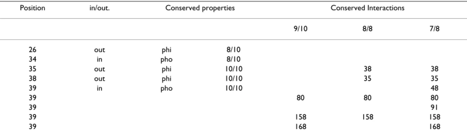
corre-Table 2: Conserved properties and interactions for Helix 1.
Position in/out. Conserved properties Conserved Interactions
9/10 8/8 7/8 26 out phi 8/10 34 in pho 8/10 35 out phi 10/10 38 38 38 out phi 10/10 35 35 39 in pho 10/10 48 39 80 80 80 39 91 39 158 158 158 39 168 168
Positions in H1 that are conserved and related structural information i.e. the position of the residue relative to the barrel; inside (in) or outside (out). The conserved properties (10/10 and 8/10 conservation) are also presented (see table 1 for details), as well as the conserved interaction partner (for 9/10, 8/8 and 7/8 lipocalins).
ClustalW (A) and refined (B) alignment of LIR2 and 1QFT
Figure 4
ClustalW (A) and refined (B) alignment of LIR2 and 1QFT. The secondary structure of 1QFT and the prediction for
LIR2 (PROF) are shown. helix is in black (text in white) and β in gray. The numbering of 1QFT refers to that of Figure 1. Posi-tions at the extremities of the secondary structure elements are numbered to facilitate the reading. Residues interacting with histamine in the structures of 1QFT are underlined. Those belonging to the H site are bold. Boxes indicate examples of rea-ligned regions.
sponds to an Arg, while being a hydrophobic residue in lipocalins. The cysteine from βB, implicated in a disulfide bridge between the C-ter part and βB in 1QFT, is also not conserved.
Modification of the alignment
To alleviate those misalignments, the alignment has been manually modified taking into account the prediction of secondary structures, the conserved interactions and posi-tions for lipocalins and the cysteines implicated in disulfide bridges for 1QFT.
H1
To align H1 the conserved interaction between residues 34 and 37 is used (see the window on Figure 4). The cor-responding residues for lipocalins and 1QFT are both hydrophilic. Positions 35 and 39 are also used: for 1QFT, residue 35 (Ala) is small allowing to decrease the steric constraints between H1 and, βD and βE. A bulkier residue at that position would interfere with the interaction with βD-βE. Residue 39 makes a hydrophobic interaction with 91. In the modified alignment, residues 34, 35, 37 and 39 now correspond respectively to Asn, Ala, Arg and Val. For the N-ter region, the secondary structure and informa-tion about disulfide bonds did not aid the alignment since, it is predicted as β-strand by PROF for LIR2 but with a low reliability and is predicted as an α-helix by NPSA (data not shown).
βA
Residues 48 and 49 are implicated in conserved interac-tions: position 48 is a conserved Trp interacting with 80, 190 and 192 and 49 is a bulky hydrophobic residue inter-acting with 194. They both were used to align βA together with secondary structure predictions (see the window on Figure 4). The latter helps to obtain a global alignment but is not sufficient to avoid ambiguity, since it can be aligned in different ways. Adding restraints for residues 48 and 49 permit the ambiguity to be eliminated. In the modified alignment, they respectively correspond to Trp and Val for LIR2. Residues 80, 190, 192 and 194 are hydrophobic and susceptible to interactions with 48 and 49.
βB
To align βB, positions 75 and 80 were used together with secondary structure predictions. Residue 75 is a conserved Cys for arthropod lipocalins and residue 80 makes a con-served hydrophobic interaction with 93. Secondary struc-ture predictions permit a global alignment to be obtained and residues 75 and 80 eliminate ambiguity.
βC and βD
Strands βC and βD were not realigned as the secondary structure predictions are in good correspondence with
that of 1QFT and as positions 91, 93 and 115 are con-served hydrophobic residues. The corresponding residues for LIR2 are Phe, Tyr and Leu respectively. Residues 91 and 115 make conserved interactions with 80, 115 and 131. They all correspond to hydrophobic residues in the mod-ified alignment.
βE
In strand βE, the secondary structure predictions are in good correspondence with that of 1QFT. Furthermore, residues 131 and 133, both implicated in conserved hydrophobic interactions, are conserved residues in the ClustalW alignment. Nevertheless, an uncertainty remains about the alignment of that region. Indeed, if a gap is sup-pressed in N-ter to βE, the secondary structures are still in good correspondence and residues 131 and 133 are still hydrophobic residues.
To determine the correct alignment, position 129 is used. In 1QFT, Asn 129 makes two H-bonds with the NH and CO groups of the backbone of the twisted βD-βE loop. This loop is of equivalent length in 1QFT and LIR2, and is longer than for other lipocalins. In the case where a gap is suppressed in N-ter, residue 129 corresponds to Asn for LIR2. For this reason, the latter was chosen. In that case, residues 131 and 133 correspond respectively to Met and Phe.
βF, βG and βH
Strands βF, βG and βH were not realigned as the second-ary structures are in good correspondence and as residues 158, 167, 169, 170, 189 and 190 (respectively Val, Cys, Ile, Leu, Leu and Trp in LIR2) are potentially able to make the conserved interactions.
H2 and βI
H2 and βI were not realigned, as positions 205 and 227 are both Cys, as in 1QFT. In the latter, two disulfide bridges are present. One joins the C-ter part to βB and is conserved for arthropod lipocalins (75–227; 1QFT, 1PEE, 1I4U). The other bridges H2 to βG (167–205; also present for 1I4U). The corresponding Cys of LIR2 are conserved; furthermore, LIR2 possesses two supplementary cysteines that could form a disulfide bridge between H2 and βH. In the modified alignment, almost all residues in the hydrophobic internal cluster of LIR2 are conserved, only residues 156 (Asn) and 168 (Thr) are hydrophilic, as for 1QFT. In the external cluster, positions 52 and 171 are Arg. Despite their hydrophilic nature, they are able to make hydrophobic interaction through their hydropho-bic tail [29]. The sequence corresponding to loop βF-βG in LIR2 is similar to the SCR2 motif of the lipocalins. Resi-dues Thr-Asp-Tyr in 1AQB are equivalent to Ser-Asn-Tyr in LIR 2 [3].
The fairly good correspondence between the secondary structures of 1QFT and LIR2, combined with the conserva-tion of the residues implicated in the two conserved hydrophobic clusters and the conservation of the Cys involved in disulfide bridges in 1QFT, lend support that LIR2 belongs to the lipocalin family.
3D model
A 3D model was constructed using the refined alignment and the 1QFT structure as template. Modeller was used to build the model [30]. Its stereochemical validity was checked with the Procheck algorithm [31]. Only one resi-due is in the disallowed phi/psi region of the Ramachan-dran plot. Three others, located in loops, are in generously allowed region. In the model, it is noted that the two cysteines located on βH and H2, have no correspondence in 1QFT and are facing each other (residues 187 and 213 on Figure 3D). The distance between the Cα of the two residues is 6 Å, compatible with a disulfide bridge. For that reason, a model where Cys 187 and Cys 213 were restrained to form a disulfide bridge was calculated. This model is similar to that built without restraint (data not shown). In the model, all interactions conserved for lipoc-alins (9/10 study) are found for LIR2, except for that between residues equivalent to positions 48 and 80. As for 1QFT, residue 48 interacts with 78. Neither interactions with residue 91 (Phe) are conserved owing to the orienta-tion of its side chain, which points outside. Interacorienta-tions involving residues 156 and 168 are not conserved in LIR2 because of their hydrophilic nature. As for 1QFT, the resi-due equivalent to 52 (Arg) interacts through its hydropho-bic tail with 189 (Leu) and interacts with 221 (Thr). Experimental measurement of the secondary structure
FTIR measurements permitted the determination of the secondary structure of LIR2. The FTIR spectrum presents a maximum at 1632 cm-1, characteristic of β-structure (data
not shown). After deconvolution, there is 22% of α-helix, 48% of β-strand, 17% of turns and 13% of coil. This is typ-ical of lipocalins, notably 1QFT that has 19% α-helix, 43% of β-strand, 24% of turns and 13% of coil, as deter-mined on the RX structure.
Prediction of ligand binding
The analysis of the internal cavity of LIR2 reveals that the bottom of the barrel is more hydrophobic than for 1QFT and that the upper part contains almost all the hydrophilic residues of the cavity. As shown in Figure 4, the hydrophobic residues in the bottom of the barrel are conserved between LIR2 and 1QFT; i.e. Trp (48), Phe (78), Val (80), Tyr (93), Leu (115), Met (131), Phe (133), Val (158), Leu (170) and Trp (190).
When comparing the residues of the L site, that participate in the binding of the histamine in 1QFT, to the
corre-sponding residues in LIR2, it appears that the negative res-idues Asp 42 and 168 (see Figure 4) are not conserved. In 1QFT, these have been shown to interact with histamine. The corresponding residues in LIR2 are Asn (42) and Thr (168). As no negative residue is conserved in the L site for LIR2, no binding of histamine is predicted for that site. Concerning the H site in LIR2, the negative residues are pretty well conserved; only residue 156 (Asn) is not. How-ever, LIR2 contains a positive residue (Lys 50) in the cavity and two others (Arg 69 and 111) that are susceptible to belong to the ligand-binding pocket; these would repulse for histamine binding. Furthermore, the aromatic resi-dues (Trp 69 and Phe 154) that are parallel to the cycle of histamine in 1QFT are not conserved in LIR2 (respectively Arg and Thr). For RaHBP1 (a close homologue to RaHBP2), such a substitution (Phe 154 is substituted by Leu) causes a significant decrease in affinity for histamine [6]. Furthermore, in the loops surrounding the entry of the H site, the ratio of negative to positive residues is 7/1 for 1QFT and 2/3 for LIR2. In 1QFT, the presence of these negative residues in the loops were proposed to contrib-ute to the attraction of histamine to the binding site [6]. For the H site, despite the fact that most of the negative histamine-binding residues are conserved, the absence of the aromatic residues and of one negative residue should hinder high affinity binding of histamine for LIR2. Experimental determination of the affinity of LIR2 to histamine LIR2 and RaHBP2 were expressed in 293T free-serum cell medium. The ability of LIR2 to bind histamine was tested by incubating concentrated supernatant cells containing LIR2 with 3H-histamine. RaHBP2 was used as positive
control, and a concentrated supernatant of untransfected cells used as negative control. These binding assays show high affinity for RaHBP2, and no affinity for LIR2 (similar cpm value to supernatant of untransfected cells) confirm-ing that LIR2 is unable to bind histamine, as predicted from the model (Figure 5).
Discussion
The aim of the present work is to provide information to help the construction of 3D models for the weakly related proteins of the lipocalin family. The members of this fam-ily have a wide variety of functions and are hence of bio-logical importance. The identity between lipocalins can fall below 10%. Building a 3D model by homology mod-eling for proteins having an identity below the 25–30% cut-off is quite risky and requires that the selection of the template and the alignment with the target be further val-idated. This can be done by comparing predictions of sec-ondary structures, accessibility to the solvent and patterns of hydrophobic and peculiar residues. In this work, infor-mation about the structural core of lipocalins was extracted and used to build a 3D model for LIR2, a protein
from the tick Ixodes ricinus. For that purpose, a set of lipoc-alin structures was analyzed and conserved properties were identified. To capture the widest diversity it was tried to find a structure for each clade identified in the phylo-genic study of lipocalins (Ganfornina et al., 2000). Nine structures were collected. Nitrophorin 2 [PDB: 1PEE] and RaHBP2 [PDB: 1QFT] were included into the study. The latter was studied separately owing to its uncommon α-conformation of the first helix and to its hydrophilic bind-ing sites. The lipocalins were structurally aligned with the VAST method.
Conserved positions of the alignment
To analyze the conserved properties of the alignment, the amino acids were classified as hydrophobic, hydrophilic, aromatic, charged, bulky or small. For the 10 lipocalins, having a mean length of 170 amino acids, only 25 posi-tions are conserved, two are kept strictly identical (Gly (42) and Trp (48) from SCR1) and one is negative (192 from SCR3). The ratio of conserved hydrophobic versus hydrophilic positions is nearly three to one. All conserved hydrophobic positions have a solvent accessible surface less than 30%. The size of the residues is less conserved than their hydrophobicity. Only 7 positions are conserved in terms of size; however this is not unusual [32].
The side chain-side chain interactions were studied for
each structure. Interactions were divided into four classes depending on the nature of the residues implicated, i.e. hydrophobic, hydrophilic, charged or aromatic. At first, RaHBP2 was not considered in the analysis and the con-servation of interactions for 10/10 and 9/10 structures were analyzed. In both studies, no conserved electrostatic
or aromatic interactions were found and mostly all con-served interactions were hydrophobic. The pattern of hydrophobic interactions suggests the existence of two clusters, one internal to the barrel and one external. The internal cluster is composed by residues 39, 48, 80, 91, 93, 115, 131, 133, 156, 158, 168, 170, 190 and 192 (Figure 3A), and the external by residues 52, 159, 169, 171, 189, 205 and 220 (Figure 3B), when considering the 9/10 study.
In the internal cluster, the β strands are linked by 14
hydrophobic interactions (Figures 2A and 3A), forming what can be seen as a hydrophobic belt. A similar belt was detected in the 10 β-strand barrel of the lipid-binding pro-tein family [33,34]. This belt is linked to the central resi-due (39) of H1 by 5 interactions, coming from 5 different strands (Figure 2B). The central residue hence appears important in the structural core of lipocalins. The helix is further stabilized by an internal hydrophilic interaction. Among the conserved positions in the alignment and not appearing in the conserved interactions (9/10 and 7/10 study; Figure 1) are positions 34, 38, and 165. They are involved in less conserved interactions between the barrel and H1.
The external cluster implicates helix 2 and strands A,
β-F, β-G, β-H as well as the C-terminal region; six interac-tions are conserved (Figures 2A and 3B). There is also an additional hydrophilic interaction (162–166) that stabi-lizes the βF-βG loop (SCR2).
It should be noted that all conserved hydrophobic posi-tions in the alignment are implicated in conserved inter-actions, except positions 49 and 214, for which interactions are conserved for a fewer number of struc-tures. These last two positions belong to the external clus-ter. Among the 6 conserved hydrophilic positions in the alignment, three are implicated in SCR3 and one (110) seems to be involved in the stabilization of the interaction between strands βB and βC on the external surface of the barrel.
Our results were compared to those of Ragona and col. [20] who have identified by NMR the interacting residues in partially folded bovine β-lactoglobulin at pH 2. These residues located in the cavity of the barrel correspond to positions 39, 48, 77, 80, 91, 131, 156, 158, 168, 170 and 190 in our alignment. Residues 93, 95, 115, 133 could not be unambiguously detected by NMR, but they were assigned by the authors to the internal cluster using the X-ray structure. All these residues are in good correspond-ence with those identified in the present study. The inter-acting residues in the external cluster (detected by NMR) correspond to positions 52, 167, 169, 204, 205, 208. Res-idues 51, 77 were furthermore assigned to this cluster Binding assay of LIR2 with histamine
Figure 5
Binding assay of LIR2 with histamine. Binding assay was
performed with 40 µl of concentrated 293T supernatant cell culture. The negative control used was a 10 time concen-trated free-serum medium of untransfected cells. The super-natants were incubated with 100 nM 3H-histamine for 2
hours at 37°C. Protein precipitation with polyethylene glycol 8000 was used to separate bound from free histamine.
using the X-ray data. Residue 77 was not detected as mak-ing conserved interaction in our study. This is due to the presence of a beta bulge in the 77–80 region. This bulge is present in our study for β-lactoglobulin [PDB: 1EXS], the odorant binding lipocalin from nasal mucosa of pig [PDB: 1A3Y] and the mouse major urinary protein [PDB: 1DF3]. Residues 204 and 208 respectively interact with residues 167 and 169 in the NMR study. In the present work, residues 204 and 167 do not appear to interact owing to the orientation of H2 towards the barrel that dif-fers for β-lactoglobulin. Residues 208 and 169 were not detected to be involved in a strictly conserved interaction. In effect they show interaction for 6/10 structures and two structures have an Arg at position 208 interacting through its hydrophobic region with residue 169. Likewise, resi-due 208 (Lys) of RaHBP2 is also interacting with 169 through its hydrophobic region.
It was suggested that β-lactoglobulin at acidic pH is in a molten globule state, similarly to the retinol binding pro-tein [26]. Since the residues implicated in interactions in the β-lactoglobulin molten globule correspond well with those conserved for native lipocalin structures, it supports the hypothesis that residues essential in the native struc-ture of the lipocalins are also important for the folding, as suggested by Ragona et al. [35]. Clarke et al. reached a similar conclusion for the immunoglobulin-like proteins, a highly diverse protein family with no conservation of function and little or no sequence identity [16].
Greene et al. have studied the evolutionarily conserved residues (ECR) in 32 lipocalins [19]. Many of those resi-dues are hydrophobic and equivalent to those highlighted in this work (residues 34, 39, 42, 48, 49, 52, 54, 129, 131, 158, 161, 162, 163, 167, 189, 191, 192, 200, 203 and 205 in our alignment). However, no residues from βB, βC, βD are found conserved by Greene et al. Even if fewer interac-tions are conserved for these strands, our study and that of Ragona et al.[20] clearly suggest that some residues of these strands also play a role in the hydrophobic internal cluster, closing the belt. This discrepancy could be due to the fact that our alignment is based on the structures and not on the sequences alone. Some other residues, such as 161 and 163 belonging to the βF-G loop (SCR2) are described as ECR, but are not found as conserved in this study. This is because outlier lipocalins were included in the alignment. Since residues 133 and 190 are implicated in conserved interactions for 11/11 structures it is surpris-ing not to see them in the ECRs. Again this could be due to the way lipocalins were aligned.
RaHBP2
In our alignment, RaHBP2 [PDB: 1QFT] was added. The latter is an outlier lipocalin with a low similarity to the other lipocalins. It lies apart from the family since it does
not share the conserved regions of the family, binds a hydrophilic ligand and its H1 is in α-conformation. When considering RaHBP2 in the conservation analysis, it comes out that only four interactions are conserved for the 11 lipocalins, three in the internal cluster and one in the external. The interactions in the barrel link the two sheets (ABCD and EFGH) together. When comparing the inter-actions conserved for the 9/10 lipocalins to those of RaHBP2, it appears that the belt is not fully conserved owing to the hydrophilic nature of two residues (156 and 168) and to the orientation of residue 131, involved in the binding with histamine. The interaction 48–80 is not present: due to the α-conformation of H1, residue 48 moves away from residue 80. Nevertheless, there are neighboring interactions than can compensate these lacks. H1 of RaHBP2 has no central residue equivalent to residue (39) interacting with strands βA, βB, βC, βF and βG, while still interacting with those strands through dif-ferent residues.
Thus, even though H1 has a different conformation and though there are two hydrophilic binding sites, the hydro-phobic internal cluster of RaHBP2 is fairly well conserved. The external cluster of RaHBP2 is conserved except for interactions 52–189 and 52–220 because of the hydrophilic nature of residue 52, but a careful analysis reveals that residue 52 interacts through its hydrophobic moiety with residue 189. For RaHBP2 (as for α-crustacy-anin [PDB: 1I4U]), a disulfide bond bridges βG to H2.
Modeling of LIR2
To determine the homology and build a 3D model for LIR 2, a protein with only 15% identity with RaHBP2 [PDB: 1QFT], information was combined from the analyses of the structural core of lipocalins, the positions of cysteines implicated in disulfide bridges in RaHBP2 and the sec-ondary structure. In a first approach, alignment between LIR2 and RaHBP2, was carried out by ClustalW. The align-ment showed inconsistencies in the Cys bonding pattern, the secondary structures and the conserved hydrophobic residues. It was corrected for H1 and strands βA, βB and βE. The information obtained from the comparative anal-ysis enabled the alignment to be improved. Due to the low similarity between the two sequences, the secondary structure prediction of LIR2 and the cysteine bridge con-served among arthropod lipocalins did not provide enough information to obtain an unambiguous align-ment. This holds true for PSI-BLAST alignment (not shown). The information from conserved interactions has permitted to obtain a coherent alignment. It is important to note that the analysis of the structural core is not aimed to perform better than PSI-BLAST (or ClustaW), but rather to eliminate ambiguities and assess the alignment obtained by those methods.
The corrected alignment enabled the building of a 3D model for LIR2. The model shows a potential disulfide bridge, not present in RaHBP2, supporting both the assignment of the fold and the alignment (Figure 3D). This is further supported on one hand by the FTIR meas-urements that indicate a secondary structure compatible with the lipocalin fold and on the other hand by the con-servation in LIR2 of most of the conserved hydrophobic interactions. Despite its homology with RaHBP2, the analysis of the model of LIR2 does not suggest binding to histamine, as confirmed experimentally. A more detailed study of the cavity should further help to understand the nature of its natural ligand.
Conclusion
The lipocalins are part of a protein super-family with a low level of pairwise similarity, making homology mode-ling a difficult task. In this study, it was shown that the determination of the residues implicated in the hydro-phobic core of lipocalins, by analyzing the conserved interactions, enabled to assess the assignment of a lipoca-lin-like protein and to improve the "classical" alignment in ambiguous regions. Information obtained from that study should help modeling other lipocalin-like proteins. This study could be applied to other protein families with low pairwise similarity, such as the structurally related fatty acid binding proteins or avidins.
Methods
Computational methods
Lipocalin analysis
To study the lipocalin family a bank of structurally aligned lipocalins with low similarity was gathered. For this pur-pose, it was tried to obtain a structure for each of the clades identified in the phylogenic analysis of Ganfornina and col. [36]. A 3D structure was found in the Protein Data Bank (PDB) for nine out of 13 clades. Since nitro-phorin [23], from Rhodnius prolixus and RaHBP2 from Rhi-picephalus appendiculatus were not considered in the phylogenetic tree owing to their low similarity, they were added in the bank. The structural alignment was gener-ated by the VAST algorithm [37] and includes the odorant binding lipocalin from nasal mucosa of pig [PDB: 1A3Y], the retinol binding lipocalin of pig [PDB: 1AQB], the mouse major urinary protein [PDB: 1DF3], the beta-lac-toglobulin of pig [PDB: 1EXS], α-crustacyanin [PDB: 1I4U], Human Complement Protein C8 γ [PDB: 1LF7], the human neutrophil gelatinase-associated lipocalin [PDB: 1NGL], nitrophorin 2 from Rhodnius prolixus [PDB: 1PEE], the Rhipicephalus appendiculatus histamine binding lipocalin 2 [PDB: 1QFT], the bacterial outer membrane lipoprotein blc [PDB: 1QWD] and the human tear lipoca-lin [PDB: 1XKI]. The retinol-binding protein (1AQB) is one of the best characterized lipocalin. Consequently, it was used as reference for the calculation of the deviation
between the structures. The RMSD between 1AQB and the other structures is between 2 and 3 Å. For some structures, the natural sequence was not conserved thoroughly; some substitutions were introduced. For the human tear lipoca-lin, residue 161 is naturally a Cys but is a Ser in the struc-ture 1XKI. In 1QWD, some residues have been mutated in the region before H1. In 1I4U, residue 94 (Lys) has been replaced by Glu [βC, facing outside]. This could destabi-lize the electrostatic interaction with Glu 79. Residue 88 in 1LF7A (Cys) has been replaced by Gly [loop between βB and βC]. In 1DF3 residue 207 (Lys) has been replaced for Gln [H2, facing exterior]. Furthermore, for 1XKI sev-eral loops are lacking in the structure, as well as H1 and the C-ter part. For 1A3Y, H1 is missing and for 1LF7 the omega loop is not present.
Study of the interactions
The interactions were computed from PDB files with the PEX software[22]. Previously, the PDB files corresponding to the structures were renumbered, so that spatially equiv-alent residues (i.e. having the same position in the struc-tural alignment) have the same number. This procedure considers that amino acids interact when the center to center distance between their closest atoms is less than 4.5 Å. Residues which interact must be separated by at least by two residues in the sequence. The accessible surface area (ASA) was calculated using the method of Shrake and Rupley [38]. To be considered accessible (or inaccessible) to the solvent a residue has to have an ASA more (or less) to 30% of its total surface.
Modeling of LIR2
The PROF prediction of secondary structure was obtained through the PredictProtein server [25,39]). The ClustalW algorithm was used to generate the non-refined alignment between LIR2 and the sequence of 1QFT [40]. The 3D model of LIR2, comprising residues 22 to 196, was gener-ated by Modeller [30], using a refined alignment. The model was afterwards evaluated by Procheck [31].
Experimental methods
Sequencing LIR2
mRNAs from salivary glands of 30 engorged females of Ixodes ricinus were extracted using the Micro-FastTrack 2.0 mRNA Isolation Kit (Invitrogen, Carlsbad, USA). The complete cDNA sequence of LIR2 was recovered by RACE-PCR (Gene Racer Kit, Invitrogen) performed according to manufacturer's recommended procedure.
Characterizing LIR2
The molecular weight of LIR2 is of 24.2 kDa and its isoe-lectric point is of 8.97, as determined by "pepstats". The signal peptide is predicted by "SignalP" to be of 20 amino acids.
Plasmid construction, protein expression, and purification of LIR2 for FTIR measurements
The coding region of LIR2 was amplified by PCR (94°C for 30 s, 56°C for 30 s, 72°C for 1 min.; 30 cycles) with the ExTaq DNA Polymerase. The PCR product was ligated into the pCRII-TOPO vector and then excised with EcoRI and SacI sites that were added at the 5' end of the primers. The resulting DNA fragment was cloned into the pBlueBac4.5-V5-His (Invitrogen) in frame with the cod-ing sequence of the V5 and His epitopes at the C-termi-nus. Recombinant baculoviruses were made by recombination between pBlueBac/LIR2 and Bac-N-Blue linear DNA virus (Invitrogen). Recombinant viruses were selected and amplified according to the manufacturer's instruction. SF9 cells were infected with a high-titer stock of recombinant baculovirus and were incubated for 72 hours at 27°C in Sf900 II Serum-Free Medium (Invitro-gen). Recombinant LIR2 was recovered in 50 mM NaH2PO4 buffer (pH 7.5) containing 150 mM NaCl by using a Ni2+ sepharose (Qiagen) column.
Expression of recombinant LIR2 and RaHBP2 for binding assays Messenger RNAs were extracted either from salivary glands of 2-days engorged Rhipicephalus appendiculatus females or from salivary glands of 5-days engorged Ixodes ricinus females. The extraction was performed by using Micro-Fast Track 2.0 mRNA isolation kit (Invitrogen). The mRNA was reverse-transcribed into cDNA using Super-Script II Reverse Transcriptase (Invitrogen). The coding regions of both RaHBP2 and LIR2 genes were amplified by PCR (94°C for 30 seconds, 56°C for 30 seconds, 72°C for 1 minute; 30 cycles) by using the Takara ExTaq DNA polymerase. The PCR products were cloned into the pcDNA3.1/V5-His-TOPO (Invitrogen). The resulting recombinant plasmids were used to transfect 293T cells with FuGene 6 (Roche applied science, Indianapolis, USA); and the proteins were expressed in fusion with a V5 and a 6-HIS epitope tag. The proteins were produced in a serum- free medium. The supernatants were collected 72 hours after transfection, and concentrated 10 fold with an Amicon membrane (Cutoff of the membrane: 10000 NMWL). The supernatants were then dialyzed against PBS (20 mM NaH2PO4, 150 mM NaCl, pH 7.4) and ultracen-trifugated at 140 000 g before used.
Binding assay with histamine
3H-histamine was purchased form GE Healthcare (Little
Chalfont, Buckinghamshire, United Kingdom). Protein precipitation with polyethylene glycol 8000 to separate bound from free histamine followed the procedure described by Warlow and Bernard [41]. The radioactivity was counted in a Wallac 1409 scintillation counter.
Infrared spectroscopy (FTIR) measurements
Attenuated Total Reflection (ATR) infrared spectroscopy was used to determine the secondary structure of LIR2. Spectra were recorded at room temperature on a Brüker Equinox 55 equipped with a liquid nitrogen-cooled Mer-cury-Cadmium-Telluride (MCT) detector at a resolution of 2 cm-1, by averaging 512 scans. The internal reflection
element was a germanium ATR plate (50 × 20 × 2 mm, Aldrich Chimica) with an aperture of 45° yielding 25 internal reflections. Reference spectra of a Germanium plate were automatically recorded after purge of 15 min-utes with dry air and rationed against the recently run sample spectra. Seventy µg of protein was spread out on the plate and slowly dried under a stream of N2. The plate was sealed in a universal sample holder and rehydrated by flushing the holder with N2 saturated with D2O for 4 hours at room temperature.
Secondary structure determination
Vibrational bands, especially the amide I band (1600– 1700 cm-1), are sensitive to the secondary structures of the
proteins. The C=O vibration is representative of 80% of the amide I band. This band accounts for all the secondary structures that have different vibration values. The combi-nation of resolution-enhancement methods with curve-fitting procedures allows quantitatively different second-ary structures such as α-helix, β-sheets and unordered structures to be assigned. Each band was assigned accord-ing to the frequency of its maximum. The areas of all bands assigned to a given secondary structure were then summed and divided by the sum of all areas. This gives the relative ratio of each secondary structure. The bands are assigned as follows [42]: α-helix: 1662-1645 cm-1,
β-sheets: 1689-1682 cm-1 and 1637-1613 cm-1, random
1645-1637 cm-1, β-turns: 1682-1662 cm-1. It should be
noted that the proteins spread on the plate are deuterated to avoid an overlap of α-helix and random-coil structures, as previously described [42].
Authors' contributions
AB conceived the study, participated in the design of the study, carried out the lipocalin structural analysis, the alignment of LIR2 and its 3D model, and drafted the man-uscript.
CB participated in the design of the study and drafted the manuscript.
BJ participated in the design of the experimental part, car-ried out the sequencing, characterization, expression and binding assays for LIR2 drafted the experimental part of the manuscript.
VL participated in the design of the experimental part and revised the manuscript.
GE participated in the design of the experimental part and revised the manuscript.
BR participated in the design and revised the manuscript. LL conceived the study, participated in the design of the study and revised the manuscript.
All authors read and approved the final manuscript.
Additional material
Acknowledgements
We thank Philippa Talmud for helpful reading of the manuscript and Chris-telle Flore for her excellent technical assistance. This work was supported by grants 215370 (Bioflam) and 215140 (αBUSTEC) from the "Région Wal-lonne" (Belgium). R. B. and L.L are Research Director and Research Asso-ciate, respectively at the Belgian Fund for Scientific Research (FNRS). A. B. and J.B. were supported by the Ministère de la Région Wallonne (DGTRE; Grant 215370). C. B. was supported by the Ministère de la Région Wall-onne (DGTRE; Grant 215370).
References
1. Flower DR: The lipocalin protein family: structure and
func-tion. Biochem J 1996, 318 ( Pt 1):1-14.
2. Cowan SW, Newcomer ME, Jones TA: Crystallographic
refine-ment of human serum retinol binding protein at 2A resolu-tion. Proteins 1990, 8:44-61.
3. Flower DR, North AC, Attwood TK: Structure and sequence
relationships in the lipocalins and related proteins. Protein Sci
1993, 2:753-761.
4. Mans BJ, Neitz AW: Exon-intron structure of outlier tick
lipoc-alins indicate a monophyletic origin within the larger lipoca-lin family. Insect Biochem Mol Biol 2004, 34:585-594.
5. Sanchez D, Ganfornina MD, Gutierrez G, Marin A: Exon-intron
structure and evolution of the Lipocalin gene family. Mol Biol
Evol 2003, 20:775-783.
6. Paesen GA, Adams PL, Nuttall PA, Stuart DL: Tick
histamine-bind-ing proteins: lipocalins with a second bindhistamine-bind-ing cavity.
Biochim-ica et BiophysBiochim-ica Acta-Protein Structure and Molecular Enzymology 2000,
1482:92-101.
7. Ganfornina MD, Kayser H, Sanchez D: Lipocalins in Arthropoda:
Diversification and Functional Explorations. lipocalins 2006.
8. Francischetti IM, My P V, Mans BJ, Andersen JF, Mather TN, Lane RS, Ribeiro JM: The transcriptome of the salivary glands of the
female western black-legged tick Ixodes pacificus (Acari: Ixodidae). Insect Biochem Mol Biol 2005, 35:1142-1161.
9. Mans BJ, Louw AI, Neitz AW: The major tick salivary gland
pro-teins and toxins from the soft tick, Ornithodoros savignyi, are part of the tick Lipocalin family: implications for the ori-gins of tick toxicoses. Mol Biol Evol 2003, 20:1158-1167.
10. Nunn MA, Sharma A, Paesen GC, Adamson S, Lissina O, Willis AC, Nuttall PA: Complement inhibitor of C5 activation from the
soft tick Ornithodoros moubata. J Immunol 2005, 174:2084-2091.
11. Leboulle G, Rochez C, Louahed J, Rutti B, Brossard M, Bollen A, God-froid E: Isolation of Ixodes ricinus salivary gland mRNA
encoding factors induced during blood feeding. Am J Trop Med
Hyg 2002, 66(3):225-233.
12. Rost B: Twilight zone of protein sequence alignments. Protein
Eng 1999, 12:85-94.
13. Marti-Renom MA, Stuart AC, Fiser A, Sanchez R, Melo F, Sali A:
Comparative protein structure modeling of genes and genomes. Annu Rev Biophys Biomol Struct 2000, 29:291-325.
14. Rost B: Protein structures sustain evolutionary drift. Fold Des 1997, 2:S19-S24.
15. Perutz MF, Kendrew JC, Watson HC: Structure and function of
haemoglobin II. Some relations between polypeptide chain configuration and amino acid sequence. J Mol Biol 1965, 13:669-678.
16. Clarke J, Cota E, Fowler SB, Hamill SJ: Folding studies of
immu-noglobulin-like beta-sandwich proteins suggest that they share a common folding pathway. Structure 1999, 7(9):1145-1153.
17. Ptitsyn OB: Protein folding and protein evolution: Common
folding nucleus in different subfamilies of c-type cyto-chromes? Journal of Molecular Biology 1998, 278:655-666.
18. Socolich M, Lockless SW, Russ WP, Lee H, Gardner KH, Ranganathan R: Evolutionary information for specifying a protein fold.
Nature 2005, 437:512-518.
19. Greene LH, Hamada D, Eyles SJ, Brew K: Conserved signature
proposed for folding in the lipocalin superfamily. Febs Letters
2003, 553:39-44.
20. Ragona L, Pusterla F, Zetta L, Monaco HL, Molinari H: Identification
of a conserved hydrophobic cluster in partially folded bovine beta-lactoglobulin at pH 2. Folding & Design 1997, 2:281-290.
21. Flower DR, North ACT, Sansom CE: The lipocalin protein family:
structural and sequence overview. Biochimica et Biophysica
Acta-Protein Structure and Molecular Enzymology 2000, 1482:9-24.
22. Thomas A, Bouffioux O, Geeurickx D, Brasseur R: Pex, analytical
tools for PDB files. I. GF-Pex: Basic file to describe a protein.
Proteins-Structure Function and Genetics 2001, 43:28-36.
23. Montfort WR, Weichsel A, Andersen JF, Roberts SA, Walker FA, Shepley DP, Balfour C: Nitric oxide transport by a
blood-suck-ing insect: Nitrophorin structure, dynamics, and distal pocket burial. Biophysical Journal 2000, 78:41A-.
24. Altschul SF, Madden TL, Schaffer AA, Zhang JH, Zhang Z, Miller W, Lipman DJ: Gapped BLAST and PSI-BLAST: a new generation
of protein database search programs. Nucleic Acids Research
1997, 25:3389-3402.
25. Rost B, Yachdav G, Liu JF: The PredictProtein server. Nucleic
Acids Research 2004, 32:W321-W326.
26. Greene LH, Chrysina ED, Irons LI, Papageorgiou AC, Acharya KR, Brew K: Role of conserved residues in structure and stability:
Tryptophans of human serum retinol-binding protein, a model for the lipocalin superfamily. Protein Science 2001, 10:2301-2316.
27. Gasymov OK, Abduragimov AR, Yusifov TN, Glasgow BJ: Binding
studies of tear lipocalin: the role of the conserved tryp-tophan in maintaining structure, stability and ligand affinity.
Biochimica et Biophysica Acta-Protein Structure and Molecular Enzymology
1999, 1433:307-320.
28. Katakura Y, Totsuka M, Ametani A, Kaminogawa S: Tryptophan-19
of Beta-Lactoglobulin, the Only Residue Completely Con-served in the Lipocalin Superfamily, Is Not Essential for Binding Retinol, But Relevant to Stabilizing Bound Retinol and Maintaining Its Structure. Biochimica et Biophysica Acta-Protein
Structure and Molecular Enzymology 1994, 1207:58-67.
Additional file 1
Identity table for the lipocalins aligned by the VAST method. These
include the structures of the odorant binding lipocalin from nasal mucosa of pig (1A3Y, [43]), the retinol binding lipocalin of pig (1AQB, [44]), the mouse major urinary protein (1DF3, [45]), the beta-lactoglobulin of pig (1EXS, [46]), α-crustacyanin (1I4U; [47]), Human Complement Protein C8 γ (1LF7, [48]), the human neutrophil gelatinase-associated lipocalin (1NGL, [49]), nitrophorin 2 from Rhodnius prolixus (1PEE, [23]), the Rhipicephalus appendiculatus histamine binding lipocalin 2 (1QFT, [50]), the bacterial outer membrane lipoprotein blc (1QWD, [51]) and the human tear lipocalin (1XKI, [52]). On the diagonal is the number of residues for each sequence, in the upper triangle the number of identical residues and in the lower triangle the percentage of sequence identity (identities/length of the shorter sequence).
Click here for file
[http://www.biomedcentral.com/content/supplementary/1472-6807-8-1-S1.doc]