Université de Montréal
Potential pathogenicity and antimicrobial resistance of Escherichia
coli from pig and poultry feces on-farm and carcasses at the abattoir
in Vietnam
par THU MINH PHAM
Département de pathologie et microbiologie,
Faculté de médecine vétérinaire
Mémoire présenté à la Faculté de médecine vétérinaire en vue de
l’obtention du grade de maître ès sciences (M.Sc.) en sciences
vétérinaires option hygiène vétérinaire et innocuité des aliments
Décembre 2012
© Thu Minh Pham, 2012
iii
Résumé
E. coli avec potentiel zoonotique pourrait éclore dans les réservoirs porcins et avicoles. Cette étude consiste à examiner la présence de souches E. coli porteuses de gènes virulents associés aux STEC (E. coli producteurs de Shiga-toxines), EPEC (E. coli entéropathogène), et ExPEC (E. coli pathogène extra-intestinal) chez les porcs et volailles élevés au Vietnam. Des prélèvements d’excréments et de carcasses ont été effectués dans des fermes et abattoirs porcins et avicoles sélectionnés où les animaux ont été suivis de l’élevage à l’abattage. Un total de 13,1% des souches, toutes sources confondues, ont été catégorisées comme potentiellement contaminées par ExPEC, possédant un ou plusieurs gènes de virulence iucD, tsh, papC et cnf. Peu d’isolats d’autres pathotypes ont été observés. Tous les gènes de virulence ExPEC, à l’exception de cnf, ont été identifiés plus fréquemment dans les isolats de fèces et carcasses avicoles que dans les isolats porcins. Même constatation pour le groupe du phylogénétique D. Une multirésistance aux médicaments a été régulièrement observée chez les deux isolats ExPEC. Les isolats de fèces de volailles ont souvent été associés à une résistance à l’acide nalidixique et à la ciprofloxacine (P<0.05), de même qu’au gène blaTEM, alors que les gènes qnr et aac(6’)-Ib ont peu été rencontrés
des deux côtés. Cette étude démontre que les isolats ExPEC avicoles sont potentiellement plus pathogèniques que ceux porcins et que les isolats ExPEC de carcasses porcines et avicoles peuvent provenir de leurs excréments par la contamination associée au processus d'abattage. Ainsi, la volaille, particulièrement, serait un facteur de transmission de souches ExPEC zoonotiques.
Mots clé : résistance antimicrobienne, gènes virulents, porcs et volailles, fermes, abattoirs, carcasses, ExPEC, PCR.
iv
Summary
Zoonotic potential pathogenic Escherichia coli could arise from poultry and pig reservoirs. The aim of this study is to investigate the occurrence of E. coli strains carrying virulence genes associated with STEC (Shiga toxin-producing E. coli), EPEC (Enteropathogenic E. coli), and ExPEC (Extraintestinal pathogenic E. coli) in pigs and poultry on-farm and at abattoirs in Vietnam. Samples of feces and carcasses were collected at selected pig and poultry farms and abattoirs, in which animals were traced from farms to the abattoir. A total of 13.1% strains from all sources were classified as potential ExPEC, possessing one or more virulence genes iucD, tsh, papC and cnf. Few isolates of other pathotypes were observed. All ExPEC virulence genes, except cnf, were more frequently found in isolates from poultry than in isolates from pigs. A higher proportion of ExPEC isolates belonging to phylogenetic group D was observed in poultry. Multi-drug resistance was frequently observed in ExPEC isolates from both pigs and poultry. Nalidixic acid and ciprofloxacin resistance were significantly associated with poultry feces isolates (P<0.05). blaTEM
gene was more frequently associated with poultry isolates, whereas qnr and aac(6’)-Ib genes were present at low prevalence in pig and poultry isolates. This study demonstrates that poultry ExPEC isolates are potentially more pathogenic than pig ExPEC isolates, and ExPEC isolates in pig and poultry carcasses may originate from pig and poultry feces, due to contamination associated to slaughtering process. Thus, meats particularly from poultry, might be a vehicle for transmission of zoonotic ExPEC strains.
Key words: antimicrobial resistance, virulence genes, pigs and poultry, farms, abattoirs, carcasses, ExPEC, PCR.
v
TABLE OF CONTENTS
SUMMARY ... ii Résumé ... iii Summary ... iv TABLE OF CONTENTS ... vLIST OF TABLES ... viii
LIST OF ABBREVIATIONS ... ix ACKNOWLEDGEMENTS ... xiii INTRODUCTION ... 1 LITERATURE REVIEW ... 4 1. Classification of E. coli ... 5 1.1. Commensal E. coli ... 5
1.2. Intestinal pathogenic E. coli ... 5
1.2.1.Enteropathogenic E. coli (EPEC) ... 6
1.2.2. Shiga toxin-producing E. coli (STEC) ... 7
1.2.3. Enterotoxigenic E. coli (ETEC)... 8
1.2.4. Enteroaggregative E. coli (EAEC) ... 8
1.3. Extraintestinal pathogenic E. coli (ExPEC) ... 9
2. Diseases caused by ExPEC in humans ... 10
2.1. Urinary tract infections ... 10
2.2. Neonatal meningitis ... 11
2.3. Sepsis ... 11
3. Virulence factors of pathogenic E. coli ... 12
3.1. Adhesins ... 12
3.1.1. Type 1 fimbriae ... 13
3.1.2. P fimbriae ... 13
3.1.3.Temperature sensitive hemagglutinin ... 15
3.2. Capsules (K antigen) ... 16
3.3. Lipopolysaccharide... 16
3.4. Iron acquisition systems ... 17
vi
3.5.1.Hemolysin ... 18
3.5.2 Cytotoxic necrotizing factor ... 18
4. Association among virulence factors and phylogenetic groups ... 19
4.1. Associations among virulence factors ... 19
4.2. Association of phylogenetic groups and virulence factors ... 20
5. Antimicrobial resistance (AMR) of E. coli ... 21
5.1. Mechanisms of antimicrobial resistance... 22
5.2. Antimicrobial use and emergence of antimicrobial resistance in food animals ... 23
5.2.1. Antimicrobial use in animal production ... 23
5.2.2.The trend in antimicrobial resistance in E. coli isolates from food-producing animals ... 24
5.3. Resistance of E. coli to category I antimicrobials ... 25
5.3.1. Resistance to fluoroquinolone ... 25
5.3.2. Resistance to cephalosporins ... 26
5.4. Resistance of E. coli to category II antimicrobials ... 27
5.4.1. Resistance to penicillins ... 27
5.4.2. Resistance to trimethoprim/sulfamethoxazole ... 27
5.5. Resistance to category III antimicrobials ... 28
5.5.1. Resistance to tetracyclines ... 28
5.5.2. Resistance to streptomycin ... 29
6. Multi-antimicrobial resistance among E. coli... 29
6.1. Multidrug resistance associated with ESBLs ... 29
6.2. Multi-resistance associated with PMQR ... 30
7. Association between virulence and antimicrobial resistance of ExPEC ... 30
8. Food animals as a reservoir of extraintestinal pathogenic E. coli in humans ... 31
8.1. ExPEC in poultry ... 32
8.2.ExPEC in pigs ... 34
8.3 Prevalence of ExPEC in food derived from animals ... 35
9. Molecular methods for determination of pathogenic E. coli ... 37
9.1. General information on PCR ... 37
9.2. Multiplex PCR for identification of E. coli pathotype ... 37
9.2.1. Multiplex PCR for identification of intestinal pathogenic E. coli ... 37
vii
9.3. Multiplex PCR for determination of the E. coli phylogenetic group ... 38
9.4. Other techniques for identification of pathogenic E. coli ... 39
10. Rationale and objectives of the study ... 40
METHODOLOGY AND RESULTS ... 41
ARTICLE IN PREPARATION ... 43
DISCUSSION ... 65
CONCLUSION ... 76
REFERENCE LIST ... 79
ANNEXES ... 95
Annex 1 List of samples of poultry feces and carcasses collected in Vietnam ... i
Annex 2 List of samples of pig feces and pig carcasses collected in Vietnam ... ii
Annex 3. Association of virulence genes and antimicrobial resistance among 132 potential ExPEC isolates ... iii
Annex 4. Association of phylotypes and antimicrobial resistance among 132 potential ExPEC isolates ... iv
Annex 5. Association of phylotypes and antimicrobial resistance genes among 77 potential ExPEC isolates from different sources ... v
viii
LIST OF TABLES
ARTICLE IN PREPARATION
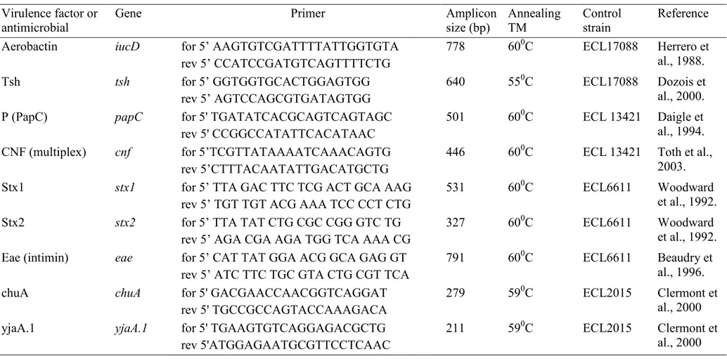
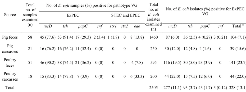
Table I. List of primers used in the PCR, single PCR conditions, and control strains ... 58 Table II. Virulence genes (VGs) associated with pathotypes of E. coli from pigs and poultry on farms and at abattoirs... 60 Table III. Distribution according to phylogenetic group and virulence gene profile of ExPEC from pigs and poultry on farms and at abattoirs ... 61 Table IV. Antimicrobial resistance of potential ExPEC E. coli isolates from pigs and poultry on farms and at abattoirs... 62 Table V. Frequency of antimicrobial resistance genes and association of AMR genes and VGs among 77 potential ExPEC isolates from pigs and poultry on farms and at abattoirs ... 63 Table VIa. Association of ciprofloxacin-resistance and PQMR genes ... 64 Table VIb. Association of ampicillin and streptomycin-resistance and AMR genes………..64
ix
LIST OF ABBREVIATIONS
A/E Attaching and effacing
Aac3 Aminoglycoside acetyltransferase Aada Aminoglycoside resistance protein AAF Aggregative adherence fimbriae AMC Amoxicillin/clavulanic acid AMK Amikacin
APEC Avian pathogenic Escherichiacoli AMR Antimicrobial resistance
AMP Ampicillin Bla Beta-lactamase
BMEC Brain microvascular endothelial cells CF Colonization factor
CIP Ciprofloxacin
CIPARS Canadian Integrated Program for Antimicrobial Resistance Surveillance CHL Chloramphenicol
Cnf Cytotoxic necrotizing factor ColV Colicin V
x
CLSI Clinical and Laboratory Standards Institute CT Cholera toxin
DAEC Diffusely adherent Escherichia coli Dfr Dihydrofolate reductase
EAEC Enteroaggregative Escherichia coli E. coli Escherichia coli
ECOR E. coli reference collection
EHEC Enterohemorragic Escherichia coli EIEC Enteroinvasive Escherichia coli EPEC Enteropathogenic Escherichia coli ESBLs Extended-spectrum beta-lactamases ETEC Enterotoxigenic Escherichia coli
ExPEC Extra-intestinal pathogenic Escherichia coli Fim Fimbriae
FIS Sulfisoxazole FOX Cefoxitin
GEN Gentamicin
GyrA DNA gyrase (type II topoisomerase), subunit A GyrB DNA gyrase (type II topoisomerase), subunit B
xi
Iss Increased serum survival Iuc Iron- Uptake Chelate Iut Iron-Uptake Transport
KAN Kanamycin
kDA Kilodalton
LEE Locus of enterocytes effacement LPS Lipopolysaccharide
LT Heat-labile enterotoxin
MLEE Multilocus enzyme electrophoresis MLST Multilocus sequence typing
NAL Nalidixic acid
OMP Outer membrane protein PAI Pathogenicity island
Pap Pyelonephritis associated fimbriae PCR Polymerase chain reaction
PFGE Pulsed-field gel electrophoresis
PMQR Plasmid-mediated quinolone resistance ST Heat-stable enterotoxin
xii Stx Shiga-toxin STR Streptomycin SXT Trimethoprim-sulphamethoxazole TET Tetracycline TIO Ceftiofur Tsh Temperature-sensitive hemagglutinin UPEC Uropathogenic Escherichia coli UTI Urinary tract infection
VF Virulence factor VG Virulence gene
ACKNOWLEDGEMENTS
Firstly I would like to thank my supervisor Professor John Morris Fairbrother for his guidance, support and regular enlightening discussions throughout the project. Thank you especially for the interest you have shown in this work, investing a lot of time in my scientific training and welcoming me in your laboratory.
I would like to thank Dr Ann Letellier, who together with Professor Fairbrother gave me the opportunity to carry out this project, for your encouragement and for your participation in scientific quality of this work.
I would like to thank Clarisse Desautels, Ghyslaine Vanier and Brigitte Lehoux for their technical assistance and availability in the Ecl Laboratory.
Thanks to the laboratory technicians, the administrative technicians, students at GREMIP for their collaboration to the success of this work.
Thanks to Dr Nadia Bergeron, member of the jury committee and Dr Philippe Fravalo, President of the jury committee, for their contribution to the scientific quality of the work.
Thanks to Dr Sylvain Quessy without whom this project would not have been possible.
Moreover, I would like to thank all my fellow students at Ecl Lab, for their encouragement and friendship, especially Rashin, Jessie and Flavien.
Special thanks to Marc-André Gagnon who always encouraged me to complete and improve my thesis.
Finally, I sincerely thank Helene Bergeron for all her supports, especially for submission of my thesis.
2
Escherichia coli (E. coli) is a common micro-organism in the gastrointestinal tract of humans and animals. Most E. coli are harmless; however, some are pathogenic and can cause disease in humans and animals (Johnson, 2003; Kaper, Nataro, & Mobley, 2004). From a genetic and clinical perspective, E. coli strains of biological significance to humans can be broadly categorized as (1) commensal strains, (2) intestinal pathogenic (i.e. enteric or diarrheagenic) strains, and (3) extraintestinal pathogenic E. coli (ExPEC) strains (Russo & Johnson, 2003). In addition, E. coli are classified into phylogenetic groups A, B1, B2, and D (Clermont et al., 2000). Intestinal pathogenic E. coli such as enteropathogenic E. coli (EPEC) and Shiga toxin-producing E. coli (STEC) may cause gastrointestinal disease, mostly consisting of more or less severe diarrhea, in humans and animals. EPEC is considered as one of leading causes of infant diarrhea in developing countries, whereas STEC is one of the most common pathogens associated with outbreaks of foodborne illness, causing bloody diarrhea and hemolytic-uremic syndrome, particularly in North America, Japan and European countries (Kaper et al., 2004). ExPEC strains do not produce enteric disease; however, they may cause diverse extraintestinal infections, including urinary tract infections (UTIs), meningitis and septicemia, in both humans and animals (Smith et al, 2007). ExPEC express a variety of virulence-associated genes instead of having a common virulence mechanism (Johnson et al, 1991, Smith et al 2007). Typical virulence factors of ExPEC include diverse adhesins, siderophores for iron acquisition, capsules, toxins, proteases, invasins, and serum resistance proteins (Kaper et al, 2004). ExPEC isolates from urinary tract infections in humans most commonly belong to phylogenetic groups B2 or D.
Animal-derived raw foods are commonly contaminated with E. coli and have long been known as an important vehicle for transmitting diarrheagenic E. coli, but their role in spreading ExPEC has not been explored extensively (Xia et al., 2011). ExPEC can asymptomatically colonize the animal and human intestinal tract, thus could be transmitted to humans through food consumption (Bélanger et al., 2011; Fairbrother & Nadeau, 2006). Recent studies indicate retail meat may be an important vehicle for transmission of ExPEC that share characteristics with human clinical ExPEC strains (Johnson et al., 2003a, 2005b; Jakobsen et al., 2010; Vincent et al., 2010). In
3
addition, it was suggested that meat consumption was epidemiologically associated with infection by antimicrobial-resistant ExPEC that causes UTI (Johnson, et al., 2005b). These findings suggest the hypothesis that potentially zoonotic ExPEC in contaminated meat could arise from poultry and pig reservoirs.
There have been no published studies on the prevalence of STEC, EPEC, and ExPEC in pigs and poultry on-farm in Vietnam. In addition, few current data are available on the prevalence of antimicrobial-resistant E. coli from animals at the farm level and from carcasses in abattoirs in this country. The aim of this study is to investigate the prevalence of E. coli strains carrying virulence genes associated with STEC, EPEC, and ExPEC in pigs and poultry on farms and at abattoirs, and to characterize these potentially pathogenic strains for phylogenetic group and antimicrobial resistance profile.
5
1. Classification of E. coli
1.1. Commensal E. coli
Commensal E. coli constitute part of the gastrointestinal microflora in humans, mammals, and birds. The commensal strains are generally benign, do not cause intestinal tract disease, and can be beneficial to the host (Russo and Johnson, 2003). However, commensal strains may cause illness if the host is compromised immunologically or medically (Picard et al., 1999; Russo and Johnson, 2003).
Commensal E. coli generally lack the specialized virulence traits that enable intestinal and extraintestinal E. coli to cause disease within or outside the gastrointestinal tract, respectively. However, commensal E. coli can participate in extraintestinal infections when an aggravating factor is present, such as a foreign body (e.g. urinary catheter), host compromise (e.g. local anatomical or functional abnormalities such as urinary or biliary tract obstruction, or immunocompromise), or a high or a mixed bacterial species inoculum (e.g. with fecal contamination of the peritoneal cavity).
Generally, human commensal E. coli strains belong to phylogenetic groups A and B1 and typically lack the specialized virulence determinants found in pathogenic strains that cause intestinal or extraintestinal diseases (Picard et al., 1999; Russo and Johnson, 2000).
1.2. Intestinal pathogenic E. coli
Intestinal pathogenic E. coli have evolved a special ability to cause gastrointestinal disease, mostly consisting of more or less severe diarrhea in humans and animals. Intestinal pathogenic E. coli are currently classified in six categories based on virulence attributes that help bacteria to cause diseases, including: enteropathogenic E. coli (EPEC), enterohaemorrhagic E. coli (EHEC), enterotoxigenic E. coli (ETEC), enteroaggregative E. coli (EAEC), enteroinvasive E. coli (EIEC) and diffusely adherent E. coli (DAEC) (Kaper et al., 2004). EHEC is also known as the pathogenic
6
subgroup of Shiga toxin-producing E. coli (STEC) that cause hemorrhagic colitis and hemolytic uremic syndrome (HUS) (Nataro & Kaper, 1998)
1.2.1.Enteropathogenic E. coli (EPEC)
EPEC is considered as one of leading causes of diarrhea in developing countries (Kaper et al, 2004). The central mechanism of EPEC pathogenesis is a lesion called attaching and effacing (A/E), which is characterized by microvilli destruction, intimate adherence of bacteria to the intestinal epithelium, pedestal formation, and aggregation of polarized actin and other elements of the cytoskeleton at sites of bacterial attachment (Kaper et al., 2004). Ability to produce A/E lesions has also been detected in strains of Shiga toxin–producing E. coli (Nataro & Kaper, 1998). The genetic determinants for the production of A/E lesions are located on the locus of enterocyte effacement (LEE), a pathogenicity island that contains the genes encoding intimin, a type III secretion system, a number of secreted (Esp) proteins, and the translocated intimin receptor named Tir. Intimin, a 94-kDa outer membrane protein encoded by the eae gene, is responsible for the intimate adherence between bacteria and enterocyte membranes. The Esp molecules (EspA, B, and D) are involved in the formation of a translocon that delivers effector molecules to the host cell and disrupts the cytoskeleton, subverting the host cell functions. Tir, which is one of the EPEC translocated proteins, is inserted into the host cell membrane, where it acts as a receptor to intimin (Kaper et al., 2004; Trabulsi et al., 2002)
Typical EPEC strains possess a plasmid of 70–100 kb called the EAF (EPEC adherence factor) plasmid, whereas atypical EPEC do not contain the EAF plasmid (Trabulsi et al., 2002). This plasmid encodes a type IV pilus called the bundle-forming pilus (BFP), which mediates interbacterial adherence and possibly adherence to epithelial cells (Kaper et al., 2004). Thus far, typical EPEC strains are identified by the presence of both eae and bfp, whereas atypical EPEC strains have been defined as those which possess only eae (Hernandeset al., 2009). Atypical EPEC serotypes have been isolated from different animal species suggesting that atypical EPEC may be a potential zoonotic cause of human diarrhea (Trabulsi et al, 2002).
7
Atypical enteropathogenic E. coli (EPEC) were identified among E. coli isolates from pork, indicating that pigs may also be potential reservoirs for the pathogen (Xia et al., 2010). Analysis of phenotypic and genotypic markers of atypical EPEC isolated from diarrheic and non-diarrheic dogs demonstrated that isolates of serotypes O4:H16 and O51:H40 were similar to those found in human disease (Almeida et al., 2012).
1.2.2. Shiga toxin-producing E. coli (STEC)
Shiga toxin-producing E. coli refers to those strains of E. coli that produce at least one member of a class of potent cytotoxins called Shiga toxin, also called verocytotoxin (Gyles, 2007). STEC have been characterized by a variety of methods, including serotyping. Isolates of O157:H7 serotype are the most common STEC pathogens associated with outbreaks of foodborne illness in North America, but several other serotypes, particularly those of the O26 and O111 serogroups, can also cause disease and are more important than O157:H7 in other countries (Kaper et al., 2004). It is well established that cattle are a major reservoir of STEC O157:H7 in North America but in countries such as Australia, sheep are of greater significance (Gyles, 2007).
Shiga toxin is the key virulence factor in STEC diseases. Currently, Shiga-like toxins are categorized into two immunologically distinct groups: Stx1 and Stx2, which show approximately 55% sequence homology (Kaper et al., 2004). Stx1 and Stx2 share a similar function and have the same genetic operon structure, encoding an A (enzymatic toxin) and a B (cell receptor binding) subunit. Stx1 and Stx2 are further subdivided into distinct genetic variants which differ in their biological activity and association with disease (Gyles, 2007).
Adherence to intestinal epithelial cells is an early feature of STEC infection and two patterns of attachment and interaction have been observed, associated with eae-positive and eae-negative STEC isolates. The eae-eae-positive STEC possess a pathogenicity island called the locus of enterocyte effacement (LEE), which encodes the bacterial proteins necessary for formation of the AE lesion (Kaper et al., 2004;
8
Nataro & Kaper, 1998). The combined presence of the eae and stx2 genes has been
indicated as an important predictor of HUS (Ethelberg et al., 2004). 1.2.3. Enterotoxigenic E. coli (ETEC)
ETEC is a pervasive cause of diarrhea in children and travellers in developing countries (Kaper et al, 2004). ETEC produce one or both of two enterotoxins, heat-labile enterotoxin (LT) and heat-stable enterotoxin (ST) which are major virulence factors. LT was found to be very similar physiologically, structurally, and antigenically to cholera toxin (CT) and to have a similar mode of action (Svennerholm, 2011). The molecular mass (84 kDa) and the subunit structure of the two toxins were essentially identical, with an active (A) subunit surrounded by five identical binding (B) subunits. LT can stimulate prostaglandin synthesis and stimulate the enteric nervous system; both of these activities can also lead to stimulation of secretion and inhibition of absorption (Kaper et al, 2004). STs are low-molecular-weight peptides, that are classified into two unrelated classes — STa and STb — which differ in both structure and mechanism of action. Only toxins of the STa class have been associated with human disease whereas the STb toxin is associated with animal disease (Kaper et al, 2004).
Other important virulence factors in ETEC include one or more colonization factors (CFs), which usually are fimbriae. More than 25 CFs have been recognized on human ETEC so far, and additional ones are likely to be recognized (Svennerholm, 2011). The CFs promote colonization of ETEC in the small bowel, thus allowing expression of either or both LT and ST in close proximity to the intestinal epithelium (Quadri et al., 2005). ETEC are also an important cause of diarrhoeal disease in animals and these animal strains express fimbrial intestinal colonization factors, such as K88 and K99, which are not found in human ETEC strains.(Kaper et al, 2004)
1.2.4. Enteroaggregative E. coli (EAEC)
EAEC is an emerging pathogen which is increasingly recognized as a cause of persistent diarrhea in adults and children. EAEC are characterized by the ability to
9
aggregate intimately with each other, adhere to human HEp-2 cells, and also attach to abiotic surfaces when grown in tissue culture plates (Okhuysen & Dupont, 2010). EAEC pathogenesis is determined by the organism's ability to adhere to intestinal cells, produce enterotoxins and cytotoxins, and induce inflammation. A number of studies described several virulence factors associated with EAEC pathogenesis. These are (i) heat stable toxin-1 (EAST-1), (ii) aggregative adherence fimbriae I and II (AAF/I and AAF/II) and AAF/III. In addition, different pathogenicity islands have been identified within the EAEC group, including Shigella she pathogenicity island, containing enterotoxin and mucinase genes and Yersinia high-pathogenicity island, containing the yersiniabactin siderophore gene (Weintraub, 2007)
1.3. Extraintestinal pathogenic E. coli (ExPEC)
E. coli strains that induce extraintestinal diseases are termed extraintestinal pathogenic E. coli (ExPEC) (Russo and Johnson, 2000). In humans, ExPEC strains are often found in the normal intestinal flora and do not cause gastroenteritis. ExPEC strains are phylogenetically and epidemiologically distinct from intestinal pathogenic strains. They do not produce enteric disease; however, they can asymptomatically colonize the human intestinal tract and may be the predominant E. coli strains in 20% of normal individuals (Johnson and Russo, 2002; Russo and Johnson, 2000, 2003). ExPEC may cause diverse infections in both humans and animals, including urinary tract infections (UTIs), meningitis and septicemia (Smith et al, 2007). The sub-set of ExPEC causing UTI has been designated uropathogenic E. coli (UPEC).
Most of the human ExPEC strains are found in the B2 and D phylogenetic groups and have acquired various virulence genes such as genes encoding attachment factors (pap, sfa, and afa), transmembrane protein involved in neonatal meningitis (ibe10), and alpha-hemolysin (hly) that allow them to induce extraintestinal infections in both normal and compromised hosts (Picard et al., 1999). The majority of the virulence factors present in ExPEC strains are distinct from those found in the intestinal pathogenic strains; Russo and Johnson, 2000, 2003).
10
ExPEC were defined by Johnson et al. as E. coli isolates containing two or more of the following virulence markers as determined by multiplex PCR: papA (P fimbriae structural subunit) and/or papC (P fimbriae assembly), sfa/foc(S and F1C fimbriae subunits), afa/dra(Dr-antigen-binding adhesins), kpsMTII (group 2 capsular polysaccharide units), and iutA (aerobactin receptor) (Johnson et al., 2003a). Other virulence markers are associated with ExPEC status, including fimH (type I fimbriae), ibe (outer membrane protein promoting invasion), and hly (hemolysin) (Kaper et al, 2004).
2. Diseases caused by ExPEC in humans
2.1. Urinary tract infections
Extraintestinal pathogenic E. coli are a frequent cause of urinary tract infections (UTIs), which are among the most common bacterial infections found in humans (Johnson and Russo, 2002, 2005). In fact, UTIs are a heterogeneous group of disorders, which are classified by the site of infection, the bladder (cystitis), kidney (pyelonephritis) and urine (bacteriuria) (Foxman, 2002). Risks of UTIs are increased in specific groups including infants, pregnant women, the elderly and compromised patients (Foxman, 2002).
Since the major means of transmission of UTI-causing ExPEC to the urinary tract is the ascending route from the fecal site into the bladder (Smith et al., 2007), the cause of ExPEC contamination in human has been debated. Johnson et al. indicate that meats, particularly poultry, can be an important source of ExPEC contamination (Johnson et al., 2003a, 2005a, 2005b).
Treatment of UTIs usually consists of administration of antimicrobials, including beta-lactams, quinolones, trimethoprim in combination with sulfamethoxazole, and nitrofuranes (Wagenlehner et al., 2005). Notably, resistance of E. coli to commonly prescribed antimicrobial agents used in the treatment of UTIs has been significantly increasing (Smith et al., 2007).
11
2.2. Neonatal meningitis
Bacterial neonatal meningitis is an inflammation of the membranes of the brain or spinal cord and consists of a purulent exudate of the membranes, perivascular in-flammation, and brain edema. In the United States, more than 50% of cases of neonatal meningitis caused by Gram-negative enteric organisms were due to ExPEC, and approximately 80% of these cases were caused by strains carrying the K1 capsular antigen (Pong and Bradley, 1999).
The pathogenesis of ExPEC-induced neonatal meningitis occurs in several steps: bacteremia, binding of bacteria to the surface of brain microvascular endothelial cells, bacterial invasion of brain microvascular endothelial cells, and invasion of the meninges (membranes that surround the brain and spinal cord) and the central nervous system (Smith et al., 2007).
Many of the virulence genes associated with ExPEC neonatal meningitis strains are present on pathogenicity islands (PAIs) (Bonacorsi et al., 2003). For example, the genes involved in facilitating blood-brain barrier penetration sfaS (fimbrial adhesin S), ibeA (invasin IbeA) and cnf1 (cytotoxic necrotizing factor) are located on PAIs (Bonacorsi et al., 2003).
Holt et al. reported that the most common antimicrobial regimes used for neonatal meningitis treatment based on third generation cephalosporins such as cefotaxime in a combination with a penicillin or ampicillin with or without an aminoglycoside (Holt et al., 2001). The use of third generation cephalosporins decreased mortality but not morbidity (Harvey et al., 1999).
2.3. Sepsis
Sepsis can be caused by a microbial infection that originates from the kidneys (UTI), bowel (peritonitis), skin (cellulitis), or lungs (pneumonia). Russo and Johnson estimated that E. coli is the cause of 17% of the cases of severe sepsis (dysfunction of at least one organ system) in the United States (Russo and Johnson, 2003). They
12
estimated there were 127,500 cases of E. coli-induced severe sepsis, with 40,000 deaths, in 2001; the mortality rate was approximately 30%.
Individuals at the extremes of age are the most susceptible to bacterial-induced, community acquired septicemia; E. coli was found to be the most frequent cause of septicemia in infants up to 1 year of age and in individuals aged 65 or greater in North America (Diekema et al., 2002). Putative virulence factors for septicemia-inducing strains of E. coli serogroups O2 and O78 include iron uptake systems (aerobactin, yersiniabactin, and IroN receptor), serum resistance, and adhesins (type 1 fimbriae, curli, and P fimbriae). Non-fimbrial adhesins were present only in strains of serotype O78, and the K1 capsule was present only in O2 (Mokady et al., 2005). Hyde et al. noted that among the infants with early-onset sepsis, the proportion of E. coli infections that were resistant to ampicillin increased during the surveillance period (1998-2000) and mortality was higher with ampicillin-resistant E coli strains than with ampicillin-sensitive strains (Hyde et al., 2002).
3. Virulence factors of pathogenic E. coli
ExPEC strains have acquired genes encoding diverse extraintestinal virulence factors that enable them to cause infections outside of the gastrointestinal tract. Characteristic virulence traits that are present in most ExPEC include various adhesins (e.g. P and type I fimbriae), factors to avoid or subvert host defense systems (e.g. capsule, lipopolysaccharide), mechanisms for nutrient acquisition (e.g. siderophores), and toxins (e.g. hemolysin, cytotoxic necrotizing factor 1).
3.1. Adhesins
The adherence step contributes to extra intestinal virulence by promoting colonization and by facilitating bacterial interactions with host cells and matrix elements. Most E. coli adhesins are fimbrial (ie, discrete hair-like structures), but some are amorphous fibers or are capsule-like. Irrespective of morphology, most adhesins contain defined molecular regions that interact with specific host receptor
13
epitopes in a lock-and-key or lectin-like fashion (Johnson, 2003). The diverse adhesins of ExPEC are categorized primarily according to receptor specificity. The broadest functional division is between mannose-sensitive and mannose-resistant adhesins, reflecting the early observation that mannose blocks adherence for a subset of E. coli isolates that attaches to erythrocytes or epithelial cells (Johnson, 1991).
3.1.1. Type 1 fimbriae
Type 1 fimbriae are non-flagellar, filamentous appendages of bacteria, were classified into type I depending on haemagglutination and sensitivity activity with D-mannose (Antao et al., 2009)
Type 1 fimbriae are produced by more than 80% of all UPEC (Kucheria et al., 2005). The role of type 1 fimbriae in attachment of UPEC strains to host cells was demonstrated through in vitro and animal studies. Type 1-fimbriae mediated for specific binding of one E. coli strain to monkey kidney cells (Salit & Gotschlich, 1977). Further research showed that type 1-fimbriae accounted for binding of E. coli strains to human urinary tract epithelial cells (Eden & Hansson, 1978).
Type 1 fimbriae are encoded by the chromosomally located fim gene cluster, including genes for a structural subunit (FimA), an adhesin (FimH) andseveral accessory proteins involved in subunit transport and assembly, and regulatory proteins (Connell et al, 1996). FimH was found to be the gene responsible for the monomannose-containing receptors shown to be adaptive for UPEC which results in increased adhesion to vaginal epithelial cells (Antao et al., 2009).
Type 1 fimbriae also play an important role in the infection process is the adherence of APEC to the epithelium of the trachea. This fimbriae in colibacillosis has been associated with mucus adherence, colonization of the trachea and the intestinal tract, and interactions with lung epithelial cells (de Pace et al., 2010)
3.1.2. P fimbriae
The P fimbriae family is the best known group of mannose-resistant adhesins, so named because of their binding specificity for the Gal(α 1-4) Gal- β disaccharide
14
galabiose, which is present in the antigens of the human P blood system (Johnson, 2003). P fimbriae are important in the pathogenesis of UTI, primarily because they mediate Gal-Gal-specific bacterial adherence to epithelial cells within the human urinary tract, thereby permitting bacterial colonization and stimulating inflammation (Johnson, 1991).
P fimbriae are composed of different polymerized subunits, with one major subunit, PapA, constituting the bulk of the fimbria. Three minor adherence-related fimbrial subunits (PapE, PapF, and PapG) are present in minor amounts at the fimbrial tips. These fimbrial proteins, as well as a number of accessory proteins, are encoded by a chromosomal gene cluster termed pap or pyelonephritis-associated fimbriae genes since these were typical of strains isolated from human urinary tract infections (Johnson, 1991). The adhesion of the fimbriae is conferred by the adhesin papG located at the distal end fimbriae. There are three alleles of adhesins papG (I, II, and III). Molecular epidemiological studies have shown that allele III of PapG is usually the predominant variant among E. coli isolates from women and children with cystitis, whereas the PapGII variant is associated with pyelonephritis in humans ( Johnson et al., 2000). Several other proteins are important in P-fimbrial synthesis. PapD is present in the periplasmic space and may complex with fimbrial subunits, stabilizing them during translocation across the periplasmic space to the outer membrane prior to assembly. PapC assists in the transport of subunits out of the cell and in their assembly into complete fimbriae (Johnson, 1991). The papC gene was observed at a high prevalence among E. coli strains associated with pyelonephritis, as determined by PCR (Le Bouguenec et al, 1992).
The P fimbriae are not restricted to uropathogenic E. coli (UPEC) causing UTI and newborn meningitis-causing E. coli (NMEC), but are also prevalent in avian pathogenic E. coli (APEC) strains. In poultry, P fimbriae are primarily associated with colibacillosis strains, but P fimbriae are also found in healthy poultry. PapC has been found at a low prevalence in E. coli from healthy chickens in Denmark (Jakobsen et al., 2010), whereas the prevalence of papC in APEC strains was greater than 20% (Ewers et al., 2007; Rodriguez-Siek et al., 2005). Until now, studies on the
15
frequency of P fimbriae in healthy pigs are scarce. However, some previous studies have suggested that P fimbriae-positive E. coli are more frequent in diseased pigs than healthy pigs (Jakobsen et al., 2010; Tan et al., 2012).
3.1.3.Temperature sensitive hemagglutinin
The mannose-resistant haemagglutinin of an avian pathogenic E. coli (APEC) isolate, which was best expressed at low temperatures, was first reported in 1994 (Provence & Curtiss, 1994). This temperature-dependent haemagglutination phenotype was termed Tsh for temperature sensitive haemagglutinin (Provence & Curtiss, 1994). Tsh and Tsh/Pap (P fimbriae)/ Iuc (aerobactin) pathotypes were suggested as important virulence factors of APEC (Ngeleka, et al., 2002).
Tsh is synthesized as a 140-kDa precursor protein, whose processing results in a 106-kDa passenger domain (Tshs) and a 33-kDa β-domain (Tshβ). The presence of a
conserved 7-amino-acid serine protease motif within Tshs classifies the protein in a
subfamily of autotransporters, known as serine protease autotransporters of the Enterobacteriaceae (Kostakioti & Stathopoulos, 2004). The passenger domain (Tshs) is secreted into the extracellular environment, being temporarily in the outer
membrane of the bacteria where it is capable of adhering to red blood cells, hemoglobin, and the extracellular matrix proteins fibronectin (Antao et al., 2009; Kostakioti & Stathopoulos, 2004)
Tsh is encoded by the gene tsh which is located on a ColV-type plasmid in many of the APEC strains. It has also been shown that tsh is more frequently observed in high-lethality isolates than in low-lethality isolates among APEC (Dozois et al., 2000). In another study, it was shown that the tsh gene was present in more than 50% of APEC, 4.5% UPEC and 11.5% NMEC isolates tested (Ewers et al., 2007). In pigs, tsh was found significantly more frequently in highly virulent ExPEC strains belonging to group B2, which might indicate that tsh is involved in the pathogenic mechanisms of ExPEC (Picard et al., 1999; Tan et al., 2012).
16
3.2. Capsules (K antigen)
Over 80 polysaccharide capsules (K antigen) have been described in E. coli (Orskov & Orskov, 1992). The capsules of most ExPEC strains belong to group 2 and 3 polysaccharides which are characterized by low-molecular-weight and high-charge density ( Johnson, 1991; Johnson & O'Bryan, 2004). The capsule protects ExPEC bacteria against phagocytosis and complement-mediated killing, thereby contributing to extraintestinal virulence (Burns & Hull, 1999; Russo, Liang, & Cross, 1994). K1 capsule is the most commonly encountered capsular type among both urinary and fecal strains in humans ( Johnson, 1991). APEC strains possessing the K1 capsule also showed greater resistance to serum and ability to colonize the internal organs in poultry than K1-negative mutants (Mellata et al., 2003).
Group 2 and group 3 capsules are encoded by kps operons which share moderately to highly conserved regions (e.g., kpsDMTE) encoding transport and assembly functions. Therefore, Johnson & O'Bryan developed a rapid technique for detection of K antigen in E. coli based on PCR amplification of the kpsM gene (Johnson & O'Bryan, 2004). The kpsM has been designated as one of virulence markers for identification of ExPEC in epidemiological studies (Johnson et al., 2005b).
3.3. Lipopolysaccharide
Lipopolysaccharide (LPS) is a structural component of the outer membrane of E. coli (Johnson, 1991). LPS, also known as endotoxin, consists of three components: the hydrophobic membrane anchor lipid A region, which is associated with the toxicity of the LPS and is well conserved among Gram-negative bacteria, the distal O-antigen polysaccharide region that is exposed to the surface and the core polysaccharide region that connects the two LPS components. The lipid A is largely responsible for stimulation of the innate immune system (DebRoyet et al., 2011). LPS is recognized by the Toll-like receptor 4 (TLR4) present on the surface of monocytes, macrophages, neutrophils and dendritic cells, cells of the innate immune system (Johnson, 2003). O antigens may contribute to the virulence of the organism, since certain O groups are associated with with specific diseases (DebRoy et al., 2011). A
17
study on E. coli O18:K1 mutants lacking either the lipopolysaccharide O antigen showed that loss of the O antigen resulted in the strain becoming more sensitive to the bactericidal effects of the classical complement pathway (Pluschke et al., 1983).
3.4. Iron acquisition systems
Uptake of iron in Gram-negative bacteria is achieved primarily through the synthesis, export, and uptake of small iron-chelating molecules termed siderophores. Recognized siderophore systems include the catecholates enterobactin, salmochelin, the hydroxamate aerobactin, and yersiniabactin (Henderson et al., 2009; Johnson, 2003). Enterobactin is efficient at sequestering iron in vitro; however, it is less effective than aerobactin and salmochelin in competing for iron during infection, as it is inhibited by serum albumin and specifically binds to the host innate defense protein neutrophil lipocalin (Caza et al., 2008).
The salmochelin-encoding system comprised of two divergently transcribed sets of genes, iroBCDE encoding the enzymes accounting for salmochelins production, and iroN encoding the outer membrane siderophore receptor. The specific roles of iroBCDE genes in salmochelins production as well as in contribution to APEC virulence have been identified (Caza et al., 2008). The iroN gene did not effect salmochelin production, however, it was required for the colonization of uropathogenic ExPEC (Russo et al., 2002), and the bacteremic step of E. coli neonatal meningitis (Negre et al., 2004).
The aerobactin system is encoded by a five-genes operon, with four genes iucABCD encoding the enzymes needed for aerobactin synthesis and a fifth gene iutA encoding the outer membrane receptor protein. In E. coli, this operon is found both on plasmids and on the bacterial chromosome, with the chromosomal location predominating among human clinical isolates (Johnson, 1991). Due to the high prevalence of aerobactin-encoding genes among isolates from poultry with colibacillosis, the iucD gene was selected as one of virulence markers for the rapid detection and characterization of APEC (Ewers et al., 2005).
18
3.5. Cytotoxin:
3.5.1.Hemolysin
There are two common types of this toxin, αhaemolysin and βhaemolysin. The α -haemolysin is commonly produced by strains isolated from cases of human UTI and other extra-intestinal infections. It is a heat-labile extracellular protein and its production can be plasmid or chromosomally determined. It is a pore forming cytolysin, and lyses erythrocytes by causing dissipation of transmembrane ion gradients. β-haemolysin is a cell associated haemolysin with a similar range of haemolytic activity ( Johnson, 1991).
In addition to lysing erythrocytes, hemolysin is toxic to a range of host cells, probably contributing to inflammation, tissue injury, and impaired host defenses. Hemolysin production is encoded by a four-gene operon termed hly ( Johnson, 1991). Plasmid and chromosomal hly regions differ with respect to flanking and regulatory sequences and to the precise sequence of hlyA, the gene encoding the structural hemolysin protein.
3.5.2 Cytotoxic necrotizing factor
Cytotoxic necrotizing factor type 1 (CNF1), presents in certain diarrheagenic E. coli and UPEC, is a member of a family of bacterial toxins that target the Rho family of small GTP-binding proteins in mammalian cells (Johnson, 1991). The role of CNF1 in the pathogenesis of UPEC-mediated urinary tract infection has been demonstrated in several studies. In a mouse model of ascending urinary tract infection, a CNF1-expressing UPEC, compared to its isogenic cnf1 mutant, caused a greater acute inflammatory response in the bladder, colonized the bladder more extensively in a coinfection experiment, and survived better when coincubated with human neutrophils (Rippere-Lampe et al., 2001). Epidemiological data showed that cnf1 gene is significantly associated with clinical strains related to cystitis, pyelonephritis and prostatitis in humans (Yamamoto, 2007). Strains of UPEC that produce CNF1 also often produce two established urovirulence determinants, α-hemolysin and P
19
fimbriae. The cnf1 gene encoding cytotoxic necrotizing factor and the genes encoding these other virulence factors are typically linked on the same pathogenicity island in certain UPEC strains (Mills et al,, 2000).
4. Association among virulence factors and phylogenetic groups
4.1. Associations among virulence factors
Although several important ExPEC virulence factors and their role during pathogenesis have been described (Johnson, 1991; Smith et al., 2007), many ExPEC cannot be unambiguously distinguished from commensal E. coli based on a set of discriminatory virulence factors (Kohler & Dobrindt, 2011). Nevertheless, ExPEC classification has been proposed based on the detection of two virulence-associated genes typical of the specific pathotype (Johnson and Russo, 2005), and two virulence-associated genes for non-host samples such as food samples (Johnson et al., 2005b).
Certain VFs commonly occur together in patterns suggesting either co-selection or direct genetic linkage. A major virulence pattern, which was characterized by the presence of F1 variant fimbriae; S fimbriae; IbeA; and the aerobactin system was identified in 75% of the O18:K1 ExPEC isolates of human origin and avian origin (Moulin-Schouleur et al., 2006). Chromosomally located virulence-associated genes such as iron acquisition genes, for example the chromosomal sitD variant, chuA, fyuA, and irp2, were frequently found in APEC and also among UPEC and NMEC (Ewers et al., 2007). These results demonstrated that very closely related clones can be recovered from extraintestinal infections in humans and poultry.
Currently recognized putative VFs of ExPEC include adhesins, siderophores, toxins, protectins, and invasins, has and have been demonstrated within pathogenicity-associated islands (PAIs) and on plasmids. Ewers et al. suggested that ColV plasmids, commonly found in APEC, and might be the source of the plasmid in human UPEC and NMEC strains, were associated with virulence genes tsh, iss, and the episomal sit locus. (Ewers et al., 2007). Rodriguez-Siek et al. suggested that a
20
typical member of the APEC pathotype is likely to contain several iron transporter-encoding genes, like irp2, fyuA, iutA, iroN, and sitA, and plasmid-associated genes, including cvi/cvaC, tsh, and iss (Rodriguez-Siek et al., 2005b). Several PAIs and genetic islands harbouring ireA and pap operon genes have been identified in APEC including PAI I (APEC-O1) (Kariyawasam et al., 2006). PAI I (APEC-1)-related genes were observed in strains belonging to the APEC pathotype and also in UPEC and NMEC (Ewers et al., 2007).
4.2. Association of phylogenetic groups and virulence factors
Phylogenetics is the study of genetic distance among groups of organism. Phylogenic relationships have been traditionally studied based on morphological data. An initial phylogeny, obtained through cluster analysis of multilocus enzyme electrophoresis (MLEE) data for 35 enzyme-encoding loci of the standard reference collection of 72 E. coli strains (ECOR), defined six major phylogenetic groups, designated A, B1, B2, C, D and E (Tenaillon et al., 2010, Lindstedt et al., 2007). This phylogeny was reconstructed within 4 phylogenetic groups A, B1, B2, and D with a subsequent analysis of the ECOR strains based on polymorphisms of 38 loci (Tenaillon et al., 2010; Herzer et al., 1990).
Among human E. coli isolates, the phylogenetic B2 group and, to a lesser extent, the phylogenetic D group strains have been shown to exhibit numerous extraintestinal virulence determinants compared to phylogenetic A and B1 strains (Clermont et al., 2000). Among ECOR strains, the most common pattern of phylogenetic distribution exhibited by the various VFs, including papG allele III, sfa/foc, sfaS, focG, hlyA, cnf1, cdtB, kpsMT-III, rfc, and ibeA, was associated with group B2. Other VFs were sufficiently found in other phylogenetic groups, but were still significantly associated with phylogenetic group B2 (e.g., “K5,” fyuA, ompT, and the PAI marker), or with both groups B2 and D (e.g., papAH, papC, papEF, papG,and kpsMTII) or with group D only (e.g., papG allele II, iha, K1, and iutA) (Johnson et al., 2001). Johnson et al. reported most of E. coli isolates from bacteremic patients (65.7%) belonged to phylogenetic group B2, whereas 11.6%,
21
10.5%, and 12.2% belonged to groups A, B1, and D, respectively. More than half (97/181, 53.6%) of the bacteremic-inducing E. coli strains were of urinary or pulmonary tract origin; 78/97 (80.4%) of these strains belonged to group B2 (Johnson et al., 2002a). Picard et al. reported that strains of the B2 phylogenetic group were highly virulent, killing in mice, and possessed the greatest number of virulence determinants. In contrast, commensal strains belong mainly to phylogenetic groups A and B1, were devoid of virulence determinants, and did not kill mice (Picard et al., 1999). Moulin et al. reported that most avian and human strains belonging to phylogenetic subcluster B2-1 expressed the K1 antigen and presented no significant differences concerning the presence of other virulence factors (Moulin-Schouleur et al., 2007). Johnson et al. reported that group B2 neonatal meningitis isolates had a significantly higher prevalence of many VFs than even the group B2 ECOR strains ( Johnson et al., 2002b).
However, the phylogenetic distribution of the various ExPEC virulence factors varied among ExPEC strains from animals and retail food. Among all ExPEC isolates recovered from retail meat, papC was significantly positively associated with phylogenetic group B1 and negatively associated with group B2, whereas kpsMTII was negatively associated with groups B1 and A and positively associated with group B2. Other virulence genes, including afa/dra, sfa/foc, iutA, papA, were widely dispersed and were not significantly associated with any phylogenetic group (Xia et al., 2011). Phylogroup B2 and D isolates from pigs, pork, broiler poultry and broiler poultry meat carried more virulence genes in comparison with phylogroup A and B1 isolates (Jakobsen et al., 2010). A significant association was observed between the presence of sitA and group D and also the presence of tsh and group D in the APEC isolates (Wang et al., 2010).
5. Antimicrobial resistance (AMR) of E. coli
In recent years, antimicrobial resistance in bacteria of animal origin and the potential for transmission of resistant bacteria from animals to humans have become a public health concern (de Jong et al., 2012). E. coli is a candidate vehicle for such transfers because of its diversity and also because it constitutes part of the microflora in the
22
gastrointestinal tracts of both humans and animals. It is sensitive to selection pressure exerted by antimircrobial use and carries genetic mobile elements to achieve such transmission (Hammerum & Heuer, 2009). Thereby, E. coli of animal origin may act as a donor of antimicrobial resistance genes for other pathogenic bacteria. Johnson et al. concluded that some drug-resistant human fecal E. coli isolates may originate from poultry (Johnson et al., 2007a). Acquired antimicrobial resistance is particularly problematic when it occurs in ExPEC, the distinctive E. coli strains that possess the specialized virulence factors (VFs) required for extraintestinal disease (Johnson & Russo, 2002).
5.1. Mechanisms of antimicrobial resistance
Bacteria may acquire resistance to antimicrobials through several mechanisms. First, resistance can be mediated by chromosomally located genes encoding enzymes that inactivate the antimicrobial agent before it can have an effect (Tenover, 2006). For example, beta-lactamases play a major role in intrinsic and acquired resistance to beta-lactams in bacteria, predominantly in Gram-negative bacteria. Chromosomal beta-lactamases were found in E. coli that showed significant resistance to penicillins, monobactams and cephalosporins (Li et al, 2007) Second, bacteria may acquire efflux pumps that extrude the antibacterial agent from the cell before it can reach its target site and exert its effect (Tenover, 2006). For example, the efflux pump system AcrAB-TolC confers intrinsic resistance to quinolones in E. coli (Li, 2005). Third, bacteria may acquire mutations that change the binding site of the antimicrobial agents. Mutations on gyrA encoded DNA gyrase and parC encoded topoisomerase IV, which are the targets of the quinolone drugs, were associated with resistance to quinolones in E. coli isolates (Chen et al, 2001).
Bacteria also develop resistance through the acquisition of mobile genetic elements such as resistance plasmids including those transposon- and integron-associated (Li, Tenover). This provides an efficient mechanism for rapid horizontal and vertical dissemination of antimicrobial resistance determinants among bacteria. The well-known plasmid-mediated mechanisms have led to resistance to almost every class of
23
clinically important antimicrobials, such as beta-lactams, aminoglycosides, macrolides, quinolones, tetracyclines, chloramphenicols, sulphonamides and trimethoprim (Li, 2005, Tenover, 2006, Li et al., 2007, Skold, 2001).
5.2. Antimicrobial use and emergence of antimicrobial resistance in food animals
5.2.1. Antimicrobial use in animal production
Antimicrobial agents have been used for the prevention and treatment of bacterial infections in animals and are also still used for growth promotion in animal husbandry in many countries (Fabrega et al., 2008). The use of antimicrobial agents, however, can cause the emergence and dissemination of antimicrobial resistance genes in bacteria (Harada & Asai, 2010).
Because of the concern for the spread of antimicrobial-resistant bacteria from the large reservoirs in food animals, European countries have banned the use of antimicrobials as growth promoters by January 2006 (Hammerum & Heuer, 2009). To assist the microbiological safety assessment of pre- and post-market evaluation of veterinary antimicrobials, Health Canada has categorized antimicrobial agents based on the indication and the availability of alternative antimicrobials for the treatment of infections in human medicine. According to this categorization, antimicrobials are classified into 4 categories as i) very high importance, ii) high importance, iii) medium importance and iv) low importance in human medicine (http://www.hc-sc.gc.ca/dhp-mps/vet/antimicrob/amr_ram_hum-med-rev-eng.php).
For E. coli, first- and second-generation cephalosporins and sulfonamides are listed as highly important antimicrobial agents for the treatment of E. coli infection in humans (Hammerum & Heuer, 2009). Several cephalosporins are widely used in veterinary clinical therapy. Sulfonamides are commonly used for the treatment of un-complicated urinary tract infection in humans. Sulfonamides, in combination with trimethroprim, are also commonly used for treatment of diarrhea in weaner pigs. Fluoroquinolones are broad-spectrum antimicrobial agents that are highly effective for the treatment of a variety of infections in humans and animals. Legal restrictions
24
aimed at reducing the use of fluoroquinolones in food animals have been introduced in certain countries such as the United States and Denmark. In 2005, The US Food and Drug Administration withdrew the fluoroquinolones sarafloxacin and enrofloxacin, which were used to treat E. coli infections in poultry (
www.gpo.gov/fdsys/pkg/FR-2005-08-01/html/05-15224.htm). In Canada,
antimicrobial use via feed was the most common route of exposure. The most common classes of antimicrobials administered via feed were macrolides/lincosamides and tetracyclines (Deckert et al., 2010). Fluoroquinolones are not licensed for use in pigs or chickens, but since October 2012, injectable enrofloxacin is licensed in Canada for treating swine respiratory disease (http://www.swinehealth.ca/newsarchives.php).
In Vietnam, few data are available on antimicrobial use as veterinary drugs and feed additives, but it is known to be widespread. Antimicrobials are the most common registered drugs, accounting for 70% of all drugs included in the list of veterinary
drugs approved for circulation
(http://www.cddep.org/sites/cddep.org/files/publication_files/VN_Report_web_1.pdf ?issuusl=ignore). Currently, there are eleven groups of antimicrobials used in animals, including: β-lactams, aminoglycosides, macrolides, tetracycline, quinolones and fluorquinolones, phenicols, polymyxins (colistin), pleuromutilins, lincosamides, sulfamides and trimethoprim. Results of a survey on antimicrobial use in pig farms in South Vietnam showed that amoxicillin, gentamicin, enrofloxacin, norfloxacine, tetracycline, ampicillin, and florfenicol are the most commonly used antimicrobials. 5.2.2.The trend in antimicrobial resistance in E. coli isolates from food-producing animals
The prevalence of antimicrobial resistance in E. coli isolated from food-producing animals has been monitored in several countries. The prevalence of resistance in E. coli to ampicillin, sulfisoxazole and tetracycline was reported similarly by Canadian Integrated Program for Antimicrobial Resistance Surveillance (CIPARS) Retail Meat Program (http://www.phac-aspc.gc.ca/cipars-picra/2008/) and National Antimicrobial Resistance Monitoring System (NARMS) in the United States
25
(http://www.fda.gov/AnimalVeterinary/SafetyHealth/AntimicrobialResistance/Natio nalAntimicrobialResistanceMonitoringSystem/ucm293578.htm). Resistance in E. coli isolates to ampicillin, streptomycin, chloramphenicol and tetracycline was reported at higher rates in CIPARS Farm results in compared with than the results from European surveillance systems (Deckert et al., 2010). Resistance to fluoroquinolones which are regarded as critically important antimicrobials in human and animal medicine has been broadly observed in animals and meat, thus, the trends of their resistance would need continuous and careful observation (Hammerum & Heuer, 2009).
Although the use of chloramphenicol in food-producing animals has been banned since the 1990s, current monitoring data showed that chloramphenicol resistance is still prevalent in E.coli isolates from animals in China and Japan (Harada & Asai, 2010; Wang et al., 2010). The use of thiamphenicol and florfenicol, which belong to the same family as chloramphenicol, may be one of the factors causing the persistence of chloramphenicol resistance in food-producing animals.
In Vietnam, a high prevalence of antimicrobial resistance in E. coli isolates from poultry, poultry meat, beef and pork was reported in 2008 (Van et al., 2008). The rates of multi-resistance in E. coli isolates which showed resistance to three or more different classes of antimicrobials were up to 89.5% in poultry meat, 95% in poultry faeces and 75% in pork isolates. Resistance in E. coli was most frequently observed to tetracycline, sulfafurazole, ampicillin, amoxicillin, trimethoprim, chloramphenicol, streptomycin, nalidixic acid and gentamicin. Additionally, E. coli isolates also displayed resistance to fluoroquinolones, with poultry isolates showing the highest rates of resistance.
5.3. Resistance of E. coli to category I antimicrobials
5.3.1. Resistance to fluoroquinolone
Quinolones and fluoroquinolones are groups of antimicrobial compounds that are commonly used for the treatment of many bacterial infections. However, resistance to fluoroquinolones has increased globally, particularly in members of the
26
Enterobacteriaceae (Li, 2005). Quinolones target the DNA gyrase and topoisomerase IV enzymes of the bacteria, thus preventing DNA replication (Higgins et al., 2003). The main resistance mechanisms to quinolones are mutations in the gyrA and parC genes that alter the conformation of target amino acid residues within the protein (Jacoby, 2005).
Plasmids can also directly produce resistance to quinolones due to the presence of plasmid-mediated quinolone resistance (PMQR) genes (Jacoby, 2005). The first PMQR gene to be described was named qnrA and encodes a 218 aa pentapeptide repeat protein that is capable of protecting the DNA gyrase from the activity of quinolones (Robicsek et al., 2006a). Since the discovery of qnrA, two related PMQR proteins have been described; these proteins are thought to act in a manner comparable to qnrA, as they share 40 and 59 % amino acid similarity and have been named qnrB and qnrS, respectively (Jacoby et al., 2006). The qnr determinants were identified in ciprofloxacin-resistant E. coli strains isolated from clinical cases in China (Wang et al., 2003). A high prevalence of qnr determinants was also identified, particularly the qnrS gene, in both E. coli community- and hospital-associated strains in Vietnam (Le et al., 2009).
An additional PMQR determinant has also been described, which has resistance mechanisms distinct from that of the qnr genes. The aac(6’)-Ib-cr gene, which encodes a variant aminoglycoside acetyltransferase capable of modifying ciprofloxacin and reducing its activity, have been emerged more recently, but might be even more prevalent than the qnr genes (Robicsek et al., 2006b). The aac(6’)-Ib-cr gene was detected in E. coli and K. pneumoniae from both hospital and community strains in Vietnam, albeit at a comparatively higher frequency in clinical strains (Le et al., 2009).
5.3.2. Resistance to cephalosporins
Extended-spectrum β-lactamases (ESBLs) are bacterial enzymes that degrade oxyimino-cephalosporins such as cefotaxime and ceftazidime (Paterson & Bonomo, 2005). Genes encoding extended-spectrum β -lactamases (ESBL) are often located
27
on conjugative plasmids (Pitout & Laupland, 2008). These most common plasmid-mediated ESBLs include the TEM-, SHV- or CTX-M-type. The CTX-M enzymes, named due to the potent hydrolytic activity of these β -lactamases against cefotaxime, have become the most prevalent family of ESBLs among Enterobacteriaceae (Hammerum & Heuer, 2009). To date, more than 80 CTX-M enzymes have been isolated (Paterson & Bonomo, 2005). Of the different CTX-M– type ESBLs, CTX-M-15 was the most common ESBL associated with human ExPEC strains isolated from UTI (Coque et al., 2008).
Many studies have demonstrated the presence of the genes encoding ESBLs in E. coli isolates from animals and meat. The genes blaTEM-1, blaCTX-M-14, blaCTX-M-1, bla CTX-M-2 were found in ESBLs-producing E. coli isolates from swine and poultry (Tang et
al., 2011, Giufre et al., 2012). blaTEM-52 and blaCMY-2 were found in E. coli recoverd
from retail meats (Jensen et al., 2006, Yan et al., 2004).
5.4. Resistance of E. coli to category II antimicrobials
5.4.1. Resistance to penicillins
A variety of beta-lactamases have been identified in bacteria derived from food-producing and companion animals. Beta-lactamase are classified on the basis of their primary structure into four molecular classes A through D (Jacoby & Munoz-Price, 2005). Gram-negative bacteria usually produce chromosomal AmpC-type beta-lactamases belonging to the Ambler class C (Li et al., 2007) . Other plasmid-encoded b-lactamases in Escherichia coli and other Enterobacteriaceae were identified such as TEM-1 or SHV-1 belonging to class A enzymes (Jacoby and Munoz-Price, 2005). BlaTEM was found frequently in E. coli derived from pigs and poultry in Vietnam and
China (Van et al,. 2007, Tang et al., 2011). Substitution of amino acid sequences at TEM-1 or SHV-1 resulted in TEM or SHV-type ESBLs (Li et al., 2007).
5.4.2. Resistance to trimethoprim/sulfamethoxazole
Sulfonamide and trimethoprim have been used for treatment of E. coli infections in humans, and the presence of sulfonamide and trimethoprim resistance can lead to