Université de Montréat
THE PREEMPTIVE USE 0f INHALED NITRIC OXIDE
DURING CARDIOPULMONARY BYPASS IN AN
EXPERIMENTAL PIG MODEL
par
Daniel Pang
Département de biomédecine vétérinaire Faculté de médecine vétérinaire
Mémoire présenté à la Faculté des études supérieures En vue de l’obtention du grade de
Maître ès sciences (M.Sc.) en sciences vétérinaires option sciences cliniques
Avril 2005 © Daniel Pang, 2005
lÇ
Direction des bibliothèques
AVIS
L’auteur a autorisé l’Université de Montréal à reproduire et diffuser, en totalité ou en partie, par quelque moyen que ce soit et sur quelque support que ce soit, et exclusivement à des fins non lucratives d’enseignement et de recherche, des copies de ce mémoire ou de cette thèse.
L’auteur et les coauteurs le cas échéant conservent la propriété du droit d’auteur et des droits moraux qui protègent ce document. Ni la thèse ou le mémoire, ni des extraits substantiels de ce document, ne doivent être imprimés ou autrement reproduits sans l’autorisation de l’auteur.
Afin de se conformer à la Loi canadienne sur la protection des renseignements personnels, quelques formulaires secondaires, coordonnées ou signatures intégrées au texte ont pu être enlevés de ce document. Bien que cela ait pu affecter la pagination, il n’y e aucun contenu manquant.
NOTICE
The author of this thesis or dissertation has granted a nonexclusive license allowing Université de Montréal to reproduce and publish the document, in part or in whole, and in any format, solely for noncommercial educational and research purposes.
The author and co-authors if applicable retain copyright ownership and moral rights in this document. Neither the whole thesis or dissertation, nor substantial extracts from it, may be printed or otherwise reproduced without the author’s permission.
In compliance with the Canadian Privacy Act some supporting forms, contact information or signatures may have been removed from the document. While this may affect the document page count, it does flot represent any loss of content from the document.
Université de Montréal Faculté des études supérieures
Ce mémoire intitulé
THE PREEMPTIVE USE 0F INHALED NITRIC OXIDE DURING CARDIOPULMONARY BYPASS IN AN
EXPERIMENTAL PIG MODEL
présenté par
Daniel Pang
a été évalué par un jury composé des personnes suivantes
Jean-Pierre Lavoie, président-rapporteur
Eric Troncy, directeur de recherche
Gilbert Biaise, codirecteur de recherche
Sophie Cuvelliez, codirectrice de recherche
Abstract
Introduction: hihaled nitric oxide (INO) is clinically approved by the Food and Dnig Administration (FDA) in the treatment of persistent pulmonary hypertension
of the newbom. Additionally, it has achieved widespread use in a range of other
conditions. Its use is flot without risks, particularly rebound pulmonary hypertension following weaning of ll\TO.
Aim: To describe the effects of a controlled weamng protocol of INO on hemodynamic and respiratory parameters.
Methods: 13 pigs were randomly spiit into two groups; one to undergo cardiopulrnonary bypass (CPB) with continuous llJO, the other to undergo CPB with INO with weaning. Both groups were subjected to a CPB procedure lasting 90 minutes and maintained anesthetised and mechanically ventilated for a total duration of 24 hours. INO weaning began afler CPB. The following parameters were measured or calculated: mean pulmonary arterial pressure (MPAP), mean systemic arterial pressure (MAP), cardiac index (CI), pulmonary vascular resistance (PVR), systemic vascular resistance (SVR), Pa02:F102, shunt and pulrnonary compliance.
Resuits: A significantly higher CI was detected in the weaned ll\TO group and a significantly greater SVR was detected in the continuous INO group. No other significant differences parameters were detected between groups.
Over time, within each group, MPAP and PVR showed a significant increase. Mean systemic arterial pressure, CI, Pa02:F102 and pulmonary compliance showed a significant decrease over time. Rebound pulmonary hypertension was not observed in the weaned iNO group.
Conclusions: We have demonstrated the safety of a controlled weaning process. With regards to the major advantages observed in previous studies comparing CPB with and without continuous iNO, our INO weaning protocol looks promising for future clinical use.
Key words: cardiopulmouary bypass, nitric oxide, inhaled nitric oxide, porcine model
Résumé
Introduction: Le monoxyde d’azote inhalé (1NO) est reconnu cliniquement par la
US food and Drug Administration dans le traitement de l’hypertension
pulmonaire persistante du nouveau-né. Son utilisation s’est répandue dans la gestion de diverses pathologies mais n’est pas sans risque. Le sevrage du patient en NO peut ainsi être associé à une hypertension pulmonaire rebond très délétère. But : Décrire l’effet d’un protocole contrôlé de sevrage sur les paramètres hémodynamiques et respiratoires.
Méthodologie: 13 cochons furent répartis aléatoirement en deux groupes; un
soumis à une circulation extracorporelle (CEC) avec du LNO en continu, l’autre soumis à une CEC avec NO et sevrage. La CEC a duré 90 minutes, la ventilation mécanique et l’anesthésie furent maintenues durant 24 heures. Le sevrage en INO commença après la CEC. Les paramètres suivants furent mesurés ou calculés pression artérielle pulmonaire moyenne (PAPm), pression artérielle systémique moyenne (PASm), index cardiaque tIC), résistances vasculaires pulmonaires (RVP), résistances vasculaires systémiques (RVS), ratio Pa02/Fi02, gradient alvéolo-artériel en oxygène, admission veineuse / shunt physiologique et compliance pulmonaire.
Résultats : Un IC significativement plus élevé fut observé dans le groupe INO sevré et des RVS significativement plus grandes dans le groupe [NO continu. Il n’y eut pas de différences significatives entre les deux groupes pour les autres paramètres hémodynamiques et respiratoires.
Avec le temps, dans chaque groupe, la PAPm et les RVP ont connu une augmentation significative. La PASm, l’IC, le Pa02:fi02 et la compliance pulmonaire ont montré une baisse significative dans le temps. Aucune hypertension pulmonaire rebond ne fut observée dans le groupe NO sevré.
Conclusions : Nous avons démontré la sécurité d’une procédure de sevrage contrôlé de l’INO post-CEC sur des paramètres hémodynamiques et respiratoires. En comparaison de la différence observée lors d’études antérieures en faveur du traitement continu en NO versus une CEC en absence d’INO, le protocole de
sevrage élaboré dans la présente étude s’avère extrêmement prometteur pour une future utilisation clinique dans le cadre des chirurgies cardiaques avec CEC.
Mots Clés : circulation extracorporelle, monoxyde d’azote! oxyde nitrique,
TABLE 0F CONTENTS IDENTIFICATION 0F JURY ii ABSTRACT iii RESUME iv TABLE 0F CONTENTS vi FIGURES x TABLES xi ABBREVIATIONS xii ACKNOWLEDGEMENTS xv 1. INTRODUCTION 1
2. REVIEW 0F THE LITERATURE I 3
2.1 INTRODUCTION 3
2.2 THE VALIDITY 0F PIGS AS MODELS FOR CARDIOVASCULAR
RESEARCH; PHYSIOLOGY AND ANATOMY 3
2.2.1 ANATOMY 3
2.2.2 PHYSIOLOGY 4
2.3 THE SYSTEMIC INFLAMMATORY RESPONSE AND
CARDIOPULMONARY BYPASS (CPB) 5
2.3.1 PATHOPHYSIOLOGY 0F THE INFLAMMATORY RESPONSE TO
CPB 5
2.3.1.1 CPB EFFECTS ON THE CARDIOVASCULAR SYSTEM 5
2.3.1.2 CPB EFFECTS ONHEMOSTASIS 8
2.3.1.3 CPB EFFECTS ON PULMONARY FUNCTION 10
2.3.2 THE EFFECTS 0F EXTRACORPOREAL PERFUSION ON THE
INFLAMMATORY RESPONSE 11
2.3.2.1 PERFUSATE TEMPERATURE 12
2.3.2.2 CARDIOPLEGIA TEMPERATURE 12
2.3.2.3 CIRCUIT TYPE 13
2.3.2.5 PREvHNG SOLUTION COMPOSITION .14
2.3.2.6 HEPARIN AND PROTAMINE COMPLEX 15
2.3.2.7 PULSATILE VERSUS NON-PULSATILE PERFUSION 15
2.3.2.$PUMPTYPES 15
2.3.2.9 SHEAR STRESS 16
2.3.3 INFLAMMATORY MARKER$ 16
2.3.3.1 CYTOKINES 17
2.3.3.2 MATRIX METALLOPROTENASES 19
2.3.3.4 REACTWE OXYGEN SPECIES 20
2.3.3.5 ENDOTHELIN 1 21
3 REVIEW 0F IHE LITERATURE II: INHALED NITRIC OXIDE
23
3.1 NTRODUCTION 23
3.1.1 BIOSYNTHESIS 23
3.1.2 ENZYME CHARACTERISTICS 24
3.1.3 ISOFORM ACTIVITY 24
3.1.4 NITRIC OXIDE CARRIAGE AN]) BREAKDOWN 24
3.2 PHYSIOLOGICAL AND PATHOLOGICAL ROLES 0F NO
25
3.2.1 VASCULAREFFECTS
25
3.2.2 NEURONAL EFFECTS 26
3.2.3 IMMUNE SYSTEM 26
3.2.4 NITRIC OXTDE AND PATHOPHYSIOLOGY 26
3.3 THERAPEUTICS 26
3.4 ll’10 TOXICITY 27
3.4.1NOANDDNA 2$
3.4.2 NO AN]) UPIDS 2$
3.4.3 INO AN]) INFLAMMATION 29
3.4.4 IMPORTANCE 0F TIMiNG 0F DELIVERY 30
4 PROJECT AIMS AND NULL HYPOTHESIS
31
5 METHODOLOGY 31
5.1 ANIMALS 31
5.2.1 ANESTHESIA .32
5.2.2 HEMODYNAMIC MONITORTNG AND SUPPORT 32
5.2.3 MECHANICAL VENTILATION AND RESPIRATORY
MONITORING 33
5.1.4 INHALED NO ADMINISTRATION 33
5.1.5 CARDIOPULMONARY BYPASS PROCEDIJRE 34
5.1.6 RE-WARMING AND CPB WEANING PROCEDURE 35
5.1.7 POST-OPERATWE PERIOD 36
5.1.8 STATISTICAL ANALYSIS 36
6 RESULTS 0F INO WEANING VERSUS CONTINUOUS
ADMINISTRATION 37
6.1 LOSSES 37
6.2 HEMODYNAMIC DATA 37
6.2.1 MEAN PULMONARY ARTERIAL PRESSURE 37
6.2.2 MEAN SYSTEMIC ARTERIAL PRESSURE 37
6.2.3 CARDIAC INI)EX 37
6.2.4 PULMONARY VASCULAR RESISTANCE 3$
6.2.5 SYSTEMIC VASCULAR RESISTANCE 38
6.2.6 SUMMARY 38 6.3 RESPIRATORY DATA 43 6.3.1 Pa02:fiO2 RATIO 43 6.3.2 PHYSIOLOGIC SHUNT 44 6.3.3 COMPLIANCE 44 6.3.4 SUMMARY 44 7 DISCUSSION 46
7.1 INHALED NO AND REBOUND HYPERTENSION 46
7.1.1 EFFECTIVENESS 0F WEANTNG 47
7.1.2 EFFECTS 0F CONTINUOUS 1NO VERSUS CONTROL
GROUPS 47
7.1.3 EFFECTS 0F CONTINUOUS VERSUS WEANED INO
7.1.3.1 HEMODYNAMIC DATA .48
7.1.3.2 RESPIRATORY DATA 50
7.2 VENTILATOR-INDUCED LUNGINJURY 50
7.2.1 PATHOPHYSIOLOGY 0F VILI 51
7.3 CONCLUSIONS 53
8 BIBLIOGRAPHY 54
FIGURES
Figure 1: The coagulation cascade .9
Figure 2: The fibnnolytic cascade 9
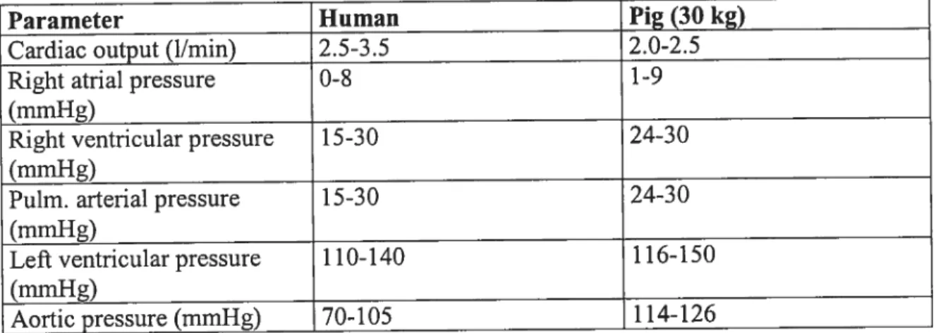
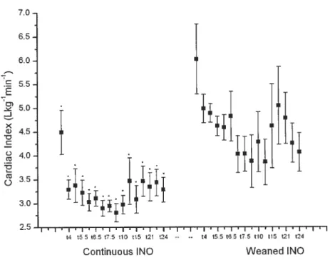
Figure 3: MPAP for continuous and weaned INO groups 40 Figure 4: CI for continuous and weaned [NO groups 41 Figure 5: PVR for continuous and weaned INO groups 42 Figure 6: SVR for continuous and weaned INO groups 43 Figure 7: Compliance for continuous and weaned INO groups 45
TABLES
Table I: Comparison of cardiovascular parameters between humans and
pigs 5
Table II: Criteria for the diagnosis ofSRS, sepsis and MODS 6
Table III: Experimental time points 35
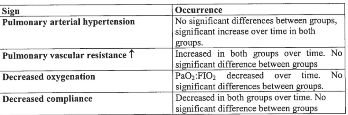
Table IV: Signs associated with rebound hypertension and occurrence during
ABBREVIATIONS
AA arachidonic acid
ALI acute lung injury
BAL bronchoalveolar lavage fluid
CABG cardiac artery bypass graft
CI cardiac index
CK-MB myocardial fraction of creatinine kinase
COMP compliance
COP colloid osmotic pressure
COX cyclooxygenase
CPB cardiopulmonary bypass
CVA cerebrovascular anornaly
CVP central venous pressure
DNA deoxyribose nucleic acid
ECG electrocardiography
EDRF endothelium - derived relaxing factor
ESR electron spin resonance
ET endothelin— 1
FAD flavin adenine dinucleotide
FDA Food and Drug Administration
FMN flavin mononucleotide
F102 inspired oxygen fraction
cGMP cyclic guanosine monophosphate
Hb hemoglobin
11CC heparin coated circuit
ICU intensive care unit
IL interleukin
INO inhaled nitric oxide
IRV inverse ratio ventilation
LT leukotriene
MAP mean systemic arterial pressure
MA? mitogen activated protein
MI myocardial infarction
MMP matrix metalloproteinase
MODS multiorgan dysfunction syndrome MPAP mean pulmonary arterial pressure
NADPH nicotinamide adenine dinucleotide phosphate NANC non-adrenergic non-cholinergic
NF-icB nuclear factor KB
NMA NG-methyl-L-arginine
NO nitric oxide
cNOS constitutive nitric oxide synthase eNOS endothelium nitric oxide synthase INOS inducible nitric oxide synthase nNOS neuronal nitric oxide synthase
02 superoxide anion
0N00 peroxynitrite
PaCO2 arterial partial pressure of carbon dioxide PAF platelet activating factor
PAH pulrnonary arterial hypertension Pa02 arterial partial pressure ofoxygen
Pa02:Fi02 arterial partial pressure of oxygen:inspired fraction of oxygen
PAP puÏmonary arterial pressure
PEEP positive end-expiratory pressure
PG prostaglandin
PMN polymorphonucleocytes
ppm parts per million
PVR pulmonary vascular resistance
ROS reactive oxygen species
SIRS systernic inflammatory response syndrome
sr soluble receptors
SVR systemic vascular resistance
TNF turnor necrosis factor
Tx thromboxane
VAL! ventilator-associated lung inj ury VILI ventilator-induced lung injury
ACKNOWLEDGEMENTS
I would like to thank the following people for their invaluable help throughout the duration ofthis MSc project:
Dr Eric Troncy for both his continued guidance during both this project and the residency program, and for his unending optimism;
Dre Sophie Cuvelliez for constant encouragement and words ofwisdom; Dr Gilbert Biaise for bis direction and support;
Drs Lavoie and Boysen for kindly agreeing to act as jury members and undertaking the significant work involved.
And finally, Dr Eugenio Rasio, as Scientific Director ofthe CHUM research Center, for financial support of this project.
At present, the use of ll\TO is clinically approved by the Food and Drug Administration (FDA) for persistent pulmonary hypertension of the newbom, following evidence from large, controïled multicentre trials.’4 It has also achieved widespread use in a multitude of other clinical situations, such as acute respiratory distress syndrome (ARDS), acute pulmonary hypertension, and pulmonary hypertensive crises during cardiopulmonary bypass
The primary benefits of INO are a selective pulmonary vasodilatation (both arteries and veins) of ventilated areas of lung resulting in decreased intrapulmonary shunt and improved Pa02. This, and the absence of effect on systemic vascular tone has been demonstrated in numerous studies.57
In addition, a reduction in neutrophil adhesion (a crucial step in acute lung injury) to both pulmonary8’° and distal” systemic vasculature has been demonstrated. It should be noted that the timing of ll\TO administration appears to be key to these beneficial effects, with contrasting results found in studies delivering INO afier pulmonary inj ury was established.12’13
The use of INO in clinical practice has flot been without problems, primarily due to the occurrence of rebound pulmonary artery hypertension (PAR) associated with an increase in right to left shunting and a resultant decrease in Pa02 following discontinuation of TNO.14 It has been proposed that downregulation of endogenous cNOS during INO could result in a lack of endogenous NO following weaning of INO,152° resulting in rebound PAR. Also, endothelin (ET) receptor
(A) blockade has been shown to prevent rebound PAR indicating a possible interaction between NO, ET, and its receptor.21’22
Circumvention ofrebound PAR is achievable through careful, stepwise weaning of INO.2325
The aim of this study was to evaluate the hemodynamic and respiratory effects of a iNO weaning protocol during a cardiopulmonary bypass procedure (CPB) procedure in a porcine model (previously reported by our group26), compared with
continuous delivery of [NO. Inhaled Nitric oxide was delivered prophylactically i.e. on induction ofanesthesia, prior to the CPB procedure.
2.1 Introduction
The aims of this review are twofold:
1. To briefly discuss the physiological similarities between the porcine and human cardiovascular system with reference to suitability for cardiovascular research.
2. To review the current knowledge ofthe effects of cardiopulmonary bypass (CPB) on the cardiovascular and respiratory systems.
2.2 The validity of pigs as animal models for cardiovascular research; anatomy and physiology
0f the large animal models currently used for cardiovascular research, the pig has particular advantages over other species with respect to its similarities to humans. These include size, digestive physioÏogy, dietary habits, kidney structure and function, pulmonary vasculature bed structure, coronary artery distribution, respiratory rate, many hematological parameters, immunology and cardiovascular anatomy and physiology.27
2.2.1 Anatomy
Anatomically, the porcine pulmonary system is similar to that ofhumans.
Grossly, the human left lung is made up of an upper and lower lobe and the right lung of an upper, middle and lower lobe. The porcine pulmonary system is cornprised ofa right lung made up ofcranial, middle and caudal lobes and a left lung made up ofa split cranial lobe and caudal lobe. There is also a left accessory lobe. In addition, the anatomy of the bronchial artery and local anastamoses closely resembles that ofhumans. This is an important feature due to the importance ofbronchial artery supply during CPB.28’29 Continuous pulmonary perfusion with oxygenated blood was associated with lower pulmonary
inflammation (reduction in polymorphonucleocytes (PMNs; neutrophuls) and neutrophil elastase concentrations in bronchoalveolar lavage fluid (BAL)), when cornpared with CPB without continuous puÏmonary perfusion in a neonatal pig model ofCPB.29
2.2.2 Physiology
The cardiovascular and pulmonary systems are similar to that ofhumans.27’3° The size of pigs often used in laboratory investigations (25-3 0 kg) have a heart size to body weight ratio of 0.005, identical to that ofhumans (70 kg). Other sirnilarities include, a very low incidence ofpre-existing collateral coronary vessels (under 2%), a sirnilar end-artery coronary anatomy, distribution and size
of coronary vessels,3’ and similar hemodynamic parameters (see Table 1).
However, their pattem of venous drainage differs from man in that there is a large hemiazyguous (left azygous) vein.31The importance ofthis during cardiac
investigations involving invasive techniques is the risk ofhemorrhage due to vessel fragility.
The electrophysiology of the heart is different in the pig compared to man though the intracardiac electrophysiologic parameters resemble man doser than any other non-primate animal.31 The porcine conduction system has the following
differences from humans: different location of AV node, shorter and more proximally branching penetrating bundle, more connective tissue, Ïess elastic tissue and a larger number ofnerve fibres, implying an important neurogenic component to conduction.32 These differences have been demonstrated in immunohistochemical studies
Importantly, with respect to CPB, the pig heart is very susceptible to ventricular fibrillation (which may or may not be preceded by atrial fibrillation), frequently requiring defibrillation andlor pre-treatrnent with anti-arrhythmic agents such as bretylium tosylate.30’35 This susceptibility highlights the importance of good CPB technique and myocardial protection when using this animal for CPB.
In addition, the metaboÏic response ofthe porcine rnyocardium to ischemia is similar to that ofhuman myocardium; rendering the porcine model relevant to the
study of post CPB ischemia and reperfusion injury, and acute myocardial infarction.3’
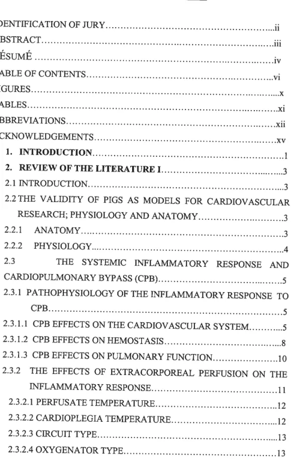
Parameter Human Pig (30 kg)
Cardiac output (hmm) 2.5-3.5 2.0-2.5
Right atrial pressure 0-8 1-9
(mmHg)
Right ventricular pressure 15-30 24-30
(mmHg)
Puim. arterial pressure 15-30 24-30
(mmHg)
Left ventricular pressure 1 10-140 1 16-150
(mmHg)
Aortic pressure (rnmHg) 70-105 1 14-126
Table I: Comparison of cardiovascular system parameters between humans and
pigs.27
Other advantages
In addition to their physiological and physical pararneters, their availability (compared to non-human primates) and breeding characteristics make them particularly suitable as large animal models. With regard to breeding
characteristics, they have large litters, reach sexual maturity rapidly (4-5 months) and ovulate every 3 weeks. They also adapt well to the laboratory environment and have few disease problems when housed conectly.
2.3 The systemic iuflammatory response and cardiopulmonary bypass (CPB)
The systemic inftammatory response syndrome (STRS) and its possible sequelae were defined at a consensus conference ofthe American College ofChest Physicians and the Society ofCritical Care Medicine.36 It has been recognised that STRS may occur as a resuit ofinfectious or non-infectious causes (such as CPB) and a clinical progression to sepsis, severe sepsis, septic shock, multiple organ dysfunction syndrome (MODS) and death has been demonstrated.37 This
levels ofmorbidity and mortality rates, highlighting the importance of non infectious causes of SRS such as CPB.
The pathogenesis of SIRS is stiil unclear due to the many complex interactions involving various facets ofthe immune system, factors associated with CPB equipment and techniques, patient factors and the generation of a reliable and consistent animal model of CPB.
At present, the progression of SRS following CPB, towards adverse sequelae is thought to depend on the balance of pro and anti-inflammatory cytokines, within each organ system. Following CPB, the theory of multiple hits states that CPB primes the immune system (PMN priming and pulmonary sequestration), and secondary minor insuits result in serious sequelae, deranging attempts of the body at re-establishing homeostasis.384’
SRS: diagnosis requires presence of 2 or more ofthe following: Temp>38°Cor<36°C
Heart rate> 90 bpm
Resp rate >20 breaths/min or PaCO2 <32 mmHg
Leukocytes> 12000, <4000/ mm3 or> 10% immature (band) fonns Sepsis: SIRS with documented infection
Severe sepsis: sepsis associated with organ dysfunction, hypoperftision or hypotension Septic shock: sepsis with hypotension despite adequate resuscitation along with the presence of perfusion abnormalities
MODS: a state of altered organ function in an acutely ill patient such that homeostasis caimot be maintained without intervention
Table II: Criteria for the diagnosis ofSIRS, sepsis and MODS.42
2.3.1 Pathophysiology ofthe inflammatory response to CPB.
The inflammatory response to CPB is mediated through contact activation, ischemia- reperfusion induced injury to various organs including brain, heart and blood, kidney, liver and lungs, and, endotoxemiavia splanclmic hypoperfusion.
2.3.1.1 CPB effects on the cardiovascular system.
The occurrence of major cardiovascular complications (cardiac death, myocardial infarction, heart failure) in cardiac artery bypass graft (CABG) patients is
considerable (10%). The overail incidence ofmyocardial infarction (MI) in a multicentre study of 566 human patients was up to 25% and the majority of cases occurred within 16 hours after the release of aortic occlusion.44 Tndependent predictors ofmyocardial infarction (MI) were intraoperative ST segment
deviation, intraventricular conduction defect, lefi bundie branch block, duration of hypotension (systolic arterial blood pressure iess than 90 mrnHg) after CPB and duration ofCPB.
Proinflammatory cytokines released as a resuit of CPB have been shown to play a roleinthe myocardial dysfunction and ischemic episodes associated with CPB procedures).45 In a study of 22 human patients,45 levels ofTNF-alpha, interleukin (IL)-6 and IL-8 were determined to exhibit two peaks; the first early in the post operative period and the second approximately 18 hours after the CPB procedure. Left ventricular wall motion abnormalities were associated with raised IL-6 and IL-8 levels and postoperative myocardial ischemic episodes were associated with raised IL-6 levels. Aortic cross clamp time was independently predictive ofthese postoperative cytokine levels. In addition, Oddis and Finkel showed that TNF alpha, IL-lbeta and IL-6, and NG-methyl-L-arginine (NMA; a NO synthase inhibitor) ail compietely biocked the positive chronotropic effects ofthe beta adrenergic agent isoproterenol on cardiac myocytes.46 Ibis demonstrates a link between cytokines and NO and myocardial beta-adrenergic desensitisation and may provide a partial explanation for the myocardial depression seen post CPB.
The leveis of production of NO by the myocardiurn may have both beneficial cardioprotective and deleterious effects. A constitutive cardiac nitric oxide synthase (cNOS) has been demonstrated in both humans and animal models and its activity linked to the contractile state ofthe heart, possibly through beta adrenergic pathways.47’48 Nitric oxide producedvia cNOS has inhibitory roles in myocardial contractility and the degree ofplatelet adhesion to endothelium.49’5° During an inflammatory response, NO production is upregulatedvia inducible NOS (iNOS) and this may have deleterious effects for myocardial function.
Increased NO concentrations have been associated with myocardial stunning in hurnans.51 There may be several mechanisms underlying the end resuit such as the inhibition of mitochondrial activity, aterations in platelet-endothelial adhesion, and the formation of radical oxygen species such as peroxynitrite and beta adrenergic desensitisation.46’48’50’52’53
Recent evidence implicates a role for ET ta potent vasoconstrictor produced by endothelial ceils) in myocardial depression foïlowing CPB as demonstrated by local production ofthis vasoactive mediator and the beneficial effects of ET receptor antagonism on rnyocardial function.54’55
In addition, myocardial cyclooxygenase (COX)-2 protein and mRNA levels have been shown to increase significantly in an animal model of CPB and cardioplegia. Inhibition ofprostaglandin production lias been associated with improved
systemic arterial blood pressure and urine production dtiring CPB.56’57
2.3.1.2 CPB effects on hemostasis
The inflammatory response to CPB affects hemostasis through contact activation ofthe coagulation and fibrinolytic cascades (figures 2 and 3), endothelial damage via inflammatory mediators, and leukocyte and platelet activation.5862
The duration of CPB, postoperative skin temperature and plasma complement (C3) levels have been positively correlated with postoperative bleeding time and levels ofblood loss.63
The use ofhemofiltration during CPB was associated with a reduction in cytokines (TNF, IL-10), rnyeloperoxidase, C3a and a reduction in postoperative bleeding.64
The involvement ofplatelets was demonstrated with significantly less postoperative blood loss and improved levels ofpulmonary function (post extubation gas exchange, lowered ventilation times) following the post CPB infusion ofpre-operatively harvested platelet rich plasma.65
Extrinsic pathway Intrinsîc pathway Tissue tacr VII la PL C a2 Platelets Pibrin
Figure 1: The coagulation cascade. Adapted from Rang etal.66
OMBU
t
fribrin degradatio products
J
2.3.1.3 CPB effects on pulmonary function.
Cardiopulmonary bypass is well known for its effects on the pulmonary system, namely Acute Lung Injury (ALI) and the more extreme form ofthis syndrome, the Acute Respiratory Distress Syndrome (ARDS). This syndrome, in addition to the effects of CPB on other organ systems puts the patient at risk of developing MODS.67 The incidence and mortality rates ofARDS vary between surveys with currently accepted figures of an incidence between 1 - 3% with a mortality of 30
-50%.426869Mortality rates have dropped markedly from approximately 90% in the early 1970s despite no major advances in pharmacological therapy in 30 years ofresearch. Improved ventilation strategies have been responsible for the majority of this improvement.69’7°
The current definition ofALI is hypoxemia (Pa02:F102 300 rnmHg), bilateral pulmonary infiltrates (indicating inflammatory change) on thoracic radiographs and normal pulmonary capillary wedge pressure.7’ The progression of ALT to ARDS has been described as involving a proliferative phase when fibroblasts infiltrate and remodel areas of inflammation followed by fibrosis and
consolidation ofthe lung parenchyma and a Pa02:F102 200 mmHg.69
The risk ofdevelopment, and severity ofALI, has been positively linked to CPB duration72
Pulmonary dysfunction below the threshold of ALT classification occurs in 12% of patients and includes perfusionlventilation mismatch, reduced oxygenation index and reduced lung compliance.73
In addition, and perhaps relevant to MODS, an association between early pulmonary dysfunction (defined by mechanical ventilation with an Pa02:F102 150 mmHg and pulmonary infiltrates) and renal and netirological dysfunction, nosocomial infections, prolonged mechanical ventilation, increased intensive care unit (ICU) and hospital stay and mortality has been identified.72
Many studies have demonstrated the role ofthe inflarnmatory response in pulmonary dysfunction.74’75 However the importance of various inflammatory mediators within the inflammatory process is still the subject of investigation.
Histologically, the inflammatory response and pulmonary damage following CPB has been demonstrated.76 Neutrophils and their associated inflammatory
mediators play an important role; 50% of circulating PMNs are sequestered to the lungs following CPB (from the release ofthe aortic cross-clamp) and are
associated with a significant increase in oxidative activity.75 Granulocyte elastase, IL-6 and IL-8 levels correlate with a reduced respiratory function.74 Reactive oxygen species (ROS) have also been associated with an increase in pulmonary endothelial permeability.77
Sinclairet aï. studied pulmonary endothelial permeability and found a transient increase in permeability in ail patients (20 patients) post-CPB but no association between markers measured (protein accumulation index, and BAL to serum urea ratio) and the development ofARDS. Increased levels ofthese markers were associated with a longer duration of CPB. They postulated that though PMNs had a role to play in pulmonary dysfunction, celiular activation occurred at a systemic rather than local level supporting the theory of a systemic inftammatory response to CPB.78
Recently, ET has been implicated in the pathogenesis ofincreased pulmonary vascular resistance following CPB. Severai animal studies have shown increased plasma ET, ET receptor and ET mRNA concentrations within pulmonary vascular cndotheÏium and ET receptor blockade has been shown to decrease the levels of pulmonary vascular resistance.7981
The enzyme COX 2 and prostaglandin (PG) synthesis may also play a role in increased pulmonary vascular resistance, with increased levels of COX 2 protein and mRNA associated with increased pulmonary resistance.82
2.3.2 The effects of extracorporeal perfusion on the inflammatory response Almost every component of extracorporeal perfusion has been shown to have a potential effect on the inflammatory system. This includes perfusate temperature; cardioplegia temperature; circuit type; oxygenator type; priming solution
composition; the heparin-protamine complex; pulsatile versusnonpulsatile perfusion; pump types; and shear stress.
2.3.2.1 Perfusate temperature
A study involving 18 patients undergoing elective CABG compared the effects of tepid (34°C) and hypothermic (28°C) perfusate on markers of inflammation, respiratory index, systemic vascular resistance and intubation time.83 Tepid CPB resulted in a reduction in respiratory dysfunction with a shorter intubation tirne compared with that ofthe hypothermie group.
Menasché et al. suggested allowing the core temperature of patients to drifi between 32-34°C during CPB following the resuits ofa clinical study of 30 patients. This study dernonstrated increased levels of cytokines and an increased requirement for vasopressor support in patients undergoing normothermic (37°C) CPB, compared with patients undergoing hypothermic CPB (28-30°C).84
An animal study (pigs) found that moderate hypothermia (28°C) was associated with the lowest histological degree of organ damage.85 This was associated with lower TNF and higher IL-10 levels.
In contrast, a recent study did not show a significant difference in the
postoperative levels of any of the inflammatory markers measured (C-reactive protein, IL-6) between normothermic (37°C) and hypothermie (26°C) CPB patients.86 Different perfusate temperatures (20°C, 32°C, 37°C) were found to have no relationship to neurological dysfunction following CPB.87
2.3.2.2 Cardioplegia temperature
Warm blood cardioplegia was found to reduce the levels ofTNF, IL-6 and IL-8 post-operatively compared with cold crystalloid cardioplegia.88 However, a study comparing the effects on complement and PMN activation between wanri and cold blood cardioplegia showed that both complement and PMN activation was higher in the warm bÏood group.89
2.3.2.3 Circuit type
The balance of cytokines is pushed in favour of inflammation as a resuit of the interaction ofblood with a CPB circuit, demonstrating the need for improvements in circuit biocompatibility (see section 2.3.1.3).°
Such improvements (a conventional circuit versus a polymer coated circuit) have been shown to decrease fibrinolysis and thrombin generation and preserve platelets.91
The use ofheparin coated circuits (HCC)versus conventional circuits in high risk patients undergoing CPB found that HCC resulted in a shorter intensive care and post-operative hospital stay, and a lower incidence of lung and renal
dysfunction.92 fi a similar study comparing HCC to conventional circuits, a reduction in post-operative morbidity (myocardial infarction, arrhythmias, respiratory insuit, neurological dysfunction) and intensive care stay was demonstrated.93
With respect to inflammation, a comparison ofthe concentrations of IL-1, IL-6, IL-8 and TNF by HCC and conventional circuits showed that levels of IL-6 and IL-8 levels were reduced in the serum of patients in the HCC group.94 In addition, the method of circuit coating and type of heparin employed have effects on the degree of activation of various components of the inflammatory cascade.95
2.3.2.4 Oxgenator type
Resuits from studies comparing the effect ofbubbleversus membrane
oxygenators on inflammation and post-operative outcome have been mixed. This may be due, at least in part, to the difference between experimental studies and the clinical situation.
Two studies by the sarne group found that though there was no difference between the degree ofcomplement activation between two groups of patients undergoing CPB with either a membrane or bubble oxygenator; evidence ofPIVIN activation (plasma lactoferrin and myeloperoxidase plasma concentrations) was significantly lower in the membrane oxygenator group.96’97 Looking at organ dysfunction in this study, cardiac (post-operative need ofinotropic support), renal
(senim creatinine levels) and pulmonary (alveolar-arterial oxygen pressure gradient), but flot cerebral function (psychometric tests and adenylate kinases levels), was improved in patients with membrane oxygenators versus those with bubble oxygenators.
The effect ofbubble oxygenators on respiratory function was associated with an increase in extravascular lung water and atelectasis (on chest radiographs), compared with membrane oxygenators.98 However, these changes had no effect on the duration ofpostoperative ventilation, mortality or hospital stay.98
A smaller, more recent study looking at complement activation, respiratory function, granulocyte activation and endothelial damage between CPB with membrane versus bubble oxygenators showed no significant differences between the groups for ail variables studied.99
A comparison of flat sheet and hollow fibre membrane oxygenators in terms of shear stress showed a positive conelation between the pressure drop across flat sheet oxygenators (shear stress generated) and the degree ofneutrophil elastase released; a marker ofPMN activation.100
2.3.2.5 Priming solution composition
The effect of crystailoidversus colloid priming solutions on colloid osmotic pressure (COP) in 20 patients during CPB found that COP decreased by a significantly lesser amount both during and after CPB in patients receiving a colloid prime.93 Measures of complement (C3b/c and C4b/c) and PMN activation (neutrophil elastase) and TNF increased similarly in both groups.
Patients with colloid prime showed an improved postoperative fluid balance, shorter duration of intubation, smaller rectal to skin temperature gradient and shorter hospital stay.
In addition, a colloid prime bas been shown to improve cardiac index and prevent extravascular lung water accumulation postoperatively when compared with a crystalloid prime)0’
2.3.2.6 Heparin and protamine compiex
Heparin and protarnine (antagonises the anticoagulant effect ofthe heparin antithrombin III complex; Figures 2 & 3) are given prior to, and at the termination of CPB respectively, in order to minimise the coagulation response to CPB. Together they fomi a complex which has been shown to activate complementvia the classical pathway with many ensuing deleterious effects.102”°3 These
complexes may cause increased pulmonary arterial pressure, decreased systolic and diastolic arterial biood pressure, myocardial oxygen consumption, cardiac output, heart rate and systemic vascular resistance.
The irnpaired balance of pro and anti-coagulants in patients following CPB lias important foies lflpostoperative coaguiopathies and multiple organ dysfunction.
2.3.2.7 Pulsatileversus non - pulsatile perfusion
A ciinical study comparing CPB with pulsatile and nonpulsatile flows
demonstrated lower leveis of endothelial damage (reflected by lower ET levels) and cytokine activation (lower IL-8 concentrations) with pulsatile flows.’°4 Levels of endogenous endotoxin are lower with pulsatile CPB whilst cNOS, which piays an important role in the modulation ofvascular compliance via NO, is activated by pulsatile ftow.105”°6
2.3.2.8 Pump types
A clinical study measuring the ievels ofinflammatory mediators TNF, IL-ibeta, IL-8, IL-6, PMN counts, neutrophil elastase and terminai complement
components in patients undergoing CPB with either a centrifugal or roller pump demonstrated that centrifugal purnps generated a greater inflammatory response in terms ofa significant increase in IL-6,PMNcounts and neutrophil elastase
concentrations.’°7 However, a recent retrospective study on the effect ofpurnp type on neurological outcomes found that patients undergoing CPB procedures with centrifugal pumps had a lower incidence of permanent neurological deficit and coma compared with roller pumps; there was no difference in mortality rates.’08
2.3.2.9 Shear stress
Within the vasculature, shear stress acts as a physiologic stimulus contributing to vasoregulation via the endothelium. An animal model (pig) comparing the effects ofpulsatileversus nonpulsatiÏe flow CPB demonstrated a reduction in endothelial NO production in the nonpulsatile group. It is known that NO contributes to vascular tone and may partially explain the increased vascular resistance seen with nonpulsatile CPB.’°9
However, the shear stress associated with CPB may be excessive and have detrimental effects. It has been demonstrated that hypothermia, plasma dilution and shear stress acted synergistically to decrease erythrocyte deformability and cause immediate and deÏayed hemolysis which may impair microcirculation and oxygen supply.”°
An in vitro study demonstrated the detrimental effects of shear stress on leukocytes. At levels ofshear stress less than that required to cause erythrocyte hemolysis, there was evidence of leukocyte destruction, disniption, aggregation, cytoplasmic granule release and increased adhesiveness.” Increased platelet activation has also been linked to shear stress.112
Endothelial injury as a resuit of shear stress may alter the interaction of these celis with the ceils ofthe immune system and underlying smooth muscle.”3
2.3.3 Inflammatory markers
Due to the cascade nature of enzyme systems and interaction between various aspects ofthe inflammatory response there are a plethora ofinflammatory markers available as indicators ofthe degree, chronicity, type ofinflammatory response and response to treatment.
In ternis ofCPB, many markers have been studied but only those ofparticular relevance to CVS and respiratory dysfunction will be discussed.
2.3.3.1 Cytokines
Cytokines are soluble paracrine mesengers that play a key role in the homeostasis ofthe immunological and physiological systems. The actions ofindividual cytokines may be loosely described as pro or anti-inflammatory though specific actions are dependent on each situation. For instance, IL-6, described as a pro inftammatory cytokine has been demonstrated to play a protective role in hyperoxic lung injury in mice through the inhibition of celi death and matrix metalloproteinases (MMP) expression.”4
Interleukin-ibeta and IL-6 plasma concentrations have been shown to be predictive of a poor outcome in ARD$ and plasma IL-8 and IL-18 levels were raised in non survivors following cardiac surgery and the subsequent development of SRS compared with survivors.”5’116
Bimodal serumpeaks for TNF(2 and 1$ hours post CPB), IL-6 (irnmediately and 12-18 hours post CPB) and IL-8 (early and 16-18 hours post CPB) have been documeflted.’”17
Striking a balance with proinflammatory cytokines are antiinflammatory
cytokines, cytokine receptors and cytokine receptor antagonists; in particular, IL-10, IL-1 receptor antagonist (ra), TNF soluble receptors 1 and 2 (TNF5r 1 and 2) and transforming growth factor beta (TGF3).64’9°
In a sirnilar fashion to pro-inflammatory cytokines, elevations of components of the anti-inftammatory response are staggered in tirne following CPB. In pediatric CPB, the plasma response began with IL-10 (increased levels during CPB and peaking 24 hours post CPB), followed by IL-ira (increased levels 2 hours post CPB and peaking at 24 hours post CPB) and TNFsr (increased levels 2 hours post CPB and peaking at 24 hours post CPB). From BAL samples, only IL-8 and IL-10 were significantly elevated following CPB.118
In aduit CPB patients, IL-10, TGF-beta and IL-ira plasma levels increased early following CPB (levels peaked within 2 hours post CPB and had decreased by 24 hours post CPB). Turnour necrosis factor soluble receptors 1 and 2 levels
increased following CPB and remained elevated at the 24 hour sample
90,119,120
time.
Pulmonary dysfunction following CPB is a frequent complication and believed to be largely mediated through local PMN sequestration and activation, through adherence, degranulation of enzymes and superoxide production.’21-126 A comparison of BAL and plasma specimens from patients with ARDS,
dernonstrated that the lungs may be the primary source of IL-8.’27 Furthermore, Donnelly et al. provided evidence of a link between IL-8 in BAL and the development ofARDS.’28 No sucli link was established with plasma IL-8.
Increased concentrations of IL-8 and IL-ibeta (at levels ten times in excess of IL ibeta ra) are present for prolonged periods in the BAL of patients with ARDS.’29 Interleukin-8 showed a strong correlation with the PMN concentration ofthe BAL. Elevated IL-Ibeta levels on day 7 after the onset ofARDS was correlated with increased mortality.
A comparison between two subsets of CPB patients (postoperative cardiovascular dysfunction and postoperative lung injury) found that increased plasma
concentrations of IL-8, IL-6 and platelet activating factor (PAF) were associated with cardiovascular dysfunction.’3° Patients with severe lung injury had increased plasma thromboxane (Tx) B2 and decreased plasma PAF, PG E2 and 6-keto PG Flaipha. BAL samples were flot taken.
Tumour necrosis factor may play a pivotal role in cardiac insufficiency following CPB through its release following ischernia and reperfusion; this has been
demonstrated by the comparison ofblood samples from peripheral arterial blood, coronary sinus blood and mixed venous blood in patients undergoing elective CABG.’3’ They showed increased levels ofTNF and IL-6 in coronary sinus blood following aortic declamping cornpared with peripheral arterial blood indicating the myocardium as an important source ofTNF and IL-6. The effect ofthe inflammatory process on the heart following CPB is myocardial depression with decreased contractile function and myocyte apoptosis.’32”33 Tumour necrosis factor is believed to create these effects through at least two different
mechanisms; an early depressive phase that is sphingosine mediated and a later component mediated through inducible NOS.’33
2.3.3.2 Matrix metalloproteinases (MMPs)
Matrix metalloproteinases are involved in the degradation of collagen and the remodelling ofthe extracellular matrix. They are believed to play an important role in MI, cardiac ischernia-reperfusion injury, ventricular dilation and heart
failure, ALI and ARDS.’34136 Use of an animal model ofALI has demonstrated the importance ofMMPs (MMP 2 and MMP 9) in the mediation ofALI.’36 Patients with ARDS have increased levels ofMMPs (2 and 9) in BAL samples.’37 Matrix metalloproteinase 9 is released as part ofthe inflammatory response in humans during CPB.’38 Matrix metalloproteinases 2 and 9 appear to play prominent roles following CPB and the associated inflammatory and ischernia
134 139-142
reperfusion injuries.
Cytokines play an important role in modulating MMP gene expression; in
particular TNF and IL-ibeta both increase the production ofMMP.’43 1n addition, they increase the activity and stability of MMPs, and the activation of CPB primed PMNs by cytokines leads to the release ofMMPs.
Measurements ofMMP have been undertaken in both the plasma and BAL of patients and animal models with pulmonary dysfunction. fticreased BAL concentrations ofMMP 2 and MMP 9 in patients with ARDS compared with healthy controls has been demonstrated and MMP 9 levels correlated with BAL PMNconcentrations.’37 There have been similar findings in the lungs of rats with hyperoxic lung injury.’44
The importance ofMMPs in different clinical conditions is still under
investigation. Though their roles in cardiac and pulmonary dysfunction have been demonstrated (as referenced above), their role in the dysfunction ofother organs is not clear. Ziswiler et aÏ. (2001) found no evidence for a role ofMMP in an animal model ofrenal ischemia reperfusion injury.’45
Appendix 1 contains an accepted article from our group entitled, “Increased alveolar and plasma gelatinases activity following porcine cardiopulmonary
bypass: liihibition by inhaled nitric oxide”. This offers further discussion on the relationship between INO and MMPs.
2.3.3.3 Reactive oxygen species (ROS)
Reactive oxygen species may play an important role in the damage and
dysfunction associated with the inflammatory response to CPB and the associated iscliemia reperfusion injury.14648 The sources ofROS and their relative
importance have been difficult to identify due to their highly reactive and short lived nature, difficulties in direct measurement and unreliabiltiy of indirect measurements.146
It has been demonstrated that ROS are generated by the myocardium as a result of ischemia reperfusion and play a role in myocardial dysfunction.149
A study in humans undergoing CPB using a direct method of detecting ROS [electron spin resonance (ESR) spectroscopy] has showed a release ofROS, independent ofmyocardial ischemia and reperfusion; detection ofROS occurred from the onset of CPB until the end ofthe CPB part ofthe procedure.’46 This provides evidence of the role of CPB (and possibly decreased perfusion) in the generation ofROS, as no increases were associated with any preparatory surgery (sternotorny, vascular graft preparation) prior to the onset of CPB.
Reactive oxygen species generated as a result of CPB may play roles in
rnyocardial dysfunction and systemic PMN activation, feeding the inflammatory response. 150,151
More cornmonly, indirect measures of oxidation have been used; signs of oxidative protein damage in BAL ofARDS patients (chlorotyrosine,
orthotyrosine, nitrotyrosine; Lamb etal.), plasma antioxidant levels (ascorbate, aipha-tocopherol, retinol, beta-carotene, selenium, lipid peroxidation products) and the plasma antioxidant enzymeactivity of catalase, superoxide dismutase and glutathione-peroxidase in erythrocytes.’52’153
Reactive oxygen species have been shown to play an important role in ALI and ARDS and it has been suggested that an oxidant-antioxidant imbalance exists)52 Reactive oxygen species take part in chain reactions involving celi membranes
resulting in the destruction of membrane integrity, the release of cytotoxic substances and DNA damage with resultant alterations in protein synthesis. Reactive oxygen species affect vascular endothelium through PMN - dependent
and - independcnt mechanisms resulting in dysfunctional vascular tone. In animal models ofpulmonary vascular smooth muscle, the effects ofROS have been mixed with reports ofboth vasodilatation and constriction.’54
The origin ofROS in ALI and ARDS maybe from arange of sources. The stimulation ofPMNs and other leukocytes by cytokines (TNF-alpha) and
endotoxin leads to ROS production. Complement plays a role in the stimulation of ROS production from primed leukocytes following contact activation associated with extra-corporeal circulation and endotoxin stimulation. A major source of ROS for ail organs following CPB is ischemia reperfusion injury. Here, ROS is endogenousÏy produced and it has been demonstrated that 20-40% ofthe free radicals detected following heart-lung bypass are produced by endotheliai cells.’56 finally, the aggessive oxygen therapy used in the management ofARDS can result in hyperoxic toxicity, caused by ROS.’57 In an animal model ofcerebral ischemia reperfusion it was demonstrated that PMN production ofROS (superoxide anion) increased in fine with the duration ofischernia, suggesting their role in cerebral ischemia reperfusion injury.’58
2.3.3.4 Endothelin 1 (ET)
Endothelin 1 is a vasoactive peptide with fibrogenic properties whose production, by endothelial ceils, has been investigated widely in various cardiovascular disorders (ischemic heart disease, hypertension, cardiogenic and endotoxic shock).’59 It may exert its effects through stimulation of arachidonic acid (AA) metabolism, and eicosanoids have been used as markers of ET activity.’6° Increased plasma concentrations following CPB have been associated with an increase in length of intensive care stay and prolonged pharmacological management.’6’ However, its role in inflammation is flot fully understood; whether its presence is a reflection of vascular injury or a cause of
vasoconstriction. In a study of patients with mixed connective tissue disease, plasma ET reflected damage and correlated with an increase in von Willebrand’s factor (a recognised marker of endothelial damage). The plasma concentrations measured were flot enough to cause vasoconstrictionin vitro.159
A comparison of ET levels ftom various sites has demonstrated that plasma ET levels were a systernic product and flot ofcardiac origin.’62 In a similar study of patients with sepsis, with and without ARDS, the lung did not appear to be a major site of ET production. In one study of CPB patients, no association was demonstrated between increases in plasma ET levels and pulmonary
vasoconstriction. On the contrary, plasma ET was associated with pulmonary vasculature vasodilation.’63 However, another study in CPB patients, found an association between pulmonary plasma concentrations of ET and an increase in pulmonary vascular resistance.64
ET levels have been positively correlated with an increased organ failure score and oxygen consumption and negatively correlated with the Pa02:F102 ratio.’65 More recently, in several animal models (pigs), ET production and rises in systemic plasma levels have been associated with local tissue production (myocardial and pulmonary vascular endotheÏial celis) and spiil over to the systemic circulation. The release of ET was documented to begin immediately following the onset of circulatory arrest and continue throughout this period.54’8’ In further animal models, ET has also been demonstrated play an important rob in myocardial depression and increases in pulmonary vascular resistance folbowing CPB
Inlialed Nitric Oxide 3.1 Introduction
Nitric oxide (NO)
Initially described as endothelium - derived relaxing factor (EDRF) before its
identification as NO.’66”67 Its discovery resuited in award of the Nobel Prize in Physiology or Medicine in 1998 to RF Furchgott, U Ignarro and F Murad. Its potentiai physiological, pathological and therapeutic roles have received widespread interest over the last 25 years. What follows is a brief sumrnary of the current state ofknowledge of NO and its potential and actual therapeutic roles.
3.1.1 Biosynthesis
The foie of NO is as an endogenous activator of soluble guanylate cyclase resulting in the formation of cyclic guanosine monophosphate (cGMP), a second messenger ofrnany ceils (nerves, smooth muscle, monocytes, platelets).
The biosynthesis of NO is controlled by nitric oxide synthase (NOS) enzymes, catalyzing a reaction between molecular oxygen and L-arginine. There are 3 known isofoms of this enzyme; inducible (iNOS), expressed in macrophages, Kupffer celis, neutrophils, fibroblasts, vascular smooth muscle and endotheiial ceils, and two constitutive (cNOS) forms, one in the endothelium (eNOS; also found in cardiac rnyocytes, renal mesangial ceils, osteoblasts, osteoclasts and platelets) and the other in neurons (nNOS). Levels of iNOS and hence NO production, increase in response to pathological stimuli as part of the immune system, whereas the primary role of cNOS is in normal physiological states. The amount of NO produced as a resuit of catalysis by iNOS, eNOS and nNOS varies; constitutive forrns producing picornolar concentrations of the molecule and iNOS capable ofproducing millimolar concentrations.
3.1.2 Enzyme characteristics
The NOS isoforms exist as dimers, each containing heme, flavin adenine dinucleotide (FAD), flavin mononucleotide (FMN) and tetrahydrobiopterin as prosthctic groups. In addition, they have binding sites for L-arginine, nicotinamide adenine dinucleotide phosphate (NADPH) and calcium-calmodulin. Activation of the dimers is controlled by these prosthetic groups in conjunction with ligand binding.
NOS enzymes are associated with the Golgi apparatus and plasma membranes. The details of the enzymatic process are unclear but it appears that NOS is bimodal in combining oxygenase and reductase activities at distinct structural dornains catalyzing the following reaction.
02 +L-arginine —> NO +citrulline (NOS)
Enzymatic activity is the rate-limiting step as substrates are abundant in the cytoplasm.
3.1.3 Isoform activity
cNOS activity is controlled by intracellular calcium-calmodulin, the production of which is stimulated by a variety of stimuli.
Vascular resistance control under physiological conditions is likely in response to pulsatile ftow and shear stress.
iNOS activity is calcium independent (though calcium-calmodulin will stimulate its activation) and induced instead, by lipopolysaccharide (LPS) and! or cytokines synthesized in response to LPS or inflammation.
3.1.4 Nitric oxide carnage and breakdown
Nitric oxide is freely diffusible across ceil membranes thus explaining its local paracrine effects (on smooth muscle, monocytes and platelets adherent to
endothelium). NO carnage in the bloodstream is through a high affinity for herne (as part ofHb). In the absence ofoxygen, bound NO is relatively stable. However, in the presence of oxygen, NO is rapidly inactivated by conversion to nitrate and the heme (to which it is attached) oxidized to metHb. Outside Hb, NO can react with oxygen resulting in nitrate and nitrite formation which are excreted in urine. Reversible binding of NO to globinvia its —SH groups (nitrosothiol formation) is also possible, resulting in S-nitrosylated Hb allowing NO to be effective at sites distant from its production.1’
3.2. Physiological and pathological roles of NO
The effect of NO may be mediated through either autocrine or paracrine action resulting in effects such as vasodilatation in vascular smooth muscle, decreased platelet adhesion and aggregation and the inhibition of monocyte adhesion and migration. These effects are through its action on cGMP. Cytotoxic actions are rnediated through combination with superoxide anion (O2), with which it reacts very rapidly, out competing superoxide dismutase and producing peroxynitrite (ONOO).
3.2.1 Vascular effects
The physiological control of peripheral (both pulmonary and systemic) vascular resistance through eNOS is constantly present and active. Endothelial NOS expression is increased by vascular shear stress and eNOS activity is increased by mediators which increase intracellular calcium, such as bradykinin and acetylcholine.
At a cellular level, NO produced by endothelial cells (or administered as INO) diffuses into vascular smooth muscle cells. Here, NO activates soluble guanylate cyclase, which catalyses cGMP. Through cGMP-rnediated protein kinases, NO exerts its clinical effects of vascular srnooth muscle relaxation. In addition, protein kinases inhibit leukocyte adhesion, platelet adhesion and activation, and cell prolifereation.
The action of cGMP is balanced by its hydrolysis to GMP. This is catalysed by
phosphodiesterase 1 (brain, heart, lung, testis) and 5 (lung, platelets, vascular
smooth muscle and kidney), in humans. Inhibition of phosphodiesterase 5, as a
dmg target (zaprinast, siidenafil, dipyridamole), increases endothelium-dependant
vasodilation.
3.2.2 Neuronal effects
As a non-adrenergic non-cholinergic (NANC) neurotransmitter it has a role in neuronal development and plasticity.
3.2.3 Immune system
iNOS appears to play an important role in the innate immune system, responding to a variety of pathogens. Here its activity is through free radical generating,
nitrosylation and heme-binding properties.
3.2.4 Nitric oxide and pathophysiology
The number of pathological conditions in which NO is known to play a part is likeiy to increase with further research due to the systemic distribution of NOS and the large number of cell types upon which NO has been shown to act. For example, sepsis states invoive the overproduction of NO through iNOS
recruitment leading to systemic vasodilatation. Whilst inadequate NO production
by cNOS is associated with artherogenesis (resultant from hypercholesterolernia and cigarette smoking) and impotence.
3.3 Therapeutics
Inhaled NO (INO) has found use in a range of conditions (acute pulrnonary hypertension, right ventricular failure, pulmonary hypertensive crises following CPB and in neonates, heart, lung and liver transplantation), ail ofwhich have two factors in common: the desired effect is within the pulmonary system (the effect
shunting or, is gained secondary to changes in the pulmonary circulation, such as reduced workload on the riglit side ofthe heart.
Pulmonary hypertensive crises following CPB is part of a range of vasomotor dysfunction following CPB.’68 Nitric oxide plays a central role to post-CPB vasomotor function; the activity of eNOS is decreased as a resuit of several rnechanisms, including alterations in ceil membrane potential, substrate and cofactor depletion, alterations in intracellular calcium, and injury to cellular components.168
The clinical effect of INO is limited to the pulmonary vasculature, with a haif life of a few seconds. Inactivation of NO is primarily as a resuit of binding of NO to O2 and to the heme component of hemoglobin, resulting in the release of NO3-. Binding of NO to thiols is another source of NO inactivation.
A potentially serious drawback to the delivery of INO is the risk of rebound pulmonary hypertension.16971 This complication may resuit in hypoxernia and right ventricular overloading and failure.
The delivery and monitoring of NO will be discussed later, suffice to say that concentrations of NO less than 50 parts per million (ppm) do not appear to be toxic.169
Traditional NO donors, such as nitroglycerin, nitroprusside and S nitrosoglutathione have systemic effects that may or may not be desirable depending on the condition being treated.
NO inhibition is currently limited to experirnental studies though this may have therapeutic potential in cases of NO over production such as sepsis. These compounds are primarily L-arginine analogues and include N-monomethyl-L arginine (L-NMMA) and N-nitro-L-arginine methyl ester (L-NAME).
3.4. INO toxïcity
There are three potentially toxic products associated with INO; NO2 resulting from combination with oxygen, ONOO from combination with superoxide ion and metHb following the oxidation ofnitrosylHb.
NO2 inhalation studies have found that inhalation of concentrations greater than 10 ppm resuits in pulmonary edema, alveolar hemorrhage, alteration in surfactant properties, hyperpiasia of Type II alveolar epithelial celis, intrapulmonary accumulations of fibrin, neutrophils and macrophages and death.172 Acute overdose of INO Ïeads to rapid NO2 accumulation, methemoglobinemia, pulmonary edema and hemorrhage, hypoxernia and death)73 The Occupational safety and Health Administration of the United States lias set $ hour time weighted average exposure limits in the workplace at 5 ppm. Chronic (9 weeks), low level exposure of 05- 1.5 ppm caused focal degeneration of pulmonary interstitial ceils and mild emphysematous changes in rats.174
Peroxynitrite is readily formed from the reaction of NO and superoxide. Under physiologie conditions this is limited by the superoxide scavengers, primarily superoxide dismutase. However, under pathological conditions when superoxide concentrations may be increasd or scavengers exhausted, peroxynitrite concentrations increase.175 Endogenous peroxyiiitrite production is useful through its ability to cause oxidation, peroxidation and nitration of lipids, proteins and DNA.’76”77 This enables the destruction ofmicroorganisms and tumour ce11s178”79 but is a double edged sword, resulting in surfactant alteration (decrease in surface tension,’80 macromolecular structural changes and apoptosis induction.176”77 MetHb production is limited in the nonnal individual by reduction back to Hb by cytochrome b5, methemoglobin reductase within erythrocytes, and by glutathione.
3.4.1 NO and DNA
NO lias several effects on DNA; the formation of mutagenic nitrosamines,’76 DNA strand breakage’8’ and inhibition ofDNA enzyme repair systems)78
3.4.2 NO and lipids
The in vivo effects of NO on lipids are unclear, with the production of both pro
peroxynitrite). These effects appear to depend on relative concentrations of the various molecules involved.’82”83
Evidence from iNO clinical trials between 198$-1997 show that signfficant methemoglobinemia or NO2 formation was uncommon and a reduction in INO was usually sufficient to decrease levels. Discontinuation of iNO was required in only 0.6 ¾ of patients.’74
The side effects of long terrn INO usage are complicated by the possible multiple sources of markers of NO related However, no changes in surfactant properties’86 or pulmonary function either during or after mechanical ventilation as a result of INO have been observed.
3.4.3 INO and inflammation
There is stiil conflicting evidence regarding the role of endogenous and INO in inflammation. Much of this appears to depend on the concentration of NO and timing ofits delivery.42”74
The low concentrations of NO normally produced by cNOS are essential for the maintenance ofvascular tone plus effects on platelet and leukocyte
It lias been reported that inhibition of cNOS during inflammatory insult worsens the resultant inflammation.’88
By contrast, iNOS leads to much higher concentrations of NO and it is this which is believed to act in a pro-inflammatory fashion.189’92
Inhaled NO is capable of decreasing elevated pulmonary artery pressure (PAP), improving hypoxemia by reducing intra-pulmonary shunt, and optimizing ventilation perfusion (V/Q) matching. Inhaled NO can also inhibit the inflammatory process by reducing cytokine synthesis and inactivating nuclear factor KB (NF-KB), as several cytokines contain a binding site for NF-KB in their promoter gjfl5•93 Nitric oxide can also decrease the expression of adhesion molecules preventing neutrophil adhesion and migration. Inhaled NO canexert its effect on the lung, on leukocytes trapped in the pulmonary area but as it is
transported by red blood celis to the general circulation, INO could have extrapulrnonary effects.
3.4.4 Importance of timing ofdelivery
Pretreatrnent with INO or delivery early in the onset of the insuit appears to be the key to maximizing the anti-inflammatory effects of NO.8’171”94’195
Though the rote of NO in inflammation is undoubtedly complex, much ofthe conflicting data regarding the pro- and anti-inflammatory effects of INO can be explained by the timing of delivery ofINO.
Appendix 2 contains an article submitted to Nitric Oxide-BioÏogy and Chemistiy (Impact factor of 2.545), represents resuits ofa previous series ofexperiments. It demonstrates the safe use ofpre-emptive, continuous INO and its protective effects against lung ischernial reperfusion associated with CPB. Stich resuits are very promising for promoting the use ofpre-emptive [NO therapy during cardiac surgery with CPB. Before expecting such clinical advance, one point remained to be solved: Could [NO be reasonably and safely weaned for the cardiac patient after CPB?
The aim of this study was to evaluate a 1NO weaning protocol during a CPB procedure in a porcine mode! (as va!idated by our group26), and use this mode! to compare the hemodynamic and respiratory effects of a weaning INO protocol against continuous IISTO.
Nuil hypothesis: step-wise weaning ofINO does not resuit in ciinically significant rebound pulrnonary hypertension. There is no difference between weaned and continuous TNO treatment.
5. Methodology
This study was performed at the Notre Dame Hospital (CHUM) anima! centre with the approva! of the institutional animal care committee (Centre de recherche du CHUM) in comp!iance with the guide!ines ofthe Canadian Council on Anima! Care.
5.1 Animais
Pigs weighing between 30 and 35 kilograms, free of c!inical pu!monary disease were purchased from Vadnais Fami Inc. (Drurnrnondville, Québec, Canada). The animais were allowed an acclirnatization period of at least 3 days in our housing facilities (Notre-Dame Hospital research laboratories) prior to the procedure. Anima!s were housed individua!!y with twice daiiy access to proprietary feed and ad libituin access to water.
5.2 Experimental procedure
The foi!owing sections on anesthesia, venti!ation, LNO administration, monitoring, CPB procedure and post-operative protocol were carried out as published by our group.26 Animais were randomized into 2 groups. One group
(weaned INO; n=6) had 1NO weaned foilowing the end of the CPB procedure (weaning began at T4, i.e. 4 hours after inducing CPB) whiist the rernaining group (continuous iNO; n=7) had iNO continued until termination of the experimental period at T24 (24 hours after induction of CPB). Individuals collecting data were blinded to the INO protocol.
5.2.1 Anesthesia
Animais were fasted 12 hours prior to surgery. Premedication consisted of intramuscular atropine (0.04 mg/kg), azaperone (4 mg/kg) and ketamine (25 mg/kg). Anesthesia was induced thirty minutes later with intravenous fentanyl (5 g/kg) and thiopentai (5 mg/kg) deiivered through a 20 gauge catheter placed in an ear vein. Intubation was performed with an $ mm endotracheal tube (Mailinckrodt Company, Mexico city, Df, Mexico) facilitated with a laryngoscope and surgical preparation (washing, shaving and first disinfection) followed. The animais were attached to the operating room table in a supine position. A single dose of antibiotic (enrofloxacin 5 mg/kg) was given intravenously at this time.
A total intravenous anesthesia technique was used for the maintenance of anesthesia with an infusion of thiopental (5 mg/kg/hr) and fentanyl (20 tg/kg/hr) in the same syringe. Neuromuscular biockade was perforrned with pancuronium through a loading dose of 0.2 mg/ kg followed by an infusion of 0.2 mg/kg/hr. Lactated Ringer’s solution (Baxter Corporation, Toronto, Ontario, Canada) was given at a basal rate of 10 ml/kg/hr throughout.
5.2.2 Hemodynamic monitoring and support
Monitoring was by continuous ECG, rectal temperature, urine output (Foley catheter placed by cystotomy), and systemic arterial blood pressure [via a 20 gauge arterial catheter (Arrow International Inc., Reading, PA, USA) piaced in the left carotid artery]. Puimonary arterial pressure, pulmonary capillary wedge pressure, central venous pressure, cardiac output (through thenriodilution) and blood temperature were monitored via a Swan-Ganz pulmonary artery catheter (Abbot Laboratories, Chicago, IL, USA) placed in the right internai jugular vein.