Direction des bibliothèques
AVIS
Ce document a été numérisé par la Division de la gestion des documents et des archives de l’Université de Montréal.
L’auteur a autorisé l’Université de Montréal à reproduire et diffuser, en totalité ou en partie, par quelque moyen que ce soit et sur quelque support que ce soit, et exclusivement à des fins non lucratives d’enseignement et de recherche, des copies de ce mémoire ou de cette thèse.
L’auteur et les coauteurs le cas échéant conservent la propriété du droit d’auteur et des droits moraux qui protègent ce document. Ni la thèse ou le mémoire, ni des extraits substantiels de ce document, ne doivent être imprimés ou autrement reproduits sans l’autorisation de l’auteur.
Afin de se conformer à la Loi canadienne sur la protection des renseignements personnels, quelques formulaires secondaires, coordonnées ou signatures intégrées au texte ont pu être enlevés de ce document. Bien que cela ait pu affecter la pagination, il n’y a aucun contenu manquant.
NOTICE
This document was digitized by the Records Management & Archives Division of Université de Montréal.
The author of this thesis or dissertation has granted a nonexclusive license allowing Université de Montréal to reproduce and publish the document, in part or in whole, and in any format, solely for noncommercial educational and research purposes.
The author and co-authors if applicable retain copyright ownership and moral rights in this document. Neither the whole thesis or dissertation, nor substantial extracts from it, may be printed or otherwise reproduced without the author’s permission.
In compliance with the Canadian Privacy Act some supporting forms, contact information or signatures may have been removed from the document. While this may affect the document page count, it does not represent any loss of content from the document.
Influence du système endocrinien de la vitamine D dans la régulation de la vitamine D3 2S-hydroxylase CYP27A hépatique et intestinale
chez l'humain et le rat
par
Catherine Theodoropoulos
Département de pharmacologie Faculté de médecine
Thèse présentée à la Faculté des études supérieures En vue de l'obtention du grade de
Philosophire Doctor (Ph.D.) En Pharmacologie
Août 2002
©Catherine Theodoropoulos, 2002
Université de Montréal Faculté des études supérieures
Cette thèse intitulée:
Influence du système endocrinien de la vitamine D dans la régulation de la vitamine D3 25-hydroxylase CYP27A hépatique et intestinale
chez l'humain et le rat
présentée par
CATHERINE THEODOROPOULOS
A été évaluée par un jury composé des personnes suivantes:
Dr Pierre Haddad Président-rapporteur Dr Marielle Gascon-Barré Directrice de recherche Dr Pierre D'Amour ···Mëmb~ë·dü·}üry··· Dr Harriet Tenenhouse ... Examinateur externe
RÉSUMÉ
La vitamine D3 25-hydroxylase CYP27 A est exprimé principalement dans le foie. Cependant, son expression est également détecté dans les tissus extra-hépatiques tel l'intestin. Le but de notre étude est de vérifier l'effet du statut nutritionnel en vitamine D3 (D3) et de la supplémentation en D3 chez le rat et chez l'humain sur l'expression génique du
CYP27A hépatique et intestinal, et en corollaire sur l'expression des cytochromes CYP24
(vitamine D3 24-hydroxylase) et CYP27Bl (vitamine D3 lcx-hydroxylase). Nos études chez l'humain ont révélées par analyse de Northem la présence du CYP27 A non seulement dans le foie adulte, mais également dans le rein adulte ainsi que le foie et le rein fétal. Les niveaux des ARNm du CYP27A sont augmentés dans des specimens d'hépatocarcinome et de métastases intrahépatiques d'origine du colon. Chez le rat, les niveaux des ARNm du
CYP27A hépatique ne sont pas influencés par le statut nutritionnel en D3 mais sont regulés
par la 1,25 dihydroxyvitamine D3 (1,25(OHhD3) au niveau de la transcription. Par ailleurs, les ARNm du CYP27 A intestinal sont hautement régulés in vivo par tous les métabolites de la D3, soient la D3, la 25-dihydroxyvitamin D3 (25(OH)D3) et la 1,25(OHhD3 chez le rat. Chez l'humain, les trois hydroxylases ainsi que le récepteur nucléaire de la D3 (VDRn) sont exprimés tout au long de l'intestin et du colon humain fétal. Une comparaison entre les niveaux d'expression des ARNm du CYP27Bl entre le jejunum et le colon indique une expression génique du CYP27Bl significativement plus grande dans le colon. Finalement, par culture organo-typique, nous avons observé que la 1,25(OH)2D3 affecte les trois hydroxylases, en abaissant les ARNm des CYP27A et CYP27Bl, et en augmentant les niveaux des CYP24 et CYP3A4. En conclusion, nos études montrent clairement qu'une carence en D3, l'administration de D3, de 25(OH)D3, de 1,25(OHhD3, ainsi que certaines pathologies peuvent influencer l'expression du CYP 27 A hépatique et intestinal, à la fois chez l'humain ainsi que chez le rat. Les résultats de ces études représentent un ajout important à la compréhension des effets de la carence en D3, un phénomène encore largement répandu à travers le monde. De plus, dans l'optique d'une utilisation croissante de certains analogues de la D3 dans le traitement de différentes pathologies, ces résultats permettront de mieux comprendre les effets de l'administration de ces produits sur l'homéostasie en D3, ainsi que sur certains autres cytochromes impliquées dans le rnétabolisme des médicaments.
IV
Mots clés: vitamine D3, CYP27 A (vitamine D3 25-hydroxylase), CYP24 (vitamine D3
SUMMARY
The vitamin D3 25 hydroxylase CYP27 A is predominantly located in the liver. However, its expression is also detected in extrahepatic tissues such as the intestine. The aim of our studies was to investigate the effect of the vitamin D3 (D3) status and of D3 repletion both in the rat (in vivo) and in hum an (ex vivo) on CYP 27 A in the liver and the intestine. In paraIlel, studies with CYP24(vitamin D3 24-hydroxylase) and CYP27Bl (vitamin D3 la-hydroxylase) were also carried out. The human studies reveal through Northem blot analyses the presence of CYP27A rnRNA not only in adult liver, but also in the adult kidney and in the fetalliver and the kidney. The studies also illustrate that CYP27A can be significantly upregulated in certain pathological situations such as hepatic carcinoma and intrahepatic colon metastasis. In the rat, hepatic CYP27 A rnRNA levels are not influenced by the D3 nutritional status but are transcriptionaUy regulated by 1,25 dihydroxyvitamin D3 (1,25(OHhD3). Furthermore, intestinal CYP27A is highly regulated in vivo by all three D3 metabolites, namely D3, 25-hydroxyvitamin D3 (25(OH)D3) and 1,25(OHhD3. The human fetal intestine expresses aU three D3 hydroxylases, CYP3A4, and the nuclear vitamin D3 receptor (VDR) aIl along the small intestine and colon, with only CYP27Bl rnRNA levels significantly higher in the colon compared to the levels observed in the jejunum. Moreover, when human fetal intestine was harvested and subjected to organotypic culture conditions, aIl three D3 hydroxylases and
CYP3A4 were found to be sensitive to 1,25(OH)2D3 administration. lndeed, CYP27A and
CYP27Bl rnRNA levels were significantly downregulated by the hormone, whereas CYP24 and CYP3A4 were upregulated. Our studies indicate that D3 deficiency, D3, 25(OH)D3, 1,25(OH)2D3 administration as weIl as malignant transformation can modulate hepatic and intestinal CYP27A rnRNA expression in the rat and human. These findings are of important relevance for our further understanding the effects of D3 deficiency, an ever-present widespread problem in today's societies. Furthermore, the increased use of certain D3 analogues in the treatment of various pathologies will benefit in the better understanding of the effect of D3 metabolites on the D3 endocrine system and overall D3 homeostasis, as well as on other cytochromes such as those involved in drug metabolism.
VI
Key words: vitamin D3, CYP27A (vitamin D3 25-hydroxylase), CYP24 (vitamin D3 24-hydroxylase), CYP27Bl(vitamin D3 lcx-24-hydroxylase), CYP3A4 (cytochrome P450 3A4).
TABLE OF CONTENTS
RÉSUMÉ (français) ... iii
SUMMARY (english) ... v
LIST OF FIGURES AND TABLES ... xi
LIST OF ABBREVIATIONS ... xv DÉDICACE ... xvi ACKNOWLEDGEMENTS/REMERCIEMENTS ... xvii CHAPTERl:GENERALINTRODUCTION 1.1 CYTOCHROME P450s 1.1.1 BIOCHEMICAL PROPERTIES ... 1 1.1.2 CLASSIFICATION ... 5
1.1.3 INHIBITION AND INDUCTION OF CYTOCHROME p45 Os ... 5
1.1.4 CYTOCHROMEP450s IN VITAM IN D3 METABOLISM ... 7
1.2 VITAMIN D METABOLISM 1.2.1 VITAMIN D3 25-HYDROXYLASES
1.2.1.1 Evidencefor mitochondrial and microsomal vitam in D3 25-hydroxylase activity ... 1 0
1.2.1.2 Biochemical and molecular aspects ofmitochondrial and microsomal vitam in D 25-hydroxylases
Vlll
1.2.1.3 CYP27A (mitochondrialvitaminD325-hydroxylase) ... 11
1.2.1.3.1 CYP27A gene ... 12
1.2.1.3.2 CYP27A promoter and regulation studies ... 12
1.2.1.3.3 CYP27Afunction and regulation not related to vitamin D metabolism ... ... 13
1.2.1.3.4 CYP27A regulation related to vitamin D metabolism ... 13
1.2.1.3.5 Vitamin D and minerai metabolism in CTX humans and in the CYP27-/- mouse ... 14
1.2.2 VITAMIN D3 24-HYDROXYLASE (CYP24) 1.2.2.1 The vitamin D3 24-hydroxylase gene ... 15
1.2.2.2 Catabolism of 1, 25dihydroxyvitamin D3 ... 16
1.2.2.3 Cellular express ion ... 16
1.2.2.4 Regulation of express ion ... 17
1.2.3 VITAMIN D31cx.-HYDROXYLASE (CYP27B1) 1.2.3.1 The vitamin D3 1cx.-hydroxylase gene ... 18
1.2.3.2 Renal distribution ... 18
1.2.3.3 Extra-renal expression ... 19
1.2.3.4 Regulation ofvitamin D3 metabolism ... .20
1.2.4 VITAMIN D3 METABOLISM IN PERINATAL DEVELOPMENT ... .22
1.2.5 GENDER DIFFERENCES IN VITAMIN D METABOLISM ... 23
1.3 ROLE OF THE LIVER IN VITAMIN D3 HOMEOSTASIS AND METABOLISM 1.3.1 HEPATICSTRUCTUREANDFUNCTION ... 24
1.3.2 VITAMIN D3 UPTAKE: REGIONALISATION ALONG THE HEPATIC ACINUS ... 24
);
J
1.3.3.1 Presence ofvitamin D receptor in the liver ... 28
1.4 THE INTESTINE: A CLASSIC VITAMIN D RESPONSIVE ORGAN 1.4.1 ANATOMY AND FUNCTION OF SMALL INTESTINE ... 28
1.4.2 ONTOGENY OF THE SMALL INTESTINE ... 29
1.4.3 DRUG METABOLIZING ENZYMES IN THE SMALL INTESTINE ... 31
1.4.4 BIOLOGICALACTIONS OF VITAMIN D3 ... 31
CHAPTER 2: EXPERIMENTAL SECTION (STUDIES IN LIVER) 2.1 EFFECT OF THE VITAMINE D3 HORMONAL AND NUTRITIONAL STATUS ON CYP27A, THE HEPATIC VITAMIN D3 25-HYDROXYLASE 2.1.1 Preliminary findings ... 35
2.1.2 Hypotheses ... 35
2.1.3 Objectives ... 36
2.2 RESULTS 2.2.1 Article 1: High sensitivity of the rat hepatic vitamin D3 25-hydroxylase CYP27A to 1, 25-dihydroxyvitamin D3 administration. ... 37
2.2.2 Article 2:Expression ofCYP27A, a gene encoding a vitamin D-25 hydroxylase in human liver and kidney. . ... 90
2.2.3 Additional Results ... 124
2.3 CONCLUSIONS ... 127
CHAPTER 3: EXPERIMENTAL SECTION (STUDIES IN INTESTINE)
3.1 EFFECT OF THE VITAMIN D3 HORMONAL AND NUTRITIONAL STATUS ON CYP27A, THE INTESTINAL VITAMIN D3 25-HYDROXYLASE
x
3.1.1 Preliminary findings ... 128
3.1.2 Hypo th es es ... 129
3.1.3 Objectives ... 129
3.2 RESULTS 3.2.1 Article 3: 1, 25-Dihydroxyvitamin D3 downregulates the rat intestinal vitamin D3 25-hydroxylase CYP27A . ... 130
3.2.2 Article 4: Calcitriol regulates the expression of the gene encoding aU three vitam in D3 hydroxylases and the drug metabolizing enzyme CYP3A4 in the humanfetal intestine ... 177
3.2.3 Additional Results ... 218
3.3 CONCLUSIONS ... 219
CHAPTER 4: OVERALL DISCUSSION AND CONCLUSIONS ... 201
LIST OF TABLES AND FIGURES
CHAPTER1:GENERALINTRODUCTION
Figure 1.1: Cyc1ic mechanism for the oxygenation of a substrate by a cytochrome P450
enzyme ... 3
Figure 1.2: Molecular constituents of the cell's cytochrome P450 system ... .4
Figure 1.3: Key vitamin D sterol structures ... 9
Figure 1.4: How the liver handles vitamin D and its metabolites ... 27
Figure 1.5: Overview of the vitamin D endocrine system ... .34
CHAPTER 2: EXPERIMENTAL SECTION (STUDIES IN LIVER) Article 1: High sensitivity of the rat hepatic vitamin D3 25-hydroxylase CYP27A to 1,25-dihydroxyvitamin D3 administration ... 37
Figure 1: Steady state expression of the gene encoding the CYP27 A gene transcript in freshly isolated hepatocytes and in hepatic sinusoidal cells obtained from D depleted male rats ... 74
Figure 2: Influence of gender and cytochrome P-450 indue ers on the steady state expression ofCYP27A mRNA level. ... 76
Figure 3: Influence of D3 and 250HD3 repletion of the hepatic expression of the gene encoding CYP27A . ... 78
Figure 4: Steady state levels ofhepatic CYP27A mRNA in D-depleted rats repleted with 1,25(OH)2D3 rats following 1 to 7 days ofrepletion ... 80
Figure 5: Representative photomicrographs ofrat liver sections obtained following in situ hybridization using rCYP27A antisens riboprobe ... 82
Figure 6: Effect of 1,25(OH)2D3 on the hepatic mitochondrial C-25 hydroxylation activity ... 84
Figure 7: In vivo influence of 1,25(OHhD3 on the half-life of CYP27A mRNA ... 86
Figure 8: Rate of transcription of the CYP27A gene transcript in livers obtained from D depleted and 1 ,25(OH)2D3 injected rats ... 88
XlI
Article 2: Expression ofCYP27A, a gene encoding a vitamin D-25 hydroxylase in hum an
liver and kidney ... ; ... : ... 90
Table 1: Circulating biochemical values in adult volunteers ... 11 0 Figure 1: Subjects studied ... 113 Figure 2: Steady state expression of the gene encoding hCYP27A in specimens of normal
parenchyma obtained from adult and fetalliver and kidneys. .. ... 115 Figure 3: Photomicrographs of a human liver section obtained from a 30-year old woman showing a perivenous are a of the liver acinus. .. ... 117 Figure 4: Comparative steady state expression of the gene encoding hCYP27A in normal
parenchyma ofliver and kidney specimens in men and women. .. ... 119 Figure 5: Steady state expression of the gene encoding hCYP27A in specimens of normal
adult livers and the circulating 250HD concentrations in spring/summer and in autumn/winter. .. ... 121 Figure 6: Steady state expression of the gene encoding hCYP27A in paired specimens
obtained from normal and pathological areas adult livers and in the liver cell lines Hep3B (hepatocellular carcinoma) and HepG2 (hepatoblastoma), and in normal isolated hepatocytes ... 123
Additional results:
Figure 2.1: Effect of 1,25(OH)2D3 on CYP7a (cholesterol 7a-hydroxylase) mRNA steady
state levels ... 124 Figure 2.2: Effect ofketoconazole alone or in combination with vitamin D metabolites on
CYP27A steady state levels ... 125
CHAPTER 3: EXPERIMENTAL SECTION (STUDIES IN INTESTINE)
Article 3: 1,Dihydroxyvitamin D3 downregulates the rat intestinal vitamin D3 25-hydroxylase CYP27 A ... 130
Figure 1: Northem analysis representing the relative abundance of the CYP27A gene transcript in male liver and duodenum. . ... 159 Figure 2: Inluence of cytochrome P-450 indue ers on the duodenal expression of the gene encoding CYP27A . ... 161 Figure 3: Influence of D3 repletion on the duodenal expression of the gene encoding
CYP27A ... 163
Figure 4: Influence of 25-hydroxyvitarnin D (250HD) repletion on the duodenal expression of the gene encoding CYP 27 A. . ... 165 Figure 5: Influence of 1,25-dihydroxyvitamin D3 [1,25(OH)2D3] repletion on the duodenal expression of the gene encoding CYP27A and on the level of the CYP27A protein. . ... 167 Figure 6: Mitochondrial 25-hydroxylase activity from freshly isolated duodenal mucosal cells obtained from D-Ca- or 1 ,25(OH)2D3-repleted rats ... 169 Figure 7: Influence of acute 1,25(OH)zD3 administration on the expression of the genes encoding CYP27A and CYP24 . ... 171 Figure 8: In vivo half-life of CYP27A rnRNA in duodena of D-Ca- and 1,25(OH)2D3 injected rats ... 173 Figure 9: Rate of transcription of CYP27A gene transcript in duodena of D-Ca- and 1,25(OH)2D3 injected rats ... 175
Article 4: Calcitriol regulates the expression of the gene encoding aIl three vitamin D3
hydroxylases and the drug metabolizing enzyme CYP3A4 in the human fetal intestine ... 177
Table 1: Description of sequence primers ... .203 Figure 1: Steady sate expression of the gene encoding CYP27A in the human fetal
intestine ... 205 Figure 2: Effect of calcitriol exposure on the exposure of the gene encoding CYP27A in the human fetal jejunum ... 207
XIV
Figure 3: Steady state expression of the gene encoding CYP27Bl in the human fetal intestine ... 209 Figure 4: Effect of the calcitriol exposure on the expression of the gene encoding CYP27Bl in the human fetal jejunum. . ... 211 Figure 5: Steady state expression of the gene encoding CYP24 and CYP3A4 in the human fetal intestine ... 213 Figure 6: Effect of calcitriol exposure on the exposure of the genes encoding CYP24 and
CYP3A4 in the human fetaljejunum ... .215
Figure 7: Steady sate expression of the gene encoding VDRn in the human [etaI
intestine ... 217
Additional results:
LIST OF ABBREVIATIONS vitamin D3: D3 1 ,25(OHhD3: 1 ,25-dihydroxyvitamin D3 25(OH)D3: 25-hydroxyvitamin D3 24,25(OH)2D3: 24,25-dihydroxyvitamin D3 1,24,25(OH)3D3: 1 ,24,25-trihydroxyvitamin D3 CTX: cerebrotendinous xanthomatosis CYP450: cytochrome P450 CYP3A4: cytochrome P450 3A4
CYP27 A: mitochondrial vitamin D3 25-hydroxylase CYP27Bl: vitamin D3 lcx-hydroxylase
CYP24: vitamin D3 24-hydroxylase D-Ca-: vitamin D deficient, hypoca1cemic DBP: vitamin D binding protein
cDNA: complementary deoxyribonuc1eic acid F AD: flavin adenosine diphosphate
FMN: flavin mononuc1eotide FXR: famesoid X receptor
NAD PH: nicotinamide dinuc1eotide phosphate, reduced form PCR: polymerase chain reaction
PKC: protein kinase C PTH: parathyroid hormone PXR: pregnane X receptor
rnRNA: messenger ribonuc1eic acid
RT -PCR: reverse transcriptase polymerase chain reaction RXR: retinoic X receptor
VDR: nuc1ear vitamin D receptor VDRE: vitamin D response element
XVI
À mes parents, pour avoir toujours étés là,
ACKNOWLEDGEMENTSIREMERCIEMENTS :
Je remercie
Dr Gascon-Barré pour m'avoir accueillie au sein de son equipe. Ses connaissances, son dynamisme, son esprit et sa rigueur scientifique furent et sont pour moi une grande inspiration.
M Christian Demers pour ses conseils techniques en biologie moleculaire, son temps, son écoute et son amitié.
M Jean-Luc Petit pour son aide avec les animaux et son expertise en HPLC.
Mme Josée Bolduc pour son aide technique avec les ARN, les PCR, la précision et la qualité du travail auquel j'ai toujours eu confiance.
Dr Ali Mirshahi pour ses conseils et son aide avec les essais enzymatiques.
Mme Manon Livemois pour son excellent travail dactylographique, sa disponibilité et son efficacité.
Mes parents et mon frère pour leur comprehension, soutien et encouragement tout au long de ce défit.
Mes amis pour leur écoute.
La FES, le département de Pharmacologie, la Fondation McAbbie et le Centre de Recherche André-Viallet pour leur soutien financier.
1
CHAPTERl:
GENERAL INTRODUCTION
1.1 CYTOCHROME P450s
The cytochromes P450s (P450s) are monooxygenase enzymes which are members of a superfamily of hemoproteins that play a pivotaI role in the metabolism of a wide variety of xenobiotic and endogenous compounds. There is considerable interest in the function of these enzymes due to their involvement in the detoxification of foreign compounds and bioactivation of drugs and carcmogens. Moreover, P450s catalyze key steps ln
steroidogenesis, as weU as in the metabolism of endogenous compounds such as prostaglandins, biogenenic amines, leukotrienes, bile acids as weU as cholecalciferol (vitamin
D3) and ergocalciferol (vitam in D2). P450s are widely distributed in nature with different
isoforms present in plants, insects, sorne bacteria, yeast and mammals (1).
1.1.1 BIOCHEMICAL PROPERTIES
Reactions catalyzed by the rnonooxygenase system include hydroxylations, epoxidations, 0-, S- and N-dealkylations, and N-oxidations (2). P450s contain approximately 500 amino acids. A cysteine residue molecule located near the carboxyl-terminus of the protein provides the essential thiol-ligand for the herne iron. The amino-terminus of the prote in is rich in hydrophobic arnino acids and is believed to act as a domain for binding the prote in to cell membranes. The P450s isoforms catalyze the nicotinamide dinucleotide phosphate (NAD PH) and oxygen dependent oxidative transformation of a large number of different chemical compounds. In general, a specific P450 isoform will catalyze the metabolism of a limited number of chernical structures (such as steroids and fatty acids) while other P450s have a broad substrate specificity suggesting a role for a unique "active site geometry" for sorne but not aU P450s. In tissues such as liver, intestine and the cortex of the adrenal gland, the concentration ofP450s greatly exceeds the concentration of other hemeproteins (such as the mitochondrial cytochrornes). Indeed, in the rat liver the concentration ofP450s can be as high as 50 nmol per gram wet weight of tissue, thereby representing more than one percent of hepatic proteins.
P450s are distributed in almost every organ of the human body, although the type of P450s in a tissue or organ appears to be specific. Moreover, the cellular expression ofmany P450s is regulated by transcription factors which become activated during exposure to various chemicals.
In general, P450s undergo a cyclic series of reactions (Figure 1.1) where a ferric form of the hemeprotein initially reacts with a molecule to form a complex. This ferric P450-substrate complex is reduced by an electron transported from NADPH. The ferrous P450-substrate complex reacts with molecular oxygen to form a ternary complex offerrous P450-substrate-oxygen. Carbon monoxide can compete with molecular oxygen resulting in the formation of a carbon monoxide complex of ferrous P450. The carbon monoxide reacts with reduced P450 producing an absorbance band with a maximum at 450 nm in the optical spectrum. The ternary complex of ferrous P450, organic chemical substrate and molecular oxygen is further reduced by a second electron transferred from NADPH. This generates a two electron reduced intermediate (that has not been fully identified) that yields a molecular rearrangement. The chemistry of oxygen incorporation into the substrate results in the complex of ferric P450, which can participate in the metabolism of another substrate.
Central to the operations of the P450s is the need to provide electrons from NADPH (Figure 1.2). Marnmalian tissues have two types of electron transport systems operative for . different P450s. One type is located in the mitochondria of sorne cells and consists of a flavin adenosine diphosphate (FAD)-containing reductase and an iron-sulfur prote in (called ferredoxin or adrenodoxin). This mini-electron transport system is similar to that found in bacteria where a P450 may be functioning to break down chemicals as an energy source for growth. The second type is associated with the endoplasmic reticulum of many cell types where a unique flavoprotein containing both FAD and FMN (flavin mononuc1eotide) function as cofactors. The latter type is frequently referred to as the microsomal type ofP450 system.
Figure 1.1: Cyclic mechanism for the oxygenation of a substrate by a cytochrome P450 enzyme. After the initial binding of the substrate to the cytochrome P450 (P450)
enzyme (upper right), the iron atom (Fe) in the enzyme is reduced (from the ferric [Fe3+] to the ferrous [Fe2+] form) by the addition of an electron (e-) from another donor molecule (a flavin-containing enzyme called NADPH-P450- reductase). The reduction of the iron atom allows it to bind to molecular oxygen (02). The addition of another electron and a proton
(H+) to the iron atom forms a FeOOH complex. The loss of a molecule ofwater produces an (FeO)3+ complex, which transfers its oxygen atom to the substrate. The oxidized substrate is released freeing the P450 enzyme to repeat the cycle.
( NADPH )
\.0~
Figure 1.2: Molecular constituents of the cell's cytochrome P450 system lie within the membrane of the endoplasmic reticulum, or the inner mitochondrial membrane. Before the cytochrome P450 (P450) enzyme can oxidize a substrate, the iron atom in the enzyme's heme group must accept two electrons from either one of two molecules: NADPH, located in the cytoplasm, or cytochrome bs, which is embedded in the membrane. P450 reductase, which contains two subcomponents, F AD and FMN, acts as an intermediary molecule in the transfer of electrons from NADPH. The F AD containing reductase equivalent in mitochondria is termed ferredoxin (or adrenodoxin).
5
1.1.2 CLASSIFICATION
P450 enzymes are ubiquitous in living organisms, and more than 400 individual isoforms have been thus far identified and sequenced from plants, animaIs, bacteria and yeast (3). Based on their sequence, P450s are classified into gene families and subfamilies. Members of the same family exhibit at least 40% amino acid sequence identity, while within a subfamily the identity is greater or equal to 55% (3).
Furthermore, based on their requirements for the redox partner, P450 enzymes can be divided into 2 main groups: class 1 enzymes, found in mitochondria and class II enzymes, more abundant in endoplasmic reticulum. Class 1 enzymes require both an iron-sulfur protein (ferredoxin) and an FAD-containing NAD(P)H-ferredoxin reductase for catalysis, whereas class II P450s require an F ADIFMN-containing NADPH-P450 reductase, although they may utilize an additional electron donor, namely cytochrome b5• Finally, there are class
III enzymes that do not require any exogenous source of electrons, the latter being obtained from a peroxide substrate (i.e. prostacyclene synthase). In mammals, the main xenobiotic metabolizing enzymes belong to class II and comprise families 1-4 (3). In contrast, P450s involved in steroidogenesis are split between class 1 and class Il, with for example CYP70', CYPII and CYP27A being class 1 families and CYPI7, 19 and 21 being class II families. Over half the families (ni ne) of human P450s are associated with cholesterol and steroid hormone metabolism. Four families ofP450s are located within mitochondria and therefore use the mini-electron transport chain containing an iron sulfur protein. As mentioned above, many forms ofP450s display broad substrate specificities, yet they often exhibit strict regio-and stereospecificity toward a particular compound.
1.1.3 INHIBITIONTION AND INDUCTION OF P450 CYTOCHROMES
Impairment of any of the steps of the P450 catalytic cycle can lead to inhibition. The binding of the substrate to the ferric form of the enzyme, binding of molecular oxygen and substrate oxygenation are particularly vulnerable to inhibition (4). The mechanisms ofP450 inhibition can be grossly divided into 3 categories: reversible inhibition, quasi-irreversible inhibition and irreversible inhibition (5). Among these, reversible inhibition is probably the
most common mechanism responsible for documented drug interactions. Ketoconazole, a broad spectrum imidazole antimycotic agent, is an example of a non-specific competitive inhibitor ofP450-catalysed reactions. In mechanistic terms, reversible interactions arise as a result of competition at the P450 active site and probably involve only the first step of the P450 catalytic cycle. On the other hand, agents that act during or subsequently to the oxygen transfer step are generally irreversible or quasi-irreversible inhibitors. Both irreversible and quasi-irreversible inhibition are caused by the formation of reactive metabolites. Thus, the irreversible and quasi-irreversible inhibition require at least one cycle of the catalytic process. One of the intriguing aspects of the P450s is that only sorne of these enzymes are inducible. Unlike inhibition, which is an almost immediate response, induction is a slow regulatory process that can reduce drug concentrations in plasma, and may compromise the efficacy of the drug in a time-dependent manner. From a biological point ofview, induction is an adaptive response that protects the cells from toxic xenobiotics by increasing the detoxification activity.
Induction ofP450 enzymes occurs predominantly at the level of transcription (6-8). A notable exception is the well-studied a1cohol inducible CYP2EI gene, the induction ofwhich involves a post-transcriptional mechanism (7;9). Induction generally occurs at sites of exposure or excretion of xenobiotics, such as in the liver, lung, skin and other tissues. Depending on the chemical nature of the xenobiotic, P450 isozymes belonging to a particular sub-family are predominantly induced. For example, the major isozymes induced by polycyclic aromatic hydrocarbon, such as co al tar constituents (benzopyrenes and anthrenes) and by-products of paper bleaching (dibenzodioxins) are those of the CYP 1 A sub-family (10). In contrast, chlorinated hydrocarbon pesticides (such as DDT, chlordane, dieldrin) and therapeutics (such as the sedative phenobarbital) predominantly induce isozymes of the CYP2B sub-family (11). Members of the CYP4A sub-family are chiefly induced by the plasticizing agent 2-ethylhexyl phthalate and by other peroxisomal proliferators, such as the antilipidaemic drug clofibrate (12; 13). The glucocorticoid dexamethasone (7) and metyrapone (14) predominantly induce isozymes of the CYP3A sub-family. It should be noted that there are often species-specific P450 isozymes and that sorne P450 isozymes can be differentially induced in different species. The molecular mechanisms by which various hydrophobic foreign chemicals increase the transcriptional activation of species specific
7 P450 genes constitute a fascinating problem of gene regulation. Most of the information on P450 gene regulation has been obtained from animal studies. Important interspecies differences also exist in the response of other inducible subfamilies ofP450s. Specifically, phenobarbital induces predominantly members ofthe CYP2B subfamily in rats, whereas in humans it appears that the major form induced belongs to the CYP3A subfamily (15). Furthermore, members of the CYP3A subfamily in rats are inducible by the steroidal agent, pregnenolone-16ex-carbonitrile, but not by the antibiotic rifampicin. The opposite is true in rabbits and humans (16). Thus, drugs that induce P450 enzymes in animaIs should not be assumed necessarily to have enzyme-inducing capacity in humans and vice-versa.
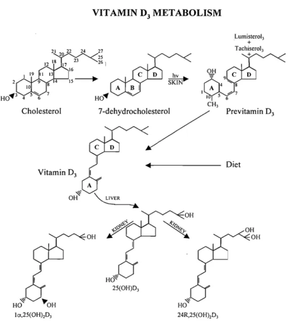
1.1.4 CYTOCHROME P450s IN VITAMIN D3 METABOLlSM
Vitamin D3 (D3) is a natural secosteroid that has, in its native form, virtually no biological activity. Once in circulation, D3 is taken up by the liver and hydroxylated at C-25 by a P450-dependent mixed function oxidase systems (Figure 1.3). At physiological concentrations, 25-hydroxyvitamin D3 (25(OH)D3) has no biological activity and must be hydroxylated at position C-l ex (principally in the kidney) into 1 ex,25 dihydroxyvitamin D3
(calcitriol, 1,25(OH)2D3) in order for the vitamin to achieve hormonal status and thus its full biological potentialThere are four P450s required for the activation and inactivation of D3 (cholecalciferol). 7 -dehydrocholesterol, a late intermediate in the cholesterol biosynthetic pathway is photolyzed by UV light producing D3' The vitamin is first hydroxylated at position C-25 by a mitochondrial and/or microsomal enzyme. The mitochondrial D3 25-hydroxylase, CYP27 A, has been identified, whereas the status and role of the D3 microsomal entity remains questionable. The product of this reaction, 25(OH)D3 is then converted to lex,25(OHhD3 by a mitochondrial P450, the vitamin D3 lex-hydroxylase, CYP27Bl. This enzyme has been known to exist in kidney mitochondria for decades but because of its very low level of expression and presumed instability, it had been virtually impossible to purify. Only within the past four years has it been c10ned and found to be a member of the CYP27 A family (17-20). The C-l ex hydroxylation generates the active and hormonal form of D3, lex,25(OH)2D3. A third mitochondrial P450, vitamin D3-24 hydroxylase (CYP24), inactivates calcitriol by hydroxylation on the side chain at position C-24 which greatly
2 Ho"J 4 Cholesterol Vitam in D3 VITAMIN
D3
METABOLISM 24 27 25 26 i HO" OH 7 -dehydrocholesterol HO~ 25(OH)D3 OH Lumisterol3 + Tachiserol3 + Previtamin DJ Diet 9Figure 1.3: Key vitamin D sterol structures. Top row: vitamin D3 (D3) is structurally
related to cholesterol (the numbering of the carbon atoms is identical). D3 is produced (in the skin) from 7-dehydrocholeserol by a UV-mediated photochemical reaction. The immediate photo che mi cal product, preD3 then thermally equilibrates (over time) into D3. Middle row: conformational representation of D3. Structure presented is an extended version derived from X-ray crystallographic analysis; Bottom row: structures of the three principal metabolites ofD3.
10
1.2 VITAMIN D METABOLISM
1.2.1 VITAMIN D3 25-HYDROXYLASES
1.2.1.1. Evidencefor microsomal and mitochondrial D3 25-hydroxylase activity
As previously mentioned 25-hydroxylation of D3 is catalyzed by P450 dependent enzyme systems in both mitochondrial (21 ;22) and microsomal fractions (23 ;24). The relative importance of the two 25-hydroxylase systems in vivo is not known, but it is generally believed that the microsomal 25-hydroxylation of D3 is more important than the mitochondrial 25-hydroxylation in the bioactivation process under normal conditions (25;26). In humans, the mitochondrial D3 25-hydroxylase was believed to be the predominant and only form expressed (27), although a microsomal form is also active (28). There is abundant evidence that the 25-hydroxyvitamin D3 1cx-hydroxylase, as weIl as the degradation system 25-hydroxyvitamin D3 24-hydroxylase, are highly regulated (29-31). However the question of whether the 25-hydroxylase is regulated remains undetermined. Interestingly, the D3 25-hydroxylase that has been described to date, in terms of gene regulation, is the enzyme CYP27 A of mitochondrial origin. The porcine microsomal D3 25-hydroxylase has been cloned (32), although its presence remains to be characterized in other species (33). This microsomal25-hydroxylase identified can catalyse 25-hydroxylation of both D3 and D2(34). A cDNA encoding the pig liver microsomal D3 25-hydroxylase, as deduced by both DNA sequence analysis, showed 70-80% identity with members of the CYP2D subfamily and has been assigned the name CYP2D25 (35). However, the only CYP2D enzyme known to be expressed in man is CYP2D6. This enzyme is polymorphically expressed and is lacking in 5-10% of the caucasian population. It is not known whether D3 25-hydroxylation in man could be catalysed by CYP2D6.
1.2.1.2 Biochemical and Molecular aspects ofmitochondrial and microsomal D metabolism
The microsomal 25-hydroxylase fraction of rat liver and was influenced by the D status (36-38) and required the presence of a cytosolic factor for optimum activity.
25-Il hydroxylation of the synthetic compound dihydrotachysterol (DHT3)was not regulated compared to that of the natural substrate D3(38). The presence of the enzyme was confrrmed to be a rat liver microsomal P-450 (24;39), requiring a soluble cytosolic factor (for full reconstitution of enzyme activity) (39). In 1983, a rat microsomal cytochrome P450 active in C-25 hydroxylation ofbile acid intermediates 5 [3-3ex,7ex,12ex-triol, cholestane-3ex,7ex-diol as weIl as D3 and lex (OH)D3 but not D2 (40). A purified and partially sequenced
P450 active in 25-hydrxylation ofD3 with an inhibitor removed from the microsomes during the purification steps (41), and did not require the presence of a cytoplasmic factor.
In 1980, Bjorkhem and Holmberg studied the activities of a P450 preparation solubilized from rat liver mitochondria obtained from rats which had been treated with phenobarbital or subjected to a rachitogenic diet (22). They observed that both phenobarbital and the rachitogenic diet increased C-25hydroxylation ofD3 but had little effect on the C-27 hydroxylation of the C-triol. These observations led the investigators to conclude that 2 distinct P450 species existed, each responsible for the hydroxylation of cholestanetriol and D3. At the same time, Masumoto et al purified to homogeneity the D3 25-hydroxylase from rat liver mitochondria and characterised it as a P450 catalysing the C-25 hydroxylation of both D3 and lex (OH)D3 (42). These researchers proposed that a single enzyme was involved in the C-27 hydroxylation of 5 [3-cholestane-3ex,7ex,l2ex-triol and in the C-25 hydroxylation of D3, although the preparation exhibited much lower activity toward D3 compounds than toward the C-27 hydroxylation ofbile acid intermediates. D3 competitively inhibited the 27-hydroxylation ofC-triol, whereas C-triol inhibited D3 25..;27-hydroxylation (42), concluding that both substrates were catalysed at a common active site on a single protein. D3 25-hydroxylation and C27 sterol 25-hydroxylation activities were located , probably exclusively, in the inner mitochondrial membrane matrix (27;43).
Heterologous cell systems confirmed the identity of sterol 27-hydroxylase and D3 25-hydroxylase as weIl as the requirement for adrenodoxin and adrenodoxin reductase but not NADPH P450 reductase (44).
J.2.1.3. CYP27A (MITOCHONDRIAL VITAMIN D3 25-HYDROYLASE)
CYP27A, the mitochondrial D3 25-hydroxylase, has been cloned in several species including the rabbit (44), rat (45;46), and human (47;48). The gene has been shown to be located on chromosome 2q33-qter (49). Northern analyses demonstrate two mRNA sizes of 1.9 and 2.3kb for CYP27A in liver and fibroblasts (46;48). Characterization of the 2.3kb mRNA indicated a sequence identical to the 1.9kb mRNA in its protein-coding region, except for ~ 400-nucleotides in an extended sequence at its S'end (50). The rat CYP27A gene contains Il exons of 80-415 nucleotides that are separated by 10 introns of 83 bases to ~ IOkb (51). The protein sequence of the human enzyme has been reported to be 72% identical to the rat and 81 % identical to the rabbit CYP27A (45;48). The mature protein contains 444 to 501 amino acids depending on the species involved and the rat enzyme molecular weight has been estimated to be 51 kDa (45).
1.2.1.3.2 CYP27Apromoter and regulation studies
Functional assays showed that the -217/-10 nucleotide region for the translation start site (minimal promoter), devoid of TATA and CAAT boxes, contains aU the elements for basal transcription (52). Possible positive transcription regulation sites are located at position -187 to -320 and -857 to 1087bp (53). A negative transcription regulator site is located in position -320 to -413bp. An enhancer sequence is located upstream to position -1087 (53). A putative bile acid response element (BARE) is also located between -110 and -86bp ofthe CYP27 promo ter ,on which HNFIcx and CIEBP bound (54) .
. Footprinting analysis of the minimal promoter showed four protected region. Three of the regions contained and SpI binding site, and one an HNF4 site. Electrophoretic mobility shi ft assays demonstrated that SPI, Sp3, and HNF4 transcription factors bind these sites (52). Mutagenesis ofany ofthese sites resulted in the loss ofpromoter activity. SpI, Sp3 and HNF4 were found to cooperate in the expression of the human CYP27 A gene in HepG2 ceUs (52). A dexamethasone responsive element was found to be located between 1087 and 678bp upstream to the putative ATG. The cyclosporin A-responsive element is mapped to between 1087 and 4000 bp upstream of the ATG (53). Cholic acid represses sterol 27-hydroxylase mRNA level by affecting the stability of its mRNA (53).
13
1.2.1.3.3. CYP27A regulation not related to vitamin D metabolism
CYP27A is a high-capacity enzyme involved primarily in the hydroxylation ofbile acid intermediates, but its substrate specificity is broad and exceeds the field of bile acid biosynthesis. The purified enzyme has been shawn ta be active on both the C-25 and C-27 hydroxylation of cholesterol as well as on other bile acid intermediates (55;56). Rat hepatic CYP27 A is highly sensitive to the prevailing concentrations of bile acids with increases in enzyme activity, steady state mRNA level and the rate of gene transcription being observed after interruption of the enterohepatic circulation (57;58). Physiological concentrations of insulin also down-regulate CYP27A gene transcription through a direct effect of the hormone
on the transcription rate in hepatocytes (59).
CYP27A is also sensitive to pituitary-regulated steroids, growth hormone and the diurnal
rhythm, with a two fold increase in enzyme activity observed in the mid-dark compared to the mid-light period (57). Both the 1.9 and 2.3kb mRNA species appear to be modulated by the physiological state of the animal and regulated by growth hormone in paraUel to the serine protease inhibitor mRNAs (50).
1.2.1.3.4. CYP27A regulation related to vitamin D metabolism
Most regulation studies on CYP27A have been linked to the metabolism of cholesterol
and bile acids. Few studies, until recently, have addressed the effect ofthese compounds on the handling ofD3.CYP27A also catalyzes the C-24, 25 and 27 hydroxylation ofD3, D2 and related compounds (47). While D3 substrates were found to be preferably hydroxylated at C-25, Guo et al., (47) observed that when the substrates exhibited the ergocalciferol side
chain, such as that found in D2 or 1 cx(OH)D2, a predominance of 24-hydroxy metabolites occurred (metabolites which have also been found in vivo in rat, cow and chicken) (60-62).
In addition, sorne production ofC-27 hydroxylated products also occured (63). Surprisingly, D2 was not, while lcx(OH)D2 was only poorly hydroxylated at position C-25 (47).
In addition, several laboratories have now shown that the enzyme prefers 1 cx-hydroxylated analogs of D3 or D2 over their non-cx-hydroxylated counterparts. This includes
the natural substrate D3, which is hydroxylated at C-25 several times less efficiently than lcx(OH)D3 (22;35;47).
Of the studies examining regulation of CYP27 A by the D3 status, Axén et ai. (64) reported that renal and hepatic CYP27A was affected by 1,25(OH)2 D3 administration but that kidney CYP27A rnRNA was decreased to a larger extent than that of the liver. Furthermore, CYP27A mRNA has been shown to be inducible by D3 in keratinocytes (65).
1.2.1.3.5 Vitamin D and minerai metabolism in CTX humans and in the CYP27-/- mouse
Mutations affecting the expression of CYP27A or its primary sequence leads to cerebrotendinous xanthomatosis (CTX) (66;67), which is an inherited disorder of sterol metabolism and storage characterized by atherosc1erosis and progressive neurological dysfunction. CTX is a rare autosomal recessive neurometabolic disease involving lipid metabolism. The c1assical phenotype, due to mutations in the CYP27 gene, is characterized by neurologie dysfunction, tendon xanthomas and juvenile cataracts.
Bone fractures occurred frequently in patients with CTX (68). Serum 25(OH)D levels were in the low to normal range whereas 24,25(OHhD levels were markedly decreased. Serum concentration of 1,25(OH)2D, Ca, inorganic phosphorus, alkaline phosphatase, parathyroid hormone and calcitonin were normal (69).Interestingly, both normal (70) and abnormal (67;69;71) D3 and/or calcium metabolism have been reported which suggests the presence of more than one enzyme active in the 25-hydroxylation of D3. However, sorne CTX patients have been shown to retain residual C-27 hydroxylase activity (72). The creation ofa CYP27A-/-mouse has not provided any further insight into the role ofCYP27A in relation to D3 homeostasis. Indeed, the CYP27A-/-mouse surprinsingly displays elevated levels of 25(OH)D, and no CTX abnormalities (73). The latter observations suggest alternative C-25 hydroxylation pathway(s) in the CYP27A ablated mouse possibly through the microsomal D3 25-hydroxylase.
The circulating concentrations of 25(OH)D was however somewhat higher in the CYP27-/- than in the CYP27 +/+ mice whereas the corresponding concentrations of
1,25(OHhD were similar in the two groups (73). Thus it seems less likely that sterol 27-hydroxylase is of importance in formation of 25(OH)D and 1,25(OH)2D in this species.
15 1.2.2 VITAMIN D3 24-HYDROXYLASE (CYP24)
1.2.2.1 The vitarnin D3 24-hydroxylase gene
The purification of the rat kidney mitochondrial D3 24-hydroxylase enzyme and the raising of a rabbit polyclonal antibody (74;75) were milestone achievements that led to the isolation of a cDNA clone for the rat 24-hydroxylase by immunological screening (76). The mRNA contains an open reading frame of 514 amino acid residues that includes the mitochondrial signal sequence. Full-Iength cDNA clones were subsequentIy isolated for the hum an (77) and mouse (78) D3 24-hydroxylase proteins. The human D3 24-hydroxylase showed about 80% amino acid sequence identity with both the rat and mouse homologs, whereas the latter two where 95% identical. DJ 24-hydroxylase was not more than 30% identical with any other P450s sequence reported to date. The highest homology (30% identity) was found to be with CYP27 A (45).
Genomic clones for rat (79) and human (77) D3 24-hydroxylase have been isolated. The rat gene is a single copy gene that spans about 15kb and contains 12 exons (79). The intron-ex on arrangement of the gene most closely resembles that of the CYP22 family (79). The transcription start site for the rat CYP24 and a likely TATA box have been located and several possible control elements have been identified in the promoter including vitamin D response elements (VDREs), CCAAT, GC, and TATA binding sites (79;80). Similar binding sites for the transcription factors are aiso present in the promoter for human CYP 24 (81).
Aithough three VDREs have been identified in the rat CYP24 promoter (82), only two functionai elements have been identified on the antisense strand (30;80;83). Initial studies established the binding ofVDR as an retinoid-X-receptor (RXR) complex at VDRE-l, which was verified and extended to include VDRE-2 through supershift analysis with a specifie monoclonal RXR antibody (82). When analyzed separately, VDRE-2 showed about 4- to 5- fold higher affinity for the VDR-RXR complex than VDRE-l. There is transcriptional synergism between the VDREs, as the wild type induction (18 fold) is greater than the sum of the individual contributions ofVDRE-1 (6 fold) and VDRE-2 (3 fold). Two VDREs for
16 the human promoter have been identified by Chen and DeLuca (81).
1.2.2.2 Catabolism of 1,25 dihydroxyvitaminDj
CYP24 directs the synthesis of 24,25 (OH)2D3 and l,24,25(OH)3D3 (84). The two D3 metabolites express less biologie al activity than 1,25(OH)2D3, although 24,25(OH)2D3 is suggested to have foeused actions in bone and cartilage. Both metabolites also represent initial reactants in the C-24 oxidation pathway that leads to metabolite inactivation via generation of23- or 24-COOH end products (85;86). Acquisition of a C-24 hydroxyl group is required for emry into the oxidation pathway, which is followed by oxidation of the 24-hydroxyl group to a keto (oxo) function. The 23- and 24-24-hydroxylase activities are usually eoexpressed in the same target cells, with the 24-hydroxylation constituting the major activity (87).
1.2.2.3 Cellular expression
The D3 24-hydroxylase enzyme displays a broad tissue distribution. A major route of enzyme induction involves the combined action of 1,25(OH)2D3 and of VDR to increase transcription of the CYP24 gene. Consequently, cellular expression of CYP24 is linked
tightly to the coexpression ofVDR. Most cells that contain VDR, therefore, express a basal level ofCYP24 or respond to increased 1,25(OH)2D3Ievels by inducing the biosynthesis of the enzyme, particularly in kidney and small intestine (88;89). Basal expression under normal calcium-homeostatic conditions has been documented for kidney, intestine, bone, placenta, skin (keratinocytes), and macrophages (88-90).
Particular attention has been given to expression of the enzyme in the kidney, which is one of the major sites of CYP24 activity. As this tissue is the major site of 1,25(OH)2D3 synthesis, it seems likely that the high basal CYP24 expression in kidney represents induction
by endogenous 1,25(OH)2D3. In the kidney, enzyme expression has been localized to the proximal tubule and does not display the more general distribution as noted for VDR (91). In parallel, CYP24 expression in bone occurs in osteoblasts and may play an important role
17 developmental expression of CYP24 is controlled by 1 ,25(OH)2D3-dependent transcriptional
induction (92). In addition, investigations with cultured cells have revealed induction ofthe
CYP24 gene in many different cell types (93-95).
This widespread distribution of the D3 24-hydroxylase supports a major role for the enzyme in regulating the local concentration and action of the hormone 1,25(OH)2D3.
1.2.2.4 Regulation of expression
Consistent with the general tissue distribution of CYP24, a broad spectrum of regulatory agents act to control cellular expression of the enzyme. Steroid and peptide hormones function through the signal transduction and transcription pathways to alter CYP24 activity. The enzyme level is also changed in response to age and several genetic mineraI metabolism disorders.
The major regulators of the D3 24-hydroxylase are PTH and 1,25(OH)2D3. In vivo
administration of PTH to thyroparathyroidectomized rats decreases renal production of 24,25(OH)2D3, whereas administration of 1,25(OH)2D3 increases 24,25(OH)2D3 production. The mechanisms of action of PTH and 1,25(OH)zD3 on D3 24-hydroxylase appear to be similar to the actions of the respective hormones on 1 cx-hydroxylase in that PTH acts by a mechanism involving cAMP and the 1,25(OH)2D3 action requires new protein synthesis.
Administration of a single dose of 1,25(OH)2D3 to rats markedly increases CYP24
mRNA level in the kidney and intestine (96;97). In contrast, CYP24 mRNA is not induced
by 1,25(OH)2D3 in hepatocytes, which do not express any 24-hydroxylase activity. Induction of CYP24 mRNA by 1,25(OH)2D3 has been further studied in primary cultures of rat tubular
cells and human colon cancer cells (98;99). This induction is inhibited by the addition of actinomycin D, 5,6-dichloro-l-cx-D-ribofuranosyl benzimidazole, and cyc1oheximide, indicating that the de novo syntheses of CYP24 mRNA and protein are required (98). It has also been reported that phorbol 12-myristate 13-acetate (TP A) alone has no effect, but TP A produces a marked increase in the expression of CYP24 mRNA in the presence of
1,25(OH)2D3 (98). TPA also shifted the stimulatory dose-response curve of 1,25(OH)2D3 to the left. It is, therefore, hypothesized that the 1,25(OH)2D3 effect leading to CYP24
18 - involving prote in kinase C (PKC).
1.2.3 VITAMIN D3lcx-HYDROXYLASE (CYP27Bl)
1.2.3.1 THE VITAMIN D3lcx -HYDROXYLASEGENE
Until recently, analysis of the expression and function of the D3 lcx-hydroxylase in the kidney has been dependent on relatively insensitive enzyme assays using tissue homogenates, mitochondrial enriched fractions, or single nephron sections obtained from D-deficient animaIs (100;101). Further studies also identified the D3 lcx-hydroxylase in the distal tubule and collecting ducts.
In 1997, Takeyama et al. (1 7), using a VDR knockout model, isolated a candidate
lcx-hydroxylase 2.5kb cDNA corresponding to a 507 amino acid P450-like protein, with a predicted size of 55kDa. The overall sequence identity of this CYP27B 1 to related mitochondriai cytochrome P450 pro teins is limited, ranging from 39% (CYP27 A) to 33% (lli)-hydroxylase) (20). CYP27Bl gene spans approximately 6kb, consists of9 exons and
has approximately 500bp of 5' untranslated mRNA.
In paraUel with the original cloning of the human gene (20), St-Arnaud et al., (102)
isolated the cDNA for the rat CYP27Bl, which was found to have 82.5% identity to human
cDNA. The latter report also confirmed the location of the human CYP27Bl gene on
chromosome 12q13-1-qI3-3. This provides further evidence that abnormal CYP27Bl gene
expression is the cause ofhereditary pseudovitamin D-defieieney rickets, a disease reported to reside on the above mentioned chromosome.
1.2.3.2 Renal distribution
Data using normal human kidneys eonfirmed the expression of mRNA and protein for the D3 lcx-hydroxylase in proximal tubules (103). However prote in and mRNA were also expressed in distal tubules and in eolleeting duets (103). The specificity of the D3 lcx-hydroxylase expression in the kidney was emphasized by stringent controls for both in situ
19 hybridization and immunohistochemistry analyses. The other key sites ofD3 lcx-hydroxylase expression along the nephron were the medullary collecting ducts and the papillary epithelium (104).
Although previous studies of the renal function of D3 have focussed primarily on the production and function of 1,25(OH)2D3 in proximal tubules, there is increasing evidence of a role for the hormone in more distal parts of the nephron. In view of studies with D3 deficient animaIs, it is speculated that production of 1,25(OH)2D3 in the proximal tubules acts in an endocrine fashion to support circulating levels of 1 ,25(OH)2D3, whereas in more distal areas of the nephron it may fulfill an autocrine or paracrine function. Indeed, previous studies have shown that 1,25(OH)2D3, as weIl as calcitonin and PTH, stimulate calcium reabsorption in the distal nephron (105-108). It is also important to recognize that in sorne cases, the impact of 1,25(OH)zD3 on renal function may occur through indirect mechanisms.
In particular, the observation that the calcium-sensing receptor is primarily regulated by 1 ,25(OH)2D3, and not by PTH or calcium suggests that this may be the key target for local production and action of 1,25(OH)2D3 in the distal nephron (109).
1.2.3.3 Extra-renal expression
The original description of extra-renal D3 1 cx-hydroxylase expression was based on studies of the granulomatous disease sarcoidosis, which is frequently associated with hypercalcaemia (110;111). Enzyme activity analyses using lymph no de homogenates and pulmonary alveolar macrophages from patients with sarcoidosis showed high levels of D3 lcx-hydroxylase activity (112-114). Furthermore, addition of exogenous 1,25(OH)zD3 did
not appear to inhibit macrophage D3 1 cx-hydroxylase as is c1assically observed with its renal counterpart. This would explain the apparently unregulated synthesis of 1,25(OH)2D3 which is characteristic ofthe more severe forms ofthis disease. However, these data also suggested that the expression and regulation of D3 1 cx-hydroxylase in extra-renal tissues was different from that observed with the kidney enzyme. It now appears that renal and extrarenal D31 cx-hydroxylase activity is due to a single gene product. Therefore, the most likely explanation is that induction of extra-renal DJ lcx-hydroxylase involves regulatory pathways that differ
20 from renal, cAMP-mediated mechanisms, less sensitive to autoregulation by 1,25(OH)2D3. Induction of extra-renal D3 1 ex-hydroxylase frequently involves antigenic mediated activators such as lipopolysaccharide or inflammatory mediators such as interferon-y. Since these agents use nuclear factor NFKB as signaling mechanism, it can be postulated that this pathway activates D3 1 ex-hydroxylase in a manner unlike that of ca1ciotrophic factors and, as a consequence show a differential sensitivity to feedback control by 1,25(OHhD3. Further analysis of signal-transduction pathways involved in regulating D3 1 ex-hydroxylase will be crucial to the understanding of the way in which 1,25(OH)2D3 functions in extra-renal tissues. Using immunohistochemistry and Western analyses with renal D3 lex-hydroxylase antisera, the enzyme was detectable in tissues such as normal skin (stratum basalis) and sarcoid lymph nodes (115). The D31ex-hydroxylase was also highly expressed in skin from sarcoid patients. Immunohistochemistry also confirmed previous enzyme activity studies which indicated that D3 1 ex-hydroxylase was expressed in decidual cells (116; 117). However, the enzyme was also detectable in trophoblasts and syncytiotrophoblasts, suggesting potentially diverse functions for the hormone in placenta and feto-placental physiology (118). Novel sites for D3 lex-hydroxylase expression include the parathyroids, pancreas, adrenal medulla, colon and cerebellum, while negative tissues include the heart, liver (hepatocytes) and adrenal cortex.
1.2.3.4 Regulation ofvitamin D3 metabolism
The apparent widespread distribution of protein and mRNA for the D3 1 ex-hydroxylase in both renal and extra-renal tissues has raised important questions concerning the local enzyme activity at these sites. The relationship between expression of the CYP27B 1 and actual synthesis of 1,25(OH)2D3 in a particular tissue probably involves two specifie mechanisms, the first of these being substrate access, and the second being auto regulation ofD3 lex-hydroxylase activity by 1,25(OH)2D3 itself. The former questions the assumption that, in common with other steroid hormones, 1,25(OH)2D3 enters cells by a passive mechanism by virtue of its lipophilic nature. Circulating vitamin D (D) metabolites can bind to a variety of serum proteins, but by far the most important of these is the vitamin D binding
21 prote in (DBP), which is synthesized in the liver. DBP may play an active role in directing D responses (119) because of its relatively high capacity for binding 25(OH)D3, making it a likely key determinant of the availability of the substrate to CYP27B 1. Recent studies have shown that DBP and DBP-bound D metabolites are filteredthrough the glomerulus and reabsorbed by the luminal endocytic receptor megalin (gp330) in the proximal tubules (120). Megalin is also expressed in a variety of tissues (121). Thus megalin-mediated endocytosis of DBP-bound 25(OH)D3 may act as an additional mechanism controlling tissue-specific synthesis of 1,25(OH)2D3 by modulating the availability of substrate to the CYP27B 1 protein. Recently, cubulin, a membrane-associated protein colocalizing with megalin, has been identified. It facilitates the endocytic process by sequestering steroid-carrier complexes on the cellular surface before megalin-mediated intemalization of the cubulin-bound ligand (122). Interestingly, dogs with an inherited disorder affecting cubulin biosynthesis exhibit abnormal D metabolism (122).
During D sufficiency, 1,25(OHhD3 production by the kidney is very tightly regulated. On the other hand, there is a striking up-regulation of the D3 lcx-hydroxylase activity in proximal tubular cells in D-deficient states (123;124). This response appears to be a function of several direct and indirect mechanisms, including changes in accessory proteins such as ferrodoxin, or alterations in VDR or 24-hydroxylase expression. Studies in vivo suggest that the key activator of CYP27B 1 is PTH and that this effect is mediated, at least in part, by target-ceIl induction of cyclic adenosine-3', 5'-monophosphate (cAMP) production(125). This then acts through potential cAMP response elements in downstream areas (-1 Akb),
which have been found to be PTH responsive in promoter-reporter assays (29;126). In both of these studies, the authors were unable to show any self-regulation of basal CYP27Bl
promoter activity and no VDREs were identified in the 1Akb fragment. However, in each case 1,25(OH)2D3 was unable to suppress PTH-induced transactivation. This suggests either that the CYP27Bl gene promoter has an atypical VDRE, or that 1,25(OH)2D3 achieves its effects by an indirect mechanism. These reports contrast with analysis of the murine promoter, which demonstrated both positive (PTH) and negative (l,25(OH)2D3) responsiveness in a region downstream of -0,9kb (127). In this study, calcitonin was shown to be a potent stimulator of CYP27Bl expression, supporting previous reports in which ca1citonin was shown to stimulate CYP27Bl mRNA and enzyme activity under
22 normocalcemic conditions (128). This suggest that calcitonin (a noncoUagenous prote in secreted by osteoblasts), acting via distal areas of the nephron, may play an important role in the 'fine-tuning' of serum 1,25(OH)2D3 levels during D3 sufficiency.
Amongst the most prominent inhibitorsof D3 1 ex-hydroxylase are calcium, phosphate, and 1,25(OH)2D3 itself. It seems likely that many ofthese effects are mediated indirectly through modulation of PTH production and secretion. However, as a consequence of the tight regulation of 1,25(OH)2D3 production, analysis of the precise mechanisms involved in controlling the D3 1 ex-hydroxylase has proved difficult, due to the very low enzyme levels.
1.2.4 VITAMIN D METABOLISM IN PERINATAL DEVELOPMENT
AU three D3 metabolites 25(OH)D3, 24,25(OHhD3 and 1,25(OHhD3 circulate in mammalian fetuses. The cord concentrations of all three metabolites are consistently lower than those measured in the mother's serum. However, 24,25(OH)2D3 is the dominant metabolite formed in fetopiacentai tissues (129). Moreover, fetomaternal relationships of 1,25(OH)2D3 concentrations are quite compIex. In most studies, no correlation between fetai and maternaI concentrations has been observed (130; 131), whereas in the study by Ross et
al., (132), a highly significant correlation in both full term and preterm sheep was described.
Studies show that most of the 1,25(OH)2D3 in fetal plasma is due to the fetai kidney production of the hormone (133;134), suggesting fetai control and autonomy in hormone regulation. Indeed, cord blood concentrations of 1,25(OH)2D3 in fetal plasma from infants with Potter syndrome (renai agenesis) are one third those observed in healthy newborns. In addition, hepatic CYP27A mRNA levels have been detected in 17 to 19 week old human
fetuses (135), further implying the possibility of fetal autonomy in D metabolism. However, a maternaI contribution to fetai 1,25(OH)2D3 cannot be eliminated, since radioactive 1,25(OH)2D3 administration to pregnant monkeys (136), sheep (137) and rats (138) can be detected in fetal tissues. Parathyroid related peptide (PTHrP) is an important autocrine and
lor paracrine growth factor regulator in fetal development. Its presence has been demonstrated in the placenta and fetal tissues in various species (139;140). In addition, it is implicated in the transfer of calcium across the placenta (139).
23 With respect to calcium homeostasis, the fetal-placental unit has a remarkable ability to meet its needs irrespective of maternaI calcium or calciotropic hormones (141;142). Specifically, the fetal-placental unit has adapted to rapidly extract calcium from the maternaI blood stream in sufficient amounts to mineralise the fetaI skeleton in late gestation (143;144). Indeed, the fetus has a higher blood calcium than the ambient maternaI calcium level (130;145). Furthermore, the newboms of hypocalcemic D-depleted female rats are normocalcemic (unpublished results from our laboratory), and newbom VDR-/- mice are normocalcemic until the time ofweaning (146).
1.2.5 GENDER DIFFERENCES IN VITAMIN D METABOLISM
A microsomal D 25-hydroxylase, CYP2Cll, is known to exist in male rat liver microsomes but not in those of females (147). This observation may well explain why microsomes (or P450 purifies from the microsomal fraction) obtained from female livers have consistently been shown to exhibit significantly far less activity in C-25 hydroxylation of D3 than preparations obtained from male counterparts (148-150).
Furthermore, estrogen deficiency leads to a decrease in serum 1,25(OH)2D regardless of age (151; 152). Estrogen replacement in postmenopausal women can increase both total and free serum 1,25(OH)2D (152;153), suggesting that menopause and the accompanying estrogen deficiency may remove an important trophic factor for the maintenance of serum
1,25(OH)2D in aging women.
In parallel, serum total and free testosterone levels decrease with advancing age (154;155) and testosterone treatment has been shown to increase modestly both serum total and free 1 ,25(OH)2D in hypogonadal men (156).
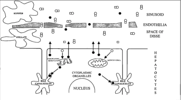
1.3.1 HEPATfC STRUCTURE AND FUNCTfON
The liver consists mainly of hepatocytes, the main parenchymal cell population. The non-parenchymal cells include sinusoidal, endothelial, Kupffer and stellate (Ito) cells. The hepatocytes represent 60% of the cell population. They are, however, more voluminous than other cells and consequently make up 80% of the parenchymal volume (157). The hepatocytes are organised in three-dimensional structures called acini (158). The hepatic acinus represents a structural and functional unit of the hepatic parenchyme whose main function consists of regulating the metabolism of the various substances exported to the systemic circulation (159).
The liver's microcirculation is composed of sinusoids, which are specialized capillaries who se discontinuous basal membrane is bordered by the sinusoidal cell population mentioned above. The sinusoidal endothelial cells form a selective semi-permeable barrier between the blood and the hepatic parenchyme, as described by De Zanger and Wisse (160). The Kupffer cells' main role is to endocytose many substances originating from the systemic circulation such as endotoxins (161). There also exists an intercellular space between the hepatocyte's plasma membrane and the endothelial cells called the space of Disse. This space encompasses the perisinusoidal stellate ceIls, which metabolize and store vitamin A (162), and synthesize the hepatocyte growth factor (HGF) (163). Stellate cells are also responsible for the synthesis of collagen, thus preventing cirrhosis of the liver.
The cells in zone 1 (or periportal area) are situated close to the supplying vessels and are bathed by blood of a composition similar to that in the afferent vessels. The cells in zone 3 (or perivenous area) are situated at the microcirculatory periphery of the acinar unit and receive blood that has already exchanged gases and metabolites with cells in zones 1 and 2 (midzonal).
1.3.2 VITAMIN D3 UPTAKE: REGIONALISATION ALONG THE HEPATIC ACINUS
There exist two distinct me chanis ms that can provide D3 to the body. First, synthesis from UV light-dependent biosynthetic sites in the skin and secondly, through intestinal absorption from dietary sources of the vitamin.