HAL Id: hal-02318063
https://hal.archives-ouvertes.fr/hal-02318063
Submitted on 25 Oct 2019HAL is a multi-disciplinary open access archive for the deposit and dissemination of sci-entific research documents, whether they are pub-lished or not. The documents may come from teaching and research institutions in France or abroad, or from public or private research centers.
L’archive ouverte pluridisciplinaire HAL, est destinée au dépôt et à la diffusion de documents scientifiques de niveau recherche, publiés ou non, émanant des établissements d’enseignement et de recherche français ou étrangers, des laboratoires publics ou privés.
Monitoring the crystal structure and the electrochemical
properties of Na3(VO)2(PO4)2F through Fe3+
substitution
Long H. B. Nguyen, Jacob Olchowka, Stéphanie Belin, Paula Sanz Camacho,
Mathieu Duttine, Antonella Iadecola, François Fauth, Dany Carlier, Christian
Masquelier, Laurence Croguennec
To cite this version:
Long H. B. Nguyen, Jacob Olchowka, Stéphanie Belin, Paula Sanz Camacho, Mathieu Duttine, et al.. Monitoring the crystal structure and the electrochemical properties of Na3(VO)2(PO4)2F through Fe3+ substitution. ACS Applied Materials & Interfaces, Washington, D.C. : American Chemical Society, 2019, 11 (42), pp.38808-38818. �10.1021/acsami.9b14249�. �hal-02318063�
1
Monitoring the Crystal Structure and the
Electrochemical Properties of Na
3(VO)
2(PO
4)
2F
through Fe
3+-Substitution
Long H.B. Nguyen
a,b,e, Jacob Olchowka
a,e,f, Stéphanie Belin
c,
Paula Sanz Camacho
a, Mathieu Duttine
a, Antonella Iadecola
e, François Fauth
d, Dany Carlier
a,e,f, Christian Masquelier
b,e,f, Laurence Croguennec
a,e,f *a CNRS, Univ. Bordeaux, Bordeaux INP, ICMCB UMR 5026, F-33600, Pessac, France.
b Laboratoire de Réactivité et de Chimie des Solides, CNRS-UMR #7314, Université de Picardie Jules Verne,
F-80039 Amiens Cedex 1, France.
c SOLEIL Synchrotron, F-91192 Gif-sur-Yvette, France.
d CELLS-ALBA synchrotron, E-08290 Cerdanyola del Vallès, Barcelona, Spain.
e RS2E, Réseau Français sur le Stockage Electrochimique de l’Energie, FR CNRS 3459, F-80039 Amiens Cedex
1, France.
f ALISTORE-ERI European Research Institute, FR CNRS 3104, Amiens, F-80039 Cedex 1, France.
Key words: Na3V2(PO4)2F3, Na3(VO)2(PO4)2F, Fe3+-substitution, sodium-ion batteries, sol-gel synthesis,
synchrotron X-ray diffraction, solid-state NMR, Na+ diffusivity.
2
Abstract
We here present the synthesis of a new material, Na3(VO)Fe(PO4)2F2, by sol-gel method. Its
atomic and electronic structural description are determined by a combination of several
diffraction and spectroscopy techniques, such as: synchrotron X-ray powder diffraction,
synchrotron X-ray absorption spectroscopy at V and Fe K-edges, 57Fe Mössbauer and 31P
solid-state nuclear magnetic resonance spectroscopy. The crystal structure of this newly obtained
phase is similar to that of Na3(VO)2(PO4)2F, with a random distribution of Fe3+ ions over
vanadium sites. Even though Fe3+ and V4+ ions situate on the same crystallographic position,
their local environment can be studied separately by the use of 57Fe Mössbauer and X-ray
absorption spectroscopy at Fe and V K-edges, respectively. The Fe3+ ion resides in a symmetric
octahedral environment while the octahedral site of V4+ is greatly distorted due to the presence
of the vanadyl bond. No electrochemical activity of the Fe4+/Fe3+ redox couple is detected, at
least up to 5 V, whereas the reduction of Fe3+ into Fe2+ has been observed at ~ 1.5 V vs. Na+/Na
through the insertion of 0.5 Na+ into Na3(VO)Fe(PO4)2F2. Comparing to Na3(VO)2(PO4)2F, the
electrochemical profile of Na3(VO)Fe(PO4)2F2 in the same cycling condition shows a smaller
polarization which could be due to a slight improvement in Na+ diffusion process thanks to the
presence of Fe3+ in the framework. Furthermore, the de-sodiation mechanism occurring upon
charging is investigated by operando synchrotron X-ray diffraction and operando synchrotron
3
1. Introduction
Lithium-ion batteries (LIBs) have long been the dominant means of energy storage used
for mobile devices thanks to their high operating voltage and high energy density. Nevertheless,
the unequal presence of Li resources in the earth’s crust and the increasing price of lithium
precursors have motivated the development of alternative technologies in order to diversify the
choice of energy storage systems. The revival of Sodium-ion batteries (SIBs) in the last decade
has attracted great attention from the scientific community as the ubiquity of sodium resources
widespread over the world would reduce the fabrication expense in case of large-scale
production.1–4 Recently, great progress has been made in the field of SIBs thanks to the
similarity to their LIBs counterpart. Several layered and polyanionic compounds have already
been extensively studied as positive electrode materials for SIBs: layered oxides with the
general formula NaxMO2 (M = Transition metal), whose structure depends on the Na-content
and the nature of the transition metal M, the fluorinated oxyphosphate Na3V2(PO4)2F3 and
Na3(VO)2(PO4)2F, or the NASICON-type compositions Na3V3+2(PO4)3 and
Na4V3+Mn2+(PO4)3.5,6,15,7–14
Among all the polyanionic materials, the series of Na3V2-y(VO)y(PO4)2F3-y (0 ≤ y ≤ 2)
compositions is one of the most promising due to its high operating voltage, high reversible
capacity and excellent stability upon long-term cycling.6,16–18 Furthermore, the stability and the
flexibility of their 3D structural framework allow their electrochemical and chemical properties
to be tuned via cationic and/or anionic substitutions. The structure of Na3V2(PO4)2F3 (y = 0) is
4
Figure 1 : The 3D crystal structure of Na3V2(PO4)2F3. The bi-octahedral units V2O8F3 and the tetrahedral PO4 groups are shown as red and violet polyhedra, respectively. The Na+ ions, represented by yellow balls, occupy the empty space in the structure at the crystallographic planes z = 0 and z = ½.
The relatively mobile Na+ ions localized in the tunnels of the Na3V2(PO4)2F3 type
structure can be replaced easily by other monovalent cations, which results in Li+, K+ or Ag+
ionic conductors.19–22 The F/O anionic substitution can be performed on the terminal fluorine
positions of the V2O8F3 bi-octahedral units in Na3V2(PO4)2F3, leading to a controlled formation
of Na3V2-y(VO)y(PO4)2F3-y (0 ≤ y ≤ 2) fluorinated oxyphosphate series.6,23,24 Several examples
of cationic substitution for vanadium have also been reported in the literature. For instance,
Bianchini et al. succeeded to obtain a limited solid solution of Na3V2-zAlz(PO4)2F3 (0 ≤ z ≤ 0.3)
by solid-state synthesis. Unfortunately, the polarization upon electrochemical cycling increases
slightly for these Al-substituted phases.21 Furthermore, the capacity loss observed in these
compounds was considered as a detrimental effect of Al-substitution for V since only the
V4+/V3+ redox couple can be activated in the electrochemical window of all the currently
available electrolytes.25,26 A decrease in the charge transfer resistance and an improvement in
the capacity retention were reported when Na3V2(PO4)2F3 was doped with yttrium or
manganese ions.27,28 Nonetheless, there is no report on the effects of cationic substitution on
the electrochemical performance of Na3(VO)2(PO4)2F that can be found in the literature. In the
5
and to study the electrochemical properties of cationic-substituted Na3(VO)2(PO4)2F phases, we
here synthesized the Fe3+-substituted Na3(VO)2(PO4)2F and investigated the crystal structure
and electrochemical performance of this newly obtained phase by a combination of several
characterization techniques.
2. Experimental methods
Material preparation. Na3(VO)Fe(PO4)2F2 was obtained through a sol-gel approach
inspired by the work of Qi et al.29 by using NaF (Sigma-Aldrich; ≥ 99%), V(C
5H7O2)3
(Sigma-Aldrich; ≥ 97%), Fe(NO3)3.9H2O (Sigma-Aldrich; ≥ 98%), and NH4H2PO4 (Sigma-Aldrich; ≥
99.99%) precursors. The initial solution containing a mixture of NaF, V(C5H7O2)3,
Fe(NO3)3.9H2O, and NH4H2PO4, dissolved in 5 mL of Ethanol and 3 mL of Acetone, is dark
red due to the dark color of V(C5H7O2)3. The solution was prepared for the synthesis of 2 g of
final product. After a few hours under stirring in a closed vessel, the solution turned into a deep
blue color as V3+ ions present in the solution were gradually oxidized into V4+ by oxygen in air.
After two days under continuous stirring, the cap was opened in order to evaporate the solvent
at ambient temperature, which leads to the formation of a yellow viscous gel. This gel was
placed into a golden crucible and first annealed at 150oC during 3h to remove all the traces of
solvent and then at 300oC during 3h in order to decompose all the nitrate and
ammonium-containing sources. Afterwards, the resulting powder was finely ground in a mortar, pelletized,
and finally calcined at 550oC during 3h under Ar atmosphere.
Structural characterization. Laboratory X-ray powder diffraction (XRD) patterns
were obtained using a PANalytical Empyrean diffractometer equipped with a Cu KX-ray
source. The data were recorded from 8°-80° with a step size of 0.0167°. High angular resolution
synchrotron X-ray powder diffraction (SXRD) was performed at the MSPD beamline at ALBA
6
of 0.006o. FullProf Suite and Jana 2006 softwares were used to perform Rietveld refinements
on the obtained data.30,31,32 Operando SXRD was performed using an in situ electrochemical
cell developed by Leriche et al.33 with the electrode preparation process described in 24. The
operando cell was operated at the cycling rate of C/10 per Na+ in the potential range of 2.5 -
4.5 V vs. Na+/Na.
Elemental analysis. The pristine material was first completely dissolved in a mixture
of concentrated hydrochloric acid and concentrated nitric acid, and inductively coupled
plasma-optical emission spectroscopy was performed on the as-prepared solution in order to determine
the Na: V: Fe: P relative ratio.
Mössbauer measurement. 57Fe Mössbauer measurement was performed using a
constant acceleration Halder-type spectrometer operating in transmission geometry with a room
temperature 57Co source (embedded in Rh matrix). The refinement of Mössbauer hyperfine
parameters was performed using homemade programs and the WinNormos software
(Wissenschaftliche Elektronik GmbH). All Mössbauer signals were analyzed using a
distribution of the quadrupole splitting parameter (Δ) following the method proposed by Hesse
and Rübartsch.34
X-ray absorption spectroscopy. Synchrotron X-ray absorption spectroscopy (XAS)
spectra were recorded at the vanadium and iron K-edges’ energy in transmission mode at the
ROCK beamline of the SOLEIL synchrotron facility (France)35,36 with the experimental
conditions as described in 24,33. The ex situ reference samples were used to determine to
oxidation state of V and Fe (V3+PO4, V5+OPO4 and LiV4+PO4O for V, Fe2+SO4.7H2O and
Fe3+PO4.2H2O for Fe). Operando XAS at V K-edge was recorded on a Na//Na3(VO)Fe(PO4)2F2
half-cell operating in the voltage range of 2.5 - 4.5 V vs. Na+/Na at the cycling rate of C/10 per
Na+. In total, 37 XAS spectra were recorded and each spectrum was an average of 690 scans
7
energy calibration and normalization were done using graphical interfaces available on the
ROCK beamline.37 The extended X-ray absorption fine structure (EXAFS) analysis was
performed using the Demeter package.38 Fourier transforms of the k² weighted EXAFS
oscillations were carried out in the k range from 2.6 - 13.0 Å-1. Fitting was performed in the R
range from 1.0 - 2.2 Å. EXAFS amplitudes and phase shifts were calculated by FEFF7. The
coordination number of each vanadium or iron center (N = 6), the energy reference shift (E0 =
0 eV), and the attenuation factor (S0² =1) were kept fixed during the fit. In Na3(VO)Fe(PO4)2F2,
V resides in VO5F while Fe resides in FeO4F2 octahedral site; however, in order to simplify the
fitting model, we assume that six ligands surrounding vanadium or iron center are all oxygen
as O2- and F- are similar X-ray scatterers. The radial distance Ri and the Debye-Waller factor σ²
were allowed to vary during the fit. This leads to 4 parameters to refine (3 different populations
of bond lengths and a common Debye Waller factor for all of them) for 7 independent parameters (2.Δk.ΔR/π ~ 7).
Nuclear magnetic resonance spectroscopy. 31P solid-state nuclear magnetic resonance
(ss-NMR) spectra were recorded on a Bruker Avance III 100 MHz spectrometer, equipped with
a 2.4 T widebore magnet. The powder was packed in a 2.5 mm rotor and spun at 30 kHz MAS
rate. An aqueous H3PO4 85% (Sigma-Aldrich) solution was used as external reference. A Hahn
echo sequence with a π/2 pulse length of 1.1 μs and a recycle delay of 0.2 s was used to record the 31P NMR signals.
Density functional theory calculations. In paramagnetic materials, such as
Na3V3+2(PO4)2F3, Na3(VO)2(PO4)2F, and Na3(VO)Fe(PO4)2F2, the NMR resonance frequency
of 31P nuclei in the structure depends on the strength of the electron - nuclear spin interaction
(Fermi contact) between the unpaired electrons of V3+/V4+/Fe3+ and the phosphorus nuclei. The
Fermi contact between V3+/V4+ and phosphorus nuclei in this structural framework has recently
8
nuclear spin interaction between Fe3+ and phosphorus nuclei, and to assign the 31P MAS NMR
resonances observed in the Na3(VO)Fe(PO4)2F2, we performed first-principles density
functional theory (DFT) calculations and evaluated the electron spin density on each
phosphorus nuclei in Na3Fe2(PO4)2F3 phase. Calculations using projector augmented wave
(PAW) method were computed with the Vienna ab initio simulation package (VASP) code.40
A wave plane cutoff energy of 600 eV and a k-mesh of 2x2x2 were applied. The energy
minimization and the electron density on each atom in the structure were calculated using the
generalized gradient approximation (GGA). Further information on the DFT calculations and
their related results can be found in the supplementary information.
Electrochemical characterization. The electrochemical performance was tested in
CR2032-type coin cells. The positive electrodes were prepared by mixing the active material
with PTFE and carbon black (80: 10: 10 by wt.%), and then dried overnight at 80oC under
vacuum. All the assembled cells were cycled in an electrolyte containing 1M of NaPF6 (Strem
Chemical; 99%) in ethylene carbonate and dimethyl carbonate (1: 1) with 2 wt.% of
fluoroethylene carbonate at C/10, C/5, C/2 and C cycling rates in the voltage window of 2.5 -
4.3 V or 1.5 - 5.0 V vs. Na+/Na.
3. Results and discussions
Material preparation
The XRD pattern recorded on the as-obtained Na3(VO)Fe(PO4)2F2 revealed that all the
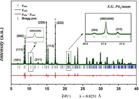
observed diffraction peaks could be indexed in the P42/mnm space group. No unindexed
diffraction peak was detected. Comparing the obtained powder to Na3V2(PO4)2F3 and
Na3(VO)2(PO4)2F, there is an obvious change in their color. Na3V2(PO4)2F3 and
Na3(VO)2(PO4)2F are dark grey and bluish green due to the presence of V3+ and V4+
9
beige (Figure S1). The ICP-OES analysis reveals a Na: V: Fe: P relative ratio of 2.9: 1: 1: 2 in
this final product, which is in good agreement with the theoretical one. The SEM images of
Figure S2 show that the powder consists of well crystalline primary particles of ca. 500 nm.
Besides the synthesis of Na3(VO)Fe(PO4)2F2 we also explored the extended
composition domain Na3(VO)2-yFey(PO4)2F1+y (0 ≤ y ≤ 2) by varying the V(C5H7O3)3:
Fe(NO3)3.9H2O molar ratio in the initial solution. The syntheses were performed in the same
conditions as those already discussed, however the obtained powders were a mixture of NVPF-
and NASICON-type phases (Figure S3). We also varied the thermal treatment conditions, but
no pure phase was successfully obtained (Figure S4).
Atomic and electronic structural description
The Na3V2(PO4)2F3 parent composition presents a subtle orthorhombic distortion of the
unit cell as detected by SXRD through the splitting of the (200)/(020) and (400)/(040)
diffraction peaks and is now indexed in the Amam space group.8 In the newly obtained
Fe-substituted Na3(VO)Fe(PO4)2F2 phase, no obvious separation of the (200)/(020) and
(400)/(040) doublets are observed suggesting that the structure should be indexed in the
P42/mnm space group. Rietveld refinements were performed in the Amam (Figure S5) and in
the P42/mnm space groups (Figure 2) in order to choose the best structural description for this
newly obtained phase. As especially highlighted by a comparison of the profile fittings, the
reliability factors, the cell parameters and the interatomic distances (given in Figure 2, Figure
S5 and Table S1), no improvement of the fit was observed by considering the orthorhombic
symmetry, and hence the tetragonal unit cell was thus retained. Three Na+ positions were
identified in the structure using Fourier difference maps, the details of this procedure are
described in Figure S6 and S7. The atomic positions and the obtained bond length values are
given in Table 1 and Table 2, respectively. The M–F(2)/O(4) distance, which corresponds to
10
value between the V=O bond length in Na3(VO)2(PO4)2F and the terminal Fe–F bond length in
Na3Fe2(PO4)2F3 (1.626(1) Å and 1.92(1) Å, respectively).24,41,42 This observation strongly
suggests the presence of V=O bonds in the structure of this newly obtained Na3(VO)Fe(PO4)2F2
phase.
Figure 2 : Rietveld refinement of the Na3(VO)Fe(PO4)2F2 structure from SXRD data, performed in the P42/mnm space group. Insets show enlargements of the (200)/(020) and (400)/(040) doublets.
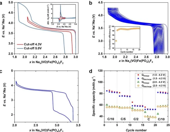
Table 1 : Structural parameters of Na3(VO)Fe(PO4)2F2 obtained from Rietveld refinement of synchrotron powder diffraction data collected at = 0.8251 Å. The Biso values of oxygen and fluorine were fixed during the Rietveld refinement.
S.G. P42/mnm Z = 4 a = b = 9.03564(6) Å c = 10.6412(1) Å V/Z = 217.195(2) Å3 RBragg = 3.46% Rp = 10.3% Rwp = 10.7% Atoms Wyckoff positions
x/a y/b z/c Occupancy Biso
V(1) 8j 0.7483(6) 0.2517(6) 0.1917(2) ½ 0.96(6) Fe(1) 8j 0.7483(6) 0.2517(6) 0.1917(2) ½ 0.96(6)
P(1) 4e ½ ½ 0.253(2) 1 0.9(1)
11 O(1) 16k 0.595(2) 0.094(2) 0.162(1) 1 0.78 O(2) 8j 0.598(2) 0.402(2) 0.160(2) 1 0.78 O(3) 8j 0.402(2) 0.402(2) 0.330(2) 1 0.78 O(4) 8j ¾ ¼ 0.360(2) ½ 1.0 F(1) 4g ¾ ¼ 0 1 0.44 F(2) 8j ¾ ¼ 0.360(2) ½ 1.0 Na(1) 8i 0.270(2) 0.022(2) 0 0.850(3) 2.1(1) Na(2) 8i 0.476(3) 0.290(3) 0 0.58(1) 4.5(4) Na(3) 4f 0.408(5) 0.408(5) 0 0.266(6) 3.0(3)
Table 2 : Bond lengths (Å) describing the coordination polyhedra of each cation in Na3(VO)Fe(PO4)2F2 determined from Rietveld refinement performed in the P42/mnm space group of synchrotron X-ray powder diffraction data collected at = 0.8251 Å.
V/Fe P(1) P(2) Na(1) Na(2) Na(3)
Coordination 6 4 4 7 7 6 O(1) 1.954(6) 1.595(8)x2 2.619(9)x2 2.27(1)x2 2.42(1)x4 O(2) 2.013(6) 1.529(6) x4 2.70(1)x2 O(3) 1.983(6)x2 1.493(8)x2 2.351(7)x2 F(1) 2.040(1) 2.464(7) 2.50(1) F(2)/O(4) 1.795(1) 2.547(6)x2 2.55(1)x2 2.50(1)x2
The 57Fe Mössbauer spectrum of this sample exhibits a single quadrupole doublet
(Figure 3a) with an isomer shift = 0.41(1) mms-1 and a mean quadrupole splitting Δ = 0.61
mms-1 corresponding to six-fold coordinated high spin Fe3+ ion (t
2g3 eg2) in a highly symmetric
environment. The rather high value of isomer shift is due to the high ionic character of the Fe–
F and Fe–O bonds. The quadrupole splitting distribution is mono-modal and very narrow
meaning that there is only one discrete site (8j) for Fe3+ ions with a regular and non-disordered
12
X-ray absorption near edge structure (XANES) at vanadium K-edge (5465 eV) and at
iron K-edge (7112 eV) spectra were recorded in order to determine the local environment as
well as the oxidation state of vanadium and iron species in the structure. Each XANES spectrum
can be divided into the pre-edge and the edge regions. For 3d transition metals, the pre-edge
signal is related to the forbidden 1s 3d transition and its intensity can be used to evaluate the local symmetry of the metal center of interest while the intense edge signal is caused by the
allowed 1s 4p transition and its position is related to the metal oxidation state and the bond length values with its surrounding ligands. If the metal center resides in a highly symmetric
octahedral site with the point group Oh, the 1s 3d transition is completely forbidden and the
pre-edge signal is very weak or completely diminished. If the metal center resides in a distorted
octahedral or tetrahedral site, the Oh symmetry is broken and the 3d orbitals can now be
hybridized with the 4p orbitals. Thanks to the 3d - 4p orbital hybridization, the electron
transition in the pre-edge region is allowed, which leads to a great increase in the pre-edge
signal.44 The pre-edge and the edge energies of the XANES spectra recorded at vanadium and
iron K-edges confirm the expected occurrence of V4+ and Fe3+ in the pristine sample (Figure
3b-c). The low intensity of the pre-edge signal on the XANES spectrum at Fe K-edge confirms
the symmetrical octahedral environments of high-spin Fe3+, while the high intensity of the
pre-edge on the V K-pre-edge spectrum implies that V local environment is highly distorted due to the
presence of the highly covalent vanadyl bond, which is in agreement with the bond length
values obtained from Rietveld refinement.
While the V4+ and Fe3+ ions share the same Wyckoff position in the structure and hence
cannot be distinguished by SXRD, the EXAFS analysis at both V and Fe K-edges provides
detailed information about the V(O,F)6 and Fe(O,F)6 local environments, even if oxygen and
fluorine cannot be distinguished. The EXAFS analysis at Fe K-edge of the Na3(VO)Fe(PO4)2F2
13
atoms in a square plane at a distance of 2.00 Å and two O/F atoms on the axial positions at
distances of 1.90 Å and 2.00 Å, with Debye-Waller factors of 4.9910-3 Å2. On the other hand, the V4+(O,F)6 octahedron is built by four equivalent O atoms in a square plane at a V–O distance
of ~ 2.00 Å, one O/F atom at 2.11 Å, and a closer O/F at 1.65 Å corresponding to the V=O
vanadyl-type bond. Smaller Debye-Waller factors (2.2410-3 Å2 vs 4.9910-3 Å2) are observed, which is in good agreement with a more covalent environment for V4+ than for Fe3+. Taking
into account the stoichiometry as well as the vanadium and iron oxidation states, the structure
of Na3(VO)Fe(PO4)2F2 can be described as FeO4F2 and VO5F octahedra which are sharing one
common fluorine atom to form FO4Fe–F–FeO4F, FO4Fe–F–VO5, or O5V–F–VO5 bi-octahedral
units. Indeed, no superstructure corresponding to the V4+/Fe3+ ordering was detected in the
SXRD data. The two ions are randomly distributed over all the transition metal sites in the
14
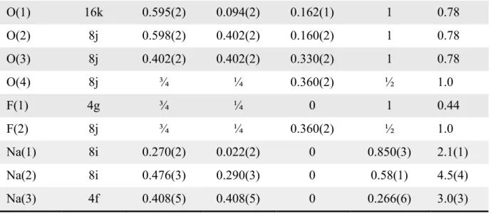
Figure 3 : (a) 57Fe Mössbauer spectrum recorded at ambient temperature with a 57Co source, (b) Fe K-edge XANES spectrum of Na3(VO)Fe(PO4)2F2. Both Fe2+SO4.7H2O and Fe3+PO4.2H2O are used as references to determine the Fe oxidation state, (c) V K-edge XANES spectrum of Na3(VO)Fe(PO4)2F2. The V3+PO
4, LiV4+PO4O and V5+OPO4 XANES spectra are reported to determine the V oxidation state, (d) 31P ss-NMR spectrum recorded at 100 MHz, MAS frequency = 30 kHz. The excitation pulse was placed at 5000 ppm. The asterisks (*) indicate the rotational spinning sidebands.
The 31P ss-NMR spectrum of the sample shows the possible presence of several
resonances in the range of 0 to 10000 ppm (Figure 3d). We have recently demonstrated that the
presence of V4+ ion in highly distorted octahedral sites in this structure will cause no Fermi
contact with the neighboring phosphorus nuclei as the orbital containing the electron spin of
V4+ does not point towards the phosphorus sites and the 31P resonance at ~ 0 ppm is associated
to the P(OV4+)
4 local environment.39 Therefore, 31P paramagnetic resonances observed here can
15
is studied through the Na3Fe2(PO4)2F3 crystal structure and is described in details in Figure S9
and Table S3. The obtained results show that the Fe3+ ions in Na3(VO)Fe(PO4)2F2 are in the
high-spin state (t2g3 eg2) with five unpaired electrons; these electron spins can be transferred
from Fe3+ to the neighboring phosphorus nuclei through the d
xz/dyz(Fe3+)−2p(O)−sp3(P) orbital
hybridization. All the phosphorus nuclei in Na3Fe2(PO4)2F3 are surrounded by four Fe3+,
forming the unique P(OFe3+)4 local environment with a paramagnetic shift of ~ 8400 ppm
(Figure S9). As we have recently shown that the paramagnetic interaction in this structural
framework is cumulative,39 which implies that the presence of one Fe3+ in the proximity of a
phosphorus nuclei will contribute to a paramagnetic shift value of ~ 2100 ppm. We thus deduce
that the P(OV4+)3(OFe3+), P(OV4+)2(OFe3+)2, and P(OV4+)(OFe3+)3 local environments, if they
exist in this crystal structure, should give rise to the 31P NMR resonances at ~ 2100 ppm, 4200
ppm, and 6300 ppm, respectively. The experimental signals are indeed observed in this range,
but unfortunately the signals are really broad due to a strong dipolar interaction between the
five unpaired electrons of each Fe3+ and the phosphorus nucleus. These broad signals also
overlap with the spinning side bands and the resolution of the recorded spectrum is not
16
Figure 4 : (a) An enlargement of the 31P NMR diamagnetic resonances (-100 ppm to 200 ppm) recorded for the Na3(VO)Fe(PO4)2F2 material. The n value indicates the number of Fe3+ in the second transition metal sites with respect to the Phosphorus nucleus. The five diamagnetic resonances observed for this material can be fitted by five different Pseudo-Voigt peak shape functions, (b) Illustration of the influence of the second neighboring metal ion belonging to the bi-octahedral unit on the 31P chemical shift value.
An enlargement on the 31P NMR diamagnetic region associated to the P(OV4+)4
environment (from -100 ppm to 200 ppm) reveals the presence of five diamagnetic resonances
equally separated by ~ 35 ppm. They can be fitted by five different Pseudo-Voigt peak shape
functions (Figure 4a) and the corresponding values for the chemical shift, peak width and
amplitude are compared in Table S4. As a V4+ ion in this structure induces almost no Fermi
17
in the second coordination sphere of 31P nucleus leading either to a weak Fermi contact shift or
to small changes in the local atomic or electronic structure around V4+ resulting in such a small
shift. These five 31P NMR resonances correspond to P(OV4+)4 local environments with different
distribution of V4+ : Fe3+ as second neighbors in the bi-octahedral units (Figure 4b). We hence
propose that each Fe3+, as second transition metal neighbor in the bi-octahedra, contributes to a
shift of ~35 ppm; therefore, the resonances at ~0 ppm, 33 ppm, 69 ppm, 105 ppm, and 139 ppm
are assigned to P(OV4+─F─V4+)4-n(OV4+─F─Fe3+)n local environments with n = 0, 1, 2, 3, 4,
respectively. The relative intensities of the five diamagnetic resonances are in agreement with
the values estimated considering a binomial distribution (Table S4). This shows that the
V4+/Fe3+ distribution in the bi-octahedral units is fully random and solely controlled by
statistics.
All the information obtained by SXRD, chemical analyses, as well as the local structure
description determined by 57Fe Mössbauer, 31P NMR and XAS at V and Fe K-edges
spectroscopies, fully support the chemical formula Na3(V4+O)Fe3+(PO4)2F2 proposed from the
very beginning of the paper.
Electrochemical properties of Na3(VO)Fe(PO4)2F2
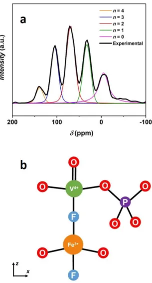
Na3(VO)Fe(PO4)2F2 was investigated as positive electrode in SIBs and cycled in the
voltage window 2.5 - 4.3 V vs. Na+/Na at the cycling rate of C/10 per Na+; only one Na+ can be
reversibly extracted in this voltage window (Figure 5a). The first derivative curve for
Na3(VO)Fe(PO4)2F2 shows the presence of a broad peak at ~ 3.64 V vs. Na+/Na (Inset of Figure
5a), which is similar to that observed for the V5+/V4+ redox couple involved during Na+
18
Figure 5 : (a) The charge/discharge curve of Na3(VO)Fe(PO4)2F2 vs. Na metal, at C/10 cycling rate with the cut-off voltage of 4.3 and 5.0 V. Inset shows the first derivative charge/discharge curve as a function of the operating voltage, (b) The long-term evolution of the charge/discharge curves at C/10 cycling rate in the potential range of 2.5 - 4.3 V vs. Na+/Na. Inset shows the evolution of the coulombic efficiency during the first twenty cycles, (c) The charge/discharge curve obtained at the second cycle for Na3(VO)Fe(PO4)2F2 vs. Na metal, at C/10 per Na+ and in the potential range of 1.5 - 4.3 V vs. Na+/Na, (d) The charge/discharge capacity of Na3(VO)Fe(PO4)2F2 at different cycle rates in the potential ranges of 2.5 - 4.3 V and 1.5 - 4.3 V vs. Na+/Na.
Several intermediate phases, stabilized by the Na+–vacancy ordering, were detected during the Na+ extraction and re-insertion from/in Na3(VO)2(PO4)2F 9; in the case of
Na3(VO)Fe(PO4)2F2, the electrochemical profile shows that the voltage increases/decreases
continuously during charge/discharge and thus suggests a solid solution mechanism. Indeed,
Fe3+ is randomly distributed among the transition metal sites, which thus prevents the formation
of charge ordering on the transition metal sites as well as the Na+–vacancy ordering within the diffusion channels of the host structure. Extra capacity is observed when the upper cut-off
19
material contains only one V4+ ion per formula unit, the extraction of one Na+ implies that all
the vanadium ions are at the pentavalent state in Na2(VO)Fe(PO4)2F2. Then, the extra capacity
can either be attributed to the oxidation of Fe3+ into Fe4+ or to the electrolyte degradation.
Nonetheless, the 57Fe Mössbauer spectrum recorded on the sample recovered at 5.0 V only
shows the presence of Fe3+ in its high-spin state (Figure S8) confirming that Fe3+ was not
oxidized into Fe4+. We thus conclude that this irreversible extra capacity is attributed to the
electrolyte decomposition.
Even without carbon-coating, this material shows very small polarization with limited
capacity fading during cycling (Figure 5b Inset). Indeed, comparing to a Na//Na3(VO)2(PO4)2F
half-cell operating in the same conditions, the polarization observed for a
Na//Na3(VO)Fe(PO4)2F2 half-cell appears to be two times smaller (Figure S10). The cell
parameters and the size of the diffusion channels of Na3(VO)Fe(PO4)2F2 are only ~ 0.1% bigger
than those of Na3(VO)2(PO4)2F; furthermore, the primary particles of Na3(VO)Fe(PO4)2F2 are
even bigger than those of Na3(VO)2(PO4)2F indicating that the easier diffusion cannot be
attributed to shorter diffusion lengths (Figure S11). These results suggest that the smaller
polarization observed for Na3(VO)Fe(PO4)2F2 is due to an improvement in the intrinsic
properties of the material. Dacek et al. reported that Na+ diffusivity in Na3M2(PO4)2X3 (M = Al,
V, Ti; X = F, O) and in similar structures is unlikely affected by the size of the diffusion
channels. It would rather depend strongly on the Na+–vacancy ordering and the Na+-defect
formation energy.45,46 A material with a high tendency to form Na+–vacancy ordering upon cycling, such as Na3V2(PO4)2F3 and Na3(VO)2(PO4)2F, will have a slower Na+ diffusion process
as extra activation energy is required to first break the Na+–vacancy ordering and then create local defects for the diffusion to occur.45 Any factor, such as cationic substitution on the
vanadium site,45,46 that can prevent the formation of Na+–vacancy ordering during charge or lower the Na+-defect formation energy will thus be a driving force for the formation of
off-20
stoichiometry compositions. Therefore, Fe3+ substitution for V with a random distribution of
Fe3+ over V sites can be considered as an appropriate substituent or dopant that can be used to
improve the Na+ diffusivity in Na3V2(PO4)2F3 and Na3(VO)2(PO4)2F-type materials.
Extra reversible capacity is obtained when the lower cut-off voltage is decreased to 1.5
V vs. Na+/Na (Figure 5c), which could be due to the activation of the Fe3+/Fe2+ redox couple
that had been reported earlier for the Na3Fe2(PO4)2F3 composition.47 The 57Fe Mössbauer
spectrum recorded on the material recovered at 1.5 V reveals the existence of 47% of Fe2+
(isomer shift δ = 1.26 mms-1) and 53% of Fe3+ (δ = 0.42 mms-1) in their high-spin state
confirming the occurrence of the Fe3+/Fe2+ redox couple in this voltage region (Figure S8).
Other hyperfine parameters of these iron species are given in Table S2. The hyperfine
parameters of Fe3+ in the pristine phase and in Na3.5(VO)Fe(PO4)2F2 are very similar implying
that there is little influence of the second coordination sphere on the Fe3+ sites. The reduction
of 0.47 Fe3+ to Fe2+ corresponds to the insertion of extra ~ 0.5 Na+ ion at the low voltage region
and leads to the composition Na3.5(VO)Fe(PO4)2F2. The insertion of more than 0.5 Na+ ion into
the structure is not achieved, even at low cycling rates, most probably due to the strong Na+– Na+ electrostatic repulsions within the channels of the structure that destabilizes the formation
of the phases with more than 3.5 Na+ per formula unit. Zhang et al. reported that Na
4V2(PO4)2F3
could be synthesized mechanically by ball milling Na3V2(PO4)2F3 with Na metal.48 Three Na+
ions were electrochemically extracted from Na4V2(PO4)2F3 upon charge; however, only two
Na+ ions could be reinserted into the structure during discharge to form Na3V2(PO4)2F3. The
initial phase, Na4V2(PO4)2F3, was never recovered even though a very low cut-off voltage was
applied.48 These observations confirm our results that the NVPF-like structure cannot
accommodate so many Na+ into its structural channels. The polarization increases greatly when
the lower cut-off voltage is set at 1.5 V (Figure 5c), suggesting that the Fe3+/Fe2+ reduction and
21
the presence of a huge polarization at low voltage does not affect the electrochemical
mechanism involving the V5+/V4+ redox couple at the high voltage region. By opening the
electrochemical window to 1.5 - 4.3 V, the Fe3+/Fe2+ redox couple can thus be activated, which
leads to an increase of the reversible capacity of ca. 50% at C/10 (Figure 5d). Furthermore, it
can recover its initial capacity delivered at C/10 after a series of cycling at higher rates (Figure
5d).
Figure 6 : The evolution of significant diffraction lines during Na+ de-intercalation from Na3(VO)Fe(PO4)2F2. Operando SXRD patterns were recorded upon cycling of a Na// Na3(VO)Fe(PO4)2F2 half-cell at = 0.8251 Å. The cell was cycled at C/10 cycling rate in the voltage window of 2.5 - 4.3 V vs. Na+/Na. The corresponding electrochemical data is given in Figure S12.
22
Operando X-ray diffraction patterns recorded upon charging of an electrochemical
Na//Na3(VO)Fe(PO4)2F2 half-cell in the potential range of 2.5 - 4.3 V show a continuous
evolution of the diffraction lines during the operation (Figure 6). This confirms the solid
solution mechanism involved in the Na+ de-intercalation process, as suggested by the
continuous evolution of the voltage profile (Figure S12). As expected for a solid solution
mechanism, the description of the structure in the tetragonal unit cell (P42/mnm S.G.) remains
valid all along the Na+ de-intercalation. The Le Bail fit of Na2.54(VO)Fe(PO4)2F2 in the P42/mnm
space group, recorded at pattern #25, is given in Figure S13. The cell parameters of Na
3-x(VO)Fe(PO4)2F2 evolve slowly during the first five patterns, which could be due to a delay in
the reaction at the beginning of charge; nevertheless, a linear evolution is observed from the
23
Figure 7 : The evolution of the cell parameters of Na3-x(VO)Fe(PO4)2F2 during the Na+ de-intercalation reaction, determined from the Le Bail fit of the SXRD patterns collected operando upon charging a Na//Na3(VO)Fe(PO4)2F2 half-cell.
The cell volume decreases with a contraction of the structure along a and b directions
and an expansion along the c direction during charge (Figure 7). As expected, the oxidation of
V4+ into V5+ with a smaller ionic radius leads to the decrease of a and b, whereas the Na+
de-intercalation decreases the screening effect between the terminal groups of two bi-octahedral units pointing directly towards each other “through” the channel (Figure 1) and thus results in an increase of the c parameter as a compensation effect. This anisotropic contraction/expansion
24
of the unit cell during the Na+ de-intercalation reaction was also observed in Na3V2(PO4)2F3,
Na3(VO)2(PO4)2F as well as many NASICON materials.9,12,13,49
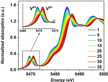
Figure 8 : Vanadium K-edge XANES spectra recorded operando upon charging a Na//Na3(VO)Fe(PO4)2F2 half-cell operating in the potential range of 2.5 - 4.5 V vs. Na+/Na (Figure S14).
Operando XAS spectra at vanadium K-edge were collected upon charging a
Na//Na3(VO)Fe(PO4)2F2 half-cell in the voltage range of 2.5 - 4.5 V at C/10 (Figure 8); note
that operando XAS experiment at Fe K-edge were not performed as 57Fe Mössbauer
measurements performed ex situ had already demonstrated that Fe3+ did not participate in the
electrochemical reaction in the high voltage region. A linear evolution of the vanadium
absorption edge is observed between 2.5 and 4.3 V (Figure S15). Above 4.3 V, no change in
the shape or the absorption edge energy is observed. The edge energy in the initial state (5481.8
eV, taken at 80% of the maximum intensity of the edge) corresponds to the presence of V4+
while at the end of the charge only V5+ is detected (5484.1 eV) (Figure S16), supporting the
25
V K-edge XAS dataset was globally analyzed using the chemometric approach, based on a
combination of Principal Component Analysis (PCA) and Multivariate-Curve Resolution
Alternating Least Squares (MCR-ALS).50
Figure 9 : The evolution of the concentration of the two principal components required to describe all the recorded Vanadium K-edge XAS spectra.
PCA allows to determine the minimum number of independent components required to
describe the whole operando dataset obtained during the electrochemical cycling. The number
of principal components is then used as the basis for the MCR-ALS analysis 51,52, allowing the
reconstruction of the principal orthogonal components needed to interpret the whole dataset.
The details about the chemometric analysis are reported in the supplementary information
(Figure S17). The PCA results show that there are two components required to describe the
whole system and the concentration profile of these two components is given in Figure 9. There
is a delay in the electrochemical reaction as the vanadium oxidation state only started to change
from the spectrum #5, and the electrochemical reaction involving the V5+/V4+ redox couple
26
reconstructed XAS spectra of these two components are identical to the spectrum #1 and
spectrum #33 recorded experimentally.
The V local environments of the two principal components were obtained by analyzing
the EXAFS oscillations of the reconstructed spectra. The local structure of the first component
is identical to those of the pristine Na3(VO)Fe(PO4)2F2 reported above, which is a highly
distorted V(O/F)6 octahedral site due to the presence of a vanadyl bond. At the end of charge,
V4+ is fully oxidized into V5+ and its local environment is even more distorted with a contraction
of the bond lengths in the equatorial plane: two of the four equatorial V–O bonds are rather
short (1.86 Å) while the other two are at located at a distance of 1.98 Å. The vanadyl bond (dV=O
= 1.62 Å) and the V–F bond (dV-F (axial) = 2.10 Å) along the c axis are unlikely to be affected by
the oxidation of V4+ to V5+. The EXAFS analysis cannot distinguish whether the two contracted
equatorial V–O bonds are in the cis- or in the trans-configuration; however, we here propose
that the two short V–O bonds would rather be formed on the trans-positions, which allows to
minimize the electrostatic repulsion between the two oxygen atoms that are approaching the
V5+ center as a result of the bond length contraction.
4. Conclusions
Na3(VO)Fe(PO4)2F2 was obtained by a sol-gel method and the structure of this newly
obtained phase was described in the P42/mnm space group. Only one Na+ can be reversibly
extracted in the potential range of 2.5 - 4.3 V vs. Na+/Na, which corresponds to the oxidation
of all V4+ ions into V5+. Limited polarization value and good capacity retention was observed
upon cycling, even if the reversible capacity decreased as the Fe4+/Fe3+ redox couple could not
be activated. On the contrary, extra 0.5 Na+ could be reversibly inserted into the structure by
27
synchrotron X-ray powder diffraction revealed there was no formation of Na+−vacancy ordering during charge (i.e. Na+ de-intercalation), which was contrary to what had been
observed for Na3V2(PO4)2F3 and Na3(VO)2(PO4)2F. Their suppression would lower the Na+
-defect formation energy, and hence increase the Na+ ionic conductivity in the diffusion
channels, and could explain the low polarization value observed for this material when used at
the positive electrode in a sodium cell. We hence suggest that Fe3+ is a promising substituent or
dopant that needs to be extensively studied in order to enhance the electrochemical performance
28
Corresponding Author:
*E-mail: Laurence.Croguennec@icmcb.cnrs.fr
Notes
The authors declare no competing financial interest.
Acknowledgements:
The authors thank the RS2E Network for the funding of LHBN’s PhD thesis. The
European Union’s Horizon 2020 research and innovation program under the Grant Agreement
No. 646433-NAIADES, the French National Research Agency (STORE-EX Labex Project
ANR-10-LABX-76-01 and SODIUM Descartes project ANR-13-DESC-0001-02) and the
Région Nouvelle Aquitaine are acknowledged for their financial support. The authors also want
to thank Cathy DENAGE, Emmanuel PETIT, Eric LEBRAUD, Alain WATTIAUX and Yohan
BIECHER (ICMCB) for their technical support. The Synchrotron diffraction experiments were
performed at the MSPD beamline at ALBA Synchrotron with the collaboration of ALBA staff
and CALIPSOplus (Grant 730872) funding. XAS experiments were performed on the ROCK
beamline at SOLEIL synchrotron, which is benefiting from a public grant overseen by the
French National Research Agency as part of the “Investissements d’Avenir” program
(Reference: ANR-10-EQPX-45).
Supporting Information
29
Comparing the color of Na3V2(PO4)2F3, Na3(VO)Fe(PO4)2F2, and Na3(VO)2(PO4)2F
powder (Figure S1); SEM images of Na3(VO)Fe(PO4)2F2 at different magnifications
(Figure S2); XRD patterns of the powder obtained at different V : Fe ratios (Figure S3);
Color of the powder obtained for different V: Fe ratios after a calcination at 550oC under
Ar atmosphere during 3h (Figure S4); Result of the Rietveld refinement performed in
the Amam space group on the synchrotron XRD data recorded on the
Na3(VO)Fe(PO4)2F2 phase (Figure S5); A comparison of the interatomic distances in
Na3(VO)Fe(PO4)2F2 obtained from Rietveld refinements by considering two structural
descriptions: Amam and P42/mnm space groups (Table S1); Calculated Fourier
difference maps with the residual charge density ranging from 0 - 3 (Figure S6);
Calculated Fourier difference maps with the residual charge density ranging from 0 - 1
(Figure S7); Room temperature 57Fe Mössbauer spectra recorded on
Na3(VO)Fe(PO4)2F2 and on the charged/discharged sample (Figure S8); Refined room
temperature 57Fe Mössbauer hyperfine parameters for Na3(VO)Fe(PO4)2F2 and for the
charged/discharged sample (Table S2); Comparison of the cell parameters between the
input model and the optimized structure (calculated by GGA method) of
Na3Fe2(PO4)2F3 (Table S3); Electron spin density calculated by GGA method at an
iso-surface value of 210-3 electronÅ-1 for Na3Fe2(PO4)2F3 (Figure S9); Refined parameters
of 31P solid-state NMR spectrum of Na
3(VO)Fe(PO4)2F2 (Table S4); Comparison of the
electrochemical properties between Na3(VO)2(PO4)2F and Na3(VO)Fe(PO4)2F2 (Figure
S10); Comparison of the primary particles’ size between Na3(VO)2(PO4)2F and
Na3(VO)Fe(PO4)2F2 (Figure S11); Electrochemical profile of Na//Na3(VO)Fe(PO4)2F2
half-cell used in the operando synchrotron XRD experiment (Figure S12); Le Bail fit of
Na2.54(VO)Fe(PO4)2F2 (Figure S13); Electrochemical profile of
30
experiment (Figure S14); Evolution the absorption edge energy (taken at the normalized
intensity = 0.8) of the Vanadium K-edge XANES spectra of Na3(VO)Fe(PO4)2F2
recorded in operando condition (Figure S15); Comparing the Vanadium K-edge
XANES spectra recorded on a Na//Na3(VO)Fe(PO4)2F2 half-cell in the initial state and
at the end of charge (Figure S16); Principal Component Analysis of the Vanadium
31
References
(1) Larcher, D.; Tarascon, J.-M. Towards Greener and More Sustainable Batteries for Electrical Energy Storage. Nat. Chem. 2015, 7 (1), 19–29. DOI: 10.1038/nchem.2085. (2) Grey, C. P.; Tarascon, J. M. Sustainability and in Situ Monitoring in Battery
Development. Nat. Mater. 2016, 16 (1), 45–56. DOI: 10.1038/nmat4777.
(3) Tarascon, J. M.; Armand, M. Issues and Challenges Facing Rechargeable Lithium Batteries. Nature 2001, 414 (6861), 359–367. DOI: 10.1038/35104644.
(4) Choi, J. W.; Aurbach, D. Promise and Reality of Post-Lithium-Ion Batteries with High Energy Densities. Nat. Rev. Mater. 2016, 1, 16013(1)-16013(16). DOI: 10.1038/natrevmats.2016.13.
(5) Delmas, C.; Braconnier, J.-J.; Fouassier, C.; Hagenmuller, P. Electrochemical Intercalation of Sodium in NaxCO2 Bronzes. Solid State Ionics 1981, 3–4, 165–169. DOI:
10.1016/0167-2738(81)90076-X.
(6) Park, Y. U.; Seo, D. H.; Kim, H.; Kim, J.; Lee, S.; Kim, B.; Kang, K. A Family of High-Performance Cathode Materials for Na-Ion Batteries, Na3(VO1-xPO4)2F1+2x (0 ≤ x ≤ 1):
Combined First-Principles and Experimental Study. Adv. Funct. Mater. 2014, 24 (29), 4603–4614. DOI: 10.1002/adfm.201400561.
(7) Oh, S. M.; Myung, S. T.; Hassoun, J.; Scrosati, B.; Sun, Y. K. Reversible NaFePO4
Electrode for Sodium Secondary Batteries. Electrochem. commun. 2012, 22 (1), 149– 152. DOI: 10.1016/j.elecom.2012.06.014.
(8) Bianchini, M.; Brisset, N.; Fauth, F.; Weill, F.; Elkaim, E.; Suard, E.; Masquelier, C.; Croguennec, L. Na3V2(PO4)2F3 Revisited: A High-Resolution Diffraction Study. Chem.
Mater. 2014, 26 (14), 4238–4247. DOI: 10.1021/cm501644g.
(9) Sharma, N.; Serras, P.; Palomares, V.; Brand, H. E. A.; Alonso, J.; Kubiak, P.; Fdez-gubieda, M. L.; Rojo, T. Sodium Distribution and Reaction Mechanisms of a Na3V2O2(PO4)2F Electrode during Use in a Sodium-Ion Battery. Chem. Mater. 2014, 26,
3391–3402. DOI: 10.1021/cm5005104.
(10) Sauvage, F.; Quarez, E.; Tarascon, J. M.; Baudrin, E. Crystal Structure and Electrochemical Properties vs. Na+ of the Sodium Fluorophosphate Na1.5VOPO4F0.5.
Solid State Sci. 2006, 8 (10), 1215–1221. DOI:
10.1016/j.solidstatesciences.2006.05.009.
(11) Zhou, W.; Xue, L.; Lü, X.; Gao, H.; Li, Y.; Xin, S.; Fu, G.; Cui, Z.; Zhu, Y.; Goodenough, J. B. NaxMV(PO4)3 (M = Mn, Fe, Ni) Structure and Properties for Sodium
Extraction. Nano Lett. 2016, 16 (12), 7836–7841. DOI: 10.1021/acs.nanolett.6b04044. (12) Chen, F.; Kovrugin, V. M.; David, R.; Mentré, O.; Fauth, F.; Chotard, J.; Masquelier, C.
A NASICON-Type Positive Electrode for Na Batteries with High Energy Density: Na4MnV(PO4)3. Small Methods 2019, 3, 1800218(1)–1800218(9). DOI:
10.1002/smtd.201800218.
(13) Abakumov, A. M.; Zakharkin, M. V.; Drozhzhin, O. A.; Antipov, E. V.; Chernyshov, D.; Tereshchenko, I. V.; Stevenson, K. J. Enhancing Na + Extraction Limit through High Voltage Activation of the NASICON-Type Na4MnV(PO4)3 Cathode. ACS Appl. Energy
32
(14) Guo, J.-Z.; Wang, P.-F.; Wu, X.-L.; Zhang, X.-H.; Yan, Q.; Chen, H.; Zhang, J.-P.; Guo, Y.-G. High-Energy/Power and Low-Temperature Cathode for Sodium-Ion Batteries: In Situ XRD Study and Superior Full-Cell Performance. Adv. Mater. 2017, 29, 1701968(1)-1701968(8). DOI: 10.1002/adma.201701968.
(15) Gu, Z. Y.; Guo, J. Z.; Yang, Y.; Yu, H. Y.; Xi, X. T.; Zhao, X. X.; Guan, H. Y.; He, X.; Wu, X. L. Precisely Controlled Preparation of an Advanced Na3V2(PO4)2O2F Cathode
Material for Sodium Ion Batteries: The Optimization of Electrochemical Properties and Electrode Kinetics. Inorg. Chem. Front. 2019, 6 (4), 988–995. DOI: 10.1039/c8qi01374h.
(16) Broux, T.; Fauth, F.; Hall, N.; Chatillon, Y.; Bianchini, M.; Bamine, T.; Leriche, J.; Suard, E.; Carlier, D.; Reynier, Y.; Simonin, L.; Masquelier, C.; Croguennec, L. High Rate Performance for Carbon‐Coated Na3V2(PO4)2F3 in Na‐Ion Batteries. Small Methods 2019, 3, 1800215(1)–1800215(12). DOI: 10.1002/smtd.201800215.
(17) Serras, P.; Palomares, V.; Goñi, A.; Gil de Muro, I.; Kubiak, P.; Lezama, L.; Rojo, T. High Voltage Cathode Materials for Na-Ion Batteries of General Formula Na3V2O2x(PO4)2F3−2x. J. Mater. Chem. 2012, 22 (41), 22301-22308. DOI:
10.1039/c2jm35293a.
(18) Park, Y.; Seo, D.; Kwon, H.; Kim, B.; Kim, J.; Kim, H.; Kim, I.; Yoo, H.; Kang, K. A New High-Energy Cathode for a Na-Ion Battery with Ultrahigh Stability. J. Am. Chem.
Soc. 2013, 135, 13870–13878. DOI: 10.1021/ja406016j.
(19) Xu, M.; Xiao, P.; Stauffer, S.; Song, J.; Henkelman, G.; Goodenough, J. B. Theoretical and Experimental Study of Vanadium-Based Fluorophosphate Cathodes for Rechargeable Batteries. Chem. Mater. 2014, 26 (10), 3089–3097. DOI: 10.1021/cm500106w.
(20) Park, Y. U.; Bai, J.; Wang, L.; Yoon, G.; Zhang, W.; Kim, H.; Lee, S.; Kim, S. W.; Looney, J. P.; Kang, K.; Wang, F. In Situ Tracking Kinetic Pathways of Li+/Na+ Substitution during Ion-Exchange Synthesis of LixNa1.5-xVOPO4F0.5. J. Am. Chem. Soc. 2017, 139 (36), 12504–12516. DOI: 10.1021/jacs.7b05302.
(21) Bianchini, M.; Lalère, F.; Nguyen, H. B. L.; Fauth, F.; David, R.; Suard, E.; Croguennec, L.; Masquelier, C. Ag3V2(PO4)2F3, a New Compound Obtained by Ag+/Na+ Ion
Exchange into the Na3V2(PO4)2F3 Framework. J. Mater. Chem. A 2018, 6 (22), 10340–
10347. DOI: 10.1039/c8ta01095a.
(22) Lin, X.; Huang, J.; Tan, H.; Huang, J.; Zhang, B. K3V2(PO4)2F3 as a Robust Cathode for
Potassium-Ion Batteries. Energy Storage Mater. 2019, 16, 97–101. DOI: 10.1016/j.ensm.2018.04.026.
(23) Broux, T.; Bamine, T.; Fauth, F.; Simonelli, L.; Olszewski, W.; Marini, C.; Ménétrier, M.; Carlier, D.; Masquelier, C.; Croguennec, L. Strong Impact of the Oxygen Content in Na3V2(PO4)2F3–yOy (0 ≤ y ≤ 0.5) on Its Structural and Electrochemical Properties. Chem.
Mater. 2016, 28 (21), 7683–7692. DOI: 10.1021/acs.chemmater.6b02659.
(24) Nguyen, L. H. B.; Broux, T.; Camacho, P. S.; Denux, D.; Bourgeois, L.; Belin, S.; Iadecola, A.; Fauth, F.; Carlier, D.; Olchowka, J.; Masquelier, C.; Croguennec, L. Stability in Water and Electrochemical Properties of the Na3V2(PO4)2F3 –
Na3(VO)2(PO4)2F Solid Solution. Energy Storage Mater. 2019, 20, 324–334. DOI:
33
(25) Bianchini, M.; Xiao, P.; Wang, Y.; Ceder, G. Additional Sodium Insertion into Polyanionic Cathodes for Higher-Energy Na-Ion Batteries. Adv. Energy Mater. 2017, 7 (18), 1700514(1) - 1700514(9). DOI: 10.1002/aenm.201700514.
(26) Olchowka, J.; Nguyen, L. H. B.; Broux, T.; Sanz Camacho, P.; Petit, E.; Fauth, F.; Dany, C.; Masquelier, C.; Croguennec, L. Aluminum Substitution for Vanadium in the Na3V2(PO4)2F3 and Na3V2(PO4)2FO2 Type Materials. Chem. Commun. 2019. Accepted.
DOI: 10.1039/C9CC05137F.
(27) Liu, W.; Yi, H.; Zheng, Q.; Li, X.; Zhang, H. Y-Doped Na3V2(PO4)2F3 Compounds for
Sodium Ion Battery Cathodes: Electrochemical Performance and Analysis of Kinetic Properties. J. Mater. Chem. A 2017, 5 (22), 10928–10935. DOI: 10.1039/C7TA03133E. (28) Zhang, Y.; Guo, S.; Xu, H. Synthesis of Uniform Hierarchical Na3V1.95Mn0.05(PO4)2F3@C Hollow Microspheres as a Cathode Material for Sodium-Ion
Batteries. J. Mater. Chem. A 2018, 6 (10), 4525–4534. DOI: 10.1039/C7TA11105C. (29) Qi, Y.; Mu, L.; Zhao, J.; Hu, Y.; Liu, H.; Dai, S. Superior Na-Storage Performance of
Low-Temperature-Synthesized Na3(VO1− xPO4)2F1+2x (0≤ x ≤1) Nanoparticles for Na-Ion
Batteries. Angew. Chemie 2015, 127 (34), 10049–10054. DOI: 10.1002/ange.201503188.
(30) Rodríguez-Carvajal, J. Recent Advances in Magnetic Structure Determination by Neutron Powder Diffraction. Phys. B Condens. Matter 1993, 192, 55–69. DOI: 10.1016/0921-4526(93)90108-I.
(31) Petříček, V.; Dušek, M.; Palatinus, L. Crystallographic Computing System JANA2006 : General Features. Z. Krist. 2014, 229 (5), 345–352. DOI: 10.1515/zkri-2014-1737. (32) Momma, K.; Izumi, F. VESTA: A Three-Dimensional Visualization System for
Electronic and Structural Analysis. J. Appl. Crystallogr. 2008, 41 (3), 653–658. DOI: 10.1107/S0021889808012016.
(33) Leriche, J. B.; Hamelet, S.; Shu, J.; Morcrette, M.; Masquelier, C.; Ouvrard, G.; Zerrouki, M.; Soudan, P.; Belin, S.; Elkaïm, E.; Baudelet, F. An Electrochemical Cell for Operando Study of Lithium Batteries Using Synchrotron Radiation. J. Electrochem.
Soc. 2010, 157 (5), A606-A610. DOI: 10.1149/1.3355977.
(34) Hesse, J.; Rubartsch, A. Model Independent Evaluation of Overlapped Mossbauer Spectra. J. Phys. E. 1974, 7 (7), 526–532. DOI: 10.1088/0022-3735/7/7/012.
(35) Briois, V.; La Fontaine, C.; Belin, S.; Barthe, L.; Moreno, T.; Pinty, V.; Carcy, A.; Girardot, R.; Fonda, E. ROCK: The New Quick-EXAFS Beamline at SOLEIL. J. Phys.
Conf. Ser. 2016, 712, 1-6. DOI: 10.1088/1742-6596/712/1/012149.
(36) Fonda, E.; Rochet, A.; Ribbens, M.; Barthe, L.; Belin, S.; Briois, V. The SAMBA Quick-EXAFS Monochromator: XAS with Edge Jumping. J. Synchrotron Radiat. 2012, 19 (3), 417–424. DOI: 10.1107/S0909049512009703.
(37) Lesage, C.; Devers, E.; Legens, C.; Fernandes, G.; Roudenko, O.; Briois, V. High Pressure Cell for Edge Jumping X-Ray Absorption Spectroscopy: Applications to Industrial Liquid Sulfidation of Hydrotreatment Catalysts. Catal. Today 2019, 336, 63– 73. DOI: 10.1016/j.cattod.2019.01.081.
(38) Ravel, B.; Newville, M. ATHENA, ARTEMIS, HEPHAESTUS: Data Analysis for X-Ray Absorption Spectroscopy Using IFEFFIT. J. Synchrotron Radiat. 2005, 12 (4), 537–
34
541. DOI: 10.1107/S0909049505012719.
(39) Nguyen, L. H. B.; Sanz Camacho, P.; Broux, T.; Masquelier, C.; Croguennec, L.; Carlier, D. 23Na and 31P Solid-State NMR: A Key Tool to Study Local Environments in Na3V2(PO4)2F3-yOy (0 ≤ y ≤ 2) Materials. In ECS 235th meeting; 2019, Abstract #544.
(40) Kresse, G.; Furthmüller, J. Efficiency of Ab-Initio Total Energy Calculations for Metals and Semiconductors Using a Plane-Wave Basis Set. Comput. Mater. Sci. 1996, 6 (1), 15–50. DOI: 10.1016/0927-0256(96)00008-0.
(41) Tsirlin, A. A.; Nath, R.; Abakumov, A. M.; Furukawa, Y.; Johnston, D. C.; Hemmida, M.; Krug Von Nidda, H. A.; Loidl, A.; Geibel, C.; Rosner, H. Phase Separation and Frustrated Square Lattice Magnetism of Na1.5VOPO4F0.5. Phys. Rev. B - Condens. Matter
Mater. Phys. 2011, 84 (1), 2–13. DOI: 10.1103/PhysRevB.84.014429.
(42) Le Meins, J.-M.; Crosnier-Lopez, M.-P.; Hemon-Ribaud, A.; Courbion, G. Phase Transitions in the Na3M2(PO4)2F3 Family (M = Al3+, V3+, Cr3+, Fe3+, Ga3+): Synthesis,
Thermal, Structural, and Magnetic Studies. J. Solid State Chem. 1999, 148, 260–277. DOI: 10.1006/jssc.1999.8447.
(43) Bill, E. 57Fe-Mössbauer Spectroscopy and Basic Interpretation of Mössbauer Parameters. In Practical Approaches to Biological Inorganic Chemistry; Elsevier, 2013; 109–130. DOI: 10.1016/B978-0-444-56351-4.00005-1.
(44) Penner-Hahn, J. E. X-Ray Absorption Spectroscopy. In Comprehensive Coordination
Chemistry II; Elsevier, 2003; 159–186. DOI: 10.1016/B0-08-043748-6/01063-X.
(45) Dacek, S. T.; Richards, W. D.; Kitchaev, D. A.; Ceder, G. Structure and Dynamics of Fluorophosphate Na-Ion Battery Cathodes. Chem. Mater. 2016, 28 (15), 5450–5460. DOI: 10.1021/acs.chemmater.6b01989.
(46) Matts, I. L.; Dacek, S.; Pietrzak, T. K.; Malik, R.; Ceder, G. Explaining Performance-Limiting Mechanisms in Fluorophosphate Na-Ion Battery Cathodes through Inactive Transition-Metal Mixing and First-Principles Mobility Calculations. Chem. Mater. 2015,
27 (17), 6008–6015. DOI: 10.1021/acs.chemmater.5b02299.
(47) Chihara, K.; Kitajou, A.; Gocheva, I. D.; Okada, S.; Yamaki, J. I. Cathode Properties of Na3M2(PO4)2F3[M = Ti, Fe, V] for Sodium-Ion Batteries. J. Power Sources 2013, 227,
80–85. DOI: 10.1016/j.jpowsour.2012.10.034.
(48) Zhang, B.; Dugas, R.; Rousse, G.; Rozier, P.; Abakumov, A. M.; Tarascon, J.-M. Insertion Compounds and Composites Made by Ball Milling for Advanced Sodium-Ion Batteries. Nat. Commun. 2016, 7 (1), 10308(1)-10308(9). DOI: 10.1038/ncomms10308. (49) Bianchini, M.; Fauth, F.; Brisset, N.; Weill, F.; Suard, E.; Masquelier, C.; Croguen. Comprehensive Investigation of the Na3V2(PO4)2F3−NaV2(PO4)2F3 System by Operando
High Resolution Synchrotron X‑ray Diffraction. Chem. Mater. 2015, 27, 3009–3020. DOI: 10.1021/acs.chemmater.5b00361.
(50) Massart, D. L.; Vandeginste, B. G. M.; Buydens, L. M. C.; Jong, S. D.; Lewi, P. J.; Smeyers-Verbeke, J. Handbook of Chemometrics and Qualimetrics: Part A; Elsevier: Amsterdam, 1997, 1-867.
(51) Garrido, M.; Larrechi, M. S.; Rius, F. X.; Tauler, R. Calculation of Band Boundaries of Feasible Solutions Obtained by Multivariate Curve Resolution–Alternating Least Squares of Multiple Runs of a Reaction Monitored by NIR Spectroscopy. Chemom.
35
Intell. Lab. Syst. 2005, 76 (2), 111–120. DOI: 10.1016/j.chemolab.2004.10.001.
(52) Jaumot, J.; de Juan, A.; Tauler, R. MCR-ALS GUI 2.0: New Features and Applications.