HAL Id: hal-00309016
https://hal.archives-ouvertes.fr/hal-00309016
Submitted on 5 Aug 2008
HAL is a multi-disciplinary open access
archive for the deposit and dissemination of
sci-entific research documents, whether they are
pub-lished or not. The documents may come from
teaching and research institutions in France or
abroad, or from public or private research centers.
L’archive ouverte pluridisciplinaire HAL, est
destinée au dépôt et à la diffusion de documents
scientifiques de niveau recherche, publiés ou non,
émanant des établissements d’enseignement et de
recherche français ou étrangers, des laboratoires
publics ou privés.
Thoracic CT-PET Registration Using a 3D Breathing
Model
Antonio Moreno, Sylvie Chambon, Anand Santhanam, Roberta Brocardo,
Patrick Kupelian, Jannick Rolland, Elsa Angelini, Isabelle Bloch
To cite this version:
Antonio Moreno, Sylvie Chambon, Anand Santhanam, Roberta Brocardo, Patrick Kupelian, et al..
Thoracic CT-PET Registration Using a 3D Breathing Model. MICCAI 2007, Aug 2007, Brisbane,
Australia. pp.626-633. �hal-00309016�
Thoracic CT-PET Registration Using a 3D
Breathing Model
Antonio Moreno1 , Sylvie Chambon1 , Anand P. Santhanam2,3, Roberta Brocardo1 , Patrick Kupelian3 , Jannick P. Rolland2 , Elsa Angelini1 , and Isabelle Bloch11
Ecole Nationale Sup´erieure des T´el´ecommunications (GET - T´el´ecom Paris), CNRS UMR 5141 LTCI - Signal and Image Processing Department, Paris, France
2
Optical Diagnostics and Application Laboratory, University of Central Florida, USA
3 Department of Radiation Oncology, MD Anderson Cancer Center Orlando, USA
Abstract. In the context of thoracic CT-PET volume registration, we present a novel method to incorporate a breathing model in a non-linear registration procedure, guaranteeing physiologically plausible deforma-tions. The approach also accounts for the rigid motions of lung tumors during breathing. We performed a set of registration experiments on one healthy and four pathological data sets. Initial results demonstrate the interest of this method to significantly improve the accuracy of multi-modal volume registration for diagnosis and radiotherapy applications.
1
Introduction
Registration of multimodal medical images is a widely addressed topic in many different domains, in particular for oncology and radiotherapy applications. We consider Computed Tomography (CT) and Positron Emission Tomography (PET) in thoracic regions, which provide complementary information about the anatomy and the metabolism of the human body (Fig. 1). Their registration has a sig-nificant impact on improving medical decisions for diagnosis and therapy [1–3]. Linear registration is not sufficient to cope with local deformations produced by respiration. Even with combined PET/CT scanners which avoid differences in patient orientation and provide linearly registered images, non-linear registration remains necessary to compensate for cardiac and respiratory motions [4].
Most of the existing non-linear registration methods are based on image information and do not take into account any knowledge of the physiology of the human body. Landmark-based registration techniques do take physiology into account by forcing homologous points to match. In this direction, several breathing models were built for medical visualization, for correcting artefacts in images or for estimating lung motion for radiotherapy applications, but few papers exploit such models in a registration process (Section 2).
In this paper, we propose to integrate a physiologically driven breathing model into a 3D non-linear registration (Sections 3 and 4). The registration problem is defined between two CT volumes and one PET volume (Fig. 1).
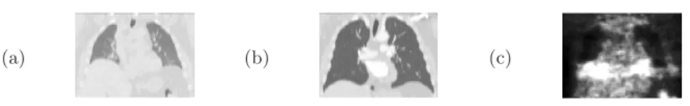
(a) (b) (c)
Fig. 1.CT images (a,b) corresponding to two different instants of the breathing cycle and PET image (c) of the same patient (coronal views).
2
Breathing models
Breathing Models and Thoracic Imaging Registration – Currently, respiration-gated radiotherapies are being developed to improve the efficiency of radiations of lung or abdominal tumors [5]. Three techniques have been proposed so far: (i) active techniques controling the patient’s breathing (airflow is blocked); (ii) passive or empirical techniques using external measurements in order to adapt radiation protocols to the tumor’s motion [6–8]; (iii) model-based techniques em-ploying a breathing model to evaluate lungs deformations during the breathing cycle [9]. Different bio-mathematical representations of the human respiratory mechanics have been developed [10]. Among Mathematical tools, the most pop-ular technique is based on Non-Uniform Rational B-Spline (NURBS), surfaces which are bidirectional parametric representations of an object. In [11], NURBS surfaces were used to correct for respiratory artifacts of SPECT images, build-ing NCAT (NURBS-based cardiac-torso) model. A multi-resolution registration approach for 4D Magnetic Resonance Imaging (MRI) was proposed in [12] with NCAT. In [13], a 4D NCAT phantom and an original CT image were used to generate 4D CT and to compute an elastic registration. Physically-based models, describing the important role of airflow inside the lungs, can be based on Active Breathing Coordinator (ABC allows clinicians to pause the patient’s breathing at a precise lung volume) [9] or on volume preservation relations [14, 15]. In [16], segmented MRI data were used to simulate PET volumes at different instants of the breathing cycle. These estimated PET volumes were used to evaluate differ-ent PET/MRI registration processes. Authors of [12, 17] used pre-register MRI to estimate a breathing model. CT registration using a breathing model was presented in [9] but a specific equipement is needed. From a modeling and sim-ulation point of view, physically-based deformation methods are better adapted for simulating lung dynamics and are easy to adapt to the patient, without the need for physical external adaptations.
Physics-Based Dynamic 3D Surface Lung Model – We employ an approach that was previously discussed in [15] and in which the two major components involved in the modeling efforts include: (1) Parameterization of PV (Pressure Volume) data from a human subject which acts as an ABC; (2) Estimation of the deforma-tion operator from 4D CT lung data sets. In step (1) a parameterized PV curve, obtained from a normal human subject, is used as a driver for simulating the 3D lung shapes at different lung volumes. In step (2), the computation takes as in-puts the nodal displacements of the 3D lung models and the estimated amount of
force applied on the nodes of the meshes (which are on the surface of the lungs). Displacements are obtained from 4D CT data of a normal human subject. The direction and magnitude of the lung surface point’s displacement are computed using the volume linearity constraint, i.e. the fact that the expansion of lung tissues is linearly related to the increase in lung volume. The amount of applied force on each node (that represents the air-flow inside lungs) is estimated based on a PV curve and the lungs’s orientation with respect to the gravity, which controls the air flow. Given these inputs, a physics-based deformation approach based on Green’s function (GF) formulation is estimated to deform the 3D lung surface models. Specifically the GF is defined in terms of a physiological factor, the regional alveolar expandability (elastic properties), and a structural factor, the inter-nodal distance of the 3D surface lung model. To compute the coeffi-cients of these two factors, an iterative approach is employed and, at each step, the force applied on a node is shared with its neighboring nodes, based on local normalization of the alveolar expandability, coupled with inter-nodal distance. The process stops when this sharing of the applied force reaches equilibrium. For validation purposes, a 4D CT dataset of a normal human subject with four instances of deformation was considered [18]. The simulated lung deformations matched the 4D CT dataset with 2 mm average distance error.
3
Combining Breathing Model and Image Registration
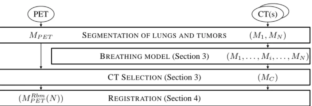
We have conceived an original algorithm in order to incorporate the breathing model described above in our multimodal image registration procedure. Fig. 2 shows the complete computational workflow. The input consists of one PET vol-ume and two CT volvol-umes of the same patient, corresponding to two different instants of the breathing cycle (end-inspiration and end-expiration, for example, collected with breath-hold maneuver). The preliminary step consists in segment-ing the lung surfaces (and, eventually, the tumors) on the PET data and on the two CT data sets, using a robust mathematical-morphology-based approach [19], and extracting meshes corresponding to the segmented objects.
SEGMENTATION OF LUNGS AND TUMORS
(M1, . . . , Mi, . . . , MN) (MC) (M1, MN) MP ET PET (MRbm P ET(N )) CT SELECTION(Section 3) REGISTRATION(Section 4) CT(s)
BREATHING MODEL(Section 3)
Computation of a Patient-Specific Breathing Model – For each patient, we only have two segmented CT datasets, therefore we first estimate intermediate 3D lung shapes between these two datasets and then, the displacements of lung surface points. Directions are given by the model (computed from a 4D CT normal data set of reference) while magnitudes are “patient-specific” (computed from the given 3D CT lung datasets). With known estimations of applied force and “subject-specific” displacements the coefficients of the GF can be estimated (Section 2). Then, the GF operator is used to compute the 3D lung shapes at different intermediate lung volumes.
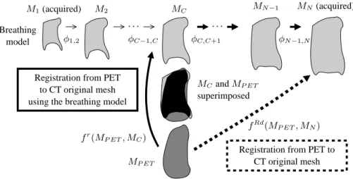
CT Selection – Let us denote the CT simulated meshes M1, M2,. . . , MN with M1 corresponding to the CT in maximum exhalation and MN to maximum inhalation. By using the breathing model, the transformation φi,j between two instants i and j of the breathing cycle can be computed as: Mj= φi,j(Mi). Our main assumption is that even if the PET volume represents an average volume throughout the respiratory cycle, using a breathing model, we can compute a CT volume that can be closer to the PET volume than the original CT volumes. By applying the continuous breathing model, we generate simulated CT meshes at different instants (“snapshots”) of the breathing cycle. By comparing each CT mesh with the PET mesh (MP ET), we select the “closest” one (i. e. with the most similar shape). The mesh that minimizes a measure of similarity C (here the root mean square distance) is denoted as MC: MC= arg miniC(Mi, MP ET).
Deformation of the PET – Once the appropriate CT (MC) is selected, we com-pute the registration, fr, between the MP ET mesh and the MC mesh as:
MP ETr (C) = f
r(MP ET, MC), (1) where Mr
P ET(C) denotes the registered mesh. Then, the transformation due to the breathing is used to register the PET to the original CT (continuous line in Fig. 3) incorporating the known transformation between MC and MN:
ΦC,N= φN −1,N◦ . . . ◦ φC+1,C+2◦ φC,C+1. (2) We apply ΦC,N to Mr
P ET(C) in order to compute the registration with MN: MP ETRbm(N ) = ΦC,N(M r P ET) = ΦC,N(f r (MP ET, MC)), (3) where MRbm
P ET(N ) denotes the PET registered mesh using the breathing model. A direct registration, denoted fRd, can also be computed between MP ET and the original CT mesh MN(dashed line in Fig. 3): MRd
P ET(N ) = fRd(MP ET, MN), where MRd
P ET(N ) is the result of registering the PET directly to the CT mesh MN (note that this could be done with another instant Mi). In the direct approach the deformation itself is not guided by any anatomical knowledge. In addition, if the PET and the original CT are very different, it is likely that this registration procedure will provide physically unrealistic results.
. . . . M2 MN −1 MP ET fRd(MP ET, MN) M1(acquired) Breathing model MN(acquired) φ1,2 superimposed Registration from PET
to CT original mesh using the breathing model
Registration from PET to CT original mesh MC φC,C+1 φN −1,N φC−1,C MCand MP ET fr(MP ET, MC)
Fig. 3.Registration framework on PET (MP ET) and CT mesh (MN) – The MCmesh
is the closest to the MP ET mesh. We can register MP ET to the MN mesh (original
CT) following one of the two paths.
4
Registration Method Adapted to Pathologies
The algorithm described in Section 3 can be applied with any type of registra-tion method, to estimate fRdand fr. These functions may be computed by any registration method adapted to the problem. We show here how the proposed ap-proach can be adapted for registration of multi-modality images in pathological cases.
Registration with Rigidity Constraints – We have previously developed a reg-istration algorithm for the thoracic region taking into account the presence of tumors, while preserving continuous smooth deformations [20]. We assume that the tumor is rigid and that a linear transformation is sufficient to cope with its displacements between CT and PET scanning. This hypothesis is relevant and in accordance with the clinicians’ point of view, since tumors are often compact masses of pathological tissue. The registration algorithm relies on segmented structures (lungs and tumors). Landmark points are defined on both datasets to guide the deformation of the PET volume towards the CT volume. The defor-mation at each point is computed using an interpolation procedure where the specific type of deformation of each landmark point depends on the structure it belongs to, and is weighted by a distance function, which guarantees continuity of the transformation.
Registration with Rigidity Constraints and Breathing Model – Here, the following procedure is used to compute fr(in our example MN is the original CT):
1. Selection of landmark points on the CT mesh MC (based on Gaussian and mean curvatures and uniformly distributed on the lung surface) [21];
2. Estimation of corresponding landmark points on the PET mesh MP ET(using the Iterative Closest Point (ICP) algorithm [22]);
3. Tracking of landmark points from MCto the CT mesh MN using the breath-ing model;
4. Registration of the PET and the original CT using the estimated correspon-dences with the method summarized in the previous paragraph.
The breathing model used in step (3) guarantees that the corresponding land-marks selected on the original CT are correct (and actually they represent the same anatomical point) and follow the deformations of the lungs during the respiratory cycle.
5
Results and Discussion
We have applied our algorithm on a normal case and on four pathological cases, exhibiting one tumor. In all cases, we have one PET (of size 144 × 144 × 230 with resolution of 4×4×4 mm3
or 168×168×329 with resolution of 4×4×3 mm3) and two CT volumes (of size 256 × 256 × 55 with resolution of 1.42 × 1.42 × 5 mm3to 512 × 512 × 138 with resolution of 0.98 × 0.98 × 5 mm3), acquired during breath-hold in maximum inspiration and in intermediate inspiration, from individual scanners. The breathing model was initialized using the lung meshes from the segmented CT. Ten meshes (corresponding to regularly distributed instants) are generated and compared with the PET. The computation time can reach two hours for the whole process (a few seconds for segmentation, a few minutes for landmark point selection and about ninety minutes for registration). Although this is not a constraint because we do not deal with an on-line process, this computation time will be optimized in the future.
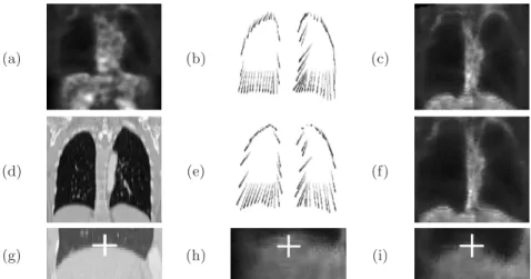
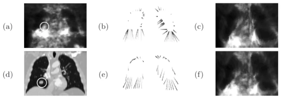
As illustrated in Fig. 4 and 5 (one normal case and one pathological case), the correspondences between landmark points on the original CT and the PET are more realistic in the results obtained with the breathing model (images (e) and (f)) than without (images (b) and (c)). Using the model, it can be observed that the corresponding points represent the same anatomical points and that the uniqueness constraint is respected, leading to visually better looking PET reg-istered images. In particular, the lower part of the two lungs is better regreg-istered using the model, the lung contour in the registered PET is closer to the lung contour in the original CT, cf. Fig. 4(g–i). In the illustrated pathological case, the tumor is well registered and not deformed. Moreover, the distance between the registered PET lungs and the original CT lungs is lower than using the direct approach.
In this paper, we consider the impact of the physiology on lung surface de-formation, based on reference data of normal human subjects. Therefore the methodology presented in this paper will further benefit upon the inclusion of patho-physiology specific data once established. The use of normal lung phys-iology serves to demonstrate improvements in CT and PET registration using a physics-based 3D breathing lung model. Current work includes a quantitative comparison and evaluation on a larger database, in collaboration with clinicians. Acknowledgments: This work has been partially funded by an ANR grant: ANR-05-BLANC-0081.
(a) (b) (c)
(d) (e) (f)
(g) (h) (i)
Fig. 4.(a) Original PET, (d) CT images in a normal case. Correspondences between selected points in the PET image and in the CT image are shown in (b) for the direct method and (e) for the method with the breathing model (corresponding points are linked). The registration result is shown in (c) for the direct method and in (f) for the method with the breathing model. Details of registration on the bottom part of right lung, (g) CT, (h) PET registered without breathing model, (c) with breathing model. The white crosses correspond to the same coordinates.
References
1. Lavely, W., et al.: Phantom validation of coregistration of PET and CT for image-guided radiotherapy. Medical Physics 31(5) (2004) 1083–1092
2. Rizzo, G., et al.: Automatic registration of PET and CT studies for clinical use in thoracic and abdominal conformal radiotherapy. Physics in Medecine and Biology 49(3) (2005) 267–279
3. Vogel, W., et al.: Correction of an image size difference between positron emission tomography (PET) and computed tomography (CT) improves image fusion of dedicated PET and CT. Physics in Medecine and Biology 27(6) (2006) 515–519 4. Shekhar, R., et al.: Automated 3-Dimensional Elastic Registration of
Whole-Body PET and CT from Separate or Combined Scanners. The Journal of Nuclear Medicine 46(9) (2005) 1488–1496
5. Sarrut, D.: Deformable registration for image-guided radiation therapy. Zeitschrift f¨ur Medizinische Physik 13 (2006) 285–297
6. McClelland, J., et al.: A Continuous 4D Motion Model from Multiple Respiratory Cycles for Use in Lung Radiotherapy. Medical Physics 33(9) (2006) 3348–3358 7. Nehmeh, S., et al.: Four-dimensional (4D) PET/CT imaging of the thorax. Physics
in Medecine and Biology 31(12) (2004) 3179–3186
8. Wolthaus, J., et al.: Fusion of respiration-correlated PET and CT scans: correlated lung tumour motion in anatomical and functional scans. Physics in Medecine and Biology 50(7) (2005) 1569–1583
9. Sarrut, D., et al.: Non-rigid registration method to assess reproducibility of breath-holding with ABC in lung cancer. International Journal of Radiation Oncology– Biology–Physis 61(2) (2005) 594–607
(a) (b) (c)
(d) (e) (f)
Fig. 5.Same as in Fig. 4(a–f) for a pathological case (the tumor is surrounded by a white circle).
10. Mead, J.: Measurement of Inertia of the Lungs at Increased Ambient Pressure. Journal of Applied Physiology 2(1) (1956) 208–212
11. Segars, W., et al.: Study of the Efficacy of Respiratory Gating in Myocardial SPECT Using the New 4-D NCAT Phantom. IEEE Transactions on Nuclear Sci-ence 49(3) (2002) 675–679
12. Rohlfing, T., et al.: Modeling Liver Motion and Deformation During the Respi-ratory Cycle Using Intensity-Based Free-Form Registration of Gated MR Images. Medical Physics 31(3) (2004) 427–432
13. Guerrero, T., et al.: Elastic image mapping for 4-D dose estimation in thoracic radiotherapy. Radiation Protection Dosimetry 115(1–4) (2005) 497–502
14. Zordan, V., et al.: Breathe Easy: Model and Control of Human Respiration for Computer Animation. Graphical Models 68(2) (2006) 113–132
15. Santhanam, A.: Modeling, Simulation, and Visualization of 3D Lung Dynamics. PhD thesis, University of Central Florida (2006)
16. Pollari, M., et al.: Evaluation of cardiac PET-MRI registration methods using a numerical breathing phantom. In: IEEE International Symposium on Biomedical Imaging, ISBI. (2004) 1447–1450
17. Sundaram, T., Gee, J.: Towards a Model of Lung Biomechanics: Pulmonary Kine-matics Via Registration of Serial Lung Images. Medical Image Analysis 9(6) (2005) 524–537
18. Santhanam, A., et al.: Modeling Simulation and Visualization of Real-Time 3D Lung Dynamics. IEEE Transactions on Information Technology in Biomedicine (2007) In press.
19. Camara, O., et al.: Explicit Incorporation of Prior Anatomical Information into a Nonrigid Registration of Thoracic and Abdominal CT and 18-FDG Whole-Body Emision PET Images. IEEE Transactions on Medical Imaging (2007) To appear. 20. Moreno, A., et al.: Non-linear Registration Between 3D Images Including Rigid
Objects: Application to CT and PET Lung Images With Tumors. In: Workshop on Image Registration in Deformable Environments (DEFORM), Edinburgh, UK (2006) 31–40
21. Chambon, S., et al.: CT-PET Landmark-based Lung Registration Using a Dynamic Breathing Model. In: International Conference on Image Analysis and Processing, Modena, Italy (September 2007) To appear.
22. Besl, P., McKay, N.: A Method for Registration of 3-D Shapes. IEEE Transactions on Pattern Analysis and Machine Intelligence 14(2) (1992) 239–256