1
Evolution of severe sleep-wake cycle disturbances
1following traumatic brain injury: A case study in both
2acute and subacute phases post-injury
3Catherine Duclos1-2, Marie Dumont1-2, Marie-Julie Potvin3, Alex Desautels,1,4 Danielle Gilbert3, 4
David Menon5, Francis Bernard 3,6 Nadia Gosselin1,7 5
1. Center for Advanced Research in Sleep Medicine, Hôpital du Sacré-Coeur de Montréal, Montréal, Canada 6
2. Department of Psychiatry, Université de Montréal, Montréal, Canada 7
3. Traumatology program, Hôpital du Sacré-Coeur de Montréal, Montréal, Canada 8
4. Department of Neuroscience, Université de Montreal, Montréal, Canada 9
5. Division of Anaesthesia, University of Cambridge, Cambridge, United Kingdom 10
6. Department of Medicine, Université de Montreal, Montréal, Canada 11
7. Department of Psychology, Université de Montréal, Montréal, Canada 12
13 14
Nadia Gosselin, PhD – CORRESPONDING AUTHOR 15
Institutional address: 16
Center for Advanced Research in Sleep Medicine 17
Hôpital du Sacré-Cœur de Montréal 18
5400 boul. Gouin Ouest, local E-0330 19 Montréal, Québec 20 H4J 1C5 21 Canada 22 Email address: 23 nadia.gosselin@umontreal.ca 24 25 26 27
2
ABSTRACT
28
Background: Sleep-wake disturbances are frequently reported following traumatic brain injury
29
(TBI), but they remain poorly documented in the acute stage of injury. Little is known about 30
their origin and evolution. 31
Case presentation: This study presents the case of a patient in the acute phase of a severe TBI.
32
The patient was injured at work when falling 12 m into a mine and was hospitalized in the 33
regular units of a level 1 trauma centre. From days 31 to 45 post-injury, once he had reached a 34
level of medical stability and continuous analgosedation had been ceased, his sleep-wake cycle 35
was monitored using actigraphy. Results showed significant sleep-wake disturbances and severe 36
sleep deprivation. Indeed, the patient had an average nighttime sleep efficiency of 32.7 ± 15.4%, 37
and only an average of 4.8 ± 1.3 hrs of sleep per 24-hr period. After hospital discharge to the 38
rehabilitation centre, where he remained for 5 days, the patient was then readmitted to the same 39
neurological unit for paranoid delusions. During his second hospital stay, actigraphy recordings 40
resumed from days 69 to 75 post-injury. A major improvement in his sleep-wake cycle was 41
observed during this second stay, with an average nighttime sleep efficiency of 96.3 ± 0.9% and 42
an average of 14.1 ± 0.9 hrs of sleep per 24-hr period. 43
Conclusion: This study is the first to extensively document sleep-wake disturbances in both the
44
acute and subacute phases of severe TBI. Results show that prolonged sleep deprivation can be 45
observed after TBI, and suggests that the hospital environment only partially contributes to 46
sleep-wake disturbances. Continuous actigraphic monitoring may prove to be a useful clinical 47
tool in the monitoring of patients hospitalized after severe TBI in order to detect severe sleep 48
deprivation requiring intervention. The direct impact of sleep-wake disturbances on 49
3
physiological and cognitive recovery is not well understood within this population, but is worth 50
investigating and improving. 51
Keywords: traumatic brain injury, sleep disorders, actigraphy, circadian rhythms, neurocritical
52
care, neuropsychiatry 53
4
BACKGROUND
54
Chronic sleep-wake disturbances, such as insomnia and hypersomnia, are among the 55
most widely-reported sequelea following traumatic brain injury (TBI), and have been 56
documented across all levels of TBI severity, until several years post-injury [1]. Less attention 57
has been paid to sleep-wake disturbances that occur in the first weeks post-injury. This might be 58
explained by the challenges of performing sleep studies in an acute care setting, where most 59
patients are confused and are not able to evaluate their own sleep quality. 60
A first group of studies that aimed at documenting sleep disturbances in post-acute TBI 61
used nurse observations in individuals admitted to rehabilitation centres. One study found that of 62
31 patients, 21 (68%) had two or more hours awake during the night [2]. Similarly, a second 63
study showed that mild to severe sleep disturbances were present among 84% of TBI patients 64
upon rehabilitation admission, and persisted for 66% of patients one month post-injury [3]. This 65
research group used item one of the Delirium Rating Scale-Revised-98 to classify the severity of 66
sleep-wake cycle disturbance as none, mild, moderate, or severe. 67
With the aim of using more objective methods to document the sleep-wake cycle of 68
patients in the acute and post-acute phases of moderate-severe TBI, a second group of studies 69
used actigraphy, which measures physical motion over time, to derive a rest-activity pattern. It 70
has been shown that the rest-activity cycle measured with actigraphy strongly correlates with the 71
sleep-wake cycle [4]; consequently, the rest-activity cycle derived from actigraphy is often 72
referred to as the sleep-wake cycle. Within this context, a study carried out during early 73
rehabilitation found that 11 of 14 moderate-severe TBI patients had an average 1-week sleep 74
efficiency lower than 63%, pointing to pervasive sleep-wake disturbances [5]. More recently, 75
Gardani and colleagues evaluated 30 patients with chronic severe TBI in an inpatient 76
5
rehabilitation setting, using actigraphy and self-report measures [6]. The authors found that 67% 77
of patients had sleep-wake cycle disturbances, 50% of which met diagnostic criteria for a sleep 78
disorder, according to the International Classification of Sleep Disorders (2nd edition). 79
Additionally, we recently used 10-day actigraphy recordings with 16 TBI patients hospitalized in 80
a level 1 trauma centre in order to quantify the clustering of activity during the daytime and of 81
rest during the nighttime as an estimate of their sleep-wake cycle consolidation. We found that 82
patients had a poor sleep-wake cycle consolidation, which gradually improved over time [7]. 83
However, using a threshold of ≥80% of all 24-h activity occurring in the daytime, only half of 84
the patients reached an acceptable sleep-wake cycle consolidation during the recording period. 85
Patients who reached an acceptable sleep-wake cycle consolidation (≥80%) were more likely to 86
emerge from posttraumatic amnesia (PTA) and to have lower disability at hospital discharge. 87
Despite the high prevalence of acute and subacute sleep-wake disturbances in TBI 88
patients, their aetiology is not well understood. Furthermore, no study has yet documented the 89
sleep-wake cycle during both the acute and subacute phases of TBI, while the patient was 90
hospitalized in the same environment, which is of interest given that the hospital environment 91
itself may be a contributing factor to disturbed sleep and wake. The aim of this article is to 92
document the case of one of our TBI patients from the abovementioned study [7], who suffered 93
severe sleep-wake cycle disturbances during his acute hospital stay. Since this patient was 94
readmitted five days post-discharge and wore the actigraph during his second hospital stay, his 95
case enables us to document the evolution of his sleep-wake cycle over time and to juxtapose the 96
sleep-wake cycle recorded during two different hospital stays in a similar environment, during 97
the acute (measured days 31-45 days post-injury, starting 4 days following discharge from the 98
ICU) and subacute (measured days 69-75 post-injury) phases. 99
6 CASE PRESENTATION
100 Biographical History 101
LC is a 43-year-old right-handed Caucasian male, who resides with his spouse and two 102
teenage daughters. Prior to his injury, LC was in good physical health, had no previous history of 103
TBI, chronic disease, drug or alcohol abuse, or psychiatric, neurological or sleep disorders. 104
Injury
105
LC suffered a severe TBI when falling 12 m into a well of a mine during work hours. LC 106
lost consciousness, had an initial Glasgow Coma Scale (GCS) score of 6 [8], and was 107
immediately transported by ambulance to the nearest hospital, located approximately 160 km 108
from the site of injury. Upon arrival at the regional hospital, his GCS score was 8. Following 109
clinical evaluation, he was immediately transferred by ambulance to a level 1 trauma centre 110
located over 500 km from the site of injury. A level 1 trauma centre provides the highest level of 111
surgical and specialized care to trauma patients, is comprised of a full range of equipment and 112
specialists dedicated to the care of patients having suffered TBI or orthopaedic injuries, and 113
generally receives the most severe cases within a large geographical area. 114
First admission 115
LC was admitted to the trauma centre approximately 15 hours after injury. His GCS score 116
was 3 (intubated) upon admission to the Emergency Room, and he was taken to the Intensive 117
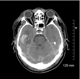
Care Unit (ICU). A computed tomography (CT) scan revealed diffuse subarachnoid haemorrhage 118
in the left hemisphere, left parieto-occipital subdural hematoma, right temporal intraparenchymal 119
hematoma (3 cm), intrapeduncular, left intrapontine and temporal petechiae, as well as left 120
frontal and right parieto-occipital contusions (see Figure 1). His Marshall score was 2 [9], and 121
his Rotterdam score was also 2 [10]. LC also suffered multiple facial fractures, a C4 cervical 122
7
fracture, D6, D8 and D12 thoracic fractures, a fracture of the left 9th rib, a spleen laceration, a 123
pseudo-aneurysm of the aorta (4 mm), and a left pneumothorax. 124
LC was hospitalized in the ICU for 27 days. Overall, he was under continuous sedation 125
for 16 days, during which time he received an average daily dose of 4.79 ± 2.33 g of propofol, 126
and 6.2 ± 2.2 mg of fentanyl. During 11 of those 16 days of continuous sedation, LC also 127
received an average daily dose of 0.55 ± 0.25 mg of midazolam. He was intubated 25 days, had 128
elevated intracranial pressure (≥ 20 mmHg) during 13 days with a peak at 46.3 ± 14 mmHg, and 129
had on average 7.3 ± 14 episodes of elevated intracranial pressure per day. 130
LC began responding to simple orders 15 days post-injury, when sedation was interrupted 131
briefly to assess his level of response, and he opened his eyes 16 days post-injury. Subsequent to 132
ICU discharge (27 days post-injury), LC was transferred to a six-patient room in the neurological 133
ward. LC suffered akinetic mutism and moderate-severe oropharyngeal dysphagia throughout the 134
first 46 days post-injury, and he then began to whisper, reaching a normal voice level 2 days 135
prior to hospital discharge. At this point, he could walk unassisted and was fully functional in all 136
bed and chair transfers. 137
LC was discharged from the trauma centre 55 days post-injury and admitted to a 200-bed 138
inpatient rehabilitation centre, specialized in the care of TBI, orthopaedic injuries and neurology. 139
Within the 72 hours prior to hospital discharge, LC had a score of 10 out of 29 on the Disability 140
Rating Scale [11], reflecting confused communication ability, partial cognitive disability for 141
grooming, a markedly dependent level of functioning (mental, emotional, or social), and a non-142
competitive level of employability. The neurological examination carried out 8 days prior to 143
hospital discharge (47 days post-injury) yielded a score of 5 on the Neurological Outcome Scale 144
8
for Traumatic Brain Injury without the supplemental items [12]. Deficits arose when LC was 145
asked the current month and his age, which he both answered incorrectly, as well as when asked 146
to identify odours or name objects for stimulus cards. This could either be the result of mild to 147
moderate aphasia, or PTA, which would account for an inability to recall the words associated to 148
various stimuli. Due to persistent akinetic mutism throughout most of the hospitalization period, 149
neuropsychological evaluations were only carried out during the second hospital stay. 150
Second admission 151
Five days after his admission to the inpatient rehabilitation centre, LC was readmitted to 152
the trauma centre by ambulance for persecutory paranoid delusion, as per clinical observations at 153
the inpatient rehabilitation centre, and remained hospitalized for 43 days in a two-patient room of 154
the same neurological ward on which he had previously been hospitalized. 155
During this second hospital stay, LC continuously suffered retrograde and anterograde 156
memory deficits with confabulations, severe temporal and spatial disorientation, verbal 157
desinhibition, distrusting and suspicious behaviour, paranoia, and anosognosia. LC’s condition 158
was attributed the diagnosis of post-TBI psychotic disorder. Neuropsychological evaluation 159
carried out on days 87 and 89 post-injury showed severe dysfunctions in all cognitive domains 160
(see Table 1). 161
[Insert Table 1 about here] 162
LC was discharged 43 days after this second admission (102 days post-injury), and was 163
readmitted to the inpatient rehabilitation centre. The occupational therapy report from LC’s final 164
evaluation, carried out 1 week prior to this second discharge, described him as completely 165
9
dependent for domestic activities of daily living (for timeline of injury and hospital stays, see 166
Figure 2). 167
Given his lengthy second admission, his psychiatric complications and persistent 168
cognitive and functional sequelea, LC’s case does not represent one of typical post-TBI 169
recovery, but rather depicts a slower and complexified recovery process. 170
Methods
171
Actigraphy protocol 172
LC was recruited as part of a larger longitudinal study taking place at Hôpital du Sacré-173
Coeur de Montréal, which was approved by the hospital ethics committee. Consent for 174
participation was obtained from LC’s spouse, since he was unable to provide informed consent 175
on his own. 176
LC wore a wrist actigraph on his non-dominant (left) arm during his first and second 177
hospital stays (Actiwatch-2 during the first stay, and Actiwatch Spectrum during the second stay; 178
MiniMitter Philips Healthcare, Andover, MA, USA). The actigraph is a small, watch-like device 179
that contains an accelerometer, which records physical motion in all directions with a sensitivity 180
of 0.05 g. Motion is then converted to an electric signal, which is digitally integrated to derive an 181
activity count per 1-min epochs. During the first hospital stay, the actigraphy recording began 31 182
days following the injury, 4 days after discharge from the ICU. Continuous intravenous or 183
subcutaneous administration of a sedative drug was ceased 11 days prior to the start of 184
actigraphic recording. LC was no longer intubated and had reached a level of medical stability 185
defined by the absence of elevated intracranial pressure, of hemodynamic instability, and of 186
fever or active infections. When the actigraph was installed, LC had also reached a Rancho Los 187
10
Amigos score of IV, indicative of a confused/agitated state [13]. LC could follow simple 188
commands for motor action inconsistently and with delay, would turn his head when his name 189
was called. Data was acquired for 15 days during hospitalization in the regular unit, during 190
which time he received no sedatives or analgesics. Approximately every 3 days, data were 191
uploaded into dedicated software (Actiware 5.0). 192
During the second hospital stay, LC wore the actigraph for seven days, beginning 69 days 193
post-injury. During this recording period, LC received a daily dose of 3 mg of lorazepam (1 mg 194
at 8:30 hrs, 17:00 hrs, and 22:00 hrs). 195
Data analyses 196
For each day the actigraph was worn, each minute of recording was scored as “sleep” or 197
“wake” using the automatic scoring system of the dedicated software (Actiware 5.0). A 198
particular 1-min epoch was scored as wake by comparing the activity counts of this epoch to 199
those immediately surrounding it. The threshold chosen to score a 1-min epoch as wake was > 20 200
activity counts per minute. A smaller number yielded to a score of sleep. For each epoch the 201
actigraph was not worn, due to the removal of the actigraph for data downloads or bathing, the 202
epoch was scored as wake since the patient was awake in both contexts. 203
A sleep bout was defined as a period of 5 or more consecutive epochs scored as sleep by 204
Actiware 5.0. To reduce the artificial fragmentation of rest periods, isolated 1-min epochs scored 205
as wake were manually converted to sleep, similar to the smoothing method suggested by Sitnick 206
et al. [14]. 207
Sleep efficiency was calculated for the nocturnal period (22:00 hrs to 6:59 hrs), and was 208
defined as [(number of epochs scored as sleep / total number of nocturnal epochs)*100]. 209
11
Sleep-wake cycle consolidation, or the clustering of activity during the daytime and of 210
rest during the nighttime, was estimated with the ratio of daytime activity to total 24-hr activity, 211
as previously described [7]. Briefly, for each 24-hr period, the activity counts were summed 212
separately for daytime (07:00 hrs -21:59 hrs) and nighttime (22:00 hrs - 6:59 hrs) periods. Total 213
24-hr activity (07:00 hrs - 06:59 hrs) was the sum of the daytime and nighttime periods. The 214
percentage of total 24-hr activity occurring in the daytime was calculated to obtain the daytime 215
activity ratio [daytime activity ratio = (daytime activity/24-hr activity) x100]. 216
Statistical analyses 217
Descriptive statistics (mean and standard deviation) were computed for the total quantity 218
of sleep per 24-hr period, mean duration of daytime and nighttime sleep bouts (“sleep bout 219
duration”), the nocturnal sleep efficiency, and the daytime activity ratio. Student’s t-tests were 220
carried out to assess differences in these results between the first and second hospital stays. 221
Results
222
The actigraphy recordings for the first and second hospital stays are presented in Figure 223
3. During the first hospital stay, high levels of activity were dispersed throughout 24-hr periods 224
for most of the 15 days of recording, and very brief periods of sleep are observed. As for the 225
second hospital stay, prolonged periods of sleep are observed, mostly during nighttime. 226
Total quantity of sleep per 24-hr period
227
During the actigraphy recording of the first hospital stay, LC had an average of 4.8 ± 1.3 228
hrs of sleep per 24-hr period, which significantly increased to 14.1 ± 0.9 hrs during the second 229
hospital stay (t(20) = -16.8, p < 0.001) (see Figure 3). 230
12
Duration of sleep bouts
231
During the first hospital stay, sleep bouts had an average duration of 14.9 ± 11.9 min and 232
the longest sleep bout over the 15 days of actigraphy was 97 min (occurring at 22:39 hrs on day 233
41 post-injury). During the second stay, sleep bouts were on average 38.4 ± 59.0 min, which 234
represents a significant improvement compared to the first hospital stay (t(400)=-6.2, p < 0.001). 235
The longest sleep bout started at 21:16 hrs on day 72 post-injury and was of 342 min in duration. 236
During the night, averaged sleep bout was significantly longer during the second hospital stay, 237
increasing from 16.6 ± 13.7 min in the first stay to 90.1 ± 88.6 min in the second stay (t(192) = -238
9.9, p < 0.001). The average duration of daytime sleep bouts also increased from the first to the 239
second hospital stay, from 12.5 ± 8.3 min to 17.9 ± 17.3 min (t(206) = -2.9, p = 0.005). 240
Sleep efficiency
241
Nocturnal sleep efficiency increased significantly from the first to the second hospital 242
stay (32.7 ± 15.4% vs. 96.3 ± 0.9%, t(20) = -10.27, p < 0.001). 243
Rest-activity cycle consolidation
244
When all days of recording were considered for each hospital stay, daytime activity ratio 245
was 67.8 ± 9.8% during the first hospital stay, and 96.2 ± 1.0% during the second hospital stay, 246
which represents a significant improvement in sleep-wake cycle consolidation (t20) = -7.53, p < 247
0.001)). 248
CONCLUSIONS
249
We presented the case of a 43-year old male, who suffered significant sleep-wake 250
disturbances in the first 3 months post-TBI. LC’s first hospital stay was marked by an average of 251
only 4.8 ± 1.3 h of sleep per 24-hr for the 15 days of recording. Importantly, this short sleep 252
13
duration measured with actigraphy probably overestimates the quantity of sleep LC actually 253
experienced. In fact, actigraphy is known to underestimate wakefulness [15-18], particularly 254
when individuals lie in bed immobile but awake [19], and especially among a critically ill 255
population [20]. On the other hand, it is not impossible that LC may have slept during periods of 256
motor activity. However, the recorded levels of activity were very high (see Figure 3), 257
suggesting that if sleep did occur, it was agitated and most likely not restful. Taken together, our 258
results suggest severe and persistent sleep deprivation during the first hospital stay. 259
Aside from sleep deprivation, this study also suggests that LC suffered severe sleep 260
fragmentation. The patient was not able to stay asleep for a long period of time (mean nighttime 261
sleep bout duration of 16.6 ± 13.7 min), and the mean sleep efficiency of 32.7% measured during 262
the first hospital stay was well below the 85% mark that is generally considered pathological 263
[21]. Altogether, these results demonstrate that sleep was highly disturbed during the first 264
hospital stay. Such a pattern of sleep is most likely incompatible with the deeper sleep stages 265
associated with recovery, although this cannot be confirmed with actigraphy measures alone. 266
When LC’s sleep-wake cycle was re-evaluated during the second hospital stay, LC was 267
able to have significantly longer periods of continuous bouts of sleep, especially during the night. 268
Sleep efficiency improved significantly, increasing from 32.7 ± 15.4% to 96.3 ± 0.9%. Total 269
quantity of sleep per 24-hr period also increased from 4.8 ± 1.3 hrs during the first stay to 14.1 ± 270
0.9 hrs during the second stay. Moreover, periods of activity were mainly concentrated during 271
the daytime (daytime activity ratio of 96.2 ± 1.0%), suggesting the presence of a well-272
consolidated sleep-wake cycle. During the second hospital stay, LC’s sleep pattern may be more 273
closely aligned with hypersomnia, which is reported in approximately 10-30% of TBI patients in 274
the post-acute and chronic phases of injury [1,22]. 275
14
During the actigraphy recording of his first hospital stay, LC was hospitalized in the 276
neurological ward, in a room of 6 patients, and was re-hospitalized in the same ward during his 277
second hospital stay, in a two-patient room. In this ward, hallway lights are generally turned on 278
from 7:00 hrs to 22:00 hrs, and the hospital personnel attempts to keep noise and light levels as 279
low as possible between 22:00 hrs and 7:00 hrs. Considering the significant improvement in 280
sleep-wake cycle consolidation during the second hospital stay, despite LC being hospitalized in 281
the same ward, this case study suggests that the hospital environment cannot entirely account for 282
the sleep deprivation and sleep disturbances occurring in patients with TBI. 283
Being under the effects of sedatives, analgesics, narcotics, anticonvulsants and 284
antipsychotics may also influence sleep characteristics during acute hospitalization following 285
TBI [23]. Furthermore, withdrawal from such medications may also influence sleep and wake. 286
As LC was discharged from the ICU only 4 days prior to the start of actigraphy, the sleep-wake 287
cycle measured during his first hospital stay may have been influenced by withdrawal from the 288
sedatives and analgesics administered while he was in the ICU. Conversely, improvements in 289
sleep-wake cycle consolidation during the second hospital stay, including longer nighttime sleep 290
periods, could partially be due to the effect of lorazepam, as LC was not taking analgosedative 291
medication during the 15 days of actigraphy recording of his first stay. However, since equal (1 292
mg) doses were administered three times daily (8:30 hrs, 17:00 hrs, 22:00 hrs) during the second 293
stay, and not exclusively prior to bedtime, LC’s consolidated daytime wakefulness and nighttime 294
sleep cannot be due solely to the effect of medication. 295
Pain may also be an important contributing factor to sleep disturbances following TBI. 296
LC had multiple fractures, which most likely generated significant pain. In the chronic phase of 297
TBI (all severities), pain is known to negatively influence sleep [24-27], as early as the post-298
15
acute period [28]. Among ICU patients without TBI, pain has also been associated to sleep 299
disturbances [29-31]. The influence of pain on LC’s sleep may have been stronger during the 300
first hospital stay, as pain may have gradually subsided with time, though no pain evaluations 301
were systematically carried out due to akinetic mutism. 302
Clinical implications
303
This case report is the first to extensively document sleep-wake disturbances in both the 304
acute and subacute phases of severe TBI. Indeed, this was the only case we encountered of a
305
patient being readmitted shortly after discharge, providing us with a unique opportunity to
306
follow-up on our actigraphy measures. This successive monitoring of LC’s sleep-wake cycle,
307
while in the same hospital ward, distinguishes the present study from previously published TBI
308
sleep studies [2,3,5,6], including our own [7].Results revealed the presence of severe sleep 309
deprivation and the absence of normal 24-h sleep-wake organisation during the acute phase after 310
a severe traumatic brain injury. Severe sleep deprivation is bound to have negative consequences 311
on physical, psychological and cognitive recovery following TBI. Indeed, post-TBI sleep 312
disturbances have been shown to heighten cognitive, mood and communication impairments, in 313
addition to intensifying pain and compromising recovery [32,33]. In a more general manner, 314
partial or chronic sleep deprivation has been shown to negatively impact cognitive, behavioural, 315
immune, inflammatory, cardiovascular, endocrine and metabolic functions [34-38]. In the case of 316
LC, severe and persistent sleep deprivation and fragmentation, as well as the severe disturbance 317
of the sleep-wake cycle in the first hospital stay, may have contributed to the psychiatric 318
condition having led to his second hospital admission. Indeed, sleep and circadian disturbances 319
are associated to mental health and psychiatric symptoms and disorders [39-41], while sleep 320
deprivation has been associated with psychotic symptomatology [42]. 321
16
The sleep deprivation experienced by LC was much more severe and prolonged and than 322
that of other moderate-severe TBI evaluated within our larger study [7]. Interestingly, the case of 323
LC differs from previously observed cases of TBI patients for whom improved sleep and wake 324
seem to coincide with improved cognitive functions in the weeks following injury [5,7,43] 325
Rather, LC had persistent PTA, cognitive deficits and psychiatric symptoms, despite significant 326
improvement of sleep-wake cycle consolidation from the first to the second hospital stay. This 327
may suggest that severe and prolonged sleep deprivation in acute TBI could possibly exacerbate 328
cerebral damage and have persistent effects on cognitive sequelea and recovery. 329
No sleep medication was given to LC during the actigraphy recording period of the first 330
hospital stay, during which he was suffering from severe sleep deprivation, probably because he 331
was not able to communicate his sleep problem. Systematic monitoring of sleep by observation 332
are difficult to conduct and quite time-consuming. It is therefore rarely included in the nursing 333
care, especially in patients in such severe medical conditions. Actigraphy may be particularly 334
useful among patients with confusion or communication deficits, as it objectively identifies sleep 335
patterns and may contribute to providing timely and adequate treatment if sleep disturbances 336
arise. The sleep disturbances experienced by LC could probably have been attenuated, though 337
the means through which sleep can be facilitated within this population still need to be further 338
investigated.
339
This report highlights the importance of monitoring the sleep-wake cycle in acute care, as 340
it may inform or influence patient recovery, though more studies are needed to define this 341
relationship and determine whether it is causal or bidirectional. Even though actigraphy cannot 342
distinguish rest from sleep, it remains a useful tool for the prolonged measurement of sleep-wake 343
17
disturbances in a hospital setting, even among patients who may lack the cognitive capacity to 344
identify and/or report sleep-wake problems to healthcare personnel. 345
Limitations
346
One limitation to this study is that no magnetic resonance imaging (MRI) was performed. 347
Given its superior spatial resolution compared with CT [44], MRI would have enabled a more 348
precise detection of alterations in cortical and subcortical structures and networks involved in the 349
regulation of sleep and wake. However, with the CT scan at admission, we were still able to 350
detect petechiae within the pons, which is a region highly involved in sleep-wake regulation 351 [45,46]. 352 List of abbreviations 353 CT – computed tomography 354
GCS – Glasgow coma scale 355
ICU – Intensive Care Unit 356
MRI – magnetic resonance imagine 357
PTA – posttraumatic amnesia 358
TBI – traumatic brain injury 359
360
Ethics approval and consent to participate
361
This study was approved by the ethics committee of Hôpital du Sacré-Cœur de Montréal, named
362
Comité d'éthique de la recherche et de l'évaluation des technologies de la santé (protocol no.
363
2011-690).
364
Written and informed consent for study participation was provided by the patient’s wife. A copy 365
of the written consent is available for review by the Editor of this journal. 366
Consent for publication
18
Written informed consent was obtained from the patient for publication of this Case report and 368
any accompanying images. A copy of the written consent is available for review by the Editor of 369
this journal. 370
Availability of data and materials
371
The raw actigraphy data supporting the conclusions of this article are included (as supplementary
372
materials).
373
Competing interests
374
The authors declare that they have no competing interests. 375
Funding
376
The research was supported by the Canadian Institutes of Health Research (CIHR), by the Fonds 377
pour la recherche du Québec, Santé (FRQS), which both provided funding for materials, data 378
collection and research assistants. Studentship to CD was also provided by the CIHR, University 379
of Montréal, by the Fondation Neurotrauma Marie-Robert, and by the J. A. De Sève foundation. 380
Authors' contributions
381
C. Duclos : recruited the patient, acquired the data, participated in the analysis and interpretation
382
of the data, drafted and critically revised the manuscript.
383
M. Dumont : contributed to the conception and design of the study, contributed to interpreting
384
the data, drafting and critically revising the manuscript.
385
M-J. Potvin : acquired data, participated in the interpretation of data, and critically revised the
386
manuscript.
387
A. Desautels : contributed to the conception of the study, interpretation of data, and critically
19
revised the manuscript.
389
D. Gilbert : contributed to the interpretation of data and critically revised the manuscript
390
DK. Menon : contributed to the conception and design of the study, interpretation of data, and
391
critically revised the manuscript.
392
F. Bernard : contributed to the conception and design of the study, interpretation of data, and
393
critically revised the manuscript.
394
N. Gosselin : led the conception and design of the study, obtained funding for the study,
395
substantially contributed to analysis and interpretation of data, and was involved in drafting and
396
rcritically evising the manuscript.
397
All authors have given final approval of the version to be published and have agreed to be
398
accountable for all aspects of the work in ensuring that questions related to the accuracy or
399
integrity of any part of the work are appropriately investigated and resolved.
400
Acknowledgments
401
We would like to thank LC and his family for their cooperation during LC’s first and second 402
hospital stays at Hôpital du Sacré-Coeur de Montréal. 403
REFERENCES
404
1. Duclos C, Dumont M, Wiseman-Hakes C, Arbour C, Mongrain V, Gaudreault PO, 405
Khoury S, Lavigne G, Desautels A, Gosselin N: Sleep and wake disturbances following 406
traumatic brain injury. Pathol Biol (Paris) 2014, 62(5):252-261.
407
2. Makley MJ, English JB, Drubach DA, Kreuz AJ, Celnik PA, Tarwater PM: Prevalence 408
of sleep disturbance in closed head injury patients in a rehabilitation unit.
409
Neurorehabil Neural Repair 2008, 22(4):341-347. 410
3. Nakase-Richardson R, Sherer M, Barnett SD, Yablon SA, Evans CC, Kretzmer T, 411
Schwartz DJ, Modarres M: Prospective evaluation of the nature, course, and impact 412
20
of acute sleep abnormality after traumatic brain injury. Arch Phys Med Rehabil
413
2013, 94(5):875-882. 414
4. Martin JL, Hakim AD: Wrist actigraphy. Chest 2011, 139(6):1514-1527. 415
5. Makley MJ, Johnson-Greene L, Tarwater PM, Kreuz AJ, Spiro J, Rao V, Celnik PA: 416
Return of memory and sleep efficiency following moderate to severe closed head
417
injury. Neurorehabil Neural Repair 2009, 23(4):320-326.
418
6. Gardani M, Morfiri E, Thomson A, O'Neill B, McMillan TM: Evaluation of Sleep 419
Disorders in Patients With Severe Traumatic Brain Injury During Rehabilitation.
420
Arch Phys Med Rehabil 2015, 96(9):1691-1697.e1693. 421
7. Duclos C, Dumont M, Blais H, Paquet J, Laflamme E, de Beaumont L, Wiseman-Hakes 422
C, Menon DK, Bernard F, Gosselin N: Rest-Activity Cycle Disturbances in the Acute 423
Phase of Moderate to Severe Traumatic Brain Injury. Neurorehabil Neural Repair
424
2013, 28(5):472-482. 425
8. Teasdale G, Jennett B: Assessment of coma and impaired consciousness. A practical 426
scale. Lancet 1974, 2(7872):81-84.
427
9. Marshall LF, Marshall SB, Klauber MR, Van Berkum Clark M, Eisenberg H, Jane JA, 428
Luerssen TG, Marmarou A, Foulkes MA: The diagnosis of head injury requires a 429
classification based on computed axial tomography. J Neurotrauma 1992, 9 Suppl
430
1:S287-292.
431
10. Maas AIR, Hukkelhoven CWPM, Marshall LF, Steyerberg EW: Prediction of outcome 432
in traumatic brain injury with computed tomographic characteristics: acomparison
433
between the computed tomographic classification and combinations of computed
434
tomographic predictors. Neurosurgery 2005, 57(6):1173-1182; discussion 1173-1182.
435
11. Rappaport M, Hall KM, Hopkins K, Belleza T, Cope DN: Disability rating scale for 436
severe head trauma: coma to community. Arch Phys Med Rehabil 1982,63(3):118-123.
437
12. Wilde EA, McCauley SR, Kelly TM, Weyand AM, Pedroza C, Levin HS, Clifton GL, 438
Schnelle KP, Shah MV, Moretti P: The Neurological Outcome Scale for Traumatic 439
Brain Injury (NOS-TBI): I. Construct validity. J Neurotrauma 2010, 27(6):983-989.
440
13. Hagen C, Malkmus D, Durham P: Rancho Los Amigos levels of cognitive functioning 441
scale. In: Professional Staff Association. Downey, CA; 1972.
442
14. Sitnick SL, Goodlin-Jones BL, Anders TF: The use of actigraphy to study sleep 443
disorders in preschoolers: some concerns about detection of nighttime awakenings.
444
Sleep 2008, 31(3):395-401. 445
15. Paquet J, Kawinska A, Carrier J: Wake detection capacity of actigraphy during sleep. 446
21
Sleep 2007, 30(10):1362-1369. 447
16. Blood ML, Sack RL, Percy DC, Pen JC: A comparison of sleep detection by wrist 448
actigraphy, behavioral response, and polysomnography. Sleep 1997, 20(6):388-395.
449
17. de Souza L, Benedito-Silva AA, Pires MLN, Poyares D, Tufik S, Calil HM: Further 450
validation of actigraphy for sleep studies. Sleep 2003, 26(1):81-85.
451
18. Kushida CA, Chang A, Gadkary C, Guilleminault C, Carrillo O, Dement WC: 452
Comparison of actigraphic, polysomnographic, and subjective assessment of sleep
453
parameters in sleep-disordered patients. Sleep Med 2001, 2(5):389-396.
454
19. Acebo C, LeBourgeois MK: Actigraphy. Respir Care Clin N Am 2006, 12(1):23-30, viii. 455
20. Beecroft JM, Ward M, Younes M, Crombach S, Smith O, Hanly PJ: Sleep monitoring in 456
the intensive care unit: comparison of nurse assessment, actigraphy and
457
polysomnography. Intensive Care Med 2008, 34(11):2076-2083.
458
21. Edinger JD, Bonnet MH, Bootzin RR, Doghramji K, Dorsey CM, Espie CA, Jamieson 459
AO, McCall WV, Morin CM, Stepanski EJ: Derivation of research diagnostic criteria 460
for insomnia: report of an American Academy of Sleep Medicine Work Group.
461
Sleep 2004, 27(8):1567-1596. 462
22. Ouellet MC, Beaulieu-Bonneau S, Morin CM: Sleep-wake disturbances after 463
traumatic brain injury. Lancet Neurol 2015, 14(7):746-757.
464
23. Dispersyn G, Pain L, Challet E, Touitou Y: General anesthetics effects on circadian 465
temporal structure: an update. Chronobiol Int 2008, 25(6):835-850.
466
24. Ponsford JL, Parcell DL, Sinclair KL, Roper M, Rajaratnam SM: Changes in Sleep 467
Patterns Following Traumatic Brain Injury: A Controlled Study. Neurorehabil
468
Neural Repair 2013. 469
25. Khoury S, Chouchou F, Amzica F, Giguere JF, Denis R, Rouleau GA, Lavigne GJ: 470
Rapid EEG activity during sleep dominates in mild traumatic brain injury patients
471
with acute pain. J Neurotrauma 2013, 30(8):633-641.
472
26. Fogelberg DJ, Hoffman JM, Dikmen S, Temkin NR, Bell KR: Association of sleep and 473
co-occurring psychological conditions at 1 year after traumatic brain injury. Arch
474
Phys Med Rehabil 2012, 93(8):1313-1318. 475
27. Beetar JT, Guilmette TJ, Sparadeo FR: Sleep and pain complaints in symptomatic 476
traumatic brain injury and neurologic populations. Arch Phys Med Rehabil 1996,
477
77(12):1298-1302.
478
28. Fichtenberg NL, Millis SR, Mann NR, Zafonte RD, Millard AE: Factors associated with 479
22
insomnia among post-acute traumatic brain injury survivors. Brain Inj 2000,
480
14(7):659-667.
481
29. Raymond I, Nielsen TA, Lavigne G, Manzini C, Choiniere M: Quality of sleep and its 482
daily relationship to pain intensity in hospitalized adult burn patients. Pain 2001,
483
92(3):381-388.
484
30. Raymond I, Ancoli-Israel S, Choiniere M: Sleep disturbances, pain and analgesia in 485
adults hospitalized for burn injuries. Sleep Med 2004, 5(6):551-559.
486 487
31. Manian FA, Manian CJ: Sleep quality in adult hospitalized patients with infection: an 488
observational study. Am J Med Sci 2015, 349(1):56-60.
489
32. Wiseman-Hakes C, Murray B, Moineddin R, Rochon E, Cullen N, Gargaro J, Colantonio 490
A: Evaluating the impact of treatment for sleep/wake disorders on recovery of 491
cognition and communication in adults with chronic TBI. Brain Inj 2013,
492
27(12):1364-1376.
493
33. Wiseman-Hakes C, Victor JC, Brandys C, Murray BJ: Impact of post-traumatic 494
hypersomnia on functional recovery of cognition and communication. Brain Inj
495
2011, 25(12):1256-1265. 496
34. Goel N, Rao H, Durmer JS, Dinges DF: Neurocognitive consequences of sleep 497
deprivation. Semin Neurol 2009, 29(4):320-339.
498
35. Shamsuzzaman AS, Winnicki M, Lanfranchi P, Wolk R, Kara T, Accurso V, Somers 499
VK: Elevated C-reactive protein in patients with obstructive sleep apnea. Circulation 500
2002, 105(21):2462-2464. 501
36. Spiegel K, Leproult R, Van Cauter E: Impact of sleep debt on metabolic and endocrine 502
function. Lancet 1999, 354(9188):1435-1439.
503
37. Mullington JM, Haack M, Toth M, Serrador JM, Meier-Ewert HK: Cardiovascular, 504
inflammatory, and metabolic consequences of sleep deprivation. Prog Cardiovasc
505
Dis 2009, 51(4):294-302. 506
38. Spiegel K, Sheridan JF, Van Cauter E: Effect of sleep deprivation on response to 507
immunization. JAMA 2002, 288(12):1471-1472.
508
39. Barczi S, Teodorescu M: Medical and Psychiatric Disorders and the Medications 509
Used to Treat Them. In: Principles and Practice of Sleep Medicine, 5th edition. edn.
510
Edited by Kryger M, Roth T, Dement W. St. Louis, MO: Elsevier; 2011: 1524-1535. 511
40. Soehner AM, Kaplan KA, Harvey AG: Insomnia comorbid to severe psychiatric 512
illness. Sleep medicine clinics 2013, 8(3):361-371.
23
41. Sutton EL: Psychiatric disorders and sleep issues. Med Clin North Am 2014, 514
98(5):1123-1143.
515
42. Gulevich G, Dement W, Johnson L: Psychiatric and EEG observations on a case of 516
prolonged (264 hours) wakefulness. Arch Gen Psychiatry 1966, 15(1):29-35.
517
43. Holcomb EM, Towns S, Kamper JE, Barnett SD, Sherer M, Evans C, Nakase-Richardson 518
R: The Relationship Between Sleep-Wake Cycle Disturbance and Trajectory of 519
Cognitive Recovery During Acute Traumatic Brain Injury. J Head Trauma Rehabil
520
2015. 521
44. Coles JP: Imaging after brain injury. Br J Anaesth 2007, 99(1):49-60. 522
45. Peplow M: Structure: the anatomy of sleep. Nature 2013, 497(7450):S2-3. 523
46. Espana RA, Scammell TE: Sleep neurobiology from a clinical perspective. Sleep 2011, 524
34(7):845-858.
24
Table 1. Scores on Neuropsychological Tests carried out 87 and 89 days post-injury (second hospital stay)
Tests
Mini-Mental State Examination 17**
Boston Naming Test (abbreviated form of 30
items) 3**
Semantic verbal fluency (Animals 90 s)
- total (errors) 13 (9)**
Phonological verbal fluency (P & F 90 s)
- total (errors) 8 (8)**
Category switching verbal fluency D-KEFS
- total (errors) 0 (2)**
Writing to dictation Dysorthographia
Clock Drawing (Rouleau scoring system)
6/10
Conceptual deficits and planning difficulties
Copy of the House Normal
Mesulam Cancellation task
- time in s 123**
Trail making test - part A (time in s) - part B (time in s)
78** 215**
Mental Control WMS-III 20*
Longest Digit span forward WMS-IV 4*
Longest Digit span backward WMS-IV 3*
25
- immediate free recall - delayed free recall
3** 0** Hopkins verbal learning test
- total immediate free recall - delayed free recall
13** 0** Victoria Stroop test – interference
- time in s - errors
51** 5**
Matrix Reasoning WAIS-IV 12*
Key Search BADS 9
BADS: Behavioural Assessment of the Dysexecutive Syndrome ; D-KEFS: Delis–Kaplan Executive Function System ; WAIS: Wechsler Adult Intelligence Scale ; WMS: Wechsler Memory Scale
* ≥ 1 ≤ 2 standard deviations away from expected mean for age and/or years of education and/or gender, according to the standards of each test ** > 2 standard deviations away from expected mean for age and/or years of education and/or gender, according to the standards of each test
26
Figure legends
Figure 1. CT scan at admission
Initial CT scan taken at admission, showing right temporal parenchymal hematoma, diffuse subarachnoid haemorrhage in the left hemisphere, and diffuse axonal injury.
Figure 2. Timeline of injury, hospital stays and actigraphy
Timeline of relevant injury information, admissions and transfers, detailing the first and second hospital stays in the level 1 trauma centre, during which actigraphy monitoring took place.
27
Figure 3. Actigraphy recordings of the first and second hospital stays
Each of the 15 and 7 days of recording are represented on a separate line, from 07:00 to 07:00 hrs. Total activity counts for each minute of recording is illustrated by vertical dark lines. The same scale of 0 to 1000 activity counts was used for all days of both hospital stays. Hours included in the day period (07:00 to 22:00 hrs) are shown in yellow and those included in the night period (22:00 to 07:00 hrs) are in blue at the top of each graph. The number on the left of each day of recording corresponds to the day post-injury. Nocturnal sleep efficiency is indicated on the right column of each actigram.