OATAO is an open access repository that collects the work of Toulouse
researchers and makes it freely available over the web where possible
Any correspondence concerning this service should be sent
to the repository administrator:
[email protected]
This is an author’s version published in:
http://oatao.univ-toulouse.fr/27428
To cite this version:
Sábio, Rafael Miguel
and Santagneli, Silvia Helena and Gressier, Marie
and Caiut, José Maurício Almeida and Pazin, Wallance Moreira and Ribeiro,
Sidney José Lima and Menu, Marie-Joëlle
Near-infrared/visible-emitting
nanosilica modified with silylated Ru(II) and Ln(III) complexes. (2019)
Nanotechnology, 31 (3). 035602. ISSN 0957-4484
https://doi.org/10.1088/1361 6528/ab494f
Near-infrared/visible-emitting nanosilica
modified with silylated Ru(II) and Ln(III)
complexes
Rafael Miguel Sabio
1•2o
,
Silvia Helena Santagneli
1,Marie Gressier
2,José Maurfcio Almeida Caiut
3,
Wallance Moreira Pazin
4,
Sidney José Lima Ribeiro
1and Marie-Joëlle Menu
21 lnstitute of Chemistry, Sao Paulo State University, UNESP, CP355 Araraquara SP, Brazil 2CIRIMAT Université de Toulouse, CNRS, INPT, UPS, Toulouse, France. 118 route de Narbonne, F 31062 Toulouse Cedex 9, France
3 Departarnento de Quimica, Faculdade de Filosofia, Ciências e l..etras de Ribeiriio Preto, Universidade de Sao Paulo, 14040 901 Ribeirao Preto, SP, Brazil
4 Departarnento de Ftsica, Faculdade de Filosofia, Ciências e Letras de Ribeirao Preto, Universidade de Siio Paulo, 14040 901 Ribeirao Preto, SP, Brazil
E mail: [email protected]
Abstract
Three luminescent silica-base.d nanohybrids were fabricated by grafting of silylated Ru(I]) and Nd/Yb(lll) complexes onto mesoporous silica nanoparticles obtained by microemulsion method. The prepared nanohybrids were characteriz.ed by Fourier transform-Raman spectroscopy, solid state-nuclear magnetic resonance, high resolution-transmission electron microscopy and scanning and transmission electron microscopy techniques. The chemical integrity and the grafting of all complexes inside MSNs nanopores as well as a good distribution of metal complexes onto MSNs surface were achieve.d for all nanohybrids. Photophysical results reveale.d that by monitoring the excitation on Ru(Il) moieties from SiO2-RuNd and SiO2-RuYb nanohybrids, the sensitiz.ation of NIR-emitting Nd/Yb(Ill) ions were successfully detected via energy transfer processes. Energy transfer rates (kEnT) of 0.20 x 10⁷ and 0.11 x 10⁷ s I and efficiencies of energy transfer (1JE0T) of 40% and 27.5% were obtained for SiOrRuNd and SiOrRuYb nanohybrids, respectively. These results confirm the preparation of promising dual (near-infrared/visible)-emitting silica-base.d nanohybrids as new nanotools for applications as nanosensores and nanomarkers.
Supplernentary material for this article is available online
Keywords: silylate.d Ru(Il) and Ln(lll) complexes, mesoporous silica nanoparticles, dual emitting silica-base.d nanohybrids
(Sorne figures may appear in colour only in the online journal) 1. Introduction
Lanthanide complexes have been extensively studied due their fascinating optical properties usually governe.d by interaction with light By using organic chromophores as coordinating agents, the excitation process could be improve.d and the emission of lanthanide centers will be intensified
[1, 2). Intense and narrow e1D1ss10n bands, high quantum yields and long emission lifetirnes could be achieve.d been suitable for industrial applications such as optical fibers, amplifiers, displays and lasers [3, 4) as well as biological applications including in vitro and in vivo biomarkers [5, 6), biosensors [7], magnetic ressonance contrasts (MRI) [8], photothermal and photodynarnic therapies (P'IT and PDT)
[9,10] and so forth. Specifically, near-infrared emitting
lan-thanide(III) complexes such as Nd(III) and Yb(III) have been garnered great attention due to their emission bands ranging from 700 to 1100 nm, spectral domain of high transparency in biological systems extremely interesting for biomedical applications[4,11]. However, antenna ligands are commonly
excited by ultraviolet(UV) light, which can generate simul-taneous autofluorescence and damage of biological systems besides to be opaque in living tissues[7,12].
In order to overcome these drawbacks, ruthenium(II) polypyridyl complexes are versatile compounds typically used in several research fields due to their tunable photo-physical and photochemical properties been attractive for applications in solar cells[13], biosensores [14], bioimaging
[15], catalysis [16], PDT [17], among others. In addition,
Ru(II) polypyridyl complexes are used as d-block chromo-phores to generate sensitized luminescence from Ln(III) ions with low energy f f excited states[12,18 21]. The long-lived
excited states known as triplet metal-to-ligand charge-transfer
(3MLCT) possesses high absorption of visible light suitable to match the low energy f f excited states been efficient sensi-tizers for NIR-emitting Nd(III)/Yb(III) ions [22]. Several
research groups have reported the synthesis of Ru Ln het-erobinuclear complexes as strategy to overcome the limitation of UV excitation [12, 18 20, 22 25]. Although these
com-plexes exhibit efficient sensitized NIR-emitting lanthanides by monitoring the excitation on 3MLCT of Ru(II) moieties, the most of them show low solubility and transport in aqueous or biological media making it difficult to apply them as luminescent sensors and markers.
In this sense, mesoporous silica matrices appear as a good and fashionable alternative to explore luminescent properties of metal complexes in biological applications. Specifically, mesoporous silica nanoparticles (MSNs) exhibit low toxicity, good biodistribution, biodegradability and easy elimination make them suitable for in vitro and in vivo assays [26 28]. Beyond that, tunable physico-chemical
properties as chemical stability, size, pores volume and
Scheme 1.RuL1 (a, i) and LnL3 (a, ii) complexes and synthesis routes to fabricate SiO2 Ru (b) and SiO2 RuLn (c) nanohybrids (described the numbers in red and blue).
diameter, morphology, loading and release capacities, hydroxyl groups available and surface modification (inner and outer of pores) become them interesting nanoplatforms for cargo protection against premature leakage, deactivation processes and fast degradation[28 30]. Menu et al [31 36]
have demonstrated the preparation of several luminescent silica-based nanohybrids and their successfully application as nanomarkers on in vitro assays[34,36].
Herein, as a strategy to explore the potential of the Ru(II) and Nd/Yb(III) complexes and the advantages of the MSNs, we report the fabrication of three new luminescent silica-based nanohybrids: (i) RuL1 grafting onto MSNs (labeled SiO2–Ru) and (ii) simultaneous grafting of RuL1 and NdL3 or YbL3 onto MSNs (labeled SiO2–RuNd and SiO2 RuYb, respectively). Structural characterization are successfully performed by using FT-RAMAM, SS-NMR and STEM techniques confirming the chemical integrity and the grafting of all complexes inside MSNs nanopores. Photophysical properties of SiO2–RuNd and SiO2 RuYb nanohybrids are studied and discussed in this work regarding to the efficiency of energy transfer processes from Ru(II) to NIR-emitting Nd/Yb(III) moieties when both d and f complexes are sub-mitted to the silica nanopores. The SiO2–RuNd and SiO2–RuYb are exhibited as a promising dual (NIR visible)-emitting nanotools for biomedical applications as nanosensors and nanomarkers.
2. Experimental
2.1. Materials
All reagents were purchased from Acros, Aldrich, SDS, New Biochem, Fluka, or Quimis and were used as received. The RuL1 silylated complex were synthesized and successfully characterized as described in previous work [35, 36]. The
NdL3 and YbL3 silylated complexes were synthesized similarly as previously described [33]. All silylated
com-plexes are shown in scheme1(a). Spherical mesoporous SiO2
nanoparticles (labeled MSNs) was obtained as described by Nandiyanto et al [37]. MSNs with an average size of 47 ±
5 nm, a specific surface area value (SBET) of 675 m2g 1and average pore size of 9 nm were obtained. Dichloromethane and ethanol were purified by distillation in an inert atmos-phere before use. The Schlenk system was used to prevent the hydrolysis reaction of the alkoxysilyl groups in the grafting reactions.
2.2. Characterization
Elemental analyses of C, H, N and S were performed on a Fisons EA1108 Instrument CHNS/O elemental analyzer. Nitrogen and Sulfur contents allow the determination of the grafting ratio, R, expressed in millimoles of complex per gram of silica. Nitrogen adsorption isotherms were measured on a Surface and Porosity Analyser Tristar 3020 Micromeritics apparatus, and the experiments were carried out on samples degassed at 90°C under vacuum for 20 h. Surface areas were determined from the BET method. The presence of lantha-nides and ruthenium on the mesoporous SiO2nanoparticles were evaluated by HR-TEM and STEM, using a JEOL JEM-ARM200F Cold FEG EDS/EELS microscope. A drop of sol was diluted in ethanol. A carbon-coated grid was dipped in the solution and allowed to air-dry at room temperature. Raman spectra of the samples were collected on a RFS 100 Fourier transformed Raman Bruker spectrometer equipped with a Ge detector using liquid nitrogen as the coolant and a Nd:YAG laser emitting at 1064 nm. For each spectrum, an average of 1024 scans was performed at a resolution of 4 cm 1 over a range from 3500 to 50 cm 1. The 29Si{1H}CP-MAS and 13C{1H}CP-MAS spectra were obtained on a Ascend III 400WB HD spectrometer, operating at 9.4 T, using a commercial 4 mm double resonance MAS-NMR probe. The frequencies of nuclei are 400.13, 79.49 and 100.6 MHz for 1H, 29Si and 13C and spinning speeds of 15 and 10 kHz, respectively. 29Si{1H}CP-MAS spectra were measured with1H 90° pulse length of 2.9 μs, a contact time of 2.5 ms, and a relaxation delay of 5 s. 13C{1H}CP-MAS spectra were measured with 1H 90° pulse length of 3.1 μs, a contact time of 3.5 ms, and a relaxation delay of 5 s. All spectra were acquired with TPPM proton decoupling during the data acquisition applying decoupling pulses of 5.8μs length (π pulses). Chemical shifts are reported relative to TMS. Luminescence spectra were measured at room temp-erature using a Jobin-Yvon Model Fluorolog FL3-22 spectrometer equipped with a H10330-75 Hamamatsu detector, TE: cooled NIR-photomultiplier module and a 450 W Xe excitation lamp. Excitation and emission spectra were recorded under CW excitation and were corrected with respect to the Xe Lamp intensity and spectrometer response. Fluorescence intensity decays for the nanohybrids were obtained using the time-correlated single-photon counting technique. The excitation source was a mode-locked Ti:sap-phire laser (Tsunami 3950 pumped by Millennia X Spectra
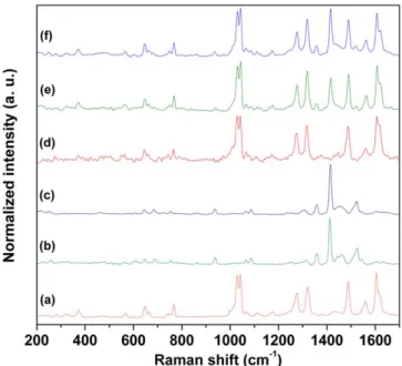
Figure 1.Raman scattering spectra of(a) RuL1, (b) YbL3, (c) NdL3, (d) SiO2 Ru,(e) SiO2 RuYb and(f) SiO2 RuNd.
Physics) producing 5 ps FWHM pulses ranging from 0.5 to 8.0 MHz repetition rate, regulated by the 3980 Spectra Phy-sics pulse picker. The laser was tuned to give output at 892 nm and a second harmonic generator BBO crystal (GWN-23PL Spectra Physics) gave the 448 nm excitation pulses that were directed to an Edinburgh FL900 spectrometer adjusted in L-format configuration. The solid samples were placed in a holder perpendicular to the excitation source. The emission wavelength at 610 nm was selected by a mono-chromator and emitted photons were detected by a cooled Hamamatsu R3809U microchannel plate photomultiplier. The whole instrument response function was typically 100 ps. Energy transfer rate constant(kEnT) and efficiency of energy transfer(ηEnT) were obtained using ruthenium3MLCT decay values from the SiO2–Ru (used as a standard) and the SiO2–RuNd and SiO2 RuYb nanohybrids.
2.3. Synthesis
Two types of luminescent nanohybrids were obtained. First, by grafting of one complex (RuL1) onto MSNs (labeled SiO2–Ru) and second, by simultaneously grafting of two complexes (RuL1/NdL3 or RuL1/YbL3) onto MSNs (labeled SiO2–RuNd and SiO2 RuYb), schemes 1(b)
and (c).
2.3.1. SiO2–Ru nanohybrid. 0.32 mmol(285 mg) of RuL1
complex was dissolved in ethanol (10 ml) and reacted with (285 mg) MSNs. The mixtures were stirred for 72 h at 295 K in N2 atmosphere. The resulting suspensions were dialyzed for 72 h, and the solids were isolated by centrifugation at 15 000 rpm for 15 min The SiO2 Ru obtained was washed with water, ethanol and diethyl ether. All solids obtained were dried under vacuum for 4 h. SiO2 Ru: Elemental analysis %, found(calcd): R = 0.18 mmol g 1: C, 10.51(8.98); H, 1.29 (0.90); N, 1.05 (1.05).
2.3.2. SiO2–RuNd and SiO2–RuYb nanohybrids. 0.20 mmol
(178 mg) of RuL1 complex, 0.20 mmol (260 mg) of NdL3 complex and 0.20 mmol (289 mg) of YbL3 complex were dissolved in ethanol (20 ml) and reacted with (450 mg) MSNs. The mixtures were stirred for 72 h at 295 K in an N2 atmosphere. The resulting suspensions were dialyzed for 72 h, and the solids were isolated by centrifugation at 15 000 rpm for 15 min The obtained solids were washed with water, ethanol, dichloromethane, and diethyl ether and then dried under vacuum for 4 h. SiO2 RuNd: Elemental analysis %, found (calcd): RRu= 0.14 mmol g 1and RNd= 0.16 mmol g 1: C, 15.34 (14.65); H, 1.74 (1.54); N, 1.32 (1.32); S, 1.53 (1.53). SiO2 RuYb: Elemental analysis %, found (calcd): RRu= 0.11 mmol g 1 and
Figure 2.13C{1H}CP MAS NMR (I) and29Si{1H}CP MAS NMR spectra (II) of (a) SiO2 Ru,(b) SiO2 RuYb and(c) SiO2 RuNd. 12: carbon atoms in methoxysilyl groups not grafted(Described the numbers in red and blue).
Figure 3.Nitrogen adsorption/desorption isotherms of (a) MSNs and(b) SiO2 Ru,(c) SiO2 RuYb and(d) SiO2 RuNd nanohybrids.
RYb= 0.15 mmol g 1
: C, 15.52(12.67); H, 1.74 (1.34); N, 1.05(1.05); S, 1.41 (1.41).
3. Results and discussion
3.1. Characterization of the silica-based nanohybrids
RuL1 complex (scheme 1(a, i)) and LnL3 (Ln = Nd(III),
Yb(III)) complexes are displayed in schemes1(a, i) and (a, ii),
respectively. The silica-based nanohybrids labeled SiO2–Ru and SiO2–RuLn were successfully prepared as shown in schemes1(b) and (c), respectively.
FT-RAMAN measurements show characteristic bands ascribed to the complexes and bands relating to Ln O and Ru N bonds. Figure 1 displays Raman scattering similar spectra obtained for SiO2–Ru (figure 1(a)), SiO2–RuYb
(figure1(b)) and SiO2–RuNd (figure1(c)) nanohybrids
The characteristic bands of the RuL1 and LnL3 com-plexes were detected also in the nanohybrids as displayed in figure1. Bands in the 640 685 cm 1region were assigned to ν(Ln O) and ν(Ru N) from LnL3 and RuL1 complexes, respectively [38 41]. Additionally, at 372 and 1030 1045
cm 1 bands attributed to ν(Ru N) were detected [40, 41].
Bands observed at 1605 1629 cm 1were ascribed toν(C=N) and at 1490 and 1560 cm 1assigned toν(C=C) present in the RuL1 complex. Bands relating to LnL3 complexes were found at 1357 and 1415 cm 1corresponding toν(C=O and C=C) [38,39]. These results corroborate DRIFT analysis (not
show) confirming RuL1 and LnL3 complexes grafted onto MSNs.
The chemical integrity of the complexes grafted onto MSNs was verified by13C{1H}CP-MAS NMR spectroscopy. Figure2(I) shows13C{1H}CP-MAS spectra for the nanohy-brids obtained by grafting of RuL1 and LnL3 complexes. The spectrum ascribed to the SiO2 Ru nanohybrid (figure 2(I, a)) shows signals of carbon atoms of the RuL1
complexes grafted in the silica matrix. Signals at 125, 139, 151 and 157 ppm were attributed to the carbon atoms of the bpy ligands, CIII-CV, CIV, CVI and CII, respectively [36].
The signals ascribed to the carbon atoms of the propyl chain were observed with low intensity at 10 (C11), 22 (C10 and C7), 55 (C9) and 58 (C8) ppm [36]. It is worth noting that the
characteristic signals expected from TTA ligands were not observed in the figures 2(I, b and c). The low sensibility in
both spectra (figures 2(I, b and c)) can be ascribed to the Figure 4.HR TEM images of(a) MSNs and different nanohybrids: (b) SiO2 Ru,(c) SiO2 RuYb and(d) SiO2 RuNd.
paramagnetic interactions, compromising the efficiency of the cross-polarization process owing to very short 1H spin-lattice relaxation times in the rotating frame [42]. For the
SiO2 RuNd and SiO2–RuYb nanohybrids (figures2(I, b and
c)), only signals ascribed to the bpy ligands, ethoxysilyl and methoxysilyl groups, present in the ruthenium and lanthanide complexes could be observed. The spectra were similar for both nanohybrids with signals at 125, 139, 151 and 157 ppm assigned to the carbon atoms of the bpy ligands, CIII-CV, CIV, CVI and CII, respectively, for the ruthenium complexes. Signals at 10, 22, 55 and 58 ppm, C11, C10-C7, C9 and C8, respectively, were attributed to the ethoxysilyl groups present in the RuL1 complexes. The signals at 47, 26 and 10 ppm were ascribed to the carbon atoms of the methoxysilyl groups, C9, C10 and C11, respectively, present in the LnL3 com-plexes. Methoxysilyl groups not grafted were detected in figures2 (I, c) and assigned to C12.
Figure3shows nitrogen adsorption/desorption isotherms from MSNs, SiO2 Ru, SiO2–RuYb and SiO2 RuNd sam-ples that exhibits a type IV shape, which is characteristic of mesoporous materials [35]. Values of specific surface area
(SBET) of the SiO2–Ru nanohybrids (SBET= 654 m2 g 1)
decreased relating to the MSNs (SBET= 675 m2g 1). For the SiO2–RuYb and SiO2–RuNd, these values of SBETdecreased significantly with 348 and 268 m2g 1, respectively, con-firming that the nanopores are filled up with complexes.
HR-TEM measurements were carried out to evaluate the morphology and nanopores structure after grafting of the silylated complexes. Figure 4 shows HR-TEM images of the MSNs (a) and SiO2–Ru (b), SiO2–RuYb (c) and SiO2–RuNd (d) nanohybrids. The MSNs (figure 4(a))
dis-plays a random distribution of the nanopores clearly observed as previously described[37]. However, according to the
HR-TEM images, all nanohybrids present spherical morphology without changing of the structure besides show the nanopores filled up, confirming the most of complexes are grafting inside de nanopores. These results corroborate DRIFT, FT-RAMAN, N2 adsorption/desorption and solid-state NMR analysis exhibiting the presence of the complexes inside de nanopores of the matrices.
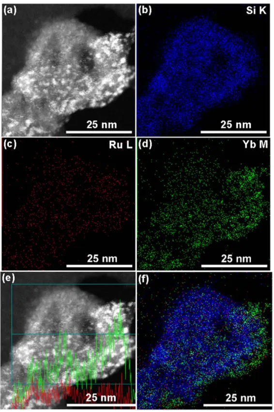
In STEM measurements, atoms with high electron den-sity as ruthenium, neodymium and ytterbium can be easily distinguished from Si atoms. The elemental mapping were obtained by EDX spectroscopy on STEM microscopy, and
Figure 5.Electron microscope images and elemental mapping of the SiO2 Ru:(a) STEM image, (b) Si mapping, (c) Ru mapping and (d) Si (blue color) and Ru (red color) mapping.
the obtained results for SiO2 Ru, SiO2–RuYb and SiO2 RuNd are shown in figures5 7
SiO2–Ru nanohybrid kept the spherical shape. Elemental mapping of Si atoms confirm that these elements are homo-genously dispersed to form the MSNs. Ru atoms are also detected and its appear homogenously well dispersed inside the
nanopores. Grafting efficiency observed by STEM are in agreement with the EA data(SiO2–Ru: 0.18 mmol g 1of silica). SiO2–RuYb and SiO2–RuNd nanohybrids displayed grafting efficiencies higher than SiO2–Ru nanohybrid dis-cussed above due to the grafting of two types of complexes onto MSNs. Concerning SiO2 RuYb and SiO2 RuNd
Figure 6.Electron microscope images and elemental mapping of the SiO2 RuYb:(a) STEM image, (b) Si mapping, (c) Ru mapping, (d) Yb mapping,(e) Si (blue color), Ru (red color) and Yb (green color) mapping. (f) STEM image with a line profile of Ru (red line) and Yb (green line) atoms in the selected area (blue line).
nanohybrids, elemental mapping of Si atoms confirms that these elements are homogenously dispersed to form the MSNs. Figure 6 exhibits Ru and Yb atoms appear hetero-geneously distributed inside and at the surface of the MSNs nanopores. The higher Yb concentration compared with Ru
atoms at the nanoparticles surface is in agreement with the EA data(SiO2 RuYb: 0.11 and 0.15 mmol g 1of silica using N and S contents for the calculation, respectively).
On the other hand, concerning SiO2–RuNd, Ru and Nd atoms are homogenously dispersed inside the nanopores
Figure 7.Electron microscope images and elemental mapping of the SiO2 RuNd:(a) STEM image, (b) Si mapping, (c) Ru mapping, (d) Nd mapping and(e) Si (blue color), Ru (red color) and Nd (green color) mapping. (f) STEM image with a line profile of Ru (red line) and Nd (green line) atoms in the selected area (blue line).
(figure7). STEM data confirm grafting efficiencies as obtained
by EA (SiO2–RuNd: 0.14 and 0.16 mmol g 1of silica, cal-culated from Ru(II) and Nd(III) moieties, respectively).
The grafting of Ru(II) and Ln(III) complexes (in mmol of complex g 1of silica) was calculated according to the N and S contents, respectively, as described by Menu et al [32, 33, 36]. The number of complexes nm2 of silica were calculated using grafting efficiency, Avogadro number and
specific surface area values of the matrix before grafting of the complexes as exhibited in table1.
The grafting efficiencies using MSNs with 765 m2g 1of silica were higher for SiO2 RuYb and SiO2–RuNd than for SiO2 Ru nanohybrid. Additionally, 0.09 0.16 complexes per nm2were grafted onto MSNs. Despite similar concentration of RuL1 and LnL3 complexes added for grafting reactions, EA results display more grafted LnL3 than RuL1, for both SiO2–RuYb and SiO2–RuNd nanohybrids.
3.2. Photophysical properties of the silica-based nanohybrids
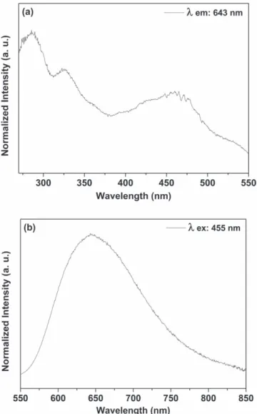
Figure 8 shows the excitation (a) and emission (b) spectra from SiO2–Ru nanohybrid. Bands at 285, 325 and 455 nm are observed in the excitation spectrum related to transitions centered on the ligand (π → π* transitions) on the metal (πM→ σ*
M transitions) and 1MLCT (d → π* transitions), respectively [35]. The characteristic emission band of
SiO2 Ru nanohybrid centered at 643 nm was ascribed to the transition from the3MLCT excited state to the ground state [35]. The blue shift compared to the free RuL1 complex is
related to the rigido-chromism phenomenon[43,44] due the
interaction between the complexes and the silanol groups of the rigid network avoid the spatial reorientation of complex. When RuL1 complex was grafted onto mesoporous surface, the solvent present into the pores was unable to reorient around the excited complex. As a consequence, the Franck Condon excited state was not completely stabilized, and emission occurred from a higher energy level than free complex[35]. Luminescence spectra from RuL1 (figure S1),
YbL3 (figure S2) and NdL3 (figure S3) complexes are exhibited in the supplementary material.
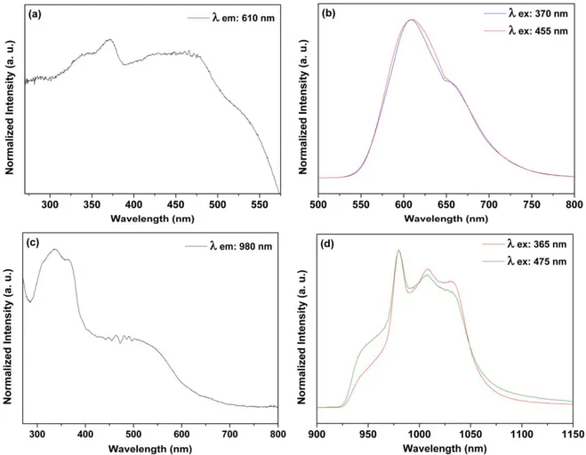
Figure9shows excitation and emission spectra obtained from SiO2 RuYb nanohybrid. A broad emission band was observed at 610 nm and assigned to the Ru(II) 3MLCT emission [35] in a similar way observed for SiO2–Ru
(figure 8). Comparing with free RuL1 complex, the3MLCT energy was blue shifted as previously described(SiO2–Ru in the figure8). The excitation spectra obtained by monitoring
the Ru(II) 3MLCT emission shows bands ascribed to the d → π* 1MLCT transitions, transitions centered on ligands (π → π* transitions) and transitions centered on the metal (πM→ σ*
M transitions), [35] in agreement with the free RuL1 complex.
The IR emission band peaking at 980 nm with broader components at 1010 and 1030 nm (figure 9(d)) could be
ascribed to the Yb(III)2F5/2→2F7/2transition[45 47]. The excitation spectrum (figure 9(c)) shows the broad ligand
centered band below 400 nm and the Ru(II) related band from 400 to 700 nm. Considering the excitation spectrum obtained by monitoring the Ru related visible emission, the sensitiza-tion of the Yb(III) IR emission is clearly shown here.
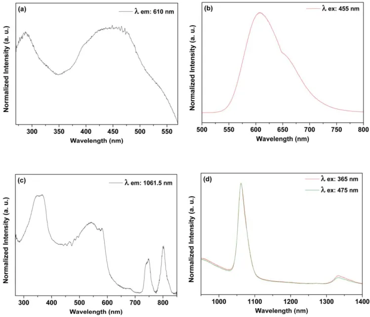
Figure 10 depicts results detected from SiO2–RuNd
nanohybrid. The Ru(II) related broad emission band is observed peaking at 610 nm (figure 10(b)). Excitation
spec-trum obtained by monitoring the broad emission bands shows the broad bands assigned to the ligands and Ru(II) in similar way obtained for the SiO2 RuYb.
Table 1.Grafting efficiencies, in mmol of Ru(II) and Ln(III) complexes g−1of silica, and amount of Ru(II) and Ln(III) complexes nm−2of silica for the nanohybrids.
Samples R(mmol g−1of SiO2)
Number of complex per nm2
RRu RLn Ru Ln
SiO2 Ru 0.18 0.16
SiO2 RuYb 0.11 0.15 0.09 0.13
SiO2 RuNd 0.14 0.16 0.12 0.14
Figure 8.Room temperature excitation((a), λemission= 643 nm) and emission((b), λexcitation= 455 nm) spectra of the SiO2 Ru nanohybrid in solid state.
Two emission bands were detected in the infrared region at 1061.5 nm(attributed to Nd(III)4F3/2→4I11/2transition) and at 1333 nm (Nd(III) 4F3/2→4I13/2 transition) [45 47].
The excitation spectrum obtained when monitoring emission at 1061.5 shows Nd(III) above 700 nm. Two broad bands are observed below 600 nm. In the same way observed for the SiO2–RuYb nanohybrid, here the Ru(II) to Nd(III) energy transfer is evidenced. The energy levels diagram presented in scheme2 illustrates the observed energy transfer processes.
Decay time results observed for the Ru(II) MLCT level are presented in table 2. Decreasing decay times in the pre-sence of lanthanide ions put in evidence Ru Ln energy transfer(EnT) processes.
Energy transfer rate of 0.11× 10⁷s 1and efficiency of energy transfer of 27.5% were obtained for SiO2–RuYb. EnT rate of 0.20× 10⁷s 1 and efficiency of energy transfer of 40% could be evaluated for SiO2–RuNd. The higher values observed for the Nd(III) hybrid is well explained by the matching of donor and acceptor energy levels as shown in scheme2.
Therefore, three new luminescent nanohybrids were prepared. The photophysical properties of SiO2 RuNd and SiO2–RuYb nanohybrids were evaluated and energy transfer processes were confirmed for lifetimes data obtained from Ru(II) emission decay and emission/excitation spectra.
4. Conclusion
We have fabricated three new luminescent silica-based nanohybrids by grafting of silylated Ru(II) and Nd/Yb(III) complexes onto MSNs. The silica matrix prepared by microemulsion method displayed interesting features for post modification such as high surface area with high porosity and silanol groups available, detected by HR-TEM, DRIFT, N2 adsorption/desorption analysis. Structural characterization of all nanohybrids were successfully achieved by using DRIFT, FT-RAMAN, SS-NMR, HR-TEM and STEM techniques, confirming the presence of the Ru(II) and Nd/Yb(III) inside the SiO2 nanopores. Luminescent properties were evaluated by monitoring the excitation on MLTC states from Ru(II) moieties. SiO2 Ru nanohybrid displays the characteristic emission band at 643 nm. For SiO2 RuNd and SiO2 RuYb nanohybrids, emissions in the visible correspond to the Ru(II) complexes (from the 3MLCT excited state) while the NIR emissions were assigned to the Nd(III) (4F3/2→4I11/2 and 4
F3/2→4I11/2transitions) and Yb(III) (2F5/2→2F7/2 trans-ition) ions. From Decay times of the Ru(II) moieties in the SiO2 RuNd and SiO2 RuYb nanohybrids, energy transfer processes were revealed with kEnTvalues of 0.20× 10⁷and 0.11× 10⁷s 1 and ηEnT of 40% and 27.5%, respectively. These results confirm Nd(III) levels more suitable for energy
Figure 9.Room temperature excitation((a), λemission: 610 nm and(c), λemission: 980 nm) and emission ((b), λexcitation: 370 and 455; and(d), λexcitation: 365 and 475 nm) spectra of the SiO2 RuYb nanohybrid in solid state.
transfer than Yb(III) from Ru(II) 3MLTC excited states. Finally, the photophysical investigation suggests the dual (NIR/visible)-emitting SiO2 RuNd and SiO2 RuYb nano-hybrids as potential nanolabels for biological assays.
Figure 10.Room temperature excitation((a), λemission: 610 nm and(c), λemission: 1061.5 nm) and emission ((b), λexcitation: 390 and 455 nm; and(d), λexcitation: 365 and 455 nm) spectra of the SiO2 RuNd nanohybrid in solid state.
Scheme 2.Schematic energy transfer processes(Ru(II)) to Nd(III) and Yb(III) (A = Absorption; ISC = Inter System Cross;
P= phosphorescence; EnT = energy transfer; F = Fluorescence) in the SiO2 Ru, SiO2 RuNd and SiO2 RuYb nanohybrids.
Table 2.Photophysical properties for MLCT based visible emission and Ln(III) based NIR.
MLCT based emission(Ex: 448 nm)
λ/nm τ/ns a kEnT/107s−1 bηEnT/% SiO2 Ru 610 344.7 SiO2 RuNd 610 206.2 0.20 40.2 SiO2 RuYb 610 249.8 0.11 27.5 a
kEnT(energy transfer rate constant) 1/τq 1/τu(τqandτurefer to
the‘quenched’ and ‘unquenched’ lifetime of Ru(II) complexes before and after coordination with Ln(III)).
b
Acknowledg ments
This work was supported by the Brazilian agencies FAPESP, CNPq, CAPES and CAPES-COFECUB Brazil-France coop eration program for grant to R M Sabio.
ORCID iDs
Rafael Miguel Sabio Cl!) https:fforcid.org/0000-0002-3852-2184
References
[1] Bunzli J C G and Piguet C 2005 Taking advantage of luminescent lanthanide ions Chem. Soc. Rev. 34 1048 77 [2] Carlos L D, Fecreira R A S, Becmudez V, de Z and Ribeiro S J L
20()() Lanthanide containing light emitting organic inorganic hybrids: a bet on the future Adv. Mater. 21 509 34 [3] Bünzli J C G 2015 On the design of highly luminescent
lanthanide complexes Coord. Chem. Rev. 293 294 19 47 [4] Bünzli J C G and Eliseeva S V 2010 Lanthanide NIR
luminescence for telecommunications, bioanalyses and solar energy conversion J. Rare Earths 28 824 42
[5] Huang P, Zheng W, Zhou S, Tu D, Chen Z, Zhu H, Li R, Ma E, Huang M and Chen X 2014 Lanthanide doped
LiLuF4 upconversion nanoprobes for the detection of
disease biomarkers Angew. Chem., /nt. Ed. 53 1252 7 [6] Ma Q, Wang J, Li Z, Lv X, Liang L and Yuan Q 2019 Recent
progress in lime resolved biosensing and bioimaging based on lanthanide doped nanoparticles Small 1804969
[7] Bünzli J C G 2016 Lanthanide light for biology and medical diagnosis J. Lumin. 170 866 78
[8] Clough T J, Jiang L, Wong K L and Long N J 2019 Ligand design strategies to increase stability of gadolinium based magnetic resonance imaging contrast agents Nat Commun. 10 1420
[9] Sun X et al 2019 Noninvasive temperature monitoring for dual modal tumor therapy based on lanthanide doped up conversion nanocomposites Biomaterials 201 42 52 [10] Feng Y, Wu Y, Zuo J, Tu L, Que I, Chang Y, Cruz L J,
Chan A and Zhang H 2019 Assembly of upconversion nanophotosensitizer in vivo to achieve scatheless real lime imaging and selective photodynamic therapy Biomaterials
201 33 41
[11] Hamblin M R and Demidova T N 2006 Mechanisms of low level light Proc. SPIE 6140 614001
[12] Chen F F, Chen Z Q, Bian Z Q and Huang CH 2010 Sensitized luminescence from lanthanides in d f bimetallic complexes Coord. Chem. Rev. 254 991 1010
[13] Wang P, Zakeeruddin SM, Moser JE, Nazeeruddin M K, Sekiguchi T and Griitzel M 2003 A stable quasi solid state dye sensitized solar cell with an amphiphilic ruthenium sensitizer and polymer gel electrolyte Nat. Mater. 2 402 7 [14] Gill M R and Thomas J A 2012 Ruthenium(ü) polypyridyl
complexes and DNA from structural probes to cellular imaging and therapeutics Chem. Soc. Rev. 41 3179
[15] Gill M R, Garcia Lara J, Foster S J, Smythe C, Battaglia G and Thomas J A 2009 A ruthenium(II) polypyridyl complex for
direct imaging ofDNA structure in living cells Nat. Chem 1
662 7
[16] Famey E P, Chapman S J, Sworos WB, Torelli MD, Hamers R J and Yoon T P 2019 Discovery and elucidation
of counteranion dependence in photoredox catalysis J. Am. Chem. Soc. 141 6385 91
[17] Albani B A, Peiia B, Leed N A, de Paula N A B G, Pavani C, Baptista M S, Dunbar K R and Turro C 2014 Marked
improvement in photoinduced cell death by a new tris heteroleptic complex with dual action: single! oxygen sensitization and ligand dissociation J. Am. Chem. Soc. 136 17095 101
[18] Lazarides T, Adams H, Sykes D, Faulkner S, Calogero G and Ward MD 2008 Heteronuclear bipyrimidine bridged Ru Ln and Os Ln dyads: low energy 3 MLCT states as energy donors to Yb(üi) and Nd(üi) Dalton Trans. 0 691 8 [19] Lazarides T, Sykes D, Faulkner S, Barbieà A and Ward MD
2008 On the mechanism of d f energy transfer in Rull/Lnlll and Osll/Lnlll dyads: dexter type energy transfer over a distance of 20 À Chemistry Eur. J. 14 9389 99
[20] Lazarides T, Tart N M, Sykes D, Faulkner S, Barbieri A and Ward MD 2009 [Ru(bipybh+ and [Os(bipybh+
chromophores as sensitisers for near infrared luminescence from Yb(üi) and Nd(üi) in d/f dyads: contributions from Forster dexter, and redox based energy transfer mechanisms
Dalton Trans. 3971 9
[21] Waro MD 2007 Transition metal sensitised near infrared luminescence from lanthanides in d f heteronuclear arrays Coord. Chem. Rev. 251 1663 77
[22] Waro MD 2010 Mechanisms of sensitization of lanthanide (III) based luminescence in transition metal/lanthanide and anthracene/lanthanide dyads Coord. Chem Rev. 254
2634 42
[23] Zhang L Y, Hou Y J, Pan M, Chen L, Zhu Y X, Yin S Y, Shao G and Su C Y 2015 Near infrared (NIR) emitting Nd/ Yb(lll) complexes sensitized by MLCT states of Ru(ll)/Ir (III) metalloligands in the visible light region Dalton Trans.
44 15212 9
[24] Wei Q H, Lei Y F, Xu W R, Xie J M and Chen G N 2012 Ru
(ü) sensitized lanthanide luminescence: synthesis, photophysical properties, and near infrared luminescent
detennination of alpha fetal protein (AFP) Dalton Trans. 41
11219
[25] Singaravadivel S, Babu E, Velayudham M, Lu K L and Rajagopal S 2013 Sensitized near infrared luminescence of Ndlll, Ybill and Erill complexes by energy transfer from a ruthenium antenna J. Organomet. Chem. 738 49 54 [26]Matuà F et al 2019 Luminescent mesoporous silica nanohybrid
based on drug derivative terbium complex Materials 12 933
[27] Lu J, Liong M, Li Z, Zink J I and Tamanoi F 2010
Biocompatibility, biodistribution, and drug delivery efficiency of mesoporous silica nanoparticles for cancer
therapy in animais Small 6 1794 805
[28] Sabio R M, Meneguin A B, Ribeiro T C, Silva R R and
Chorilli M 2019 New insights towards mesoporous silica nanoparticles as a technological platform for
chemotherapeutic drugs delivery /nt. J. Pharm. 564 379 409
[29] Rocha L A, Do J, Freiria C, Mauricio J, Caiut A, Ribeiro S J L, Messaddeq Y, Verelst M and Dexpert Ghys J 2015
Luminescence properties of Eu complex formations into ordered mesoporous silica particles obtained by the spray pyrolysis process Nanotechnology 26 335604
[30] Rocha L A, Caiut J M A, Messaddeq Y, Ribeiro S J L, Martines M AU, Freiria J, do C, Dexpert Ghys J and
Verelst M 2010 Non leachable highly luminescent oroered mesoporous SiO2 spherical particles Nanotechnology 21 155603
[31] Cousinié S, Gressier M, Reber C, Dexpert Ghys J and Menu M J 2008 Europium(lll) complexes containing
organosilyldipyridine ligands grafted on silica nanoparticles Langmuir 24 6208 14
[32] Cousinié S, Mauline L, Gressier M, Kandibanda S R, Datas L, Reber C and Menu M J 2012 Bulk or swface grafted silylated Ru(ü) complexes on silica as luminescent nanomaterials New J. Chem. 36 1355
[33] Duarte A P, Gressier M, Menu M J, Dexpert Ghys J, Caiut J M A and Ribeiro S J L 2012 Structural and luminescence properties of silica based hybrids containing new silylated diketonato Europium(III) complex J. Phys. Chem. C116 505 15
[34] Duarte A P et al 2013 Organosilylated complex [Eu(TTA)3 (Bpy Si)]: a bifunctional moiety for the engeneering of luminescent silica based nanoparticles for bioimaging Langmuir29 5878 88
[35] Sábio R M, Gressier M, Caiut J M A, Menu M J and Ribeiro S J L 2016 Luminescent multifunctional hybrids obtained by grafting of ruthenium complexes on mesoporous silica Mater. Lett.174 1 5
[36] Mauline L, Gressier M, Roques C, Hammer P, Ribeiro S J L, Caiut J M A and Menu M J 2013 Bifunctional silica nanoparticles for the exploration of biofilms of Pseudomonas aeruginosa Biofouling29 775 88 [37] Nandiyanto A B D, Kim S G, Iskandar F and Okuyama K
2009 Synthesis of spherical mesoporous silica nanoparticles with nanometer size controllable pores and outer diameters Microporous Mesoporous Mater.120 447 53
[38] Malta O L, Brito H F, Menezes J F S, Silva F R G E, Alves S, Farias F S and de Andrade A V M 1997 Spectroscopic properties of a new light converting device
Eu(thenoyltrifluoroacetonate)3 2(dibenzyl sulfoxide). A theoretical analysis based on structural data obtained from a sparkle model J. Lumin.75 255 68
[39] Binnemans K 2009 Lanthanide based luminescent hybrid materials Chem. Rev.109 4283 374
[40] Ishida H, Tobita S, Hasegawa Y, Katoh R and Nozaki K 2010 Recent advances in instrumentation for absolute emission
quantum yield measurements Coord. Chem. Rev.254 2449 58
[41] Shavaleev N M, Pope S J A, Bell Z R, Faulkner S and Ward M D 2003 Visible light sensitisation of near infrared luminescence from Yb(iii) Nd(iii) and Er(iii) complexes of 3,6 bis(2 pyridyl)tetrazine Dalton Trans. 2003 808 14 [42] Ilibi M, de Queiroz T B, Ren J, De Cola L,
de Camargo A S S and Eckert H 2014 Luminescent hybrid materials based on covalent attachment of Eu(iii) tris (bipyridinedicarboxylate) in the mesoporous silica host MCM 41 Dalton Trans.43 8318
[43] Innocenzi P, Kozuka H and Yoko T 1997 Fluorescence properties of the Ru(bpy)32 + complex incorporated in sol gel derived silica coatingfilms J. Phys. Chem. B101 2285 91
[44] Matsui K and Momose F 1997 Luminescence properties of Tris (2,2′ bipyridine)ruthenium(II) in sol gel systems of SiO2 Chem. Mater.9 2588 91
[45] Sun L N, Zhang H J, Meng Q G, Liu F Y, Fu L S, Peng C Y, Yu J B, Zheng G L and Wang S B 2005 Near infrared luminescent hybrid materials doped with lanthanide(Ln) complexes(Ln = Nd, Yb) and their possible laser application J. Phys. Chem. B109 6174 82
[46] Feng J, Song S Y, Deng R P, Fan W Q and Zhang H J 2010 Novel multifunctional nanocomposites: magnetic mesoporous silica nanospheres covalently bonded with near infrared luminescent lanthanide complexes Langmuir26 3596 600 [47] Liu Y, Sun L, Liu J, Peng Y X, Ge X, Shi L and Huang W 2015 Multicolor(vis NIR) mesoporous silica nanospheres linked with lanthanide complexes using 2(5
bromothiophen)imidazo[4, 5 f][1, 10] phenanthroline for in vitro bioimaging Dalton Trans.44 237 46

![Figure 3 shows nitrogen adsorption /desorption isotherms from MSNs, SiO2 Ru, SiO2–RuYb and SiO2 RuNd sam-ples that exhibits a type IV shape, which is characteristic of mesoporous materials [ 35 ]](https://thumb-eu.123doks.com/thumbv2/123doknet/2964652.81829/7.892.167.730.101.664/nitrogen-adsorption-desorption-isotherms-exhibits-characteristic-mesoporous-materials.webp)

![DNA intercalating near-infrared luminescent lanthanide complexes containing dipyrido[3,2-a:2′,3′-c]phenazine (dppz) ligands : synthesis, crystal structures, stability, luminescence properties and CT-DNA interaction](data:image/gif;base64,R0lGODlhAQABAIAAAP///wAAACH5BAEAAAAALAAAAAABAAEAAAICRAEAOw==)