Open Archive Toulouse Archive Ouverte (OATAO)
OATAO is an open access repository that collects the work of some Toulouse
researchers and makes it freely available over the web where possible.
This is
an author's
version published in:
https://oatao.univ-toulouse.fr/23111
Official URL :
https://doi.org/10.1016/j.otsr.2018.04.026
To cite this version :
Any correspondence concerning this service should be sent to the repository administrator:
tech-oatao@listes-diff.inp-toulouse.fr
Dauzere, Florence and Delclaux, Stéphanie and Pham, Thuy Trang and Rongières, Michel and
Mansat, Pierre Combined median and ulnar nerve palsy complicating distal radius
fractures. (2018) Orthopaedics & Traumatology: Surgery & Research, 104 (6). 871-875. ISSN
1877-0568
OATAO
Combined
median and ulnar nerve palsy complicating distal
radius
fractures
Florence
Dauzere
a,
Stéphanie Delclaux
a,∗,
Thuy Trang Pham
b,
Michel Rongières
a, Pierre Mansat
aa Orthopaedics Department, Toulouse Purpan Teaching Hospital, Toulouse, France b Orthopaedics Pediatric Department, Toulouse Purpan Teaching Hospital, Toulouse, France
Keywords: Fracture Radius Palsy
a b s t r a c t
Background: Fractures of the distal radius only rarely give rise to complications in the immediate post-operative period. Combined median and ulnar nerve palsy is a complication that can be missed by the surgeon.
Materials and methods: Three cases diagnosed early after surgery are reported here. The patients were 15, 16, and 30 years of age, respectively. None had preoperative neurological deficits. The youngest patient was injured during sports and the other 2 patients during traffic accidents. All 3 patients had a displaced fracture of the distal radius combined with a fracture of the distal fourth of the ulna or ulnar styloid process and were treated by anterior plate fixation. Operative times were 47, 62, and 120 minutes, respectively. Compartment syndrome was ruled out based on low pain intensity and absence of forearm tightness to palpation.
Results: The electrophysiological study performed 1 month post-injury in all 3 patients showed severe impairments of both median and ulnar nerve function. Median and ulnar nerve release surgery was performed in the 15-year-old 6 weeks post-injury. No nerve damage or fibrosis was seen during the procedure. All patients recovered fully within 3 months and had normal findings from follow-up elec-trophysiology testing after 6 months.
Discussion: Combined median and ulnar nerve palsy has rarely been reported and is among the rare complications of distal radial fractures that can develop in the event of a high-energy trauma and/or major displacement. Both previously published data and our experience indicate that surgical nerve release is unnecessary. Clinical recovery within 3 months is the rule.
Level of evidence: IV, case-reports.
1. Introduction
Fractures of the distal radius are rarely accompanied with neu-rological complications. Median nerve injury has been reported in 5% to 7% of cases. Ulnar nerve injury is far more uncommon, with 2% of 330 articular fractures of the distal radius studied by Melone[1]
and a single case (0.05%) among 2000 distal radial fractures studied by Bacorn and Kurtzke[2]. Combined damage to the median and ulnar nerve is an exceedingly rare event for which few published case-series studies are available and no pathophysiological mech-anism has been established. Isolated ulnar nerve palsy is usually ascribed primarily to contusion.
∗ Corresponding author. Orthopédie, CHU de Toulouse Purpan, TSA 40031, place du Dr Baylac, 31059 Toulouse cedex 9, France.
E-mail address:stephanie.delclaux@laposte.net(S. Delclaux).
Here, 3 patients with combined median and ulnar nerve palsy after a fracture of the distal radius are described. All 3 patients were managed over a period of only 1 year, suggesting that this complication may be less uncommon than believed.
The objective of this study was to describe combined median and ulnar nerve injury after a distal radius fracture and to suggest a treatment strategy for this rare but dramatic complication. Surgical nerve release is not universally recommended in the literature. The severity of the clinical impairments requires a standardised man-agement strategy, particularly as the patients are often severely distressed by their symptoms for which they frequently demand revision surgery.
2. Material and method
Three patients with combined median and ulnar nerve palsy diagnosed shortly after surgery for distal radius fractures were seen
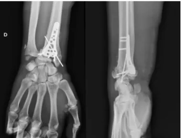
Fig. 1. Patient #1, antero-posterior and lateral radiographs before surgery.
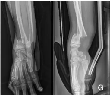
Fig. 2. Patient #2, antero-posterior and lateral radiographs before surgery.
in 2014 at a single orthopaedic and trauma surgery department in a university hospital in Toulouse, France. The 3 patients were male and were aged 15, 16, and 30 years. The fractures were consistently due to high-energy trauma, sustained during sports in 1 patient and during traffic accidents in the other 2 patients.
In the AO classification, the fracture of the distal radius was 23-A3 in 2 patients (Figs. 1 and 2) and 23-C3 in 1 patient (Fig. 3). Either the distal fourth of the ulna or the ulnar styloid process was fractured also. In 1 patient, Gustilo grade 1 skin breakage was present at the dorsal aspect of the forearm.
The patients were first seen at the trauma emergency depart-ment of the university hospital, where they were examined by an orthopaedic surgery resident. No neurological deficits were found. Closed reduction with equimolar nitrous oxide/oxygen for analge-sia was performed at the emergency department in 2 patients. No radiographs were obtained after reduction. All 3 patients had their fracture immobilised in a long arm splint while waiting for surgery, which was performed 12 h, 13 h, and 6 h post-injury, respectively.
Fig. 3. Patient #3, antero-posterior and lateral radiographs before surgery.
Fig. 4. Patient #1, antero-posterior and lateral radiographs after surgery.
Surgery was performed by a senior hand surgeon under local anaesthesia and short-duration axillary brachial plexus block. A pneumatic tourniquet inflated to 250 mmHg was placed at the root of the limb in all 3 patients. Operative time was 47, 62, and 120 minutes, respectively.
2.1. Operative technique
After inflation of the pneumatic tourniquet at the root of the limb, the volar approach to the radius described by Henry was performed in all 3 patients. In 1 patient, reduction of a displaced articular dorsal fragment required an additional dorsal approach with incision of the extensor retinaculum between the third and fourth compartments. Side-to-side translation was reduced using Muller’s forceps combined with external manipulation. Fixation was with an anterior plate (Fig. 4) combined, in 2 patients, with lateral pinning of the radial styloid (Figs. 5 and 6).
The ulnar styloid process was fractured in 1 patient. The distal radio-ulnar joint was stable upon testing at the end of the procedure, indicating that specific treatment of the ulnar styloid fracture was unnecessary. Closed reduction to treat the fractures of the distal fourth of the ulna in the other 2 patients was successful and stable. Neither the median nerve nor the ulnar nerve was
Fig. 5. Patient #3, antero-posterior and lateral radiographs after surgery.
Fig. 6. Patient #2, antero-posterior and lateral radiographs after surgery.
visualised during surgery. At the end of the procedure, the skin incision was sutured by separate stitches with no subcutaneous suturing or drainage.
2.2. Postoperative care
All 3 patients were admitted for 48 h in the orthopaedics surgery department. Immobilisation was with a long arm plaster splint with the wrist in the neutral position. The patients attended follow-up visits including a physical examination and radiographs on days 1, 15, 45, 90, and 180 after surgery. Electrophysiological studies were performed after 30 and 180 days.
3. Results
The day-1 postoperative visit performed after dissipation of all effects of the local anaesthesia and nerve block showed combined distal median and ulnar nerve palsy manifesting as impairments in flexion of the metacarpo-phalangeal joints of all five digits, extension of the inter-phalangeal joints of digits 2 through 5, and opposition of the thumb. Flexion of the inter-phalangeal joints of
all 5 digits was unimpaired. Hypaesthesia was noted at the volar aspect of the fingers. All distal pulses were normal to palpation.
Compartment syndrome was ruled out based on the absence of tightness to palpation and of severe pain upon mobilisation of the extended rays. No significant haematoma was noted postop-eratively. Additional immobilisation at night in the intrinsic plus position was used. However, 2 weeks after surgery the median and ulnar nerve palsies persisted, with claw deformity of digits 2 through 5. Healing was complete, with no discharge or dehiscence. A diagnostic electrophysiological study was performed about 1 month after surgery. The results consistently showed combined motor and sensory impairment of the median and ulnar nerves, with diminished amplitudes of the sensory and motor responses and axonal loss. In the youngest patient, the ulnar nerve abnormal-ities were classified as severe (Table 1).
Six weeks after surgery, an improvement in the neurological deficit was noted in 2 patients. Fracture healing was satisfactory in all 3 patients. Surgical exploration with release of the median and ulnar nerves performed 6 weeks after the injury in 1 patient showed no gross nerve damage or entrapment within the fracture site or scar tissue.
Three months postoperatively, the physical examination showed nearly full recovery of sensory and motor function of the median and ulnar nerves. Clinical recovery was similar for both nerves in all 3 patients.
After 6 months, no sensory or motor deficit was detectable clinically in the distributions of the median and ulnar nerves. A follow-up electrophysiological study showed complete nerve func-tion recovery with normal conducfunc-tion velocities and latencies in 2 patients. The youngest patient had persistent axonal loss confined to the ulnar nerve, with improvements in the amplitudes of the motor and sensory responses (Table 2).
4. Discussion
Combined median and ulnar nerve injury is an extraordinarily rare complication of distal radial fractures of which few reports are available in the literature. In 1968, Siegel and Weiden[3]described 2 cases in patients with fractures of the distal fourth of both fore-arm bones. Surgical exploration showed no evidence of contusion, dilaceration, or other gross abnormalities. The neurological deficits were detected preoperatively. Similarly, Rychak and Kalenak[4]
reported a case in 1977 related to a displaced fracture of the dis-tal radius combined with a fracture of the ulnar styloid process. The neurological status before surgery was not specified. Surgical exploration 3 weeks post-injury evidenced gross contusion of the median and ulnar nerves. Kumar[5]reported a similar case, with no information on the preoperative neurological status. No con-tusion or gross nerve damage was found by surgical exploration. All these patients were young males and, when available, infor-mation on the injury consistently indicated a high-energy trauma. Our patients were also young males who sustained high-energy traumas (Table 3). However, none had preoperative evidence of nerve damage. Compartment syndrome was routinely ruled out by a senior surgeon, both intra-operatively and at the postoperative visit, based on absence of tightness to palpation. Fracture fixation was consistently performed by a senior hand surgeon.
Several hypotheses have been put forward to explain combined median and ulnar nerve injury related to distal radial fractures. In a discussion of a clinical case, Pazart et al.[6](Fig. 7) suggested that ulnar nerve injury might be caused by major dorsal and radial displacement of the distal fragment. The ulnar nerve is tethered to Guyon’s canal and therefore susceptible to severe stretching by dis-placed bone fragments. During surgery in this patient[6], the proxi-mal ulnar nerve was found to be severed at Guyon’s canal. However,
Table 1
Results of the electrophysiological study 1 month post-injury in patient #1.
Motor conduction velocity (CV)
Nerve Latency Amplitude CV
ms mV % m/s
Ulnar, cm, 1st IO motor, left
Wrist, 1st IO No response
Median, motor, left
Wrist, abductor pollicis brevis 4.04 2.2
Median, motor, right
Wrist, abductor pollicis brevis 3.94 5.3
Ulnar, abductor digiti minimi, motor, left
Wrist, adductor digiti minimi 2.71 0.19
Sensory conduction velocity (CV)
Nerve Latency Amplitude CV
ms V m/s
Median, sensory, left
Wrist, dig II 2.06 21.6 65.5
Median, sensory, right
Wrist, dig II 2.25 31.6 58.7
Radial, sensory, left
Forearm, thenar 1.56 20.0 –
Radial, sensory, right
Forearm, thenar 1.71 25.5 –
Ulnar, sensory, left
Wrist, dig V 2.38 2.4 50.4
Ulnar, sensory, right
Wrist, dig V 2.15 21.9 55.8
Table 2
Results of the electrophysiological study 6 months post-injury in patient #1. Motor conduction velocity (CV)
Nerve Latency Amplitude CV
ms mV % m/s
Ulnar, cm, 1st IO motor, left
Wrist, 1st IO 6.97 1.05
Median, motor, left
Wrist, abductor pollicis brevis 3.61 5.0
Elbow-wrist 8.50 5.0 0 53.2
Sensory conduction velocity (CV)
Nerve Latency Amplitude CV
ms V m/s
Median, sensory, left
Wrist, dig II 2.41 21.5 57.3
Ulnar, sensory, left
Wrist, dig V 2.14 5.7 55.1
Table 3
Features of the 3 patients with combined median and ulnar nerve palsies.
Patient # Age, years AO Classification Open fracture Ulnar fracture Mechanism Fixation
1 15 23-A3 No Metaphyseal Traffic accident Anterior plate
2 16 23-A3 Yes Metaphyseal Sports injury Anterior plate plus pinning
3 30 23-C3 No Styloid Traffic accident Anterior plate plus pinning
in cadaver studies, Clarke and Spencer[7]found that mobility at the wrist was greater for the ulnar than the median nerve. This greater mobility may act as a protective factor that may explain the very low frequency of ulnar nerve damage in patients with distal radius fractures. Clarke and Spencer therefore advocated routine nerve release surgery in the absence of recovery within 2–3 months.
More recently, Soong and Ring[8]argued that ulnar nerve dam-age in distal radial fractures was due, not to compression, but to stretching of the nerve tethered to Guyon’s canal. Thus, the ulnar nerve may be vulnerable to contusion and/or stretching during
high-energy traumas. In contrast, median nerve injuries may be chiefly induced by compression within the carpal tunnel. Soong and Ring therefore advocate surgical exploration and release of ulnar nerve palsy in patients with acute carpal tunnel syndrome or an open fracture. When neither of these factors is present, release can be considered if recovery does not occur within 3–6 months.
Of our 3 patients, only 1 underwent surgical nerve release, 6 weeks post-injury. No entrapment in scar tissue or other gross abnormalities were found. All 3 patients achieved a full recovery of
Fig. 7. Mechanism of ulnar nerve injury in distal radial fractures. From Pazart et al.
their nerve function, irrespective of whether surgical release was performed at a distance from the injury.
The site of the nerve lesions deserves discussion. Rubin et al.
[9]found injuries to the median and ulnar nerves at the elbow after distal radius fractures. The electrophysiological study showed that the median nerve was injured proximal to the motor branch for the pronator teres muscle and the ulnar nerve proximal to the branch for the flexor carpi ulnaris muscle. These data support injury by stretching during the accident due to the high-energy of the trauma, major displacement of the bone fragments, and tethering of the nerves at points proximal and distal to the fracture site. In our patients, however, the electrophysiological data suggested dis-tal nerve injury, as the electromyogram traces of the flexor carpi ulnaris and radialis muscles were normal.
Combined median and ulnar nerve palsy is an exceedingly rare complication of distal radius fractures. An iatrogenic mechanism is conceivable, with stretching of the nerves during surgery, pro-longed tourniquet inflation, inordinate pressure on the retractors, and/or excessive traction during reduction. Cadaver studies may shed light on the iatrogenic factors that may be involved.
The optimal management of combined median and ulnar nerve palsy in patients with distal radial fractures remains unclear. In patients without preoperative neurological deficits, we advise against nerve release at the acute phase and suggest that this procedure should be reserved for patients who do not recover within 3 months. In the reported cases, surgical exploration only rarely found nerve damage amenable to microsurgical suturing or entrapment in scar tissue warranting nerve release. In our patient managed with surgical exploration and release 6 weeks post-injury, no local causes of nerve dysfunction were identified. Finally, elec-trophysiological studies should be performed repeatedly to confirm the sensory and motor nerve dysfunction, identify the anatomic level of injury, and monitor nerve function recovery over time. The patient and family should be informed that recovery may take several months.
5. Conclusion
Combined median and ulnar nerve palsy related to distal frac-tures of the radius are exceedingly rare but nevertheless require a standardised management strategy. Published data support
stretching during the trauma as the mechanism of nerve injury. Three consistent epidemiological characteristics are young patient age, fracture displacement, and high-energy trauma. We advise against routine surgical exploration but suggest that nerve release deserves consideration in patients with no clinical evidence of recovery 3 months after the injury.
Disclosure of interest
The authors declare that they have no competing interest.
Funding
None.
Contribution
FD: data collection, literature review. SD: data collection and follow-up of patients. TTTP: follow-up of patients.
MR: literature review and help by writing.
PM: follow-up of department’s patients and help by writing.
References
[1]Melone CP. Articular fractures of the distal radius. Orthop Clin North Am 1984;15:217–36.
[2]Bacorn RW, Kurtzke JF. Colles’ fracture a study of two thousand cases from the New York State Workmen Compensation Board. J Bone Joint Surg Am 1953;35:643–58.
[3]Siegel RS, Weiden I. Combined median and ulnar nerve lesions complicating fractures of the distal radius and ulna: two case reports. J Trauma 1968;8: 1114–8.
[4]Rychak JS, Kalenak A. Injury to the median and ulnar nerves secondary to fracture of the radius: a case report. J Bone Joint Surg Am 1977;59:414–5.
[5]Kumar A. Median and ulnar nerve injury secondary to a comminuted Colles fracture. J Trauma 1990;30:118–9.
[6]Pazart F, Stindel E, Le Nen D. Fracture of the distal part of the radius associated with severed ulnar nerve. Ann Chir Main 1999;18:197–201.
[7]Clarke AC, Spencer RF. Ulnar nerve palsy following fractures of the distal radius: clinical and anatomical studies. J Hand Surg [Br] 1991;16:438–40.
[8]Soong M, Ring D. Ulnar nerve palsy associated with fracture of the distal radius. J Orthop Trauma 2007;21:113–6.
[9]Rubin M, Heise CW. Proximal neuropathy in Colles’ fracture. Can J Neurol Sci 1997;24:77–8.