VALVULAR HEART DISEASE (TL KIEFER, SECTION EDITOR)
Pulmonary Hypertension with Valvular Heart Disease: When to Treat
the Valve Disease and When to Treat the Pulmonary Hypertension
Christophe Martinez1&Toshimitsu Tsugu1 &Tadafumi Sugimoto2 &Patrizio Lancellotti1,3# Springer Science+Business Media, LLC, part of Springer Nature 2019
Abstract
Purpose of Review This article will review the current guidelines for therapeutic intervention in (pulmonary hypertension) PH related to left heart disease (PH-LHD).
Recent Findings The 6th World Symposium on Pulmonary Hypertension (WSPH) recommended that the mean pulmonary artery pressure (mPAP) should be lowered to 20 mmHg. In several randomized controlled trials performed in patients with PH-LHD, pulmonary arterial hypertension (PAH)–specific drug therapy demonstrated no evidence of beneficial effects. Furthermore, in the sildenafil for improving outcomes after valvular correction (SIOVAC) trial, the use of sildenafil in the context of PH post-valvular heart disease (VHD) intervention is associated with an increased risk of clinical deterioration and death. Therefore, medical therapy such as PAH-specific drugs is still not recommended in PH-LHD. The principle of PH-LHD therapy is the treatment of underlying VHD. It is crucial to undergo surgical intervention at an appropriate time prior the development of potentially irreversible PH. Stress echocardiography (SE) is helpful to define symptoms and can be useful to assess the systolic pulmonary artery pressure (SPAP) and stratify severity of VHD.
Summary This comprehensive review of the literature highlights the role of SE imaging to assess VHD and is needed for the asymptomatic patients with severe VHD or symptomatic patients with non-severe VHD in the context of PH-LHD. The focus of patient evaluation should be on identifying patients with significant underlying valvular heart disease and referring in a timely manner for VHD treatment per society guidelines as pharmacologic pulmonary vasodilator therapy for PH-LHD has not shown efficacy as seen in other forms of PH.
Keywords Valvular heart disease . Surgery . Stress echocardiography . Aortic stenosis . Mitral stenosis . Mitral regurgitation
Introduction
According to the guidelines of European Society of Cardiology (ESC)/European Respiratory Society (ERS), pul-monary hypertension (PH) is defined as an increase in mean pulmonary arterial pressure (mPAP) ≥ 25 mmHg at rest as assessed by right heart catheterization (RHC) [1]. Recently, it was suggested that the mPAP threshold should be lowered to 20 mmHg at the 6th World Symposium on Pulmonary Hypertension (WSPH) in Nice, 2018 [2]. This suggestion is based on data from healthy subjects in whom the normal mPAP at rest is 14 ± 3 mmHg with an upper limit of approx-imately 20 mmHg [3,4]. Two standard deviations above this mean value would indicate that a mPAP > 20 mmHg is the threshold for abnormal pulmonary arterial pressure (above the 97.5th percentile). However, mPAP of 20 mmHg alone is not sufficient to cause pulmonary vascular disease. Therefore, the task force has proposed including a pulmonary vascular resis-tance (PVR)≥ 3 Wood units (WU) into the definition [2]. The
This article is part of the Topical Collection onValvular Heart Disease * Patrizio Lancellotti plancellotti@chu.ulg.ac.be Christophe Martinez cmartinez@chu.ulg.ac.be Toshimitsu Tsugu tsugu917@gmail.com Tadafumi Sugimoto t_sugimoto_japan@hotmail.com 1
Department of Cardiology, University of Liège Hospital, GIGA Cardiovascular Sciences, CHU du Sart Tilman, Université de Liège, 4000 Liège, Belgium
2
Clinical Laboratory, Mie University Hospital, Mie, 2-174 Edobashi, Tsu, Japan
3 Gruppo Villa Maria Care and Research, Anthea Hospital, Bari, Italy https://doi.org/10.1007/s11886-019-1240-7
definition of PH may change in the future; we will consider it in line with the current guidelines and the 6th WSPH recommendations.
Mechanism of Development of Pulmonary
Hypertension Due to VHD
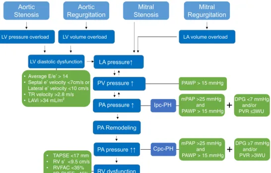
In severe valvular heart disease (VHD), a progressive increase in left ventricular (LV) filling pressure and left atrial (LA) pressure leads to a corresponding passive increase in pressure in the pulmonary veins and, subsequently, to isolated post-capillary PH (Ipc-PH), which can still be reversible at this stage. However, when pulmonary vasoconstriction or vascular remodeling ensues with Ipc-PH, PH may become irreversible and is called combined post-capillary PH (Cpc-PH). Cpc-PH occurs in 12 to 13% of patients with PH due to left heart disease (Fig.1).
Definition
In the ESC/ERS guidelines, PH related to left heart disease (PH-LHD) is classified into group 2 PH with subgroup 2.3 specifically dedicated to VHD. A definitive diagnosis of PH-LHD is made when the mPAP≥ 25 mmHg with a pulmonary artery wedge pressure (PAWP) > 15 mmHg in the context of significant VHD [1, 5]. Ipc-PH is defined as a diastolic
pulmonary pressure gradient (DPG: diastolic pulmonary ar-tery pressure minus mean PAWP) < 7 mmHg and/or PVR≤ 3 WU. An out-of-portion increase in the mPAP with respect to the PAWP suggestive of pulmonary vascular remodeling and constriction defines Cpc-PH (DPG is≥ 7 mmHg and/or PVR is > 3 WU) [1,5,6] (Fig.1).
In contrast, new defining criteria for the different hemody-namic types of PH that occur with LHD have been proposed by the 6th World Symposia on Pulmonary Hypertension (WSPH) [7•]. The proposed hemodynamic definition of PH-LHD by the WSPH is based only on hemodynamic right heart catheterization (RHC) values: (1) Ipc-PH: PAWP > 15 mmHg and mPAP > 20 mmHg and PVR < 3 WU; (2) Cpc-PH: PAWP > 15 mmHg and mPAP > 20 mmHg and PVR≥ 3 WU.
Diagnosis
Clinical signs and symptoms of PH-LHD are orthopnea and paroxysmal nocturnal dyspnea, which are generally not fea-tures of other types of PH [8]. Signs of right ventricular heart failure, such as peripheral edema, ascites, and syncope, de-pend on the severity of PH-LHD. These additional findings suggest the presence of PH-LHD: pulmonary vascular con-gestion, pleural effusion, and/or pulmonary edema on chest X-ray or computed tomography. In addition to the right ven-tricular overload findings, LV/LA hypertrophy is a distinctive sign on echocardiogram. RHC is mandatory for the
• Average E/e’ > 14 • Septal e’ velocity <7cm/s or Lateral e’ velocity <10 cm/s • TR velocity >2.8 m/s • LAVi >34 mL/m2 LV pressure overload LA pressure↑ PA Remodeling PA pressure ↑↑ Ipc-PH Mitral Regurgitation Aortic Stenosis Aortic Regurgitation Mitral Stenosis
LV volume overload LA volume overload
LV diastolic dysfunction Cpc-PH PA pressure ↑ PAWP > 15 mmHg DPG <7 mmHg and/or mPAP >25 mmHg and PAWP > 15 mmHg and/or mPAP >25 mmHg and PAWP > 15 mmHg RV dysfunction • TAPSE <17 mm • RV s’ <9.5 cm/s • RVFAC <35% • 3D-RVEF <45% PV pressure ↑
Fig. 1 Mechanisms for development of pulmonary hypertension due to valvular heart disease.LV left ventricular; LA left atrial; TR tricuspid regurgitation;LAVi left atrial volume index; TAPSE tricuspid annular plane excursion;RVFAC right ventricular fractional area change; 3D-RVEF three-dimensional 3D-RVEF; PV pulmonary vein; PA pulmonary
artery;RV right ventricular; Ipc-PH isolated post-capillary pulmonary hypertension;Cpc-PH combined pre- and post-capillary pulmonary hy-pertension;mPAP mean pulmonary artery pressure; PAWP pulmonary artery wedge pressure;DPG diastolic pulmonary gradient; PVR pulmo-nary vascular resistance;WU Wood units
confirmation of a PH diagnosis. Echocardiography also plays an important role not only for the diagnosis of PH but also for the identification of and characterization of VHD severity. A probability model for PH-LHD (low, intermediate, high) was published in the 6th WSPH (Table1) [7•]. This pre-test prob-ability matrix may aid in correct classification of PH-LHD for patients with suspected PH.
Resting Echocardiography
Transthoracic echocardiography (TTE) is the initial evaluation modality for patients with suspected PH of any etiology and is particularly informative for cases of suspected PH-LHD. TTE can estimate systolic pulmonary artery pressure (SPAP) non-invasively via Doppler ultrasound evaluation. The SPAP is the sum of right ventricular + right atrial pressure. The most com-monly used technique to estimate SPAP remains the direct measurement of peak tricuspid regurgitation jet velocity
which, when applied to the simplified Bernoulli equation (peak gradient = 4v2
), provides the estimated RV pressure. A tricuspid regurgitation jet velocity < 2.5 m/s is considered to be normal, 2.5–2.8 m/s as borderline, and > 2.8 m/s as strong-ly indicative for manifest PH. The right atrial pressure may be derived from the assessment of the inferior vena cava diameter (measured with the patient in the left decubitus position at 1.0 to 2.0 cm from the junction with the right atrium) and its respiratory changes [8]. In addition, it also provides visual assessment of the RV size and function related to potential PH. Furthermore, in cases of suspected PH-LHD, TTE can characterize the severity of valve lesions. The various valve pathologies will be discussed in greater detail below.
Stress Echocardiography
The clinical indications for stress echocardiography (SE) in the context of PH-LHD are as follows: severe VHD without symptoms, non-severe VHD with symptoms, and symptom-atic non-severe VHD with low flow [9–11]. Heart rate nor-mally increases two- to three-fold during exercise, contractil-ity three- to four-fold, and systolic blood pressure by≥ 50%, while systemic vascular resistance decreases. This often serves to reveal symptoms in patients previously considered asymptomatic or, for example, may demonstrate that mitral valve stenosis changes from moderate to severe with activity. Therefore, SE may be a useful diagnostic tool in selected cases of suspected PH-LHD leading to recommendations to treat the underlying VHD.
Therapy
There are three effective therapeutic options for PH-LHD: (1) surgical/interventional treatment; (2) non-specific pulmonary arterial hypertension (PAH) drugs; and (3) PAH-specific drugs. Indications for invasive therapy, surgery or transcathe-ter treatment, depend on the VHD etiology (see below). Some patients may benefit from non-specific vasodilators such as nitrates and hydralazine, although evidence supporting this approach is limited [12, 13]. PAH-specific drugs such as endothelin receptor antagonists and phosphodiesterase-5 in-hibitors have been reported to be effective in terms of im-provement in hemodynamics, exercise tolerance, and symp-toms, but these results did not provide enough evidence to support the use of these drugs [13]. The sildenafil for improv-ing outcomes after valvular correction (SIOVAC) trial was performed to determine whether treatment with sildenafil im-proved outcomes of patients with PH after correction of VHD. Patients underwent a successful surgical treatment (valve re-placement or repair) during the year before enrollment and were randomized to 40 mg sildenafil three times daily versus
Table 1 Probability of pulmonary hypertension related to LHD (Reproduced with permission of the © ERS 2019: European Respiratory Journal 53 (1) 1801897;https://doi.org/10.1183/13993003. 01897-2018Published 24 January 2019) [7•] Feature Low probability Intermediate probability High probability Age < 60 years 60–70 years > 70 years Obesity, systemic
hypertension, glucose
intolerance/diabetes
None 1–2 factors > 2 factors
Previous cardiac intervention*
No No Yes
Atrial fibrillation No Paroxysmal Current Structural LHD No No Present ECG Normal or signs of RV strain Mild LVH LBBB or LVH Echocardiography No LA dilatation; E/e′ < 13 No LA dilatation; grade < 2 mitral flow LA dilatation; grade > 2 mitral flow CPET HighV′E/V′
CO2slope; no EOV ElevatedV′ E/V′CO2 slope or EOV Mildly elevated VE/VCO2 slope; EOV CMR No left heart abnormali-ties LA strain or LA/RA > 1
*Coronary artery and/or valvular surgical and/or surgical producers, in-cluding percutaneous interventions
CPET cardiopulmonary exercise testing, CMR cardiac magnetic reso-nance,EOVexercise oscillatory ventilation, LA left atrial, LBBB left bun-dle branch block,LHD left heart disease, LVH left ventricular hypertro-phy,RA right atrial, RV right ventricular, V′CO2carbon dioxide
placebo for 6 months. The primary endpoint was a composite clinical score combining death, hospital admission for heart failure, change in New York Heart Association functional class, and patient global self-assessment. Improvement in the clinical score was significantly more frequent in the placebo group. In contrast, worsening of the clinical score was more common in the sildenafil group [14]. There is still no multi-center trial that suggests targeting PH-LHD with PAH-specific drugs is beneficial, and the 6th WSPH does not recommend the use of PAH-specific drugs in the treatment of group 2 PH [7•].
Aortic Stenosis
Resting PH has been reported in up to 15–30% of patients with severe symptomatic aortic stenosis (AS) [15], whereas the prevalence of PH in patients with asymptomatic severe AS patients is about 6% [16]. The negative impact on outcome of PH has been largely demonstrated in patients with symptom-atic AS. Conversely, data are limited in asymptomsymptom-atic AS. In our previously published data, asymptomatic patients with resting PH were older and had greater severity of AS, and half of them experienced cardiac events during the follow-up (in-cluding 1 death and 2 aortic valve replacements prompted by development of severe symptoms) [16]. The presence of PH in reportedly asymptomatic patients should raise suspicion about occult or unrecognized symptoms that would potentially trig-ger referral for valve replacement. Surtrig-gery is indicated in symptomatic patients with severe AS (ESC/EACTS and AHA/ACC: class 1) (Table2) [17••,18••,19]. For asymptom-atic patients with severe AS, the ESC/EACTS guidelines rec-ommend surgery when the left ventricular ejection fraction (LVEF) is < 50%, in cases with symptoms (class 1) or a de-crease in systolic blood pressure (class IIa) during exercise testing, or when a PH (SPAP > 60 mmHg) (class IIa) is con-firmed at RHC. SE has recently emerged as a potential tool to better stratify the risk of patients with asymptomatic AS. Lancellotti et al. showed that exercise PH (SPAP > 60 mmHg), observed in 55% of asymptomatic patients with severe AS, was independently associated with a 2-fold in-crease in risk of cardiac events [16]. The development of PH at exercise was mainly determined by male gender, resting SPAP, LV parameters of diastolic burden during exercise (i.e. LV end-diastolic volume, left atrial area ande′-wave ve-locity), and aortic valve area compliance (higher mean transaortic gradient). In practice, an increase in mean aortic pressure gradient (> 18–20 mmHg), a deterioration of LV sys-tolic function, the lack of LV contractile reverse, and a SPAP > 60 mmHg during SE are strong predictors of worse progno-sis in patients with asymptomatic AS [20]. Patients with ex-ercise PH require closer follow-up to rapidly identify the onset of symptoms or any changes in LV function. When peak aortic
jet velocity is > 4 m/s and exercise PH is observed, a high rate of cardiac-related death is observed in these patients (12%). While interesting from a research and prognosis perspective, exercise-induced PH is not included in the ACC/AHA or ESC valve guideline recommendations for aortic valve replace-ment. The potential role for exercise-induced PH to trigger aortic valve replacement may be studied in future prospective large-scale clinical studies.
Mitral Stenosis
Percutaneous mitral balloon commissurotomy (PMC) is indi-cated in symptomatic patients with moderate or severe mitral stenosis (MS) (mitral valve area≤ 1.5 cm2), whereas mitral valve surgery is indicated in symptomatic patients who are not suitable for PMC (AHA/ACC and ESC/EACTS: class I) [18••,19]. PMC should also be considered per the ESC guide-lines in asymptomatic patients at high risk of hemodynamic decompensation (SPAP > 50 mmHg at rest) [18••].
SE is recommended for the assessment of patients with both asymptomatic severe MS and symptomatic non-severe MS. The AHA/ACC guidelines do not define an elevated SPAP in the evaluation/management algorithm but rather uti-lize a PCWP > 25 mmHg with exercise as a class IIb recom-mendation for PMC. However, a rise in SPAP to > 60– 70 mmHg should prompt the clinician to carefully consider the patient’s symptoms (class I) [19]. EACVI/ASE recom-mends to consider PMC if exertion results in a mean gradient > 15 mmHg and SPAP > 60 mmHg [20].
The previous criteria are for rheumatic MS (RMS) but MS has not only RMS but also degenerative MS (DMS). The majority of MS is RMS, which is an inflammatory change affected by rheumatic fever, although its frequen-cy has significantly decreased in industrialized countries [21]. DMS is characterized by chronic non-inflammatory degeneration and subsequent calcification of the fibrous mitral annulus and the mitral valve leaflets, often referred to as mitral annular calcification. The accurate prevalence of DMS in the general population is unknown, but the prevalence is increasing with the aging of the population. In a large retrospective cohort study, the mean age of patients with DMS was 73 ± 14 years and SPAP ≥ 40 mmHg was found in 63%. The 5-year mortality rate of 53% due to DMS was three times the expected mortal-ity in the US general population [22]. Although RMS and DMS should be considered quite different pathological conditions, they are evaluated using the same criteria in the current guidelines. However, one should keep in mind that decision-making may be more challenging in DMS patients. Surgery for DMS is higher risk as it occurs in elderly patients with a severely calcified mitral annulus. Noteworthy, most of these patients have coronary artery
disease (49%). PMC is also higher risk in cases of DMS due to the unique feature of no commissural fusion [23]. In patients with severe DMS, preliminary experience has been reported with transcatheter valve replacement [24].
Primary Mitral Regurgitation
The prevalence of PH in mitral regurgitation (MR) varies ac-cording to symptomatic status, MR severity, and the presence
Table 2 Summary of the AHA/ACC and ESC/EACTS guidelines for rest and stress echocardiographic indications for the surgery in VHD VHD Clinical status Moderate Severe
2014/2017 ACC/AHA guidelines for surgery
2017 ESC/EACTS guidelines for surgery
2014/2017 ACC/AHA guidelines for surgery
2017 ESC/EACTS guidelines for surgery
EACVI/ASE recommendation AS Symptomatic SE: evaluate the
low-flow/low--gradient AS with LVEF < 50% (class 2a)
Patients with mild and moderate AS should be re-evaluated year-ly. SE: contraindication (class 3)
SE: contraindication SE: contraindication
Asymptomatic None Rest: LVEF < 50% (class 1) or very severe (aortic velocity≥ 5.0 m/s) and low surgical risk
SE: decrease exercise tolerance or exercise fall in BP (class 2a)
Rest: LVEF < 50% (class 1) SE: symptoms (class
1), decrease in BP below baseline (class 2a), SPAP > 60 mmHg (class 2a) SE: increase in mPG > 18–20 mmHg, SPAP > 60 mmHg, lack of LV contractile reserve, and deterioration of LV systolic function are predictors of worse prognosis Rheumatic MS
Symptomatic SE: evaluate the response of mPG and SPAP when there is a discrepancy between TTE findings and clinical symptoms (class 1). Note the symptoms when SPAP > 60–70 mmHg SE: provide additional information by assessing change in mPG and SPAP Rest: MVA≤ 1.5 cm2 (class 1) Rest: MVA≤ 1.5 cm2 (class 1)
SE: patients should be considered severe when mPG > 15 mmHg and SPAP > 60 mmHg Asymptomatic Rest: very severe
(MVA≤ 1.0 cm2) Rest: SPAP > 50 mmHg (class 2a) SE: mPG > 15 mmHg and SPAP > 60 mmHg Primary MR
Symptomatic SE: evaluate the response of symptoms when there is a discrepancy between symptoms and severity of MR at rest (class 2a)
Patients with moderate MR and preserved LVEF can be followed on a yearly basis and echocardiography should be performed every 1–2 years. Rest: LVEF > 30% (class 1) Rest: LVEF > 30% (class 1) SE: increase in MR severity (≥ 1 grade), SPAP≥ 60 mmHg, < 5% increase in EF or < 2% increase in GLS, and TAPSE < 19 mm are predictors of worse prognosis Asymptomatic Rest: LV dysfunction
(LVESD≥ 40 mm and/or LVEF 30–60%) (class 1), preserved LV func-tion (LVESD < 40 mm and LVEF > 60%) and new onset of AF or SPAP > 50 mmHg (class 2a) Rest: LV dysfunction (LVESD≥ 45 mm and/or LVEF ≤ 60%) (class 1), preserved LV func-tion (LVESD < 45 mm and LVEF > 60%) and new onset of AF or SPAP > 50 mmHg (class 2a) Secondary MR
Symptomatic Rest: undergoing other cardiac surgery
Rest: severely symptoms (NYHA 3/4) (class 2b)
Rest: LVEF < 30% but with an option for revascularization and evidence of myocardial viability (2a) Increase in ERO ≥ 13 mm2 and SPAP≥ 60 mmHg are predictors of worse prognosis
Asymptomatic None None None
AHA American Heart Association, ACC American College of Cardiology, ESC European Society of Cardiology, EACTS European Association For Cardio-Thoracic Surgery,EACVI European Association of Cardiovascular Imaging, ASE American Society of Echocardiography, VHD valvular heart disease,AS aortic stenosis, MS mitral stenosis, MR mitral regurgitation, SE stress echocardiography, LVEF left ventricular ejection fraction, BP blood pressure,SPAP systolic pulmonary artery pressure, MVA mitral valve area, mPG mean pressure gradient, LVESD left ventricular end-systolic diameter, GLS global longitudinal strain, NYHA New York Heart Association, ERO effective regurgitation orifice
of LV systolic dysfunction [25–28]. Resting PH (SPAP > 50 mmHg) is present in 20–30% of patients with severe MR and up to 64% in severely symptomatic patients [29]. Surgery, especially repair, is the standard of care for primary degenera-tive MR and is indicated (class I) for all patients with either symptoms or LV dilation/dysfunction (LVESD≥ 40 mm and/or LVEF≥ 30–60% for ACC/AHA; LVESD ≥ 45 mm and/or LVEF ≤ 60% for ESC/EACTS guidelines) [17••, 18••]. Percutaneous edge-to-edge repair (MitraClip) may also be con-sidered (class IIb) in patients with symptomatic severe primary MR who fulfill the echocardiographic criteria of eligibility and are judged inoperable or at high surgical risk by the Heart Team [18••]. Surgical mitral valve repair should also be considered (class IIa) in asymptomatic patients with severe MR, preserved LV function (LVESD < 45 mm and LVEF > 60% for ESC/ EACTS guidelines), and either new onset of atrial fibrillation or resting PH (SPAP≥ 50 mmHg) (ACC/AHA [17••] and ESC/ EACTS [18••] guidelines). The low SPAP threshold associated with outcome supports that surgery should not be delayed in the presence of significant PH. Nevertheless, waiting for SPAP to progress≥ 50 mmHg to treat MR may impact prognosis and persistent postoperative PH [26].
Surgery (ESC/EACTS) should be considered in asymptomat-ic patients with preserved LVEF (> 60%) and LVESD 40– 44 mm when a durable repair is likely, surgical risk is low, the repair is performed in heart valve centers, and the following finding is present: flail leaflet or presence of significant LA dila-tation (volume index≥ 60 mL/m2BSA) in sinus rhythm. In the ACC/AHA guidelines, mitral valve repair is also a class IIa in asymptomatic patients with chronic severe primary MR with preserved LV function (LVEF > 60% and LVESD < 40 mm) in whom the likelihood of a successful and durable repair without residual MR is > 95% with an expected mortality rate of < 1% when performed at a Heart Valve Center of Excellence. The imaging group (EACVI/ASE) recommends exercise echocardi-ography to provoke symptoms and to assess the SPAP, with an increase by≥ 1 grade in MR, a SPAP ≥ 60 mmHg, and a lack of contractile reverse (< 5% increase in EF or < 2% increase in global longitudinal strain) during exercise reported as markers of poor prognosis [20]. Whether exercise echocardiography re-sults may help in decision-making and ultimately improve pa-tient outcomes requires further analysis.
Secondary Mitral Regurgitation
Patients with chronic secondary MR and heart failure with reduced LVEF should receive standard guideline–directed medical therapy for heart failure, including ACE inhibitors, ARBs, beta-blockers, and/or aldosterone antagonists, and car-diac resynchronization therapy (CRT) as indicated. Mitral valve surgery is reasonable for patients with chronic severe secondary MR who are undergoing coronary artery bypass
grafting (CABG) or AVR (ACC/AHA class IIa). Conversely, surgery is a class I indication in the ESC/EACTS guidelines in patients with severe MR and LVEF > 30% undergoing CABG. Mitral valve repair or replacement may be considered (class IIb) for severely symptomatic patients with chronic se-vere secondary MR who have persistent symptoms despite optimal medical therapy, including CRT if indicated, for heart failure [17••,18••].
In patients with severe secondary MR and LVEF < 30% who remain symptomatic despite optimal med management (includ-ing CRT if indicated) and who have no option for revasculari-zation, the Heart Team may also consider a percutaneous edge-to-edge procedure (MitraClip) (ESC/EACTS class IIb). No similar consideration is made in the ACC/AHA guidelines. However, this is subject to change due to the recent results from the COAPT trial comparing a percutaneous mitral valve repair therapy (using a MitraClip device) plus medical therapy with medical therapy alone in carefully selected heart failure patients with reduced LVEF and more than moderate secondary MR. The absolute risk reduction in all-cause mortality in patients receiving the MitraClip in the COAPT trial was 17% which translated to a number needed to treat (NNT) of 6 to prevent one death over 2 years [30]. The US FDA approved use of the MitraClip device for secondary MR in March 2019.
Conclusions
The focus of PH-LHD therapy relates to the treatment of underlying VHD. PAH non-specific or specific drugs do not demonstrate clinical benefit in PH-LHD, and the 6th WSPH recommends against the use of PAH-specific drugs in PH-LHD. Ipc-PH proceeds over time to Cpc-PH, which represents an irreversible change. Therefore, treatment for VHD should be ideally pursued before the Cpc-PH stage. In patients with severe VHD, the onset of symptoms is a clear indication for intervention (sur-gery or transcatheter). Treatment decision in asymptom-atic patients with severe VHD or in symptomasymptom-atic with moderate VHD remains challenging. The role of PH in the management of VHD depends on the valve pathol-ogy involved with greater evidence for using the pres-ence of PH with mitral valve regurgitation to recom-mend valve surgery in the society guidelines. Future research will hopefully provide additional data on the role of PH in the timing of VHD treatment leading to better patient outcomes.
Compliance with Ethical Standards
Conflict of Interest Christophe Martinez, Toshimitsu Tsugu, Tadafumi Sugimoto, and Patrizio Lancellotti declare that they have no conflict of interest.
Human and Animal Rights and Informed Consent This article does not contain any studies with human or animal subjects performed by any of the authors.
References
Papers of particular interest, published recently, have been highlighted as:
• Of importance •• Of major importance
1. Galie N, Humbert M, Vachiery JL, et al. 2015 ESC/ERS guidelines for the diagnosis and treatment of pulmonary hypertension: the Joint Task Force for the Diagnosis and Treatment of Pulmonary Hypertension of the European Society of Cardiology (ESC) and the European Respiratory Society (ERS): endorsed by: Association for European Paediatric and Congenital Cardiology (AEPC), International Society for Heart and Lung Transplantation (ISHLT). Eur Heart J. 2016;37:67–119.
2. Galie N, McLaughlin VV, Rubin LJ, et al. An overview of the 6th World Symposium on Pulmonary Hypertension. Eur Respir J. 2019;53.
3. Kovacs G, Berghold A, Scheidl S, et al. Pulmonary arterial pressure during rest and exercise in healthy subjects: a systematic review. Eur Respir J. 2009;34:888–94.
4. Hoeper MM, Bogaard HJ, Condliffe R, et al. Definitions and diag-nosis of pulmonary hypertension. J Am Coll Cardiol. 2013;62: D42–50.
5. Rosenkranz S, Gibbs JS, Wachter R, et al. Left ventricular heart failure and pulmonary hypertension. Eur Heart J. 2016;37:942–54. 6. Wood P. Pulmonary hypertension with special reference to the
va-soconstrictive factor. Br Heart J. 1958;20:557–70.
7.• Vachiery JL, Tedford RJ, Rosenkranz S, et al. Pulmonary hyperten-sion due to left heart disease. Eur Respir J. 2019;53(1):1801897.
https://doi.org/10.1183/13993003.01897-2018Published 24 J a nuary 2 019 . T h e l a t e s t re v i e w of t h e p u l m o n a r y hypertension due to left heart disease at the 6th World Symposium on Pulmonary Hypertension.
8. Guazzi M, Arena R. Pulmonary hypertension with left-sided heart disease. Nat Rev Cardiol. 2010;7:648–59.
9. Garbi M, Chambers J, Vannan MA, et al. Valve stress echocardiog-raphy: a practical guide for referral, procedure, reporting, and clin-ical implementation of results from the HAVEC group. JACC Cardiovasc Imaging. 2015;8:724–36.
10. Picano E, Pibarot P, Lancellotti P, et al. The emerging role of exer-cise testing and stress echocardiography in valvular heart disease. J Am Coll Cardiol. 2009;54:2251–60.
11. Pierard LA, Lancellotti P. Stress testing in valve disease. Heart. 2007;93:766–72.
12. McMurray JJ, Adamopoulos S, Anker SD, et al. ESC guidelines for the diagnosis and treatment of acute and chronic heart failure 2012: the Task Force for the Diagnosis and Treatment of Acute and Chronic Heart Failure 2012 of the European Society of Cardiology. Developed in collaboration with the Heart Failure Association (HFA) of the ESC. Eur Heart J. 2012;33:1787–847. 13. Vachiery JL, Adir Y, Barbera JA, et al. Pulmonary hypertension due
to left heart diseases. J Am Coll Cardiol. 2013;62:D100–8. 14. Bermejo J, Yotti R, Garcia-Orta R, et al. Sildenafil for improving
outcomes in patients with corrected valvular heart disease and per-sistent pulmonary hypertension: a multicenter, double-blind, ran-domized clinical trial. Eur Heart J. 2018;39:1255–64.
15. Kapoor N, Varadarajan P, Pai RG. Echocardiographic predictors of pulmonary hypertension in patients with severe aortic stenosis. Eur J Echocardiogr. 2008;9:31–3.
16. Lancellotti P, Magne J, Donal E, et al. Determinants and prognostic significance of exercise pulmonary hypertension in asymptomatic severe aortic stenosis. Circulation. 2012;126:851–9.
17.•• Nishimura RA, Otto CM, Bonow RO, et al. 2017 AHA/ACC fo-cused update of the 2014 AHA/ACC guideline for the management of patients with valvular heart disease: a report of the American College of Cardiology/American Heart Association task force on clinical practice guidelines. J Am Coll Cardiol. 2017;70:252–89 Guideline for valvular heart disease by American Heart Association and American College of Cardiology.
18.•• Baumgartner H, Falk V, Bax JJ, et al. 2017 ESC/EACTS guidelines for the management of valvular heart disease. Eur Heart J. 2017;38: 2739–91 Guideline for valvular heart disease by European Society of Cardiology and European Association for Cardio-Thoracic Surgery.
19. Nishimura RA, Otto CM, Bonow RO, et al. 2014 AHA/ACC guideline for the management of patients with valvular heart dis-ease: a report of the American College of Cardiology/American Heart Association Task Force on Practice Guidelines. Circulation. 2014;129:e521–643.
20. Lancellotti P, Pellikka PA, Budts W, et al. The clinical use of stress echocardiography in non-ischaemic heart disease: recommenda-tions from the European Association of Cardiovascular Imaging and the American Society of Echocardiography. Eur Heart J Cardiovasc Imaging. 2016;17:1191–229.
21. Iung B, Vahanian A. Epidemiology of acquired valvular heart dis-ease. Can J Cardiol. 2014;30:962–70.
22. Pasca I, Dang P, Tyagi G, et al. Survival in patients with degener-ative mitral stenosis: results from a large retrospective cohort study. J Am Soc Echocardiogr. 2016;29:461–9.
23. Abramowitz Y, Jilaihawi H, Chakravarty T, et al. Mitral annulus calcification. J Am Coll Cardiol. 2015;66:1934–41.
24. Guerrero M, Dvir D, Himbert D, et al. Transcatheter mitral valve replacement in native mitral valve disease with severe mitral annu-lar calcification: results from the first multicenter global registry. JACC Cardiovasc Interv. 2016;9:1361–71.
25. Magne J, Pibarot P, Sengupta PP, et al. Pulmonary hypertension in valvular disease: a comprehensive review on pathophysiology to therapy from the HAVEC group. JACC Cardiovasc Imaging. 2015;8:83–99.
26. Magne J, Lancellotti P, Pierard LA. Exercise pulmonary hyperten-sion in asymptomatic degenerative mitral regurgitation. Circulation. 2010;122:33–41.
27. Magne J, Lancellotti P, O’Connor K, et al. Prediction of exercise pulmonary hypertension in asymptomatic degenerative mitral re-gurgitation. J Am Soc Echocardiogr. 2011;24:1004–12.
28. Le Tourneau T, Richardson M, Juthier F, et al. Echocardiography predictors and prognostic value of pulmonary artery systolic pres-sure in chronic organic mitral regurgitation. Heart. 2010;96:1311–7. 29. Ghoreishi M, Evans CF, DeFilippi CR, et al. Pulmonary hyperten-sion adversely affects short- and long-term survival after mitral valve operation for mitral regurgitation: implications for timing of surgery. J Thorac Cardiovasc Surg. 2011;142:1439–52.
30. Michler RE, Smith PK, Parides MK, et al. Two-year outcomes of surgical treatment of moderate ischemic mitral regurgitation. N Engl J Med. 2016;374:1932–41.
Publisher’s Note Springer Nature remains neutral with regard to jurisdic-tional claims in published maps and institujurisdic-tional affiliations.