A
comparative
study of class-D f-lactamases
Philippe
LEDENT,* Xavier
RAQUET,*
Bernard
JORIS,* Josef VAN
BEEUMENt
and Jean-Marie
FRERE*j
*Laboratoire d'Enzymologie, Universite de Liege, Institut de Chimie, B6, B-4000 Sart-Tilman (Liege 1), Belgium,and tRijksuniversiiteit, Gent Laboratorium voor Microbiologie, Ledeganckstraat, 35, B-9000 Gent, Belgium
Three class-D
fl-lactamases
(OXA2, OXAl and PSE2)were produced and purified to protein homogeneity. 6,-Iodopenicillanate inactivated the OXA2 enzyme without detectable turnover. Labelling of the same fl-lactamase with 6,-iodo[3H]penicillanate allowed theidentification of Ser-70 as
the active-site serineresidue.Inagreementwithpreviousreports,
the apparent Mr of the OXA2 enzyme as determined by molecular-sieve filtration, was
significantly
higher than thatINTRODUCTION
Two groupsof ,-lactamasescanbedistinguishedon the basisof
theircatalytic mechanisms.Class-B enzymes are metalloproteins
containing at least one Zn2+ ion per active subunit (Ambler,
1980).Incontrast,8-lactamases ofclasses A, Cand D are
active-site serineenzymes(JaurinandGrunstrom, 1981; Mossakowska etal., 1989). Thesethreeclassesdifferin theirprimary structures.
Membersof classesA andC have been widely studiedandsome
high-resolution three-dimensionalstructureshave been obtained
(Kelly etal., 1986; Samraoui etal., 1986; Oefner et al., 1990; Knox and Moews, 1991; Herzberg, 1991). At the present time,
five enzymesclearly belongto class D: OXAI, OXA2, OXA5, LCR1 and PSE2. The geneshave been sequenced (Dale et al.,
1985; Ouelletteetal.,1987;Huovinenet al.,1988; Couture et al.,
1992), and the deduced primary structures do not exhibit
statistically significant similarities to those of the enzymes of
classesAand C (Jorisetal.,1988).Nonetheless,a closeanalysis
(Jorisetal., 1991) shows thatsome of the motifsbordering the active sites of classes A and C enzymes and of the R61
DD-peptidase can be found in similar positions in class-D /1-lactamases. Inconsequence,itseems safe to assume that Ser-70
[numbering of Joris etal. (1991)] is theactive serine of class-D
enzymes. The class-Denzymes exhibitoriginal substrate profiles, oxacillin being themostsensitive tohydrolysis,in contrast with
the situation withmany class-A enzymes (Matagne et al., 1990) and,even moreso, withall class-Cenzymes(Galleni andFrere, 1988; Galleni et al., 1988). If these observations canbe
extra-polated, it is likely that the OXA3, OXA4, OXA6 and OXA7 enzymes also belong to class D (Medeiros et al., 1985). Immunologicalcross-reactionshave, for instance, been observed
with antibodies raised against the OXA2 and OXA3 proteins
(Hollandand Dale, 1985).
The presentinvestigationwasinitiated toobtaindetaileddata about the chemical and catalytic properties of class-D ,-lactamases. Three of the recognized members of the class have beenstudied.
deduced from the gene sequence, but this was not due to an
equilibriumbetween a monomer and a dimer. Theheterogeneity of the OXA2 ,-lactamase on ion-exchange chromatography contrasted with the similarity ofthecatalytic properties ofthe
variousforms. Afirst overview of the enzymic properties of the three 'oxacillinases' is presented. With the OXA2 enzyme,
'burst'kinetics,implying branched pathways, seemedtoprevail
with many substrates.
MATERIALS AND METHODS Bacterlal strains and plasmids
PlasmidR46(52kb)wasthesourceof the OXA2gene(Anderson
and Datta,1965), plasmid pMON234 (5.05kb)wasthesourceof the PSE2 gene(Huovinenetal., 1988) and plasmidpBGS18+ (4.4kb)wasusedasa vector(Sprattetal., 1986). Escherichiacoli
JM105 [endA sbcB15 hsdR4 rpsL thi A(lac-proAB) F' (traD36
proAB+ lacIP lacZAM15)] was used for the production of the
OXA2,f-lactamase,
E.coliHB101(supE44 hsdS20 recA13ara-14 proA2lacYlgalK2 rpsL20 xyl-5mtl-i)for theproductionof the PSE2,-lactamase
and E. coliHBIOI (pMON301)(Levesque etal., 1987) for the production of theOXAI,-lactamase.
Antibiotics
Ampicillin and oxacilinwerefrom Bristol Benelux S.A. (Brussels, Belgium), 6-aminopenicillanic acid, carbenicillin, cloxacillin, methicillin, flucloxacillin and dicloxacillin were from Beecham Research Laboratories (Brentford, Middx., U.K.),
benzyl-penicillinwasfromRhone-Poulenc(Paris, France), cefamandole,
cefazolin, cephaloglycin, cephaloridine and cephalosporin C
were from Eli Lilly and Co. (Indianapolis, IN, U.S.A),
6,i-iodopenicillanic acid was fromPfizer Central Research
(Sand-wich, Kent,U.K.), ceftazidimewasfrom GlaxoGroupResearch (Greenford, Middx, U.K.) and cefotaxime was from Hoechst-Roussel(Romainville,France).All thesecompoundswerekindly
given by the respective companies. Nitrocefin was purchased
from Oxoid (Basingstoke, Hants, U.K.) and 7-amino-cephalosporanic acid from Janssen Pharmaceutica (Beerse,
Belgium).
6fl-Iodo[3H]penicillanic
acid(2.1mCi/mmol)wasthe sample describedpreviously (DeMeesteretal., 1985). Kanamycin waspurchased fromBoehringer (Mannheim, Germany). ProteinsTos-Phe-CH2Cl ('TPCK')-treated trypsin was from Millipore
Corp. (Freehold, NJ, U.S.A.), BSA and ovalbumin were from
Abbreviations used: BH medium, brain/heart medium; TB, Terrific broth.
$
To whom correspondence should be addressed.556 P. Ledent and others
Sigma Chemical Co. (St. Louis, Mo, U.S.A.)andlysozymewas
fromBelovo (Bastogne, Belgium).
Protein concentrations were estimated bymeasuring the A280 of the solutions.
Growth media
Brain/heart (BH) medium contained, per litre, 37 g of
brain/heart infusion (bioMerieux, Charbonnieres-les-Bains, France). 'Terrific broth' medium (TB) contained,perlitre, 12 g
of Bactotryptone (Difco), 24 g of Bacto yeast extract (bio-Merieux) 4ml of glycerol and 100 ml of 0.17 M KH2PO4,
0.72 MK2HP04buffer.
Chromatography
Amberlite CG50 was from Serva (Heidelberg, Germany), Sephadex G-25, Sephadex G-75, Sephadex 75 HR 10/30, Sephadex G-100, Mono S HR5/5,QSepharoseHiLoad 26/10 andPepRPCHR5/5 werefrom Pharmacia(Uppsala, Sweden). Theagarose-phenylboronic acid (type B) columnwasprepared
asdescribedby Cartwright and Waley (1984). Spectrophotometers
Allthe spectrophotometricmeasurementswereperformed with the help of a Uvikon 860 (Kontron instruments)
spectro-photometer interfacedtoa Copam+ PC 88C microcomputer.
Gas-phase sequenator
Theamino acidsequenceofthe active-sitepeptidewasdetermined withanApplied Biosystems 470-A gas-phase sequenator.
Genetic techniques
Standard DNArecombinant techniques wereused (Maniatiset
al., 1982).
Kinetic measurements
Usually,acomplete timecourseof thehydrolysis of the substrate was recorded at482nmfornitrocefin, at260nm forthe other cephalosporins, at 240nm for carbenicillin, at 260nm for oxacillin, cloxacillin, dicloxacillin and methicillin,at272nmfor flucloxacillin, and at 235nm for the other penicillins. The interaction between 6,-iodopenicillanic acid and the OXA2 ,J-lactamase was studied by using the reporter substrate method (De Meesteretal., 1987; Galleni and Frere, 1988). When biphasic hydrolysis time courseswereobserved, the values of the initial
rates(v0), the steady-staterates
(v.,),
andthepartial inactivationand re-activation rate constants
(ki
and kr respectively) were determined withthe help of the microcomputeras describedby De Meesteretal. (1987).Dilutions of theenzymesbelowaconcentration of 0.1 mg/ml were performed with buffer solutions containing 0.1mg of BSA/ml.
RESULTS
Subcloning ofthe OXA2 genefromthe R46 plasmid Inthe
pBGS18+ multicopy vector
In orderto increase the production of the OXA2 ,-lactamase,
the OXA2 gene was subcloned by inserting the 2.5 kb PstI
50860-BamHI 1160DNAfragment of R46 into the BamHI-PstI
site of the multiple-cloning region ofpBGS18+. The ligation mixture was used totransformE.coliJM105.Ampicillin-resistant and kanamycin-resistant transformants were selected. The recombinant plasmid pDML303 was isolated, purified and its restriction mapestablished (Figure 1).
The levels of /8-lactamase production by E. coli JM105 transformed by R46 or pDML303 are compared in Table 1. Fusion of the pMON234 plasmid harbouring the PSE2 gene with thepBGS18+vector
As forOXA2,varioussubcloning experimentswereattemptedin
pBGS18+.Noampicillin- andkanamycin-resistanttransformants wereobtainedwhen the transformation was made with aligation mixture in which thePSE2 gene was on ashortfragment (i.e.the 1350pb BamHI-SphI or the 1550pb BamHI-SstI fragments).
However,each ofthe twoplasmids contains onlyoneEcoRIsite.
Ligation of the plasmids after digestion by EcoRI yielded the
recombinant plasmid pBGS18+: :pMON234 (Figure 1). After
transformation with pBGS18+::pMON234, E. coli HBO1I
overproduced the PSE2 /3-lactamase (Table 1).
Production and purification of the OXA2
f,-lactamase
Twenty 1-litre conical flasks each containing 500ml of BH
mediumwereinoculated with 10ml ofa 16hpreculture in the
same medium and stirred overnight at 37'C. The cells were
separated by centrifugation and submitted to three rounds of
sonication (2x3min). The combined supernatants were
centrifuged for30 min at 39000 g. Atotal
,-lactamase
activity of27600,umol/min was measured with benzylpenicillin as
substrate.
The isoelectricpH of theOXA2 enzyme being8.0 (±0.5), a
batch adsorption on a CG50 cation exchangerwas performed afteradjusting thepH to 6.0 with dilutedH3P04.Twosuccessive desorptions with 0.1 M Tris/H3P04 buffer, pH 8.6, containing
0.25 M K2SO4 allowed the recovery of 71 % and 10 % of the initialfl-lactamase activity respectively.
Thelaststepofthe purificationwas affinity chromatography
on anagarose-phenylboronic acid (type B) column. The OXA2
enzyme was only retarded on washing with the Tris/ H3P04/K2S04, buffer. Three successivepassages were necessary to obtain the pure enzyme. Table 2 summarizes the different
stepsof the purification.
Production and purffication of the PSE2
fi-lactamase
(Table 3) Aninitialtestof productionwasmade in 100 mlofTBmedium.Alinear increasein
,-lactamase
activitywasobserved after the end of the exponential phase.Theactivity after45 hof cultureat 37'C wasapproximatelytentimes higherthanafter 12h.The bacteria were grown for 2 days at 37 'C in a 20-litre
fermenter containing 15 litres of TB medium. After centrifugation, the cellsweresuspendedin100 mMTris adjusted
to pH 9.4 with EDTA. Four rapid freeze-thaw cycles were
performedto extractthe PSE2
,-lactamase
from theperiplasm.Abatchadsorption-desorption on aCG50 cationexchanger
wasalso the firstpurificationstep for this enzyme. The isoelectric pH oftheenzyme is 6.1 and thusadsorptionwas completed by adjustingthe mixture topH4.6with 5MH3P04.Twosuccessive
desorptionsin 50 mM sodiumphosphate buffer, pH 7.6,allowed the recoveryof46 % and 14% of the initialf,-lactamase activity
respectively.
The samplewas filtered at 4'C through a Sephadex G-100 column (80cm x4.0cm) in 50mM sodium phosphate buffer,
Aval3700 SstI3550 SphI3350 XbaI 3000 PstI EcoRl N2670
Figure1 ConstruetonoftheplasmidspDML303andpBGS18+::pMON234 pH 7.6, at aflow rate of 30ml/h. The f6-lactamase was eluted
atavolumeof 320ml.Fractionscontainingfl-lactamaseactivity werepooled and concentrated.
A Q-Sepharose Hiload column coupled with an f.p.l.c.
ap-paratuswasthenused forthe last step of thepurification:buffers
A and B were 50mM sodium phosphate, pH 7.6, and 50 mM
sodiumphosphate, pH 7.6, containing 0.5MK2SO4 respectively;
thegradientwentfrom0to 100% ofbuffer B in500ml;the flow ratewas 5ml/min.The fl-lactamase was elutedatavolume of
100mlcorrespondingto20% of buffer B.
11500 111600
558 P. Ledent and others
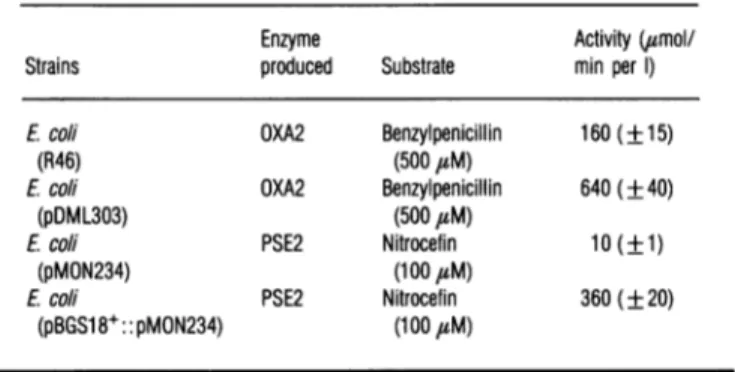
Table 1 Levelsofproducton of theOXA2and PSE20I-lactamases
The values are expressed in umol of substrate hydrolysed/min per litre of culture. They
representthemeansof fivemeasurements+S.D.
Enzyme Activity (umol/
Strains produced Substrate min per 1) E.coli (R46) Ecoli (pDML303) Ecoli (pMON234) Ecoli (pBGS18+:: pMON234) OXA2 Benzylpenicillin (500 tsM) OXA2 Benzylpenicillin (500 uM) PSE2 Nitrocefin (1 00,sM) PSE2 Nitrocefin (100
#M)
160(±15) 640(±40) 10(±1)Table 4
Purfficatlon
oftheOXAIfl-lactamase
For details, see the legend to Table 3.Total Specific
activity Total activity Purifi- Recovered
(/umol/
proteins (4tmol/minper cation activity min) (mg) mg of protein) factor (%)Crude extract 60 150 0.40 - 100
SephadexG-100 52 40 1.3 3.3 87
Mono S 36 2.7 13.3 33 60(69)
360(±20)
Table 2 PurIficaton of theOXA2 -lactamase
The values in parentheses give the yield (%) of each step. The activity is expressed as ,umol ofbenzylpenicillinhydrolysed/min per mg ofprotein.
Total Specific
activity Total activity Purifi- Recovered (umol/ proteins (,umol/min per cation activity min) (mg) mgof protein) factor (%) Crude extract CG50 Phenylboronate 27600 25000 1.1 - 100 22400 2000 11 10 81 1 19300 650 30 27 70(86) 2 15250 110 140 127 55(79) 3 14000 56 250 227 51(92)
Table 3 Purfflcaton ofthePSE2
fl-lactamase
Theactivity is expressed as,smolof oxacillinhydrolysed/min per mg of protein. The yield (%) of eachstepisgiveninparentheses.
Total Specific
activity Total activity Purifi- Recovered
(4mol/ proteins (tmol/minper cation activity min) (mg) mg ofprotein) factor (%)
Crude 17000 48000 0.35 - 100
extract
CG50 10200 6700 1.5 4.3 60
Gl00 8300 2500 3.3 9.5 49(81)
QSepharoseHL 6200 150 41 117 36(75)
Production and purfflcation of theOXAI /i-lactamase (Table 4)
A 1-litre conical flask containing 500 ml of TB medium was inoculated with 10 ml ofa 16 hprecultureinthe samemedium and stirred overnight at 37'C. The cells were separated by centrifugation, resuspendedin 100mM Trisadjusted topH9.5
with EDTA and submitted to two freeze-thaw cycles. The solutionwasfilteredat4'C throughaSephadex G-100column (80cmx4.0cm)in 10 mM sodiumphosphate buffer, pH 7.0, at
aflowrateof 40ml/h.The fl-lactamasewaselutedatavolume
of 440ml. Active fractions were pooled, concentrated and dialysed against 50mMpotassiumacetate, pH 4.5.
K k+2 k+3
E+CC± EC >- EC* - E+P
I k+4
ECi
Scheme 1 General model for the interactionoffi-lactamase(E) with
6fl-lodopenicillanate (C)EC* is the acylenzyme and EC' the rearranged adduct containing the dihydrothiazine chromophore.
The sample was then submitted to chromatography on a MonoS columncoupledto anf.p.l.c.apparatus. Buffers A and
B were 50 mM potassium acetate adjusted to pH 4.5 with concentrated acetic acid and the same buffer containing 0.5 M
K2SO4 respectively;theflowrate was 0.5ml/minand thegradient
wentfrom0 to100% of bufferB in 25ml. The/8-lactamasewas
eluted ata volumeof17 mlcorresponding to66 % of buffer B. Interaction ofthe
OXA2
f8-lactamases
with6-fp-lodopenicillanic
acid
Titration of the OXA2
fi-lactamase
with6fi-iodopenicillanic
acidA 'branched-pathway' mechanism (Scheme 1)seems to prevail in the interaction of this
fi-lactam
with several class-A/1-lactamases where the acylenzyme can rearrange into an
irreversibly inactivated speciesorundergo normal hydrolysis(De Meester etal., 1986). The hydrolysisproduct and the rearranged
inactivator moiety molecule bound to the active site of the enzyme both contain a dihydrothiazine chromophore. The
adsorption maxima are at305nm (e=8200 M-1 cm-') and at 325 nm (e= 12500
M-'
cm-') for the former and the latterrespectively (Frereetal., 1982).
Titration oftheOXA2
/J-lactamase
with6,#-iodopenicillanate
showed that this reagent inactivated the enzyme without apparent turnover
(k+4
>k+3).
Theinactivator/enzyme ratio necessary to obtain total inactivationwas 1:1(±5%). Indeed, at the end of thetitration,theabsorptionspectrumexhibitedaclear maximum at325 nm (Figure 2).The enzyme concentration determined on the basis of the titration endpoint (27.6 ,uM)was ingood agreement with that deducedfromthe absorbance valueat325nm(27.2,uM). From
the known molecular weightand the tryptophan and tyrosine contentof the enzyme, thetheoretical A280(1%)value (Cantor and Schimmel, 1980) could be computed (21.9), yielding a
100 Zg 80 60 40 20 0 (b 2 A 1 0 230
Volumeof6f-iodopenicillanate(pi)
270 2 (c) - 0 ' 320 230 250 0.04 .,- 0.02 600 500 - 400 -5 300 O 200 100 300 350 A (nm) A(nm)
Figure2 Interaction of theOXA2fp-lactamasewith
6fi-iodopenicillanic
acid(a) Titration of the OXA2 ,8-lactamase by 6,8-iodopenicillanic acid. Identical samples of inactivator(2.4 nmol in 4,ll)wereaddedto500,ul of theenzymesolution until nofurther increase inA325wasobserved. The end point of the titration correspondedto23,1u (13.8 nmol) of6fl-iodopenicillanicacid.(AA=A325after each addition of inactivator-A325of the solution without inactivator; AA tot.=A325atthetitration end point -A325 of the solution without
inactivator.) (b) and (c) Absorptionspectraof theOXA2,-lactamase before(b)and after (c) inactivation by6,f-iodopenicillanicacid.
0
[C] (PM)
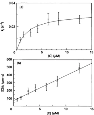
Figure 3 Inactivaton of theOXA2 f-lactamase with 6fl-iodopenicillanic
acid
(a) Pseudo-first-order inactivation rate constant (ks)as afunction of the inactivator concentration (C). Each point is themean+S.D. of five measurements. (b) Plot of[C]/kias afunctionof[C]:
r=0.985.
iodopenicillanate]= 100lM; [OXA2]= 1-10,uM),
k,
wasequalto
k+2
for whicha value of 2.5 (±0.7)x10-2 s-I was found, in goodagreementwith that determined above.25.4 uM concentration ofenzyme in the titration experiment,
thus ingoodagreementwith the valuesdetermined above. Kinetic measurements
The determination of constants
k+2
and Kwas made with oxacillin (500,uM) as a reporter substrate. Pseudo-first-orderinactivation rate constants (k0) were measured for inactivator concentrations ranging from 0.7,M to 12.6,uM (Figure 3). After correction for the protection bythe reportersubstrate,the constants
k+2
and Kwere calculated with the help ofeqn. (1)(Frereetal., 1982):
gKms+[S]8
whereS ) kl [I
(1)
where Kms and [S] are respectively the K,,, for and the
con-centration of the reporter substrate. The
k+2,
K andk+2/K
valueswere respectively 3.2 (+0.2)x10-2 s-1, 0.95 (±0.20) ,sM and
34000(±7000)M-1 s-1.Theerrorswerethose obtained for the
parameters after alinear regressionwith the Enzfitterprogram
(Leatherbarrow, 1987).
The value of
k+2
was verified by directly following the absorbance at 325nm. Under these conditions([6,8-Identification ofthe active-site serine ofthe OXA2,-Iactamase
Sequencealignments indicated Ser-70astheprobable active-site
serineresidue.Inordertoverifythisassumption,the enzyme was
labelled by adding 95nmol of 6,-iodo[3H]penicillanate to a
stoichiometric amount ofenzyme in a total volume of3.5ml. After 10 min at 30° C,thesamplewasfreeze-dried, and the solid residue redissolved in 500,1 of 8 M urea. The solution was
incubated for6 h at 37° Cand the urea concentrationdecreased
to 4 M by addition of500,1 ofwater. Trypsin digestion was realized in two steps: after addition of 250,g oftrypsin, the
sample was incubated for 12h at 37° C and a further 3 h
digestion was performed after a second addition ofthe same amountofthe same proteaseatthesame temperature.
The digest was filtered through a Sephadex G-25 column (120cmx1cm) in 10mMNH4HC03.Theelution profile showed four radioactive peaks; fractions corresponding to the smallest labelled peptides (18nmol) were pooled and freeze-dried. The
powderwas redissolved in 500 1u of 1% trifluoroacetic acidin water and further purified with the help off.p.l.c. apparatus
equippedwithaPEP-RPCHR(5/5) column. The solventswere
0.1% (v/v) trifluoroacetic acid inwater (A)and in acetonitrile
(B). Theflowrate was0.7ml/minand thegradientwentfrom 0
to60%(v/v) ofsolvent Bover42.5 ml.Thelabelledpeptidewas
elutedat avolumecorrespondingto 28% of solventB. Theresults ofgas-phase sequencing showed the presence of two peptides. Some residues were difficult to identify with
560 P.Ledent and others
Table 5 Active-sitepeptde sequencing
32 39 66 70 73
Sequence (Dale, 1985) F FS E F Q A K
Fraction 49: peptide 1 F FS E-Q A K peptide 2
* Residue not identified with certainty.
Y S P A S*T F K Y-P A- -F K
Table 6 Apparent molecular masses of the OXA2, PSE2 and OXAI
pi-lactamases
as determined onSephadex75HR 10/30,andK.,
valuesThe linear-regression analysis [log (molecular mass) =f(Ka)] was performed with BSA, ovalbumin and lysozyme (r=-0.988).
Proteins Molecular mass (kDa) Kav
BSA Ovalbumin Lysozyme OXA2 PSE2 OXAl 66 43 14.4 50.6+0.9 35.3+0.6 24.6 +1 0.118+0.001 0.183+0.001 0.602+0.002 0.171+0.002 0.293+0.003 0.415+0.015
the dihydrothiazine-labelled serine was generally poor on
gas-phase sequencing (Amicosante et al., 1988). Although the
sequences wereincomplete, they could bereplacedin theamino acid sequence deduced from the known OXA2 nucleotide
sequence(Daleetal., 1985), and,inparticular,the well-identified residues ofpeptide2allowedanunambiguous positioningin the
sequence. As predicted bythe sequence alignments, itcould be concluded that Ser-70wastheactive-siteserineresidue(Table 5). Behaviour of the
OXA1,
OXA2 and PSE2 f8-lactamases on Sephadex 75The determination ofthe apparent molecularmassof thethree class-D fl-lactamases was performed on a Sephadex 75 HR
10/30column coupled to an f.p.l.c. apparatus. The
Kav
valuesand the corresponding molecular masses are given in Table 6.
The three class-D
fl-lactamases
were characterized by quitedifferentapparentmolecularmasses.Those ofOXA2andPSE2 werehigher thanthose deduced from theirprimary structures;
that of OXAI waslower than 30 kDa.
Thehighapparent molecularmassofthe OXA2
fl-lactamase
on Sephadex 75 HR was not due to non-specific interactions between theproteinand the matrix since these wouldincreasetheK,V,
leading to a lower apparent molecular mass. It has beensuggested that the OXA2 and OXA3 enzymes could exist as
dimeric proteins (Dale and Smith, 1976), whichwould explain thehighapparentmolecularmassof the former.Inconsequence,
weanalysedthebehaviour of theOXA2enzyme underdifferent
conditions whichmightinfluence theequilibriumbetweendimer and monomer.
The
Kav
value on the Sephadex 75 HR column was notinfluenced by pH variations (from 4 to 10), high ionic
strength (0.5M Na2SO4) or the presence of 200 mM borate (a
reversibleinhibitor), 1 M NaCl04 (a dissociating agent)or 1 M guanidinium chloride. Preincubation of the enzymes with
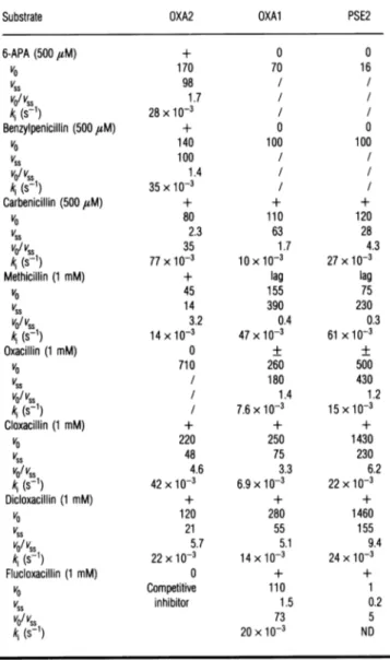
Table 7 Substrate profiles of OXA2, OXAl and PSE2
fl-lactamases:
penicillins
voand vssareexpressedaspercentages of thevssofbenzylpenicillin arbitrarilytakenas100%. Thekj value characterizes therateof transitiontothefinalsteadystate. Itcorrespondsto an
increase inactivityfor methicillin (lag) andto adecreaseinall other cases. Abbreviations: 6-APA, 6-aminopenicillanic acid; ND,notdetermined; +,burst; 0,noburst; +,very small burst;/, not relevant sincenoburstwasobserved.
Substrate OXA2 OXAl PSE2
6-APA(500 uM) + 0 0 VO 170 70 16 VSS 98 / / vo/vss 1.7 / / kj(s-1) 28x / / Benzylpenicillin (500 ,M) + 0 0 V0 140 100 100 VSS 100 / / vo/vss 1.4 kA(s-1) 35x10 / / Carbenicillin (5004M) + + + VO 80 110 120 VSS 2.3 63 28 vo/vss 35 1.7 4.3 kj(s-1) 77 x10-3 10x10-3 27x10-3
Methicillin (1 mM) + lag lag
VO 45 155 75 VSS 14 390 230 vo/vss 3.2 0.4 0.3 k, (s-1) 14x10-3 47x10-3 61x10-3 Oxacillin(1 mM) 0 + + VO 710 260 500 VSS / 180 430
vo/
vss / 1.4 1.2ki
(s-1),
7.6x10⁻³ 15x10-3 Cloxacillin (1 mM) + + + v0 220 250 1430 VSS 48 75 230 vo/vss 4.6 3.3 6.2kA (s-1)
42x10-3 6.9x10⁻³ 22 x10-3 Dicloxacillin (1 mM) + + + VO 120 280 1460 VSS 21 55 155vo/
vss 5.7 5.1 9.4 kj(s-1) 22x10-3 14 x10-3 24x10-3 Flucloxacillin(1 mM) 0 + + VO Competitive 110 1 VSS inhibitor 1.5 0.2 vo/vss 73 5 kj(s-1)
20x10-3 NDcephaloridine,
a substrateexhibiting
burst kinetics(see below),
also failedto
modify
the enzyme behaviour.Affinity chromatography
on anagarose-phenylboronate
columnof the
OXA2
f8-lactamase
partially
inactivatedby
618-lodopenicillanate
The dimericnatureof the OXA2
,-lactamase
didnotseemtobeconfirmed
by
thegel-filtration
results. The existence ofarapid
equilibriumbetween monomeric and dimeric formsofthe enzyme
was further
investigated by
chemical derivatizationexperiments
coupled with
affinity chromatography
on anagarose-phenylboronate
column.A
sample
oftheOXA2/8-lactamase
was 50% inactivatedby
6,-iodopenicillanate
sothatthe ratioofadducttointact enzymewas1.The mixturewas
applied
totheaffinity
column whichwas elutedat 20° C
with 0.1 MTris/phosphate,
pH 8.6,
containing
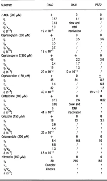
Table 8 Substate profiles of OXA2, OXAl and PSE2
pl-lactamases:
cephalosporinsFor the legend, seeTable 7. 7-ACA,7-aminocephalosporanicacid.voand
vs.
areexpressedaspercentagesof the vssvalue ofbenzylpenicillin.
Substrate OXA2 OXAl PSE2
7-ACA(200
IsM)
+ 0 0 V0 0.67 1.1 0.1 VSS 0.13 slow and / vo/vss 5.0 total / kA(s-1) 19xlO- inactivation / Cephaloglycin (200,M) + 0 0 VO 3.1 3.5 3.6 Vss 0.5 1 vo/vss 6.2 / / ki(s-1) 19xlO- / I CephalosporinC(300 ,M) + + 0 VO 46 2.2 3.0 vSS 2.4 1.7 / vo/vss 19 1.3 ki(s-1) 29x10-3 12x10-3 / Cephaloridine (150aM) + 0 + VO 45 34 8.2 VSS 1.4 / 6.8 vo/vss 32 / 1.2 kA (s-1) 42x10-3 / 19x1-3 Ceftazidime(100,M) + 0 0 V0 0.12 2.6 0.02 VSS 0.02 Slow and / v0/vss 6 total / ki(s-1) 41 x1-3 inactivation / Cefazolin (1501M) + 0 0 VO 16 13 3.1 vSS 3.6 / / vo/vss 4.4 / / k;(s-') 25x10-3 I / Cefamandole (200,M) + 0 0 VO 8.4 9.5 10 vSS 6.5 / I vo/ vss 1.3 I Iki (s-1)
4.5x10⁻³ / / Nitrocefin(150 ,uM) + 0 0 V0 80 215 165 VSS Complex / / vo/vss kinetics / / kA(s-1) / /0.25MK2SO4;the flowratewas2.5ml/min. The elution profile clearly showed thepresenceoftwopeaks. The firstone,centred
onthe deadvolume, correspondedto
,8-lactamase
labelled by the inactivator; its absorbance spectrum exhibited a secondary maximum at 325nm. The secondpeak contained non-labelledenzyme interacting with the boronate groups. The same result wasobtained withadduct/freeenzymeratios of 1: 3and 3:1; the
peak areas determined at 280nm were proportional to these ratios.
This experiment invalidated the hypothesis ofa rapid equi-libriumbetweenmonomeric and dimeric forms ofenzyme.Inthe
presenceof suchanequilibrium, onlyonepeak should have been observed,atanelutionvolume intermediate betweenthose of the non-retarded andretarded forms ofenzyme.
Heterogeneity of theOXA2 8-lactamase on Ion exchangers
Matthew and Hedges (1976) and Matthew et al. (1979) clearly showed the existence of at least three bands on isoelectric
focusing ofanOXA2 preparation. Inorder to determine if these
various forms of enzyme had similar kinetic properties, the following experiment was performed. A sample of OXA2
/3-lactamase (50mg)wasloadedon to aQ Sepharose Hiload 26/10
column with50 mMsodiumphosphate,pH7.0, at aflowrateof
5ml/min. During the washing of the column with the same
buffer, four distinct peakswereseparated. After330ml, thesame
buffercontaining 0.5 M K2S04was used and two other peaks appeared within thenext 100 ml.Thefractions centred oneach ofthepeakswerepooled, concentrated and their kinetic
proper-ties studied. The hydrolysis ofcephaloridine by the OXA2 ,-lactamasebeingbiphasic,aninitialrate/steady-state ratio(v0/v,8) and apartial inactivation rateconstant (k1) could be measured (see below). These differentparameters were the samefor all the
enzymeforms. Moreover, theproteinsinthe sixseparatefractions exhibited similar mobilitieson SDS/PAGE.
Substrate profile of OXA1, OXA2 and PSE2
f8-lactamases
Tables 7 and 8 give the relative rates of hydrolysis of several penicillins and cephalosporins respectively (benzylpenicillin being
thereference) and, whenabiphasic hydrolysiswasobserved, the
vo/v..
ratiosandthefirst-orderrateconstantki
characterizing the rateof transitionto thefinalsteady state. Ascouldbepredicted from the data available in the literature, the three enzymesconsistently hydrolysed oxacillin and cloxacillin faster than benzylpenicillin, justifying their appellation as 'oxacillinases'. However,surprisingly,in most cases,thehydrolysis time courses
didnotcorrespondtothesimple Henri-Michaelis model.'Bursts' wereoften observed and 'lags'inthehydrolysis of methicillin by
OXA1 and PSE2. In all these experiments, the enzyme
con-centration (0.04-0.1 ,M) was much lower than that of the substrate (0.1-1 mM) and the size of the burstwassignificantly larger than the enzymeconcentration, thus indicating that the
burstwasnotduetoacylenzyme accumulation butmoreprobably
tothe occurrenceofa branchedpathway.
The
vo/vS,
ratios increased with the number of Clatoms onthe isoxazolyl side chain butthe replacement of the second Cl withFproduced specific results dependingon the enzyme.
Cephalosporins,with thepossible exception of nitrocefin,were generally poorer substrates than penicillins. With these substrates, the behaviour of OXA2 was clearly different from thoseofthe twootherenzymes.Indeed, OXA2did nothydrolyse
anyof the cephalosporins accordingtosimple Henri-Michaelis
kinetics whereas OXAI and PSE2generally obeyedthis simple model.
DISCUSSION
Duringthisstudy,wehavepurifiedthree class-D
,-lactamases
toprotein homogeneity. Our protocol of purification of the OXA2 and
OXAl
enzymes gave higher specific activities and betteryields than thosepreviously published (Yamagishiet al., 1969;
Dale andSmith,1971;Monaghanetal.,1982; Holland andDale, 1984). Philippon et al. (1983) purified the PSE2 ,-lactamase 56-foldwith a 26 %yieldbut didnotindicatethespecific activity ofthe pure enzyme.
Surprisingly, despite the apparent lack of affinity for
3-aminophenylboronate, the OXA2
,/-lactamase
was retardedontheaffinitycolumn. Thisphenomenon seemedtobespecificsince theinactivationofthe enzymeby
6,8-iodopenicillanate
appearedto suppress theinteraction. Onemight arguethat the observed
results were due to anion-exchange phenomenon and that the additional negative charge supplied by the inactivator moiety
562 P. Ledent and others
seems unlikely since thechromatography wasperformed in the
presence of 0.5M
K2SO4.
The interaction of the OXA2 ,-lactamase with 6,-iodopenicillanate followed a simple linear pathway and the
k+2/K
valuewasintherange of thoseobserved with class-A fi-lactamases. Moreover, theKvaluewas strikinglylow.Isolation of an active-site peptide after labelling with
6,/-iodopenicillanate allowed the identification of Ser-70 as the
active-site serineresidue. Thiswasin agreementwiththe tentative identificationbased onthe gene sequence (Daleetal., 1985). It
couldbe argued thatpeptide 1 also contained a serine residue, but Ser-34 is not conserved in other class-D enzymes, which excludes it asthepotential active-site serine (Joriset al., 1991). Moreover, in the analogous class-A /-lactamases (Joris et al., 1991),the corresponding peptide ispartofana-helix inthea/fl
domain, faraway from the active site.
Inagreementwith the resultsof Dale (1971) andFoster(1983), the behaviour of the OXA2
,f-lactamase
during molecular-sieve chromatography indicated anMr value twiceaslargeasthat ofOXAlandconsiderably largerthanthat deducedfrom thegene sequence. Since covalent linking oftwoidentical subunits could berejected onthe basisof the normal behaviour of theenzyme on SDS/PAGE and the absence of cysteine residues in the sequence, we explored the possibility of a rapid equilibrium betweena monomerand adimer.Noconditionswerefoundthat
could increase the elution volume of the enzyme. Moreover,
when
6fl-iodopenicillanate-inactivated
enzyme was mixed withthe intact protein, no subunit interchange leading to the
formation of hybrid dimers (or polymers) could be detected. In
conclusion, thecausesof the apparently larger Mr value observed for the OXA2 enzymeduringmolecular-sieve chromatography
remain amystery, since onthebasis of the structural analogies withthe class-A,3-lactamases,onewouldnot expecttheOXA2 proteinto present anespeciallyelongated shape.
As observed with many other
/8-lactamases
(Matagne et al., 1991), several different forms of the OXA2 enzyme could be separatedbyion-exchange chromatography. The Mr values of all theseforms, determined by SDS/PAGE,weresimilaranditwassafe to conclude thatthey originated from slightly 'ragged'
N-termini or deamidation of particularly fragile asparagine residue(s). Moreover, the catalytic properties of the various forms appearedtobe identical.
Afirst overview of the catalytic properties ofthethreeenzymes
has been realized. As expected, the hydrolysis ofoxacillin was
catalysedmoreefficiently than that ofbenzylpenicillin. Moreover, themostunexpected resultwasthefinding that,atleast with the
OXA2 enzyme, 'burst' kinetics seemed to prevail with many
substrates.Infact,thisphenomenonperfectly explained the wide dispersion ofthe
Vm,.
orinitial-rate values foundinthe literature. Indeed, we observed an apparent correlation between that dispersion and the size of the 'burst'. The two other enzymes more consistently produced 'bursts' with substrates that areknowntoinduce thattypeofbehaviour, suchastheisoxazolyl penicillins. These phenomena, which imply the existence of a
branched pathway, are at present underdetailed investigation.
This work wassupported,inpart bytheBelgian GovernmentintheframeoftheP6le
d'Attraction Interuniversitaire (PAI no. 19), Actions concertees with the Belgian
Government(conventions 86/91-90 and89/94-130), the Fonds de la Recherche
Scientifique Medicale (contractno. 3.4537.88)and aconventiontripartite between the RegionWallone, Smith Kline Beecham, U.K. andthe University of Liege. P.L.
and X. R. are fellows of the Institut pour 1'encouragement de la Recherche
Scientifique dansl'Industrie etl'Agriculture(IRSIA, Brussels,Belgium)and B. J. is
'Chercheur Qualifie' of the Belgian National Foundation for Scientific Research
(FNRS). P. L. thanks everybody in the Laboratoire d'Enzymologie (Liege) for their help and encouragement and P. Cuvelier (Boehringer Mannheim, Belgium) for financial support.
REFERENCES
Ambler, R. P.(1980)Philos. Trans. R. Soc. London Ser. B 289, 321-331
Amicosante, G., Oratore, A., Joris, B., Galleni, M., Frere, J.-M. and Van Beeumen, J. (1988) Biochem. J.254,891493
Anderson, E. S. and Datta, N. (1965) Lancet 1, 407-409
Cantor,C. R. andSchimmel, P. R. (1980) Biophysical Chemistry, p. 380, W. H. Freeman and Co.,San Francisco
Cartwright, S. J. and Waley, S. G. (1984) Biochem. J. 221, 505-512
Couture, F., Lachapelle,J. andLevesque R.C.(1992)Mol. Microbiol. 6,1693-1705 Dale,J. W.(1971)Biochem. J.123,501-505
Dale,J. W. andSmith,J. T.(1971) Biochem. J.123,493-500
Dale,J. W.andSmith, J. T. (1976) Biochem. Biophys. Res. Commun. 68, 1000-1005 Dale, J. W.,Godwin, D., Mossakowska, D., Stephenson, P. and Wall, S. (1985) FEBS Lett.
191, 39-44
De Meester, F., Frere, J.-M.,Piette, J. L. and Vanderhaeghe, H. (1985) Labelled Compd. Radiopharmacol. 22,415-425
DeMeester, F., Frere,J.M., Waley, S. G., Cartwright, S. J., Virden, R. and Lindberg, F. (1986) Biochem. J.239,575-580
De Meester, F.,Joris, B.,Reckinger, G.,Bellefroid-Bourguignon, C., Frere, J.-M. andWaley, S. G. (1987) Biochem. Pharmacol. 36, 2393-2403
Frere,J.-M., Dormans, C., Duyckaerts, C. and De Graeve, J. (1982) Biochem. J.207, 437-444
Foster, T. J. (1983)Microbiol. Rev. 47, 361-409
Galleni,M.and Frere, J.-M. (1988) Biochem. J.255,119-122
Galleni,M., Amicosante,G. and Frere, J.-M. (1988) Biochem. J.255,123-129 Herzberg,0. (1991) J. Mol. Biol. 217, 701-719
Holland, S. and Dale, J. W. (1984) Biochem. J. 224, 1009-1013
Holland, S. and Dale, J. W. (1985) Antimicrob. Agents Chemother. 27,989-991 Huovinen, P., Huovinen, S. and Jacoby, G. A. (1988) Antimicrob. Agents Chemother.32,
134-136
Jaurin,B.andGrundstrom,T. (1981) Proc. Natl.Acad.Sci. U.S.A. 78,4897-4901 Joris, B.,Ghuysen,J.M.,Dive, G. Renard, A., Dideberg, O., Charlier, P.,Frere, J.-M.,Kelly,
J.A., Boyington, J.C., Moews, P. C. and Knox, J. R. (1988) Biochem. J. 250, 313-324 Joris, B.,Ledent, P., Dideberg,0., Fonze, E., Lamotte-Brasseur,J., Kelly,J.A.,Ghuysen,
J. M. and Frbre, J.-M. (1991) Antimicrob. Agents Chemother.35,2294-2301 Kelly, J. A., Dideberg, O., Wery, J. P., Libert, M., Moews, P. C., Knox, J. R., Duez, C.,
Fraipont,C., Joris, B., Dusart, J., Frere, J.-M. and Ghuysen, J. M. (1986) Science231, 1429-1 431
Knox, J. R. andMoews, P. C. (1991) J. Mol. Biol.220,435-455
Leatherbarrow, R. J.(1987) Enzfitter: A Non-linear Regression Data Analysis Program for the IBMPC/P52, Elsevier Biosoft,Cambridge,U.K.
Levesque, R.C.,Medeiros,A. A. & Jacoby, G. A. (1987) Mol. Gen. Genet.206,252-258 Maniatis, T.,Fritsch, E. F. &Sambrook,J.(eds) (1982) Molecular Cloning: ALaboratory
Manual, ColdSpring Harbor Laboratory Press, Cold Spring Harbor, NY
Matagne, A.,Misselyn-Bauduin, A. M., Joris, B., Erpicum, T., Granier, B. & Frere, J.-M. (1990) Biochem. J.265,131-146
Matagne,A.,Joris, B., VanBeeumen,J.& Frere,J.-M. (1991) Biochem. J.273, 503-510 Matthew,M. & Hedges, R. W. (1976) J. Bacteriol. 125, 713-718
Matthew,M., Hedges, R. W. &Smith,J. T. (1979) J. Bacteriol. 138,657-662 Medeiros, A. A., Cohenford, M.&Jacoby, G. A. (1985) Antimicrob. Agents Chemother. 27,
715-719
Monaghan, C., Holland, S. and Dale, J. W. (1982) Biochem. J.205,413-417 Mossakowska, D., Ali, N. A. &Dale,J. W. (1989)Eur. J. Biochem. 180,309-318 Oefner,C., D'Arcy, A.,Daly,J.J., Gubernator, K.,Charnas, R.L.,Heinze, I., Hubschwerlen,
C. &Winkler, F. K. (1990)Nature(London) 343, 284-288
Ouellette, M.,Bissonnette, L. &Roy, P. H.(1987) Proc.Nati. Acad.Sci. U.S.A.84, 7378-7382
Philippon, A. M., Paul, G. C. & Jacoby, G.A.(1983) Antimicrob. Agents Chemother.24, 362-369
Samraoui, B.,Sutton, B.J.,Todd,R.J.,Artymiuk,P.J., Waley,S. G. &Phillips, D.C. (1986)Nature (London)320,378-380
Spratt,B.G., Hedge, P.J.,teHeesen, S.,Edelman,A.& Broome-Smith,J. K.(1986) Gene 41, 337-342
Yamagishi, S.,O'Hara,K., Sawai,T. & Mitsuhashi, S. (1969)J. Biochem. (Tokyo) 66, 11-20