HAL Id: tel-02128298
https://tel.archives-ouvertes.fr/tel-02128298
Submitted on 14 May 2019
HAL is a multi-disciplinary open access archive for the deposit and dissemination of sci-entific research documents, whether they are pub-lished or not. The documents may come from teaching and research institutions in France or abroad, or from public or private research centers.
L’archive ouverte pluridisciplinaire HAL, est destinée au dépôt et à la diffusion de documents scientifiques de niveau recherche, publiés ou non, émanant des établissements d’enseignement et de recherche français ou étrangers, des laboratoires publics ou privés.
Analysis of HIV-1 cell-to-cell transfer to macrophages
Lucie Bracq
To cite this version:
Lucie Bracq. Analysis of HIV-1 cell-to-cell transfer to macrophages. Immunology. Université Sorbonne Paris Cité; University of Chinese academy of sciences, 2017. English. �NNT : 2017USPCB063�. �tel-02128298�
UNIVERSITY PARIS DESCARTES-SORBONNE PARIS CITE / INSTITUT COCHIN
UNIVERSITY OF CHINESE ACADEMY OF SCIENCE / INSTITUT PASTEUR OF SHANGHAI
PhD THESIS
Thesis to obtain the degree of PhD of University Paris Descartes and University of Chinese Academy of Sciences
Major: Biology
Specialty: Cellular biology and virology Presented publicly by Lucie Bracq
17 November 2017
Analysis of HIV-1 cell-to-cell transfer to macrophages.
Committee members:
Dr Alexandre Benmerah President of committee Pr Quentin Sattentau Principal referee Dr Helene Dutartre Principal referee
Pr Xia Jin Referee
Dr Delphine Muriaux Referee Pr Andres Alcover Referee
Dr Serge Benichou PhD Supervisor Pr Paul Zhou PhD Co-supervisor
Analysis of HIV-1 Cell-to-cell transfer to macrophages
Abstract:
Macrophages are important targets of HIV-1 and play crucial roles in physiopathology of infection. Because of their long term survival capacity, infected macrophages participate in virus dissemination and establishment of persistent virus reservoirs in numerous tissues. In
vitro, macrophage infection and analysis of the different steps of the virus cycle have been
largely documented using cell-free virus infection. However, there is a paucity in knowledge of the mechanisms that control infection and dissemination to macrophages by cell-to-cell transfer. In the work presented here, we establish a model of HIV-1 cell-to-cell transfer from infected T cells to macrophages. We observed that infected T cells are able to interact with macrophages leading to cell fusion for transfer of viral material to macrophages targets. This cell-to-cell fusion transfer, very fast and efficient, is restricted to CCR5-tropic viruses, and mediated by viral envelope-receptor interactions. Transferred viruses can then accumulate in cytoplasmic compartments of newly formed lymphocyte/macrophages fused cells but we also observed early viral assembly and budding events at the plasma membrane of these fused cells, resulting from the merging of viral material between infected T cells and macrophages. These cells then acquire the ability to fuse with neighboring non-infected macrophages for virus dissemination. Together, these two-sequential envelope-dependent cell fusion process lead to the formation of highly virus-productive multinucleated giant cells reminiscent of the infected multinucleated giant macrophages detected in vivo in lymphoid organs and the central nervous system of HIV-1 infected patients and simian immunodeficiency virus-infected macaques. These mechanisms may represent an original mode of virus transmission for viral spreading and formation of macrophage virus reservoirs during HIV-1 infection.
Keywords : HIV-1, T cell, Macrophages, Cell-to-cell transfer, Cell fusion Laboratories :
- Virus et trafic intracellulaire.
Institut Cochin – InsermU1016 – CNRS UMR8104 –Université Paris-Descartes, Sorbonne Paris-Cité.
- Antiviral immunity and genetic therapy
Analyse du transfert intercellulaire du VIH-1 vers les macrophages.
Résumé :
Les macrophages sont une cible particulièrement importante de l’infection par le VIH-1 et jouent un rôle crucial dans la physiopathologie de l’infection. Lorsqu’ils sont infectés, leur capacité de survie dans les tissus leur permet de jouer un rôle essentiel dans la dissémination virale et l’établissement de réservoirs viraux au niveau des différents territoires tissulaires. In vitro, les étapes précoces et tardives du cycle de réplication virale dans les macrophages ont été analysées dans le cadre de l’infection par des virus libres. Cependant, les modalités d’infection des macrophages lors d’une transmission intercellulaire restent largement inexplorées. Les travaux présentés ici ont permis d’établir un modèle de transmission intercellulaire du VIH-1 des lymphocytes T infectés vers les macrophages. Nous avons montré que les lymphocytes T infectés sont capables d’interagir étroitement avec les macrophages, conduisant ainsi à la fusion cellulaire de ces deux cellules et permettant le transfert de matériel viral dans les macrophages cibles. Ce transfert viral par fusion cellulaire, rapide et efficace, est restreint aux virus utilisant le corécepteur CCR5 et dépend de l’interaction entre l’enveloppe virale et le récepteur CD4. Les virus transférés sont alors stockés au sein de compartiments cytoplasmiques dans les cellules fusionnées mais nous observons également des évènements précoces d’assemblage et de bourgeonnement du VIH-1 à la membrane plasmique des cellules fusionnées, résultant de la fusion des membranes des lymphocytes T infectés et des macrophages cibles. Ces cellules fusionnées acquièrent alors la capacité de fusionner avec les macrophages non infectés environnants permettant la dissémination du VIH-1. L’ensemble de ces résultats met en évidence un nouveau mécanisme de transmission intercellulaire entre lymphocytes T et macrophages via un mécanisme de double fusion cellulaire dépendant de l’enveloppe virale et des récepteurs CD4 et CCR5. Ces évènements successifs de fusion entre lymphocytes T et macrophages puis entre macrophages permettent la formation de cellules géantes multinucléées capables de produire de grande quantité de virus infectieux. Ces cellules multinculéees pourraient correspondre aux macrophages multinucléees observés in vivo dans les organes lymphoïdes et le système nerveux central de patients infectés par le VIH-1 ou de singes infectés par le VIS. Ce mécanisme représente donc un modèle de transmission intercellulaire original permettant la dissémination virale et la formation de macrophages réservoirs durant l’infection par le VIH-1.
Mots clés : VIH-1, Lymphocytes T, Macrophages, Transfert intercellulaire, Fusion cellulaire.
Laboratoires :
- Virus et trafic intracellulaire.
Institut Cochin – InsermU1016 – CNRS UMR8104 –Université Paris-Descartes, Sorbonne Paris-Cité.
- Antiviral immunity and genetic therapy
HIV-1
6
52 HIV-1 6 HIV-1 1 HIV-1 T CCR5 / HIV-1 SIV-1 1 36 HIV-1 HIV-1 - Inserm U1016 - CNRS UMR8104 - ,-Acknowledgments
Opening these acknowledgments, I warmly thank Pr Quentin Sattentau, Dr Hélène Dutartre and Pr Xia Jin for their careful reviewing of my thesis manuscript. I am also very thankful to Dr Alexandre Benmerah, president of my thesis jury, and to Prof Andres Alcover and Dr Delphine Muriaux, examinators, for their interest in my thesis project.
J’ai eu la chance durant cette thèse en cotutelle de faire partie de deux équipes dont celle de Serge Benichou à l’institut Cochin et celle de Paul Zhou à l’Institut Pasteur de Shanghai. J’ai donc fait de nombreuses rencontres en France et en Chine à la fois sur le plan professionnel que personnel. Cette cotutelle m’a permis de voyager et de découvrir ce nouveau pays que j’affectionne désormais tant. Je tiens donc particulièrement a remercié Serge Benichou pour m’avoir donné l’opportunité de faire partie de cette aventure mais aussi Paul Zhou qui m’a accueilli dans son équipe et dont j’ai beaucoup appris.
Depuis mon arrivée dans son équipe en 2014 pour mon stage de master, Serge m’a accordé une grande confiance, et une liberté qui m’ont permis de pleinement m’épanouir durant ma thèse. Merci à toi Serge pour les nombreuses discutions scientifiques, opportunités que tu m’as donné mais aussi pour d’un point de vue plus personnel pour les nombreux fous rires (notamment au cours de chinois), les déjeuners au Cafelito, et tous les (très nombreux) bons moments passés qui ont fait que venir travailler au labo chaque matin était un véritable plaisir.
Je pense aussi à tous les membres de l’équipe de Serge Benichou et de Paul Zhou. Merci à Lihong Liu et Weiming Wang d’avoir partagé leurs savoirs avec moi et de m’avoir si bien intégré à mon arrivée à Shanghai. Je remercie aussi Maorong, Marie-christine, Trang, Héloise et Amandine pour avoir partagé de nombreux moments ensemble à l’institut Cochin. Un merci tout particulier à Jérôme Bouchet pour tous tes conseils, pour ton aide et d’avoir partagé avec moi ton expérience mais aussi tous les discutions (pas forcement scientifique) que l’on a eu aux pauses café, dans nos nombreux trajets en train (et parfois en Uber) ou autour d’une bière.
Je souhaite également remercier toutes les personnes qui ont participé à ma formation et permis de réaliser ce travail de thèse. Merci à Julie Matz qui a fait preuve d’une grande douceur et gentillesse à mon arrivée en Master à Cochin et qui a su me donner gout à ce projet sur lequel j’ai tant aimé travailler. Je tiens également à remercier Cécile
Hérate et Clarisse Vigne qui étaient également présente à mon arrivée et qui sont devenus de très bonnes amies depuis et ont beaucoup manqué après leur départ.
Au-delà des membres de l’équipe, je voudrais remercier de nombreuses personnes au sein des deux Instituts. Merci donc aux membres des équipes d’Isabelle Dusanter et Ralf Jockers mais aussi à l’équipe de microscopie électronique pour tous les déjeuners et moments passés ensemble. Du côté chinois, je remercie particulièrement l’équipe de Xia Jin dont Jiayi et Ranran ainsi que Fernando et Laure qui m’ont beaucoup aidé lorsque ma compréhension plus que limité du chinois me limitait énormément.
J’ai eu la chance lors de cette thèse de collaborer avec plusieurs équipes qui ont ainsi partagé leur savoir et leurs bonne humer avec moi. Je remercie donc toute l’équipe de Clotilde Randriamampita mais aussi l’équipe de Christel Vérollet et particulièrement Maeva qui est venu passé quelques mois à Cochin avec qui se fut un plaisir de travailler.
Je remercie aussi les membres des plateformes scientifiques de l’Institut Cochin et de l’Institut Pasteur de Shanghai qui m’ont aidé et conseiller durant ces quatres années. Mais également les différentes gestionnaires, Véronique Chauvin et Xiaoyu pour leur aide et leur bonne humeur.
Si j’ai eu le courage de partir passé de nombreux mois à Shanghai c’est grâce au soutien de ma famille et particulièrement de mes parents et mes sœurs que je remercie pour tout, mais aussi de mes amis. Un grand merci donc à Anthony, Kim, Juliette, Clara et tous les autres, de m’avoir tout d’abord poussé à partir, d’avoir toujours été présent a chacun et mes retours mais aussi d’avoir supporté mes longues tirades sur la chine a mes retours en France. Merci particulier à Kim, Clara et à ma famille qui sont venus me rendre visite à Shanghai.
Pour finir merci à tous ceux qui ont fait que chaque moment passé à Shanghai était un réel plaisir. Un immense merci à Antoine qui a été le premier que j’ai rencontré, qui m’a permis de rencontrer tant d’amis, avec qui j’ai passé tant de bons moments et qui restera le meilleur coloc que j’ai pu avoir. Merci à Clara, Cécile, Victoria Oliver et tant d’autres avec qui j’ai partagé mon quotidien, mes galères et mes bonheurs pendant ces années et qui sont devenus ma deuxième famille à l’autre bout du monde. Enfin je tiens à remercier Marie, qui suit le même parcours que le miens et avec qui nous nous sommes beaucoup soutenus pendant les moments plus compliqués mais aussi dans les bons lorsque nous allions « zapater » autour d’une marguarita. Je te souhaite bon courage pour la fin de ta thèse et espère te revoir rapidement à Shanghai.
Table of Contents
Acknowledgments ... 7 Table of Contents ... 11 Table of Figures ... 15 Abreviations ... 19 INTRODUCTION ... 23 1. The Human immunodeficiency virus ... 25 1.1. Physiopathology of HIV-1 infection ... 25 1.2. General presentation of HIV-1 ... 27 1.2.1. Structure and genome ... 27 1.2.2. Target cells of HIV-1 ... 29 1.2.2.1. T lymphocytes ... 29 1.2.2.2. Monocytes/Macrophages ... 29 1.2.2.3. Dendritic cells. ... 30 1.2.3. Viral entry and tropism ... 30 1.2.4. HIV-1 life cycle ... 31 2. Mechanisms of cell-to-cell transmission of HIV-1 ... 33 2.1. Cell-free and cell-to-cell infection ... 33 2.2. Nanotubes and trogocytosis ... 34 2.2.1. Nanotubes and filopodia ... 34 2.2.2. Trogocytosis ... 35 2.3. The Virological synapses ... 38 2.3.1. Structure of the virological synapse ... 38 2.3.2. Viral entry downstream of the virological synapse. ... 41 2.3.3. Viral transfer across the virological synapse and resistance to neutralizing antibodies and antiretroviral drugs ... 44 2.4. Heterogeneity of virological synapses ... 45 2.4.1. The DC infectious synapse ... 46 2.4.2. Transfer of HIV-1 at the virological synapse formed with macrophage targets ... 48 2.5. Engulfment of infected T cells by macrophages ... 49 2.6. Cell-to-cell fusion ... 50 2.6.1. Formation of T cell-syncytia ... 50 2.6.2. Inhibition of cell-cell fusion at the virological synapse. ... 51 2.6.3. Presence of multinucleated giant cells in infected tissue ... 52 3. Macrophages: Targets of HIV-1 ... 543.1. Origins and tissue distribution of macrophages ... 54 3.2. Functions of macrophages and impairment by HIV-1 ... 55 3.2.1. Phagocytosis ... 55 3.2.1.1. Phagocytosis under physiological condition ... 55 3.2.1.2. Impairment of phagocytosis by HIV-1 ... 56 3.2.2. Cell-cell fusion ... 58 3.2.2.1. Cell-cell fusion for osteoclast formation ... 58 3.2.2.2. Cell-cell fusion and HIV-1 ... 61 3.3. Implication of macrophages in HIV-1 infection. ... 62 3.3.1. Viral reservoirs ... 62 3.3.2. Multinucleated giant macrophages in the neurophysiopathology of AIDS. ... 62 3.4. HIV-1 replication in Macrophages ... 64 3.4.1. Early steps of the viral replication cycle in macrophages ... 64 3.4.2. Late steps of the viral life cycle in macrophages ... 66 3.4.3. Implication of HIV-1 auxiliary proteins. ... 67 3.4.3.1. The auxiliary proteins Nef. ... 68 3.4.3.2. The auxiliary proteins Vpr ... 68 3.4.3.3. The auxiliary protein Vif and APOBEC3 ... 69 3.4.3.4. The auxiliary protein Vpu and BST-2 ... 69 3.4.3.5. The auxiliary protein Vpx and SAMHDI ... 70 3.5. Macrophages and HIV-1: conclusions ... 71 4. Aims of the experimental work ... 73 4.1. Analysis of HIV-1 cell-to-cell transmission between infected T cells and macrophages . 73 4.2. Analysis of HIV-1 infection of osteoclasts. ... 74 RESULTS ... 77 Article 1: T cells-macrophages fusion triggers multinucleated giant cell formation for HIV-1 spreading. ... 79 1. Abstract ... 79 2. Presentation of the article ... 79 3. Article ... 80 4. Conclusion ... 131 Article 2: The bone degradation machinery of osteoclasts: a novel HIV-1 target that contributes to bone loss ... 133 1. Abstract ... 133 2. Presentation of the article ... 133 3. Article ... 134 4. Conclusions ... 172
DISCUSSION ... 173 1. Relevance of macrophages in HIV-1 infection. ... 175 2. Envelope and co-receptor implication for cell-cell fusion ... 176 3. HIV-1 Cell-to-cell infection of macrophages through cell-cell fusion or internalization of infected T cells. ... 178 3.1. Different experimental systems could lead to phagocytosis ... 178 3.2. Phagocytosis and cell-fusion are tightly regulated. ... 179 3.3. Absence of HIV-1 induced cell-cell fusion between T cells. ... 181 3.4. Signaling pathways involved in HIV-1 cell-cell fusion. ... 183 3.5. HIV-1 Cell-cell fusion of macrophages ... 184 CONCLUSIONS ... 186 REFERENCES ... 192 APPENDIX ... 212
Table of Figures
Figure 1: Antiretroviral therapy coverage and number of AIDS-related deaths. ... 25
Figure 2: Evolution of HIV-1 infection. ... 27
Figure 3: Genomic and structural organization of HIV-1. ... 28
Table 1: Proteins encoded by the HIV-1 genome. ... 29
Figure 4: Schematic representation of HIV-1 binding and fusion. ... 31
Figure 5: Simplify representation of HIV-1 life cycle. ... 32
Figure 6: HIV-1 transfer across nanotubes in T cells. ... 35
Figure 7: Schematic illustration of a trogocytosis process between an antigen-presenting cell (APC) and CD4+ and CD8+ T cells. ... 37
Figure 8: Schematic illustration of virological synapse formation. ... 41
Figure 9: Hypothetical mechanisms of HIV-1 entry across Virological synapse. ... 42
Figure 10 : Model for Maturation-Induced Fusion at the Virological Synapse. ... 43
Figure 11: Schematic illustration of virological and infectious synapses formed between infected DCs and T cells. ... 47
Figure 12: Recruitment and accumulation of Gag at the macrophage/macrophage and macrophages/T cell synapse. ... 49
Figure 13: inhibition of cell-cell fusion across virological synapse. ... 52
Table 2: Summary of the different cell-to-cell transmission mechanisms according to the cell type. ... 53
Figure 14: Phagocytosis of apoptotic cells by macrophages. ... 56
Figure 15: HIV-1 impairs complement-receptor mediated phagocytosis. ... 57
Figure 16: HIV-1 impairs Fc-receptor mediated phagocytosis. ... 58
Figure 18: HIV-1 infection induce formation of multinucleated macrophages. ... 61
Figure 19: Presence of multinucleated giant macrophages in HIV-1 infected patients. ... 63
Figure 20: Early steps of the HIV-1 life cycle in T cell and macrophages. ... 65
Figure 21: Late step of the HIV-1 life cycle in T cells and macrophages. ... 67
Figure 22: Effect of auxiliary proteins and cellular restriction factors of HIV-1. ... 70
Additional Figure A1: Expression of fusion proteins E-cadherin, CD9, CD81 in macrophages after cell-to-cell infection with infected T cells.Erreur ! Le signet n’est pas défini. Additional Figure A2: Implication of Vpu, Vpr and Vif in cell-to-cell infection of macrophages by infected T cells. ... Erreur ! Le signet n’est pas défini. Additional Figure A3: Fluorescence microscopy analysis of intercellular contacts and viral transfer between infected T cells and osteoclasts. . Erreur ! Le signet n’est pas défini. Additional Figure A4: HIV-1 cell-to-cell transfer from infected T cells to osteoclasts is co-receptor dependent. ... Erreur ! Le signet n’est pas défini. Additional Figure A5: Effect of fusion inhibitor T20 in HIV-1 cell-to-cell transfer from infected T cells to osteoclasts ... Erreur ! Le signet n’est pas défini. Additional Figure A6: Osteoclasts are multinucleated and express T cell specific marker after cell-to-cell infection by infected T cell. ... Erreur ! Le signet n’est pas défini. Figure 23: hypothetical regulation of cell-cell fusion and phagocytosis for HIV-1 transfer. . 180
Figure 24: hypothetical regulation of cell-cell fusion for HIV-1 transfer. ... 182
Figure 25: Summary of different model for HIV-1 cell-to-cell transfer. ... 183
Figure 26. Model for virus cell-to-cell transfer from infected T cells to MDMs and virus spreading between MDMs. ... 188
Abreviations
AIDS : Acquired immunodeficiency syndrome
AMP : Adenosine monophosphate
APC : Antigen presenting cell ART : Anti-retroviral therapy
APOBEC : Apolipoprotein BmRNA-editing catalytic polyprotein AP1 : Adaptator protein complex 1
AZT : Azidothymidine
BST2 : Bone marrow stromal antigen 2
CCR5 : C-C Chemokine receptor 5
CD4 : Cluster of differentiation 4 CXCR4 : CXC Chemokine receptor 4 CDC42 : Cell division control protein 42 CNS : Central nervous system
DC : Dendritic cell
DAP12 : DNAX activation protein of 12kb
DC-SIGN : Dendritic cell-specific intracellular adhesion molecule grabbing non-integrin DC-STAMP : Dendrocyte expressed seven transmembrane protein
dNTP : Deoxynucleoside triphosphate DNA : Deoxyribonucleoside acid
DNM2 : Dyanamin 2
EEA-1 : Early endosomal antigen 1
ELISA : Enzyme-linked immunosorbent assay EM : Electron microscopy
GMCSF : Granulocyte macrophage colony-stimulation factor HAART : Highly active retroviral therapy
HCK : Hematopoietic cell kinase HIV : Human immunodeficiency virus
HTLV : Human T-cell leukemia-lymphoma virus IL-4 : Interleukin-4
ICAM-1 : Intracellular adhesion molecule 1
IN : Integrase
IFN : Interferon
IFITM : Interferon-induced transmembrane protein KIR : Killer-cell immunoglobulin like receptor LAMP-1 : Lysosomal-associated membrane protein 1
LFMC : Lymphocyte/macrophage fused cell LFA-1 : Lymphocyte function associated antigen 1
MA : Matrix
MDM : Monocyte derived macrophage
MHC : Major histocompatibility complex MMP9 : Matrix metallopeptidase
MLV : Murine leukemia virus
MGC : Multinucleated giant cell MTOC : Microtubule organizing center
NC : Nucleocapsid
Nef : Negative factor NK : Natural killer
NRTI : Nucleosidic reverse transcriptase inhibitor NNRTI : Non-nucleosidic reverse transcriptase inhibitor
OC : Osteoclast
PtdSer : Phosphatidyl Serine
PAMP : Pathogen-associated molecular pattern PIC : Pre integration complex
RANKL : Receptor activator of nuclear factor kappa-B ligand Rev : Regulator of expression of virion proteins
RT : Reverse transcriptase
SIV : Simian immunodeficiency virus
SAMHDI : Sterile alpha motif and HD domain-containing protein- 1 SIRPa : Signal regulated protein alpha
STAT6 : Signal transducer and activator or transcription 6 SYK : Spleen tyrosine kinase
TRAIL : Tumor-necrosis-factor related apoptosis inducing ligand TNF : Tumor-necrosis factor
Tat : Trans-activator TCR : T-cell receptor
TSP : Surface-bound thrombospondin TSPAN : Tetraspanin
VCC : Virus containing compartment Vif : Viral infectivity factor
Vpr : Viral protein R Vpu : Viral protein U VS : Virological synapse
1. The Human immunodeficiency virus
1.1. Physiopathology of HIV-1 infection
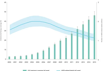
The Human Immunodeficiency Virus (HIV) is the causative agent of the acquired immunodeficiency syndrome (AIDS). In 2016, 36.7 million people were living with HIV, with 1.8 million people newly infected in 2016 (World Health Organization website). Two types of HIVs are known to cause AIDS: HIV-1 and HIV-2, exhibiting distinct biological and epidemiological attributes. Whereas HIV-1 has world wild distribution, HIV-2 remains confined to West Africa and is characterized by low transmission and slow progression to AIDS (Gilbert et al, 2003). The Sub-Saharan region of Africa is the most affected region with approximatively two thirds of HIV-1 infected people living in this area (Tebit & Arts, 2011). Therefore, HIV is a major global public health issue since more than 35 million people died so far from AIDS since the beginning of the epidemic in 1980. Until now, no cure or vaccine are available but the appearance and development of antiretroviral therapy in the early 1990s led to a significant decrease of AIDS related death and morbidity in HIV-infected patients (Figure 1).
Figure 1: Antiretroviral therapy coverage and number of AIDS-related deaths.
Representation of HIV-1 treatment coverage and AIDS-related deaths from 2000 to 2015. From UNAIDS 2016.
HIV-1 is a lentivirus transmitted through mucosa or blood. In infected patients, a common pattern of three different stages is observed with an extended incubation period (median of 10 years) varying among patients. Each stage is characterized by the viral load (number of viral RNA copy/mm3 of plasma) and the number of circulating CD4+ T cells (Figure 2).
The first stage of HIV-1 infection is an acute phase, which generally develops within 3 to 6 weeks after the initial infection and is characterized by a high viral load due to viral replication of HIV-1 in peripheral blood mononuclear cells (Pantaleo et al, 1993). This active replication leads to a rapid but transient decrease of the number of circulating CD4+ T cells. 50 to 70% of infected patients have flu-like symptoms during this phase, such as fever, headache and/or rash. This phase is also characterized by the development of an HIV-specific immune response leading to a rapid decrease of viral load after several weeks. However, this immunity is not sufficient to suppress viral replication completely since HIV-1 expression persists in lymph nodes even when plasma viral load is very low or when viral RNA is undetectable in peripheral blood mononuclear cells (Pantaleo et al, 1993).
The second stage of HIV infection is the chronic phase, and is also called asymptomatic phase or clinical latency. During this stage of infection, the virus continues to replicate in lymph nodes but at very low levels. This low viral replication is related to both humoral and cellular immune responses. During clinical latency, high levels of HIV-1 specific antibodies and cytotoxic CD8+ T cells are observed, and this immune response leads to the killing of infected CD4+ T cells, responsible, at least in part, for the progressive decrease of circulating CD4+ T cells (McCune, 2001). Without treatment with antiretroviral therapy, chronic HIV infection usually progresses to AIDS in 10 years or longer, though it may take less time for some patients. The USA centers for disease control and prevention defined this chronic stage by a CD4+ T cell number between 200 and 499 cells/mm3. The world health organization recommends starting antiretroviral therapy if the number of CD4+ T cells goes below 350 cells/mm3.
When the number of CD4+ T cells is below 200 cells/mm3, the patient is in the clinical stage of AIDS. Loss of cytotoxic CD8+ T cells and immune response are thus observed whereas the viral load increases rapidly. Severe immunodeficiency leads to severe symptoms and opportunistic infections such as pneumonia, tuberculosis, cancer or neurologic diseases leading to the death of infected patients.
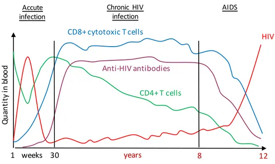
Figure 2: Evolution of HIV-1 infection.
During the first week after 1 infection, virus multiplies actively (red line) and the level of HIV-1 increases whereas the number of CD4+ lymphocyte decreases (green line). 6 weeks after infection, the immune response leads to the presence of HIV-1 targeting antibodies (purple line) and activation of CD8+ cytotoxic T lymphocytes (blue line). During chronic infection, the viral load is low, because of immune response of cytotoxic T cells however the number of CD4+ T cells decrease progressively leading to AIDS where the immune system collapses and viral load increases dramatically. From (Pantaleo et al, 1993).
1.2. General presentation of HIV-1
1.2.1.
Structure and genome
HIV is an enveloped virus that belongs to the Retroviridae family and Lentivirus genus. HIV-1 particles are spherical with an average diameter of 100 nm and are coated by the viral envelope membrane. The viral membrane is a lipid bilayer derived from the membrane of the host cell containing the viral envelope glycoproteins, the surface gp120 and the transmembrane gp41, generated from the cleavage of the viral glycoprotein precursor gp160 encoded by the env gene (Freed, 2001). These envelope glycoproteins are present on the cell surface of infected cells as the envelope complex, a trimer of heterodimers of gp120 and gp41, and are incorporated into the lipid bilayer through the transmembrane region of gp41 (White et al, 2010). During viral budding from the plasma membrane of the host cell, around 7 to 14 trimers of envelope are incorporated in a single virus particle (Chertova et al, 2002; Cosson, 1996). The inner leaflet of the envelope lipid membrane is lined with the matrix protein p17 which surrounds a conical capsid composed
1 weeks 30 years 8 12 Qu an ti ty in b lo od HIV CD8+ cytotoxic T cells Anti-HIV antibodies CD4+ T cells Accute infection Chronic HIV infection AIDS
of the capsid protein p24. This capsid encloses two copies of the viral single-stranded RNA,
approximatively 10,000 bases long, that are linked together through non-covalent
interactions. The HIV-1 RNA is part of a nucleoprotein complex, which is composed of the nucleoprotein p7 and the reverse transcriptase p66 (RT). The viral particle also contains all the enzymatic equipment that is necessary for replication: the reverse transcriptase (RT), the integrase p32 and the protease (Figure 3).
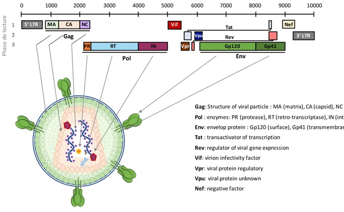
Figure 3: Genomic and structural organization of HIV-1.
The HIV-1 genome contains three major genes (Gag, Pol and Env) encoding for structural polyproteins, enzyme and envelope proteins. In addition, HIV-1 encodes for regulatory and auxiliary proteins (Tat, Rev, Vif, Vpr, Vpu and Nef). From (Ganser-Pornillos et al, 2008).
The HIV-1 genome contains the three major genes that are found in all retroviruses,
gag, pol and env encoding respectively for structural proteins of the viral core and matrix,
enzymes and envelope glycoproteins. These proteins are first synthesized as polyproteins and then cleaved resulting in all the viral proteins present in virions. The HIV-1 genome also contains nonstructural genes (or accessory) encoding for auxiliary proteins (Vif, Vpr, Vpu and Nef) and regulatory proteins (Tat and Rev) (Figure 3 and Table 1).
5’ LTR MA CA NC PR RT IN Gp120 Gp41 3’ LTR Vpr Vif Vpu Nef Tat Rev 0 1000 2000 3000 4000 5000 6000 7000 8000 9000 10000 1 2 3 Ph as e de le ct ur e Gag Pol Env Gag: Structure of viral particle : MA (matrix), CA (capsid), NC (nucleocapsid) Pol : enzymes: PR (protease), RT (retro-transcriptase), IN (integrase) Env: envelop protein : Gp120 (surface), Gp41 (transmembrane) Tat : transactivator of transcription
Rev: regulator of viral gene expression Vif: virion infectivity factor
Vpr: viral protein regulatory Vpu: viral protein unknown Nef: negative factor
Table 1: Proteins encoded by the HIV-1 genome.
The HIV-1 genome encodes for structural, regulatory and auxiliary proteins as well as enzymes. The viral proteins can be directly synthesized as primary protein products or first synthesized as polyproteins and then cleaved resulting in processed protein products.
1.2.2.
Target cells of HIV-1
1.2.2.1. T lymphocytes
One major characteristics of the HIV-1 infection is the loss of CD4+ T lymphocytes which are thus the main targets of the virus and have been the most studied target cells. HIV-1 infection and replication are highly efficient in activated CD4+ T cells compared to resting CD4+ T cells. CD4+ T lymphocytes express CD4 as well as the coreceptor CXCR4 and CCR5 leading to their infection by a large amount of viral strains (Jolly et al, 2004). CD4+ T cells can be directly infected by cell-free viruses, but they can also be infected through cell-to-cell transmission of viruses from other infected donor T cells (Jolly & Sattentau, 2005; Jolly et al, 2004), dendritic cells (DC) (Dong et al, 2007) or macrophages (Duncan et al, 2014; Giese & Marsh, 2014).
1.2.2.2. Monocytes/Macrophages
Macrophages derived from blood monocytes are important targets for HIV-1 infection since they also express the CD4 receptor and the CXCR4 and CCR5 coreceptor. However, whereas CD4+ T lymphocytes die quickly after infection (within 48 h) (Perelson
et al, 1996), macrophages are more resistant to the cytopathic effect of HIV-1 and thus may
participate in the establishment of viral reservoirs in host tissues (see 3.3.1 section – viral reservoirs). Furthermore, monocytes can migrate in a lot of different tissues where they differentiate into macrophages allowing efficient dissemination of HIV-1 in the organism (Kumar & Herbein, 2014). Differentiated macrophages can be infected by cell-free viruses but are less permissive, at least in vitro, than T cells, and they can also transmit the virus to T cells through cell-to-cell contacts. However, only a single study describing cell-to-cell Class Gene name Primary protein products Processed protein products
Gag Gag polyprotein MA, CA, SP1, NC, SP2, P6
Pol Pol polyprotein RT, Rnase H, IN, PR
Env Gp160 Gp120, Gp41 Tat Tat Rev Rev Nef Nef Vpr Vpr Vif Vif Vpu Vpu Viral structural proteins Essential regulatory proteins Accessory regulatory proteins
infection of macrophages from infected T cell was reported so far in the literature (see 2.4 section – Engulfment of infected T cells by macrophages).
1.2.2.3. Dendritic cells.
Dendritic cells (DCs) have either myeloid or lymphoid origins and are divided in different subsets depending on their anatomical distribution and functions. The main dendritic cell subset includes myeloid DCs, derived from the same cell progenitors as macrophages, plasmocytoid DCs (pDC) present in low frequency in blood, and Langerhans cells present in tissues (skin, mucosa and other stratified squamous epithelia). In the mucosa, dendritic cells as well as macrophages are present and may correspond to the first cells infected during HIV-1 infection. Myeloid DCs, pDCs and Langerhans cells are all susceptible to HIV-1 infection (Wu & KewalRamani, 2006) but HIV-1 replication in DCs is generally less productive compared to T cells, and the frequency of HIV-1 infected DCs in
vivo is often 10 to 100-fold lower (McIlroy et al, 1995).
Early studies suggested that DCs are able to bind and capture HIV-1 in mucosal tissues through the interaction between HIV-1 envelope glycoprotein and C-type lectin receptor DC-SIGN (Geijtenbeek et al, 2000; McDonald et al, 2003), which will be thus stored for weeks and maybe months in a nonacidic internal compartment without any replication (Smith et al, 2001). DCs then migrate to lymph nodes where they interact and transfer HIV-1 to CD4+ T cells. More recently, another receptor, Siglec-1 (or CD169) has been identified as a new receptor able to bind HIV-1 for capture of HIV-1 in DCs and transfer to CD4+ T cells (Izquierdo-Useros et al, 2012). Through this process, DCs can then efficiently transmit HIV-1 to CD4+ T for virus dissemination. This transfer will be described later in detail (see 2.4.1 section - The infectious synapse).
1.2.3.
Viral entry and tropism
CD4+ T cells, DCs, monocytes and macrophages, which are target cells of HIV-1, all express the CD4 receptor at the cell surface required for virus entry (Klatzmann et al, 1984). The first step of viral entry is the binding of HIV-1 envelope glycoprotein gp120 to CD4 receptor expressed on target cells (Figure 4). The binding of CD4 with gp120, which contains five conserved domains (C1-C5) and five variable loops (V1-V5), induces rearrangement of V1/V2 and subsequently V3 loop of gp120 that favors exposure of a co-receptor binding surface on gp120. Co-co-receptor binding to the binding site of gp120 leads to the exposure of the hydrophobic gp41 fusion peptide which inserts into the host cell
membrane thus tethering the viral and host membrane resulting in the formation of a fusion pore and efficient fusion between viral membrane and plasma membrane of the cell.
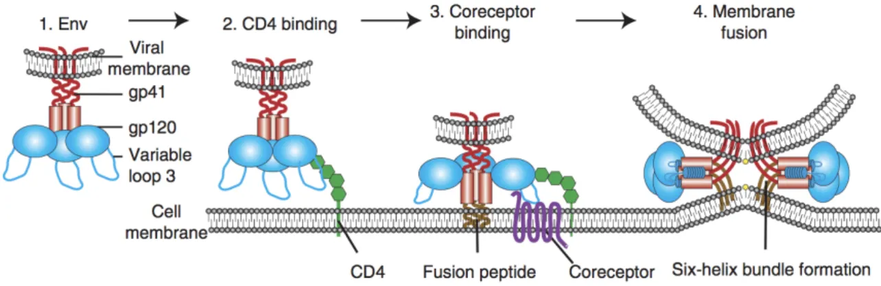
Figure 4: Schematic representation of HIV-1 binding and fusion.
During HIV-1 entry, the viral envelope (Env) binds CD4 receptor on the plasma membrane of target cells. Conformational changes in Env result in the engagement of the co-receptor, insertion of the fusion peptide and ultimately membrane fusion. From (Wilen et al, 2012).
In addition to CD4, HIV-1 also uses two chemokines receptors as co-receptor for virus entry: CXCR4 and/or CCR5. These receptors are seven-transmembrane G-protein coupled receptors and are involved in different cell-signaling pathways (Alkhatib, 2009). CXCR4 is a ubiquitously expressed protein while CCR5 in only expressed on immune cells such as macrophages, DCs and T cells. HIV-1 strains are divided according to their co-receptor usage: while X4 viruses use CXCR4 as co-receptor, R5 viruses use CCR5, and R5X4 strains use both co-receptors. The cell-tropism of the viral strains, X5, R5 or R5X4 was defined regarding the ability of the HIV-1 envelope glycoprotein gp120 to recognize respectively CXCR4, CCR5 or both (Berger et al, 1998). Usually, most of the viruses involved in primary infection are R5-tropic whereas most of the viruses during the symptomatic AIDS stage are X4-tropic viruses (Connor et al, 1997; Scarlatti et al, 1997).
1.2.4.
HIV-1 life cycle
HIV enters the cell through initial interactions between viral surface glycoproteins gp120 and cellular receptors CD4 and CXCR4 or CCR5. This binding leads to the fusion of the viral envelope with the host cell plasma membrane. Once in the cytoplasm, the viral core is uncoated, leading to the release in the cytoplasm of the viral genome and the enzyme required for the reverse transcription of viral RNA into double-strand viral DNA. As the first DNA strand is synthesized, the viral RNA is degraded by the enzyme RNase H, allowing synthesis of the complementary DNA. The newly synthesized double-stranded viral DNA, together with the integrase enzyme, and auxiliary proteins such as Vpr, form the pre-integration complex (PIC). This PIC is translocated from the cytoplasm to the nucleus
through the nuclear pore complex by an active mechanism. Once in the nucleus, viral DNA is integrated into the host cell genome by the integrase enzyme. The integrated viral genome will be then transcribed upon transcription of the host genome. The mRNAs and genomic RNA are transported to the cytoplasm where mRNAs are translated into viral proteins. After synthesis of viral proteins, HIV-1 proteins and viral genomic RNA are trafficked to the plasma membrane where they are assembled into new immature viral particles leading to budding and release of immature virions. In macrophages, assembly and budding does not take place at the plasma membrane but in internal compartments continuous with the plasma membrane. This specific step of viral replication in macrophages will be described later in more details (see 3.4 section – HIV-1 replication in macrophages). In the newly released viral particles the cleavage of the gag-pol polyprotein by the viral enzyme protease results in the maturation of the viral particles (Figure 5).
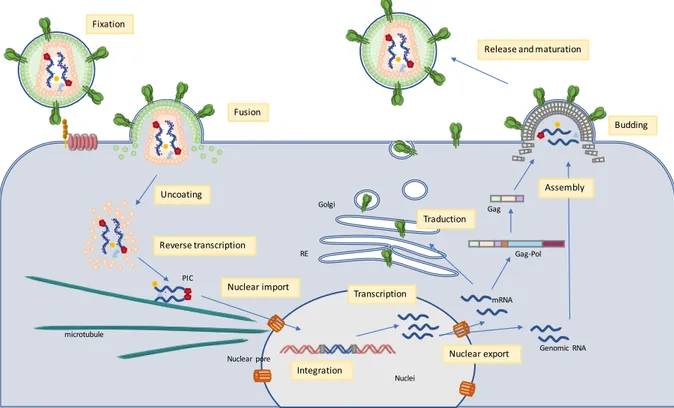
Figure 5: Simplified representation of HIV-1 life cycle.
The envelope glycoprotein (Env) recognizes the CD4 receptor and co-receptor at the surface of the target cell. Viral envelope fuses with the plasma membrane leading to the release of the HIV-1 capsid into the cytoplasm. Viral RNA is then uncoated, reverse transcribed into double-stranded cDNA, and imported into the nuclei where viral DNA is integrated in the genomic DNA of the target cell. It is then transcribed into mRNA and genomic RNA is exported back to the cytoplasm. mRNAs are translated into viral proteins which assemble at the plasma membrane with genomic RNA for budding of new immature viral particle. Once released into the extracellular space, HIV-1 becomes mature. From (Ganser-Pornillos et al, 2008).
Fixation Fusion Uncoating Nuclear import Integration Transcription Reverse transcription Nuclear export Traduction Assembly Budding Release and maturation microtubule PIC Nuclear pore RE Golgi Genomic RNA mRNA Gag-Pol Gag Nuclei
2. Mechanisms of cell-to-cell transmission of HIV-1
2.1. Cell-free and cell-to-cell infection
It is now well documented by numerous studies that in addition to cell-free infection, HIV-1 is able to infect target cells through cell-to-cell contacts with a virus infected-donor cell (Sattentau, 2008). At least in vitro, the establishment of cell-to-cell contacts between infected donor CD4+ T cells and target T cells leads to a massive and very efficient infection that may be 100-1000 times more efficient than cell-free infection (Carr et al, 1999; Chen et al, 2007; Dimitrov et al, 1993; Martin & Sattentau, 2009; Phillips, 1994). It is difficult to quantify the contribution of cell-free and cell-to-cell infection by HIV-1 in vivo, but some reports show that cell-to-cell infection could be the main route of HIV-1 infection
in vivo (Sourisseau et al, 2007; Zhong et al, 2013). Recently, using mathematical models,
Iwami et al. estimated that viral cell-to-cell transfer may correspond to about 60% of total HIV-1 infection in infected patients (Iwami et al, 2015) showing the relevance of HIV-1 cell-to-cell transmission studies.
The efficiency of cell-to-cell infection between CD4+ T cells has been related to the high multiplicity of infection (MOI) at the cell-cell contact site probably leading to the integration of multiple proviruses in the target cell (Agosto et al, 2014; Zhong et al, 2013). The high efficiency of cell-to-cell infection was also proposed to be responsible for partial escape to antiretroviral therapy (ART) and neutralizing antibodies (Sigal et al, 2011) but these results are controversial and will be discussed below in the manuscript (Agosto et al, 2015; Chen et al, 2007; Permanyer et al, 2012) (see 2.3 section - Virological synapses).
Several different structures have been described over the past years for cell-to-cell transmission of HIV-1 in vitro. Intercellular transfer of viral material can occur through cytoplasmic protrusions (tunneling nanotubes), membrane exchange (trogocytosis), establishment of infectious or virological synapses, cell fusion or cell engulfment. These different cell-to-cell structures will be described below.
2.2. Nanotubes and trogocytosis
2.2.1.
Nanotubes and filopodia
In a physiological context, cells can communicate through different pathways including cytoplasmic protrusions. Different types of membrane protrusions between two cells have been described in a large variety of cells such as neurons, glial cells and cells of the immune system (B, T and Natural Killer cells, neutrophils, dendritic cells and macrophages) both in vitro (Aggarwal et al, 2012; Rustom et al, 2004; Sherer et al, 2007) and in vivo (Chinnery et al, 2008; Lou et al, 2012; Pasquier et al, 2013; Seyed-Razavi et al, 2013).
Two different types of nanotubes have been described corresponding to close-ended nanotubes and open-ended nanotubes (also called tunneling nanotubes) (Kimura et al, 2013; Marzo et al, 2012; Onfelt et al, 2004). Intercellular communications involving tunneling nanotubes were first observed in 2004 and were described as a F-actin-containing membrane extension able to connect distant cells during minutes to hours (Rustom et al, 2004). Tunneling nanotubes are fragile and dynamic structures extended up to 100 µm in length with diameters ranging from 50 to 200 nm, and are not attached to the substratum (Rustom et al, 2004). Tunneling nanotubes can mediate and facilitate the transfer of cytoplasmic and cell-surface molecules and components between cells, but also cellular organelles (mitochondria)(Marzo et al, 2012).
Several studies showed that HIV-1 is able to use tunneling nanotube networks to move from one cell to another leading to virus cell-to-cell transfer. HIV-1 can thus be transferred through intracellular vesicles or by surfing along tunneling nanotubes (Eugenin
et al, 2009; Sowinski et al, 2008) (Figure 6). The frequency of tunneling nanotube formation
is not affected by HIV-1 in T cells (Sowinski et al, 2008) whereas, in macrophages, it was reported that the HIV-1 auxiliary protein Nef, together with the scaffolding protein M-Sec, may promote tunneling nanotubes formation (Eugenin et al, 2009; Hashimoto et al, 2016).
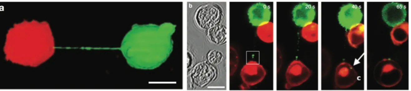
Figure 6: HIV-1 transfer across nanotubes in T cells.
a) Long membrane tethers, or membrane nanotubes, readily form between Jurkat T cells labelled with DiO (green) and DiD (red), n >500. b) Time-lapse imaging of Gag-GFP (green), expressed in the context of the fully infectious virus, along a membrane nanotube connecting infected with uninfected Jurkat T cells (stained with membrane dye DiD; red; n = 8). (Sowinski et al, 2008)
Another way of viral cell-to-cell transmission through membrane extension involving formation of filopodia was first described for transmission of the retroviral murine leukemia virus (MLV) (Sherer et al, 2007). Filopodia are F-actin-rich thin plasma membrane extensions, in contact with the substrate, that are involved in several cellular functions, such as chemo-migration, growth of nerve cones and formation of cell-cell contacts. Filopodia have been observed in HIV-1 infected cells expressing CD4 and CXCR4 where virus can “surf” on the outside of filopodia, and this process is dependent on the interaction between Env and the CD4 receptor (Lehmann et al, 2005; Sherer et al, 2007). Thus, unlike nanotubes, filopodia are attached with the substratum and dependent on the interaction between the receptor CD4 and HIV-1 envelope glycoprotein gp120.
2.2.2.
Trogocytosis
During the formation of the immunological synapse with antigen-presenting cells (APCs), membrane patches containing transmembrane proteins can be bi-directionally transferred from the surface of one cell to another one (Figure 7). This mechanism of membrane exchange is called trogocytosis (from the greek trogo meaning “gnaw”). Using trogocytosis, CD4+, CD8+ T cells or natural killer (NK) cells are able to capture membrane fragments from APC during antigen presentation (Joly & Hudrisier, 2003). Indeed CD4+, and CD8+ T cells are able to capture major histocompatibility complex class I or class II (MHC-I-and MHC-II), respectively, as well as the intracellular adhesion molecule ICAM-1 and the costimulatory molecules B7-1 (CD80), B7-2 (CD86), (Caumartin et al, 2006). Natural killer cells can also perform trogocytosis and thus acquire MHC-I or -II and transfer of the killer-cell immunoglobulin like (KIR) receptor expressed on natural killer cells, to T cells. One study reported that these membrane exchanges could also be involved in HIV-1 cell-to-cell transfer by increasing viral transfer during contacts between infected cells and
target cells (Blanco et al, 2004). Another study also reported some cases of trogocytosis between CD4+ and CD8+ T cells enabling the transfer of the CD4 receptor to CD8+ T cells (Aucher et al, 2010), suggesting that CD8+ T cells, expressing CD4 after trogocytosis from CD4+ T cells can acquire the ability to bind to HIV-1 leading to syncytia formation. The mechanism of trogocytosis is not well understood in the context of HIV-1 infection but may have important implications during the virological synapse process (described below).
Figure 7: Schematic illustration of a trogocytosis process between an antigen-presenting cell (APC) and CD4+ and CD8+ T cells.
During antigen presentation, APC, CD4+ and CD8+ T cells form immunological synapses, and upon dissociation of these synapses uproot membrane patches from APC. Thus, after immunological synapse dissociation, CD4+ and CD8+ T cells acquire APC specific components. (Caumartin et al,
2006) Immune synapse formation Immune synapse-mediated cross-talk Trogocytosis APC CD4+ T cells CD8+ T cells CD8+ T cells APC CD4+ T cells APC CD8+ T cells CD4+ Tcells TCR/CD3 CCR5 (CXCR4) MHCII CD4 CD28 CD86 MHCI
2.3. The Virological synapses
The formation of a so-called virological synapse is the major and well-established route for viral cell-to-cell infection, and was first described in the context of HTLV-1 (Human T-lympho-tropic virus) infection as a close and organized cell-to-cell contact between an infected donor cell and a target cell, enabling the transfer of viral material between the two cells (Igakura et al, 2003). The virological synapse has been named from some homologies with the immunological synapse formed between APCs and T cells for antigen presentation. During the formation of the immunological synapse, binding of the T-cell receptor (TCR) to the MHC-peptide complex expressed at the surface of APCs leads to T cell activation by transducing signals that cause transcriptional up-regulation of numerous genes and cell-proliferation (Huppa & Davis, 2003). The immunological synapse is very stable due to the interaction between the integrin Lymphocyte Function-Associated Adhesion molecule (LFA-1) and its ligand Intracellular Adhesion Molecule-1 (ICAM-1) but also through the interaction between the costimulatory receptor CD28 and its ligand CD86. The virological synapse has also been described in the context of HIV-1 infection and will be described in detail here.
2.3.1.
Structure of the virological synapse
The virological synapse between HIV-1 infected cells was defined by the group of Quentin Sattentau as a cytoskeleton-dependent, stable adhesive junction across which virus is transmitted by directed transfer (Jolly & Sattentau, 2004). The virological synapse shares several common features with the immunological synapse. Indeed, formation of both virological and immunological synapses involves the recruitment of receptors and cell adhesion molecules to an adhesive interface in an actin-dependent manner. I will here focus on the structure of the virological synapse established between a donor infected CD4+ T cell and a T cell target which has been the best documented. The specific synapses formed between T cells and DCs or macrophages will be described after (see 2.4 – heterogeneity of the virological synapses).
The virological synapse is a dynamic structure initiated by the recognition of the CD4 receptor at the surface of the target T cell by the HIV-1 envelope-surface glycoprotein gp120 expressed at the surface of the infected T cell (Figure 8). This interaction allows the recruitment of the Gag precursor to the intercellular interface (Jolly et al, 2004). The interaction between gp120 and CD4 is essential for the formation of the virological synapse
since a total inhibition of the formation of cell- cell conjugates and viral transfer was observed when gp120/CD4 interactions were blocked (Chen et al, 2007; Puigdomènech et al, 2008). This first step induces the recruitment of co-receptors CXCR4 or CCR5, adhesion molecules LFA-1, ICAM-1, actin, and other cell surface proteins such as tetraspanins to the contact site for stabilization of the virological synapse and efficient viral transfer. While some studies suggested that the formation of the virological synapse and virus transfer between CD4+ T cells at the virological synapse was independent of the co-receptor usage (Blanco et al, 2004; Chen et al, 2007; Jolly et al, 2004; Puigdomènech et al, 2008), other reports showed that the formation of the virological synapse and virus transfer required co-receptor expression and was inhibited by co-co-receptor antagonists (Dale et al, 2011; Felts et
al, 2010, 2009; Hübner et al, 2009; Martin et al, 2010). This discrepancy could be explained
by different assays since some teams looked at viral transfer when others analyzed viral production following virological synapse transfer. In this case co-receptor usage could have no effect on virological synapse formation but could be required for efficient infection after HIV-1 transfer across the virological synapse.
Similarly, the implication of LFA-1 and ICAM-1 is still a matter of debate. Initially, interaction between LFA-1 and ICAM-1, has been proposed to stabilize the virological synapse for efficient viral transfer. Jolly et al. first demonstrated that antibodies against LFA-1 and ICAM-1 were able to partially block the formation of the virological synapse (Jolly et al, 2007b). However, the percentages of inhibition were very different depending on the antibody used (40 to 90% inhibition for LFA-1 antibodies and 30% inhibition for ICAM-1 antibodies). Rudnicka et al. then confirmed the importance of adhesion molecules for the formation of the virological synapse and viral transfer (Rudnicka et al, 2009). They showed a significant three-fold decrease in virus transfer using T cells lacking the a subunit of LFA-1. Finally, a third group obtained opposite results showing that virus transfer through the virological synapse between T cells did not require LFA-1 binding to ICAM-1. Using antibodies blocking adhesion molecules LFA-1, ICAM-1 and ICAM-3, or 293T cells lacking LFA-1, no change in viral transfer through the virological synapse was observed in this study (Puigdomènech et al, 2008). The different assays used to analyze viral transfer and viral production could also explain this discrepancy. In the study by Puigdomènech et al., the LFA-1/ICAM-1 interaction could poorly affect viral transfer but seems to inhibit viral production after HIV-1 transfer through the virological synapse.
Finally, specific rearrangements of the cytoskeleton are required for the formation of the virological synapse and efficient viral transfer. Both actin and microtubules are indeed required for polarization of the Gag precursor and envelope glycoproteins to the site of
cell-cell contact (Jolly et al, 2007a; Rudnicka et al, 2009). During the immunological synapse the T cell receptor (TCR) interaction with its ligand, MHC molecules expressed at the surface of an antigen-presenting cell (APC), triggers a cascade of intracellular signals leading to cytokine gene expression, proliferation, and execution of the T cell effector functions. This cascade of events requires the activation of several protein tyrosine kinases such as ZAP-70 and cytoskeleton rearrangements. Using ZAP-70 deficient T cells, Blanchard et al, (2002) demonstrated that ZAP-70 signaling drives the T cell polarization through reorientation of the microtubule-organizing center (MTOC) at the cell-to-cell contact site (Blanchard et al, 2002). Similarly, polarization of MTOC at the virological synapse has been observed in 30 to 60% of the conjugated formed between infected donor T cells and target T cells (Jolly et
al, 2011; Sol-Foulon et al, 2007; Vasiliver-Shamis et al, 2009). An intracellular compartment
containing the viral envelope glycoproteins associates with the polarized MTOC during MTOC reorientation to the virological synapse, suggesting an active role of the microtubule network in the recruitment of the viral envelope at the virological synapse (Starling & Jolly, 2016). Polarization of organelles such as mitochondria, lysosomes, lipid bodies has also been observed at the site of cell-cell contact in 75% of the virological synapse (Jolly et al, 2011). Polarization of the cells can be mediated by ICAM1/LFA-1 signaling, which in addition to its effect on stabilizing virological synapse formation, induces a ZAP70-dependant signaling pathway for cytoskeleton remodeling, T cell polarization and efficient HIV-1 transfer across the virological synapse (Sol-Foulon et al, 2007; Starling & Jolly, 2016).
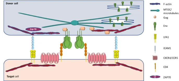
Figure 8: Schematic illustration of virological synapse formation.
HIV-1 envelope glycoproteins (Env) are expressed on the infected cell plasma membrane and interact with the receptors CD4 and CCR5 or CXCR4 on the target cell. The adhesion molecule intercellular adhesion molecule 1 (ICAM1) and lymphocyte function-associated antigen 1 (LFA1) stabilize the synapse. LFA-1/ICAM-1 interaction leads to ZAP70 signaling inducing the microtubule organizing center (MTOC) polarization to the cell-cell contact site. Microtubule and filamentous actin (F-actin) leads to recruitment of Env and Gag at the virological synapse for efficient viral transfer. (Sattentau, 2008)
2.3.2.
Viral entry downstream of the virological synapse.
The exact mechanisms of viral transfer through the virological synapse are still debated since some groups described that the virus enters in the target T cells through fusion with the plasma membrane in the inter-synaptic space, when others described endocytic-uptake of virus particles and then membrane fusion in an intracellular compartment (Blanco et al, 2004; Jolly & Sattentau, 2004) (Figure 9).
Zap 70 Zap 70 F-actin MTOC/ microtubules Gag Env LFA1 ICAM1 CXCR4/CCR5 CD4 ZAP70 Donor cell Target cell
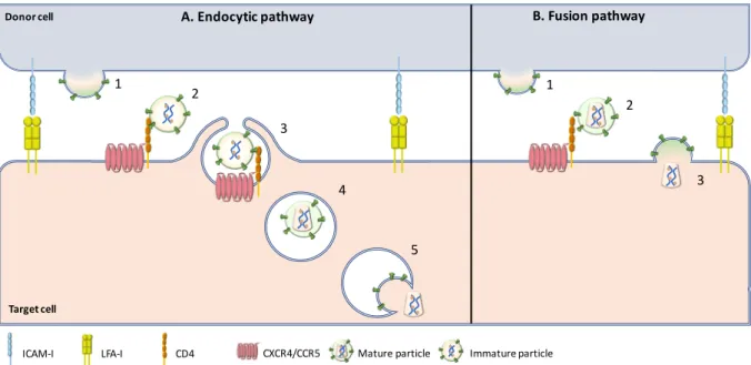
Figure 9: Hypothetical mechanisms of HIV-1 entry across Virological synapse.
(A) Endocytic pathway: HIV-1 budding from an infected T cell leads to the production of immature virus particles at a virological synapse (1). Immature virions then interact with the CD4 receptor but are incompetent for viral fusion (2). Immature particles are thus internalized by endocytosis (3). Maturation happens in internal compartments (4) leading to viral fusion and release of viral capsid into the cytoplasm of the target cell (5). (B) Fusion pathway: HIV-1 budding from an infected T cell leads to the production of mature, fusion- competent virions (1). Mature viruses can thus bind to CD4 (2) and leads to viral-cell fusion for release of viral material in the cytoplasm of the target cells for viral transfer (3)(Sattentau, 2010).
The first observation of the virological synapse by electron microscopy by Jolly et al. showed that mature HIV-1 particles were found in the synaptic space (Jolly & Sattentau, 2004), suggesting that mature viruses released from the donor T cells at the virological synapse could fuse directly with the plasma membrane of the target cells. Another group also observed HIV-1 particles in the synaptic space but suggested that these particles can be internalized by lamellipodia-like structures (Blanco et al, 2004). They also showed that HIV-1 was endocytosed in a trypsin-resistant compartment observed by electron microscopy. Several groups confirmed the mechanism of endocytosis of HIV-1 at the virological synapse. It has been shown that immature HIV-1 particles can be transferred across the virological synapse through dynamin- and clathrin-dependent endocytosis leading to productive infection of target T cells (Hübner et al, 2009; Miyauchi et al, 2009; Ruggiero
et al, 2008; Sloan et al, 2013). HIV-1 particles contained in intracellular compartments
colocalized with the early-endosomal marker EEA1 but not with the lysosomal-associated membrane protein LAMP1 suggesting that viruses are internalized in endosomal compartments but are not addressed to lysosomal degradation (Bosch et al, 2008). In accordance with these results, Dale et al demonstrated that after HIV-1 endocytosis, the
Donor cell
Target cell
A. Endocytic pathway B. Fusion pathway
1 2 3 4 5 1 2 3
cleavage of gag polyprotein by the viral protease induced maturation of viral particles in endosomal compartments (Dale et al, 2011). This cleavage restores the viral membrane fusion activity and thus leads to viral-cell membrane fusion in these compartments (Figure 10).
Figure 10 : Model for Maturation-Induced Fusion at the Virological Synapse.
(A) In the steady state, an HIV-1 infected T cell has a diffuse distribution of Env and Gag. (B) The engagement of Env on the infected donor cell, with CD4 on the target cell, induces an adhesion event and results in the recruitment of Gag, Env, and CD4 to the virological synapse. (C) An endocytic event is triggered resulting in CD4-dependent uptake of immature virus into acceptor cell intracellular compartments. (D) Bound to CD4, virus particles undergo protease-dependent maturation over time. (E) Viral particle maturation triggers viral membrane fusion, releasing the capsid to the cytoplasm. (Dale et al, 2011)
Finally, the group of Sattentau, who first described the formation of the virological synapse for HIV-1 transfer failed to observe endocytosis of HIV-1 across the virological synapse using confocal microscopy, electron microscopy or electron microscopy coupled with tomography (Jolly & Sattentau, 2004; Martin et al, 2010). Puigdomenèch et al. hypothesize that these different results could be explained by different experimental systems and the use of primary or immortalized cell lines (Puigdomènech et al, 2009). They suggest that the differences observed between primary CD4 T cells and cell lines might be associated with the kinetics of fusion events. Delayed fusion at the cell membrane may increase endocytosis in primary cells. In contrast, rapid fusion kinetics at the cell membrane in cell lines may favor transmission of HIV infection with lower levels of endocytosis. Thus they suggest that the endocytosis of HIV particles is the main mechanism of HIV transfer in primary CD4 T cells.
2.3.3.
Viral transfer across the virological synapse and resistance to
neutralizing antibodies and antiretroviral drugs
As described above (see 2.1 section, Cell free and cell-to-cell infection), viral transfer of HIV-1 across the virological synapse between an infected donor T cell and a recipient T cells is more efficient than cell-free infection for productive infection of the target T cells. Like for other viruses (Herpesviruses, poxviruses and Hepatitis C viruses), it has been proposed that HIV-1 could escape, at least partially, neutralization by specific antibodies targeting the viral envelope when it is transferred across the virological synapse. This was already suggested in 1995 by Pantaleo et al. who described that neutralizing antibodies against the glycan V3-loop of gp120 were unable to block cell-to-cell viral transfer (Pantaleo et al, 1995). However, after characterization of the virological synapse for HIV-1 cell-to-cell transfer in 2004, several different groups tried to elucidate the mechanisms of neutralizing antibody escape in virological synapse-mediated viral transfer. There is a general agreement that the potency of neutralizing antibodies is reduced during cell-to-cell transmission compared to cell-free infection and that only a subset of neutralizing antibodies can efficiently inhibit cell-to-cell transmission (Abela et al, 2012; Blanco et al, 2004; Jolly & Sattentau, 2004; Jolly et al, 2004; Malbec et al, 2013; Massanella
et al, 2009). For example, Abela et al. demonstrated that several specific anti-CD4 binding
site antibodies lost considerable potency (10- to 100-fold decrease) when HIV-1 was transferred by cell-to-cell transmission (Abela et al, 2012). However, if some antibodies are still able to block cell-to-cell transmission at high concentration, VRC01, which is one of the most potent antibodies for inhibition of cell-free infection, is particularly ineffective for blocking cell-to-cell viral transfer. While most anti-gp120-directed antibodies, and in particular those directed against the CD4 binding site of gp120, displayed a reduced activity during viral cell-to-cell transmission, the same group reported that gp41-directed inhibitor T20 and neutralizing antibodies targeting gp41 maintained their activity and are thus able to block both cell-free and cell-to-cell infection with the same efficiency. However, other groups showed that anti-gp41 antibodies failed to inhibit cell-to-cell transmission (Blanco et
al, 2004; Chen et al, 2007; Massanella et al, 2009). Globally, efficiency of neutralizing
antibodies for neutralization of the VS-mediated viral transfer is variable and some epitopes of the viral envelope glycoproteins seem more susceptible than others to neutralization of VS-mediated viral transfer.
Similarly, activities of entry inhibitors and antiretroviral drugs on HIV-1 transmission through the virological synapse is still a matter of debate. A lot of opposite results have been published regarding the effect of entry inhibitors. Whereas Chen et al. initially reported that the peptide entry inhibitor of Env-mediated membrane fusion T20, targeting the gp41 transmembrane glycoprotein, was unable to block VS-mediated viral transfer using flow cytometry analysis (Chen et al, 2007), Martin et al., then showed that cell-free and cell-to-cell infection across the virological synapse were equivalently susceptible to T20, using qPCR for detection of infection (Martin et al, 2010). These different results can be explained by the different experimental approaches used since the first study was looking for viral transfer of viral material when the other study analyzed
de-novo synthesized viral DNA after viral transfer. Thus, we can hypothesize that the T20
entry inhibitor does not affect viral transfer across the virological synapse but inhibits HIV-1 infection in the target cell after the VS-mediated viral transfer. Fusion inhibitors, such as T20, could have no effect on endocytosis of HIV-1 (viral transfer) but could then block viral fusion in the endosomal compartments leading to the inhibition of the productive infection.
Other studies reported that protease inhibitors inhibited HIV-1 VS-mediated infection similarly to cell-free infection (Agosto et al, 2014; Sigal et al, 2011). Regarding reverse transcriptase inhibitors, it seems that non-nucleoside-analog reverse transcriptase inhibitors (NNRTI) could block VS-mediated infection (Agosto et al, 2014; Sigal et al, 2011) whereas nucleoside-analog reverse transcriptase inhibitors (NRTI) were unable to do it (Agosto et al, 2014). However, other results from Sigal et al showed that NRTI were also able to block cell-to-cell infection even if the level of inhibition is lower than for cell-free infection (Sigal et al, 2011). Finally, it seems that the idea that the cell-to-cell viral transfer through the VS can escape from neutralizing antibodies and antiretroviral therapies is not so clear and is probably dependent on the inhibitors used.
2.4. Heterogeneity of virological synapses
Most of the studies regarding HIV-1 transmission through the virological synapse have been done between an infected donor T cell and a recipient target T cell. However, dendritic cells and macrophages can also perform cell-to cell transmission to target T cells through the formation of a related virological synapse. Thus, several groups investigated virological synapses formation using dendritic cells (DCs) or macrophages as infected donor cells. If cell-to-cell transfer between DCs and T cells, and between macrophages and