HAL Id: tel-02180575
https://tel.archives-ouvertes.fr/tel-02180575
Submitted on 11 Jul 2019
HAL is a multi-disciplinary open access archive for the deposit and dissemination of sci-entific research documents, whether they are pub-lished or not. The documents may come from teaching and research institutions in France or abroad, or from public or private research centers.
L’archive ouverte pluridisciplinaire HAL, est destinée au dépôt et à la diffusion de documents scientifiques de niveau recherche, publiés ou non, émanant des établissements d’enseignement et de recherche français ou étrangers, des laboratoires publics ou privés.
Identification of new genes involved in hereditary
steroid-resistant nephrotic syndrome using next
generation sequencing and in vivo functional
characterization in drosophila melanogaster
Sara De Almeida Gonçalves
To cite this version:
Sara De Almeida Gonçalves. Identification of new genes involved in hereditary steroid-resistant nephrotic syndrome using next generation sequencing and in vivo functional characterization in drosophila melanogaster. Urology and Nephrology. Université Sorbonne Paris Cité, 2017. English. �NNT : 2017USPCB030�. �tel-02180575�
Doctoral Thesis of
PARIS DESCARTES University
Doctoral School of Bio Sorbonne Paris Cité
Sara DE ALMEIDA GONÇALVES
A dissertation submitted in accordance with the requirements for the
award of the degree of PhD in Science
Identification of new genes involved in hereditary
steroid-resistant nephrotic syndrome using next generation
sequencing and in vivo functional characterization in
Drosophila melanogaster
Defense date - 28
thSeptember, 2017
Jury panel
Pr. Alain FISCHER
Dr. Clément CARRÉ
Pr. Nine KNOERS
Pr. Moin SALEEM
Pr. Corinne ANTIGNAC
1
To my father
3
Acknowledgements
5
Acknowledgements
At the end of this important chapter of my life I would like to acknowledge all the people who have, directly and/or indirectly, allowed me to grow in this passionate and always partially mysterious world of science.
First and foremost I would like to thank my mentor Corinne Antignac. Thank you for guiding me through the world of genetics, methods of research, in developing my passion for science and for all the exciting discussions. Thank you for always being there, for demanding a lot but only because you cared. Thank you for your patience and your kindness with me. Thank you for your example of strength, perseverance and passion, even when obstacles come. Thank you for your kind attitude in difficult or joyful moments of my personal life. These are things that I will always be grateful of, much more so than I can express.
To Matias Simons, thank you for accepting me into your lab whilst you were still in Freiburg, where I first met the fruit fly under a stereomicroscope. Thanks for introducing me to the world of Drosophila and for guiding me in the Drosophila experiments. Thanks for all the exciting lab meetings and also for your help in the conclusion and writing of the ADD3 and KAT2B paper.
To the reviewers of my thesis, Clément Carré and Nine Knoers, as well as to the other members of the jury, Alain Fischer and Moin Saleem, who have agreed to evaluate my work.
To Julie Patat, who helped me in my experiments and was brave enough to sequence the KAT2B gene in 73 patients and to start dangerous and new experimental procedures such as the heat-fixation (I hope you will go ahead with your patent!).
To Noëlle Lachaussée, the M2 student who worked with me in the initial phases of the ADD3 and KAT2B project. Thanks for your enthusiasm, speed and energy.
To Christelle Arrondel, with whom I learned the ABC of experimental techniques at the bench! Thank you for all of your teaching and patience. To the remaining podocyte team, both present and past members: Géraldine Mollet (for your scientific advice and joyful character), Olivier Gribouval (for all your help in and outside the lab), Evelyne Huynh Cong (the queen of podocytes in culture, for all the help with protocols and experiments), Olivia Boyer (for your encouragement and help with patient data), Guillaume Do al the )e afish o petito … D osophila is ette ! , Ma ia “e a o o e e lose to e , Lau i e Bus a a, F a es Tille ou k o I do ot a t to kill ou, do t ou? a d Giulia Menara. To Kalman Tory and his endless good humor! May all of your work reveal more and more of this octopus cell! To Da iel Poul f o the sti osis tea , fo his Audi st lé , a otu es,
é-f o t a d othe a azi g Ca a a -like jokes .
To Fred Bernard and Maria Rujano, the top Drosophilists that guided me in the passionate world of Drosophila. To the rest of the Simons team, including those who have already left, Simon Gerber (hope to see you soon again), Clara Guida (who still worked with me on the other side of the Atlantic, breaking the hearts of adult flies), Virginie Hauser and Zvoni Marelja (now you are the only one to keep the good old hu o i the la !!! …a d to the o e e e t e e s of the la : Vale ti a
6
Marchesin and Magda Cannata Serio (the Italian team) Mathilda Bedin, Gwenn Le Meur, Laure Villoing-Gaudè, Albert Pérez. Thanks to all of you for making my days always more joyful!
To Marion Delous, with whom I shared my office and my jokes day after day (imagine how tough that can be for one person alone!). Thank you for not despairing! Thank you for our scientific discussions and for checking each of my scientific projects, publications, posters and oral presentations as if they were your own!
To Rebecca Ryan, thank you for reading my thesis, for your scientific and English-native-speaker advice. Thanks for encouraging me in the dark days, especially during the writing of the sphingolipids chapter, and for your great creativity (if you ever doubt it you can just look at your knitted creations!).
To the other teams of the lab and their PIs: Sophie Saunier, Alexandre Benmerah, Cécile Jeanpierre. Thank you for your kindness and nice discussions. To the old ones, starting with Albane Bizet (who is at least 1000 years old), who looked after me from the very beginning, gave me a place to stay when I was homeless and always helped me with tough experimental procedures; to Flora Legendre al a s ith a i e s ile to e ; to G eltas Od e, e e though e fight ea h othe ou k o I ha e e e lo ed ou! I ould ot e ithout ou hat alade a d ou dé iles jokes; a d to the young ones in the lab, Hugo Garcia (may the force be with you on your new leader position in the S.F. group), Louise Reilly (may the force be with you to defeat Hugo Garcia in the S.F. group and continue making cakes), petite Marie Dupont (viva le bigeminism!), Lara De Tomasi (after all your laugh is not awkward), Charline Henry (bon courage!), Esther Porée (if you want to reconsider you can still enter the S.F. group).
To the secret friends group (you know who you are!). Thank you for your unconditional friendship and amazing cakes!
To the ones that were in the lab when I started but have already left - Maxence Macia (the sweetest friend), Valentina Grampa dea est Italia f ie d, house ate a d the o e I as té oui e of), Zuza Andrzejewska, Lucie Thomas, Fabien Nevo, Nathalie Nevo. For most of you we continue linked by the outside-lab life (yes, there is one!).
To Meriem Garfa-Traoré and Nicolas Goudin, for their help with confocal microscopy. Without you (and Rebecca Ryan) there would have been no prize for the best funny picture of the YRII congress. To Antonio Gomes da Costa, who understood my passion for research and allowed me to start this PhD.
Lastly, my gratefulness to all my family and friends: Thomas, thank you for enduring the thesis- stress with me, for the endless thesis formatting and for all your kindness to me; Mariana and mum, thank you for your infinite patience and support; Teresa, Cristina, Ines, Cristininhas, Fili, Mariana, Eunice, Filipas, Mordi, and all the others that I do not mention here, thank you for your everlasting friendship.
7
Abstract
9
Abstract
Nephrotic syndrome (NS) is a kidney disease characterized by disruption of the glomerular filtration barrier and the massive loss of proteins into the urine. Although in the majority of cases treatment with steroids leads to remission of the disease, in 15-20% of cases the disease is not responsive to this therapy and is classified as steroid-resistant nephrotic syndrome (SRNS). SRNS is a clinical condition with high morbidity leading to progressive renal failure as well as multiple metabolic and cardiovascular complications. Extensive research over the last 20 years has identified more than 40 SRNS causing genes that are crucial for function of the podocyte, a highly specialized kidney epithelial cell. However, the mutated gene is still unknown in about half of the familial cases. We have used exome sequencing to identify new genes mutated in SRNS. In order to prove the pathogenicity of the identified mutations we used the Drosophila model, assessing defects of fly viability and the structure and function of nephrocytes, podocyte like-cells. My thesis work consists of two projects. Firstly, we identified biallelic mutations in a new candidate gene, SGPL1, encoding the sphingosine 1-phosphate lyase, in individuals presenting SRNS with facultative adrenal insufficiency, ichthyosis, neurological defects and immunodeficiency. SGPL1 is the main catabolic enzyme of sphingolipids, irreversibly degrading sphingosine 1-phosphate into phosphoethanolamine and hexadecenal. In flies, these mutations were shown to decrease viability, induce nephrocyte defects and lead to the accumulation of sphingoid bases due to the loss of SGPL1 catabolic activity. Together, these results indicate that the identified SGPL1 mutations are pathogenic and cause a new syndromic form of SRNS.
Moreover, in a second project, we defined the contribution of homozygous mutations found in two different genes, ADD3 and KAT2B, to a complex phenotype found in affected individuals from one consanguineous family. These individuals presented with neurological defects, cataracts, mild skeletal defects, cardiomyopathy and SRNS. ADD3 encodes adducin, an F-actin capping protein that also links the actin cytoskeleton to the spectrin based membrane skeleton, while KAT2B encodes the lysine acetyltransferase 2B, mainly known for acetylation of histones and modulation of transcriptional programs. We found additional nonrelated patients carrying only biallelic ADD3 mutations that presented a partially overlapping syndrome but with no cardiac or renal manifestations. In the Drosophila model we found that both ADD3 and KAT2B mutations impaired fly viability and that the ADD3 mutation also impaired fly motor function. However, only the KAT2B mutation induced functional defects in Drosophila heart and nephrocytes. Altogether, these results suggest that ADD3 mutations are responsible for a neurological phenotype with facultative cataracts and skeletal defects while the KAT2B mutation induces heart and kidney defects.
These results highlight the Drosophila as a good in vivo model to test the pathogenicity of the mutations found in SRNS candidate genes.
11
Table of Figures
13
Table of Figures
Figure 1. The glomerulus and the glomerular filtration barrier. Figure 2. Ultrastructure of the podocyte.
Figure 3. Podocyte structure in normal conditions and upon injury.
Figure 4. Ultrastructural changes accompanying podocyte injury and detachment. Figure 5. Mechanisms underlying idiopathic nephrotic syndrome.
Figure 6. Histologic Variants of FSGS.
Figure 7. Progression of glomerular diseases.
Figure 8. Parietal epithelial cells participate in the progression of glomerulosclerosis. Figure 9. The slit diaphragm zipper-like structure.
Figure 10. New EM techniques suggest a novel slit diaphragm structure. Figure 11. Main molecular components of the slit diaphragm.
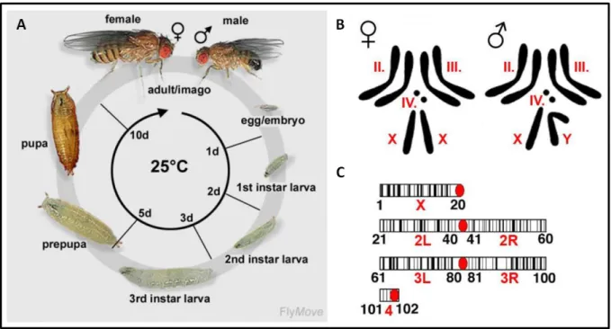
Figure 12. The life cycle of Drosophila melanogaster and its chromosomes.
Figure 13. Simplified evolutionary patterns of essential genes and their association with Mendelian disorders.
Figure 14. Genetic strategies used in Drosophila for the study of human diseases. Figure 15. Some of the strategies for genetic manipulation in Drosophila.
Figure 16. Homology between human and Drosophila excretory systems.
Figure 17. Molecular homology between the components of the podocyte and the nephrocyte slit diaphragm.
Figure 18. Distribution of endocytic vesicles in nephrocytes. Figure 19. Nephrocyte subpopulations in Drosophila. Figure 20. Nephrocyte foot process effacement. Figure 21. Chemical structure of sphingolipids. Figure 22. Overview of the sphingolipid metabolism.
Figure 23. Cellular localization and trafficking pathways of sphingolipids. Figure 24. Nucleosome structure.
15
List of Abbreviations
17
List of Abbreviations
ABC ATP-binding cassette APOL1 Apolipoprotein L1
ARHGAP24 Rho-GTPase-activating protein 24 ARHGDIA Rho GDP-dissociation inhibitor 1 ASMase Acid sphingomyelinase
ATG9A Autophagy related protein 9A
BDSC Bloomington Drosophila Stock Center bHLH Basic Helix Loop Helix
C1q Complement C1q
C3 Complement C3
CD2AP CD2 associated protein Cerases Ceramidases
CerS Ceramide synthases CERT Ceramide transfer protein
CHD2 chromodomain helicase DNA binding protein 2 ChIP Chromatin Immunoprecipitation
CNS Central nervous system CoRL Cells of renin lineage
CRB2 Crumbs homolog 2
CRISPR Clustered Regularly Interspaced Short Palindromic Repeats DAD Auto-regulatory domain
DAG Diacylglycerol
DID Diaphanous inhibitory domain DMS Diffuse mesangial sclerosis DNMT DNA methyltransferase
Dpp Decapentaplegic
DZNep S-adenosylhomocysteine hydrolase inhibitor 3-deazaneplanocin A
EM Electron microscopy
EMS ethylmethanesulfonate
ER Endoplasmic reticulum
ERK Extracellular signal-regulated kinase ESRD End-stage renal disease
EZH2 Enzyme enhancer of zeste homolog 2 FGF Fibroblast growth factor
FIB-SEM Focused ion beam-scanning electron microscopy FSGS Focal and segmental glomerulosclerosis
GAP GTP activating factors Gb3 Globotriaosylceramide
GBM Glomerular basement membrane
GDIs Guanine dissociation inhibitors GEFs Guanine nucleotide exchange factors GFB Glomerular filtration barrier
GFP Green fluorescent protein GLA Alpha-galactosidase A
18
GWAS Genome-wide association studies HAT Histone acetyltransferase
HDAC Histone deacetylase
HEK Human embryonic kidney
IgM Imunoglobulin M
INF2 Inverted formin 2
IQGAP Iq motif containing GTPase-activating protein JNK c-Jun N-terminal kinases
JUNK Jun-N-terminal protein kinase
KANK2 KN motif and ankyrin repeat domain-containing protein 2 KAT Lysine acetyl-transferase
KAT2A Lysine acetyl transferase 2A KAT2B Lysine acetyl transferase 2B
KD Knock down
KDAC Lysine deacetylase Kirre Kin-of-irre
KO Knock out
LOF Loss of function
LPS Lipopolysaccharide
MAGI2 Membrane-associated guanylate kinase 2 MCD Minimal change disease
mDia Mammalian diaphanous related formin
MELAS Mitochondrial encephalomyopathy, lactic acidosis and stroke-like episodes MEFs Mouse embryonic fibroblasts
mTOR Mammalian target of rapamycin MYO1E Myosin 1E
NGS Next generation sequencing
NS Nephrotic syndrome
Ntrk3 Neurotrophic tyrosine kinase receptor, type 3 N-WASP Neural Wiskott-Aldrich-syndrome
PAK p21 activated kinase
PAN Puromycin aminonucleoside nephrosis PCAF P300/CBP associated factor
PDGF Platelet derived growth factor
PE Phosphoethanolamine
PECs Parietal epithelial cells
PHB Prohibitin
PI3K Phosphoinositide 3-kinase PP2A Protein phosphatase 2A
PTM Post translational modifications
PTPRO Protein tyrosine phosphatase, receptor type O RFP Red fluorescent protein
RNAi RNA interference RPA Replication protein A S1P Sphingosine 1 phosphate S1PR S1P receptors
SGPL1 sphingosine 1 phosphate lyase 1 SIOD Schimke Immuno-Osseous Dysplasia
19 SMARCAL1 SWI/SNF Related, Matrix Associated, Actin Dependent Regulator of Chromatin,
subfamily A-Like 1 SMases Sphingomyelinases
SMPDL-3b Sphingomyelinase-like phosphodiesterase 3b Sns Sticks-and-stones
Sparc Secreted protein acidic and rich in cysteine SPHK Sphingosine kinases
SPNS2 Spinster 2
SRNS Steroid-resistant nephrotic syndrome SSNS Steroid-sensitive nephrotic syndrome
SUPAR Soluble urokinase plasminogen activator receptor TEM Transmission electron microscopy
TRPC6 Transient receptor potential channel, subfamily C, member 6 UAS Upstream activating sequence
VDRC Vienna Drosophila Resource Center VEGF Vascular endothelium growth factor
WAVE WASP-family verprolin-homologous protein
WES Whole exome sequencing
Wg Wingless
WGS Whole genome sequencing
WT1 Wilms tumour 1
21
Table of Contents
23
Table of Contents
Acknowledgements ... 3 Abstract ... 7 Table of Figures ... 11 List of Abbreviations ... 15 Table of Contents ... 21 Foreword ... 25 Review of the Literature ... 29 The glomerulus and the glomerular filtration barrier ... 31 The podocyte ... 33 The nephrotic syndrome ... 34 Immune forms of nephrotic syndrome: the circulating permeability factor ... 37 Histologic variants of idiopathic nephrotic syndrome ... 38 Mechanisms of podocyte injury and glomerulosclerosis ... 41 Podocyte detachment, incapacity for cell division and glomerulosclerosis ... 41 Podocyte regeneration by stem cells ... 43 The podocyte slit diaphragm, lipid rafts and actin cytoskeleton ... 46 The slit diaphragm ... 46 Podocyte lipid rafts ... 52 The podocyte actin cytoskeleton and its regulation ... 52 Genetics of steroid-resistant nephrotic syndrome ... 57 Genetic susceptibility factors and oligogenism ... 60 Mendelian forms: Non-syndromic vs syndromic SRNS ... 63 NGS and the need for high-throughput functional testing in model organisms ... 64 The Drosophila odel as a tool to stud podo topathies a d o e… ... 66 Drosophila for the study of human diseases ... 66 The Drosophila excretory system – an analogy to the human kidney ... 71 The nephrocyte as a model for human podocytopathies ... 76 Sphingolipids and kidney disease ... 80 Sphingolipids and their metabolism ... 80 Bioactive sphingolipids and their regulation ... 82 Sphingolipids in podocyte biology ... 87 Epigenetic regulation and podocyte homeostasis ... 9124
Epigenetic mechanisms ... 91 Epigenetic regulation in podocytes ... 95 Results and Discussion ... 99 Part I ... 101 A new syndromic form of SRNS caused by SGPL1 mutations ... 101
Article: Mutations in sphingosine-1-phosphate lyase cause nephrosis with ichthyosis and adrenal insufficiency ... 103 Summary of the findings and discussion ... 141 Phenotypic variability ... 142 Mechanisms of the disease in different organs ... 145 Potential therapies ... 151 Part II ... 153 ADD3 and KAT2B, two novel SRNS candidate genes? ... 153
Article: A homozygous mutation in KAT2B extends the spectrum of ADD3-dependent intellectual disability syndrome. ... 155 Summary of the findings and discussion ... 199 ADD3 mutations ... 200 KAT2B mutations ... 203 Synergic/additive effect between ADD3 and KAT2B loss of function mutations? ... 205 Possible pathogenic mechanism mediated by ADD3 loss of function ... 206 Possible pathogenic mechanisms mediated by KAT2B loss of function in podocytes ... 207 Conclusion of ADD3 and KAT2B project ... 208 Conclusion ... 209 Bibliography ... 213
25
Foreword
27
Foreword
In the last twenty years, genetic studies on familial forms of steroid-resistant nephrotic syndrome (SRNS) have allowed the identification of mutations in more than 40 genes that are crucial for the function of the podocyte, a highly specialized epithelial cell that lies in the glomerular filtration barrier (GFB). However, in more than half of the familial cases of SRNS, the underlying molecular defect is still unknown. The era of next-generation sequencing (NGS) brought new opportunities for the cases where a molecular diagnosis had not been established, however it also came with new challenges, in particular to address the functional impact of multiple rare and private variants. In this thesis we have used the Drosophila model to answer this need. Its short life cycle and the availability of advanced genetic tools make it an attractive model for studying the pathogenicity of mutations found in new candidate genes. Moreover, flies have nephrocyte cells which are homologous to podocytes, allowing us to specifically address the impact of the mutations found in SRNS patients. In the introductory chapter of this thesis I start by detailing the general physiology and structure of the GFB and particularly of the podocyte, and reviewing the concept of SRNS as well as the mechanisms known to be involved in the podocyte response to injury and progression of glomerular disease. I then briefly review the structure of the podocyte slit-diaphragm, a modified cell junction that allows maintenance of the permselectivity of the GFB, and the podocyte actin cytoskeleton. This is followed by the genetics of the podocytopathies with a focus on the syndromic forms of the SRNS. I then present the advances and pitfalls of NGS techniques and the main advantages of the Drosophila model, focusing on the nephrocytes and their similarities to podocytes. I finish the introduction by exposing two novel thematics related to the two SRNS candidate genes discovered during this thesis: the complex sphingolipid metabolism and functions, and the fine-tuning of the DNA genomic information conferred by the epigenetic machinery. In both cases I give a special emphasis to the relationship of these mechanisms to the podocyte.
The results and discussion chapter is divided into two main parts, each focusing on one of the two different projects of this thesis. For each project, results are presented in the form of a journal manuscript and a summary of the findings and discussion then follows. In the first project we have identified mutations in SGPL1, encoding sphingosine -1-phosphate lyase, as a cause of a new syndromic form of SRNS, where the main extra-renal features were adrenal insufficiency, ichthyosis, immunodeficiency and neurological defects. We defined the pathogenicity of the mutations and started to address the impact of SGPL1 loss of function on the metabolism of sphingolipids and on the function of the nephrocytes. In the discussion I mainly focus on the phenotypic heterogeneity of
28
the disease and the possible underlying pathogenic mechanisms in the affected organs. In the second project we have defined the pathogenicity of biallelic mutations found in ADD3 and KAT2B in affected individuals from one consanguineous family presenting a complex syndromic form of nephrotic syndrome. I discuss the contribution of ADD3 and KAT2B mutations to the patient phenotype, the potential pathogenic mechanisms in the podocyte, as well as the difficulties that we found during the project.
This thesis will finish with a brief conclusion that highlights the great advantages but also the pitfalls that can be encountered when using the Drosophila model in human genetics.
All the work in this thesis was done under the direction of Corinne Antignac, my thesis supervisor, and Matias Simons, who shared with us his expertise on the Drosophila model and with whom we closely collaborated.
29
Review of the Literature
31
The glomerulus and the glomerular filtration barrier
The main role of the kidney is the regulation of the hydric, acid-base and ion balance and excretion of toxic compounds produced by cell metabolism. To achieve these functions the kidney depends on small functional structures called nephrons, which number up to ~1x10⁶ per human kidney. Each nephron is composed of a glomerulus (Figure 1), responsible for the plasma filtration, and tubules that further modify this primary filtrate through secretion and reabsorption of small molecules, electrolytes and water, to finally produce urine.
Each day the kidney filters approximately 180 L of plasma. Blood enters the glomerulus through a small pre-glomerular arteriole (afferent arteriole) that then branches into a series of capillaries that form the glomerular capillary tuft. These then join again in another arteriolar, post-glomerular, vessel (efferent arteriole). This specialized vascular bed allows the fine-tuning of hydrostatic pressure inside the capillaries, the main driving force for filtration. Blood is thus filtered across the glomerular filtration barrier (GFB) to produce the primary urine.
The GFB has three layers. The first is formed by the fenestrated endothelium of glomerular capillaries. The meshwork-like glomerular basement membrane (GBM) forms the second layer, while the final layer is formed by specialized epithelial cells, termed podocytes, which extend foot processes that interdigitate and organize into a network that covers the outer surface of the capillary loops (Figure 1).
The main function of the GFB is to maintain permselectivity. This means that in normal homeostatic conditions the GFB is able to freely allow the passage of water, ions, by-products of cellular metabolism and small proteins but not blood cells or high molecular weight proteins. All three layers participate in this function (Kalluri 2006). The fenestrated endothelial cells prevent the passage of the blood cellular elements and the negative charge of the endothelial glycocalyx impedes the filtration of negatively charged molecules. Moreover the GBM dense meshwork also constitutes an obstacle to the passage of high molecular weight proteins. The GBM is around 300 nm in thickness, and is mainly composed of laminins, collagen IV, agrin and nidogen (Suh and Miner 2013, Lennon, Randles et al. 2014). Podocytes make the final layer and play a key role in GFB permeability. Interestingly, it has been proposed that filtration itself generates a local electric field at the GFB that also prevents the plasma proteins from entering or crossing the glomerular filter, explaining why the GFB does not clog (Moeller and Tenten 2013).
Besides the endothelial cells and podocytes, mature glomeruli also contain mesangial cells which are embedded in the mesangial matrix and are classically known to contribute to the structural integrity
32
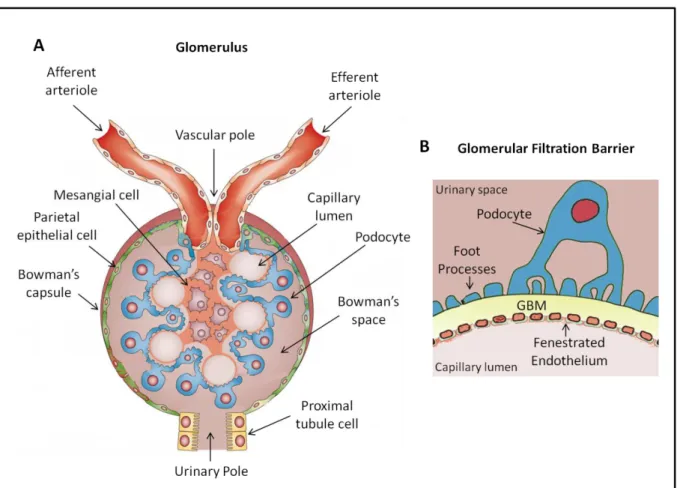
Figure 1. The glomerulus and the glomerular filtration barrier. (A) Structure of the glomerulus. The glomerulus contains a capillary tuft that receives its blood supply from the afferent arteriole. Podocytes line the outer aspect of the capillaries and their cell bodies float in the Bo a s spa e. Mesa gial ells a e e edded i the esa gial at i a d p o ide structural support to the capillary tuft. The Bowman capsule delimitates the glomerulus and is lined by parietal epithelial cells. (B) The glomerular filtration barrier. Plasma is filtrated through the glomerular filtration barrier that is composed of three layers: the fenestrated endothelial cells, the glomerular basement membrane (GBM) and the podocytes. Podocyte foot processes encircle the capillary loops. Adapted from Shankland, et al. 2014 and Patrakka et al. 2010.
of the capillary loops and to participate on the regulation of glomerular hemodynamics. Moreover, lining the glomerular Bo a s apsule, e fi d the parietal epithelial cells for which new roles have been ascertained in recent decades (Figure 1).
The spa e et ee the glo e ula tuft a d the Bo a s apsule is a ed the u i a o Bo a s space, and corresponds to the compartment into which the plasma filtrate (also called primary urine) first passes. Low molecular weight proteins that pass the GFB are then reabsorbed in the first segment of the nephron tubule, the proximal tubule, by epithelial cells with dense microvilli at their apical surface.
33
The podocyte
Podocytes are highly specialized epithelial cells that sit in the GBM, wrapping around the capillary loops. Podocytes attach to the outer aspect of the GBM mainly by integrins (α3β1 integrin that binds to laminin and integrin-α β1 and α2β2 that bind to type IV collagen) (Mathew, Chen et al. 2012) but also th ough α a d β d st ogli a s that link podocyte actin to GBM laminin, and syndecans (Perico, Conti et al. 2016).
Podocytes have a large cell body that floats in the urinary space and elongated cellular extensions called the podocyte processes (Figure 2). The first thick elongations are called the primary processes and their cytoskeleton is mainly composed of microtubules. These further divide into secondary processes, from which actin-rich foot processes project. Podocyte foot processes completely encircle the capillary loops and are linked to their neighbors by slit diaphragms, a modified adherens junctions (Tryggvason, Patrakka et al. 2006). The slit diaphragm is classically viewed as a zipper-like structure with pores that allow the passage of water and small solutes but limit the passage of large molecular weight proteins (Gagliardini, Conti et al. 2010). An anionic glycocalix composed of glycosaminoglycans and sialoproteins, including podocalyxin, covers the podocyte surface, is important for maintaining the structure of the foot processes (Kerjaschki, Sharkey et al. 1984, Pavenstadt 1998) and has recently been shown to also play a role in the size selectivity of the GFB (Lawrence, Altenburg et al. 2017).
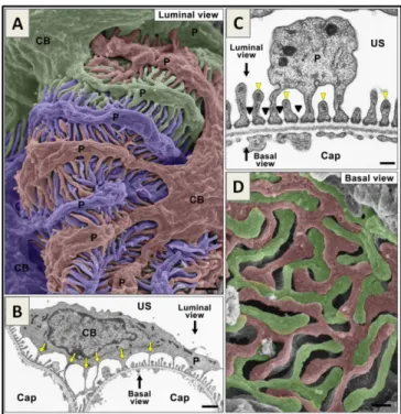
Figure 2. Ultrastructure of the podocyte. Podocytes are structurally divided in cell body (CB), primary and secondary processes (P) and foot processes. (A) Scanning electron microscopy images showing the CB, P and foot processes. Note that foot processes interdigitate with neighboring foot processes from another cell (individually colored in purple, green and red). (B and C) Transmission electron microscopy images (B) foot processes encircle the capillary loops (Cap). Arrows point to foot processes directly protruding from the podocyte CB. (C) Note the presence of a thin grey line between neighboring foot processes, corresponding to the slit diaphragm (black arrow heads), and the electron dense actin-bundle in the apical cytoplasm of foot processes (yellow arrow heads) (D) SEM images of the basal surface of podocytes. Bar scales, 1 μ i A), (B); 200 nm in (C), (D). US, urinary space. Adapted from Ichimura et al. 2015.
34
Podocytes have to withstand the shear stress from the flow of filtrate and are continuously exposed to potential toxins from the plasma filtrate. This particular situation demands high plasticity and dynamics that depend on a finely regulated cytoskeleton. In fact podocytes express mechanosensors that can respond to physical stimuli like shear stress (Huber, Schermer et al. 2007). These cells have several other important functions, namely to synthesize many extracellular matrix components of the GBM, such as type IV collagen; to help to physically support the capillary loop; and to interact with other glomerular cells, namely endothelial cells and mesangial cells, in order to guarantee their normal function. A paradigmatic example of cell to cell interaction is the production by the podocytes of the soluble vascular endothelial growth factor (VEGF) that traverses the GBM counter-flow in order to act on VEGF receptors on the endothelial cell, conditio sine qua non the fenestrated endothelial cells swell and detach from the GBM as nicely shown by S. Qwaggin group, using a conditional KO of Vegf in podocytes (Eremina, Sood et al. 2003, Eremina, Jefferson et al. 2008). The same dependence on VEGF produced in podocytes has also been shown for mesangial cells (Eremina, Cui et al. 2006). Mesangial cells are also regulated by podocyte production of PDGF (van Roeyen, Eitner et al. 2011).
The nephrotic syndrome
Any disruption of the filtration barrier leads to loss of glomerular permselectivity and urinary protein loss. When the loss of proteins is massive, this leads to secondary alterations in plasma protein levels and their turnover, alterations in lipid metabolism, endocrine disturbances and hemostasis defects. Nephrotic syndrome (NS) is thus defined as the constellation of the following clinical findings: massive proteinuria (>3.5 g/day), hypoalbuminemia (<3.0 g/dL), its clinical symptom - edema, and hypercholesterolemia (Nishi, Ubara et al. 2016). NS is a condition which carries high morbidity due to cardiovascular complications, such as thrombosis, increased infection risk and malnutrition. Moreover, when NS persists and does not respond to therapy this leads to progressive scarring of the glomeruli, a process called glomerulosclerosis, and secondary fibrosis of the tubular-interstitial compartment. Glomerulosclerosis will ultimately lead to renal failure and end-stage renal disease (ESRD), and patients will need dialysis or kidney transplant to survive. Glomerular diseases leading to secondary glomerulosclerosis are a major health problem in the Occidental world and account for ≈85% of all ESRD (Tharaux and Huber 2012).
Extensive research over the past decades has highlighted the main role of the podocyte in NS. In fact, although Mendelian cases of NS are very rare (Boyer O 2016), genetic studies have made a major
35 Figure 3. Podocyte structure in physiological conditions and upon injury. (A) Upper panel: In physiological conditions the GFB, composed of fenestrated endothelial cells (EC), GBM and podocytes, is able to prevent the spill of proteins into the urine. Lower panel: SEM and TEM images showing healthy podocytes and foot processes (FP), as well as the slit diaphragm (SD) and fenestrated endothelium (E) and a Schematic of normal podocyte foot processes (B) Upper panel: In nephrotic syndrome there is podocyte foot process effacement and protein leakage into the urine. Lower panel: SEM and TEM images showing podocyte foot process effacement and loss of SD. The corresponding schema shows a continuous sheet of cytoplasm instead of the well defined foot process. HMW, high molecular weight; BS, Bo a s spa e; CL, Capillary lumen. Adapted from Lin et al. 2016 and Mundel et al. 2010.
contribution to this field with the identification of mutations in genes encoding proteins crucial for podocyte function. Moreover, the ultrastructural phenotype common to all NS forms is podocyte foot process effacement with slit diaphragm disruption (Figure 3).
The process of foot process effacement depends on the rearrangement of the podocyte cytoskeleton in response to noxious stimuli and some authors consider that it may have developed as a mechanism to improve the attachment of these cells to the underlying GBM (Kriz, Shirato et al. 2013). If the noxious stimulus is transient, podocytes can recover their morphology and foot processes, as it occurs in self-remitting or steroid-sensitive NS. However, if the injury is persistent and/or severe enough, foot process effacement is followed by detachment of the podocyte foot processes from the GBM, the formation of pseudocysts and ultimately the detachment of these cells into the Bowman space and the nephron tubules, with resultantly high numbers of podocytes detectable in the urine (Kriz, Shirato et al. 2013). In vivo studies in the rat model have shown that
36
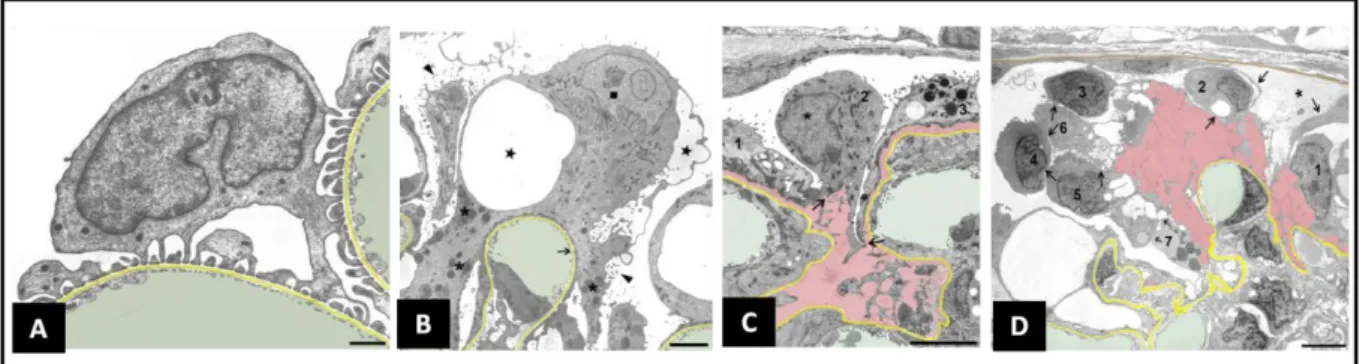
Figure 4. Ultrastructural changes accompanying podocyte injury and detachment. GBM is highlighted in yellow, capillary lumens in green and spaces between GBM and detaching podocytes in pink (A) Normal podocytes (B) Podocyte effacement in response to injury, formation of pseudocysts (stars) and microvillous transformation of the podocyte apical membrane (arrow head) (C and D) Podocyte detachment leaving areas of bare GBM. Bar scales, 1 μ i A) 3 μ in (B and C); 5 μ i D). Adapted from Kriz et al. 2013.
areas of the GFB with damaged podocytes seem to have high permeability to proteins (Peti-Peterdi and Sipos 2010). The ultrastructural changes have been clearly described by Kriz et al. (Kriz, Shirato et al. 2013) and are summarized in Figure 4.
NS may be a primary or secondary glomerular disorder, in the latter case associated with metabolic disorders such as diabetes, infections (i.e. HIV, HBV, HCV), immune disorders (i.e. systemic lupus erythematosus) drugs (pamidronate, interferon), toxins or malignancy (Eddy and Symons 2003). Among primary disorders, idiopathic NS is characterized by the absence of inflammation and immune deposits in biopsy samples and is most commonly found in children but can also be found in adults. According to the age of apparition, NS can be further classified in congenital NS (<3 months); infant (3 months–12 months); early childhood (13 months–5 years); late childhood (6–12 years); adolescent (13–18 years); and adult (>18 years old) (Brown, Pollak et al. 2014).
Idiopathic NS has two main pathophysiological mechanisms: (1) a structural defect of the GFB, as it occurs in genetic diseases, and which will be the object of this thesis; (2) an immune defect believed to be a circulating permeability factor of lymphocyte origin that secondarily leads to podocyte damage (Antignac 2002) (Figure 5). In both cases the clinical presentation of the disease is similar but some differences allow their distinction: the presence of other affected organs, and the onset before one year of age, all suggest a genetic defect (Hinkes, Mucha et al. 2007), while response to immunosuppressive therapy and early relapse after kidney transplant suggest an immune defect (Ding, Koziell et al. 2014).
Whatever the underlying defect, idiopathic NS is always initially treated with a course of corticoids a d, depe di g o the patie ts espo se to this i u osupp essi e the ap , it a e further classified into steroid-sensitive NS (SSNS) – if symptoms completely remit – or steroid-resistant NS (SRNS) – if symptoms persist. SSNS accounts for 80-90% of idiopathic NS and usually does not result
37 Figure 5. Mechanisms underlying idiopathic nephrotic syndrome. Immune dysfunction underlies steroid-sensitive forms and a subset of steroid-resistant forms, namely those that respond to treatment with other immunosuppressive drugs and those that recur after transplantation. The subset of SRNS patients that have an underlying molecular defect typically do not respond to any immunosuppressive drug and do not recur after transplantation. Adapted from Antignac et al. 2002.
in renal failure. SRNS, which represents 10-20% of idiopathic NS, shows a worse prognosis progressing to ESRD if no response is obtained with other classes of immunosuppressors (Cattran and Rao 1998). Though SSNS and most SRNS cases are immunological in nature, a small percentage of SRNS cases have a genetic/structural cause (Figure 5) (Hinkes, Mucha et al. 2007, Ding, Koziell et al. 2014, Sadowski, Lovric et al. 2015, Boyer O 2016).
Immune forms of nephrotic syndrome: the circulating permeability factor
Several lines of evidence suggest that an immune defect is the cause of most cases of SSNS and of SRNS relapse after transplantation, these include the response to immunosuppressive therapy, the NS relapse after infectious or allergic stimuli and the occurrence of N“ ith Hodgki s disease, o -Hodgki s l pho a, o th o a. Initial studies suggested that the immune defect was of T-lymphocyte origin (Shalhoub 1974). In fact, decreased T-regulatory cells and altered cytokine profiles have been identified in children with idiopathic NS (Araya, Diaz et al. 2009, Le Berre, Bruneau et al. 2009, Prasad, Jaiswal et al. 2015). The role of B-cells has also been suggested due to the efficacy of treatment with the monoclonal anti-CD20 antibody in affected individuals with steroid-dependent or frequent relapsing NS, however their exact role in the pathogenesis of the disease remains unknown (Iijima, Sako et al. 2014). Recently, hematopoietic stem/progenitor CD34-positive cells transferred from humans with idiopathic NS to immunocompromised mice have been shown to cause
38
proteinuria and podocyte effacement after three weeks (Sellier-Leclerc, Duval et al. 2007). However, the precise immune dysfunction mechanism remains unknown.
Several findings support the existence of a circulating permeability factor of immune origin, such as the proteinuria recurrence after kidney transplantation which occurs in about 30% of SRNS cases. This can occur within minutes to hours, and can be temporarily treated with plasmapheresis and immunoadsorption (Hoyer, Vernier et al. 1972). Studies in the rat model and case reports from humans have shown that when recurrence occurs following a kidney transplant and the organ is then grafted back to another recipient (or another rat strain), no proteinuria develops (Le Berre, Godfrin et al. 2002, Gallon, Leventhal et al. 2012). Moreover, kidneys from patients with SSNS that are transplanted into a patient with another type of kidney disease do not have recurrence of proteinuria (Gallon, Leventhal et al. 2012). In addition, serum from patients with NS is able to induce podocyte defects and proteinuria when injected into rats (Zimmerman 1984).
Several candidates have been proposed as circulating permeability factors. In cases of segmental glomerulosclerosis and chronic kidney disease, soluble urokinase plasminogen activator receptor (SUPAR) seemed to be the main candidate (Wei, El Hindi et al. 2011, Hahm, Wei et al. 2017). However, others have contested these findings and shown that high plasma SUPAR levels are mainly dependent on renal function (Meijers, Maas et al. 2014) and are a biomarker of cardiovascular disease (Lyngbaek, Marott et al. 2013, Meijers, Poesen et al. 2015), reflecting subclinical immunological activity and inflammation, but are not a mediator of a single disease entity (Meijers and Sprangers 2014).
Histologic variants of idiopathic nephrotic syndrome
In idiopathic NS three main types of non-specific histological lesions can be identified: minimal change disease (MCD), focal and segmental glomerulosclerosis (FSGS) and diffuse mesangial sclerosis (DMS). MCD and FSGS in particular have been proposed to correspond to two different morphological manifestations of the same underlying pathogenic process of podocyte injury (Maas, Deegens et al. 2016). These authors propose that there are several factors that will determine whether or not MCD will later evolve into FSGS lesions, including extent of injury, vulnerability of podocytes, other potential noxious factors and the response to therapy. In fact, clinical data suggest that patients with NS that are unresponsive to therapy or have a known molecular defect of genetic origin, can initially have findings of MCD by biopsy, and later develop FSGS (Tejani, Phadke et al. 1985). Moreover, the dose dependent development of FSGS lesions in the puromycin
39 aminonucleoside nephrosis (PAN) model and in other models of transient proteinuria, support the hypothesis that MCD and idiopathic FSGS are different manifestations of the same underlying injury and related to the severity of podocyte loss (Pippin, Brinkkoetter et al. 2009). This of course does not exclude the fact that several and not a single cause of podocyte injury exists.
Rodent models used for MCD and FSGS aim to induce podocyte injury and proteinuria. While in MCD the objective is to obtain a response that is transient and mild, FSGS lesion models are severe or prolonged enough to lead to the scarring process. Podocyte injury can be induced by antibodies directed against podocyte epitopes (endogenous or ectopically expressed) or by toxic agents. The last ones include the adriamycin and PAN models, protamine sulfate, and many others (Frenk, Antonowicz et al. 1955, Bertani, Poggi et al. 1982, Diamond and Karnovsky 1986, Leeuwis, Nguyen et al. 2010).
Minimal Change Disease
The pathological features include normal glomeruli on light microscopy and absence of immune deposits by immuno-fluorescence but the finding of widespread foot process effacement on electron microscopy. Clinically this lesion is associated to a higher rate of disease remission in response to corticotherapy and maintenance of kidney function (Mason 2010).
Focal and Segmental Glomerulosclerosis
FSGS is characterized by the sclerosis of a subset of the glomeruli (focal) and a portion of the glomerular tuft (segmental). Histologically a segmental obliteration of the glomerular capillaries by extracellular matrix can be observed (D'Agati 2008), as well as hyalinosis, corresponding to the accumulation of entrapped plasma proteins. Cellular adhesions, also called synechiae, can be formed between the Bowman capsule and the sclerosing segment. By electron microscopy, in addition to podocyte foot process effacement, regions of podocyte detachment can be observed. Immuno-fluorescence can show IgM and C3 deposits, corresponding to plasma proteins entrapped in areas of hyalinosis. With progression of the disease a more global and widespread glomerulosclerosis can be observed (D'Agati, Kaskel et al. 2011), and this is accompanied by tubular atrophy and interstitial fibrosis that is thought to result from the continuous exposure of the tubular cells to an excessive load of proteins (Eddy and Giachelli 1995). FSGS is associated with a higher likelihood of steroid resistance and progression to ESRD (Appel 2010). FSGS lesions have been further subdivided
40
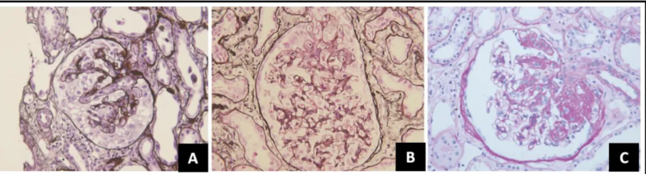
Figure 6. Histologic variants of FSGS. (A) Collapsing variant showing capillary collapse and podocyte hypertrophy (B) Tip variant showing a segmental lesion of the glomerular tuft next to the urinary pole (C) Peri-hilar variant showing sclerosis and hyalinosis of the vascular pole. Adapted from Lim et al. 2016.
in 5 subtypes according to the Columbia classification proposed in 2004 (D'Agati, Fogo et al. 2004) that is based on the localization of the lesions (urinary pole vs glomerular hilus (vascular pole)), the glomerular cellularity and presence of vascular collapse. These variants include: not-otherwise-specified (NOS), tip, peri-hilar, collapsing and cellular (Figure 6), and each is associated to a specific clinical prognosis, that is the best for tip lesions and the worst for the collapsing variants (D'Agati, Kaskel et al. 2011).
Diffuse Mesangial Sclerosis
Typically this lesion presents with mesangial matrix expansion that may be accompanied by podocyte hypertrophy over the capillary tufts (Habib, Gubler et al. 1993). In later stages widening of the GBM, global sclerosis of the glomeruli and tubular atrophy with interstitial fibrosis may supervene. Immuno-fluorescence can show non-specific deposit of IgM, C3 and C1q. By EM the common lesion to all forms of NS, foot process effacement, is observed. DMS is mostly seen in cases of Denys-Drash and Frasier syndromes (caused by WT1 mutations) Galloway-Mowat syndrome (caused by WDR73 mutations), Pierson syndrome (caused by LAMB2 mutations) as well as in cases of isolated SRNS associated to PLCE1 mutations (Hinkes, Wiggins et al. 2006).
41
Mechanisms of podocyte injury and glomerulosclerosis
Podocyte detachment, incapacity for cell division and glomerulosclerosis
Research in recent years seems to concede that podocyte depletion is the main initiator of the glomerulosclerosis process. Apoptotic death of podocytes seems to be a quite rare event in vivo (Kriz and Lemley 2015), while detaching podocytes have been found in several models of glomerular disease (Kriz, Shirato et al. 2013). The detachment is preceded by foot process effacement, and ultimately leads to the loss of viable podocytes into the urine (Kriz, Shirato et al. 2013). The remaining podocytes theoretically have two possible mechanisms of adaptation in order to cover the denuded GBM at the sites of podocyte loss: either hypertrophy or hyperplasia. However, podocytes are terminally differentiated cells; as such they are unable to proliferate or to respond to the decrease in cell number by cell division (D'Agati, Kaskel et al. 2011). In fact, it has been shown that podocytes forced to exit cell cycle arrest become polyploid and eventually enter a cellular event called mitotic catastrophe (Liapis, Romagnani et al. 2013). The cell division process itself demands complex alterations of the cytoskeleton and cell morphology. In the context of a cell that is exposed to the permanent shear stress of fluid flow across the GFB, these alterations likely also lead to reduced adhesion of the podocyte to the basement membrane and expose the cell to the risk of detachment. In fact binucleated podocytes, thought to result from an incomplete mitosis, have been observed in several types of glomerular disease and show foot process effacement (Nagata, Yamaguchi et al. 1995, Vogelmann, Nelson et al. 2003, Kriz, Shirato et al. 2013, Liapis, Romagnani et al. 2013, Hodgin, Bitzer et al. 2015). In rodent models it has been shown that in response to antibody-complement mediated podocyte injury the levels of cyclin A and cyclin dependent kinase 2 increase (Shankland, Floege et al. 1997), driving these cells to G2/M phases. However, this is limited by a simultaneous increase in cyclin kinase inhibitors such as p21 and p27 (Shankland, Floege et al. 1997), leading to an aborted mitosis. Moreover, the lack of aurora kinase B in mature podocytes also disables the formation of an efficient mitotic spindle (Lasagni, Ballerini et al. 2010). Recently in vitro studies on mouse immortalized podocytes grown in non-permissive conditions (and thus leading to differentiation) showed that re-entry in S phase due to stress stimuli induces binucleation and leaves these cells more susceptible to death by a secondary injury (Hagen, Pfister et al. 2016).
Even in normal physiological circumstances there is a continuous loss of podocytes, which can be found in urine of healthy individuals, although at a relatively low level when compared to individuals with glomerular disease. In the adult kidney each glomerulus has approximately 600 podocytes and the normal loss of podocytes has been calculated to be about 1-2 podocytes per glomerulus per year
42
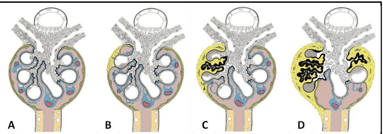
Figure 7. Progression of glomerular diseases. (A) Normal glomerulus; podocytes are shown in blue, parietal epithelial cells in green. The GBM is shown in black (B) Initial lesion: Podocyte-denuded capillary attach to Bowman's capsule. (C) Progression of the sclerotic process. The adhesion has spread to neighboring capillaries resulting in collapse or in hyalinosis (shown as a thick black line) of the involved capillaries. A paraglomerular space (shown in yellow) containing the sclerotic tuft remnants is formed. (D) Late stage of the sclerotic process. A small i ta t tuft still e ai s. Fibroblasts invade the sclerotic area, resulting in fibrous organization. Adapted from Kriz et al. 1998.
(Hodgin, Bitzer et al. 2015). In fact, in kidneys from older individuals the density of podocytes is lower than in young kidneys and this is accompanied by a compensatory mechanism of podocyte hypertrophy (Hodgin, Bitzer et al. 2015).
If an inciting injury is strong or prolonged enough to decrease the podocyte number past a threshold level where glomerular survival is no longer possible, glomerular scarring occurs. (Wiggins 2007) (Figure 7). In experimental rodent models where podocytes were genetically modified to become susceptible to specific toxins (i.e. Diphtheria toxin or Pseudomonas exotoxin), it has been shown that the loss of more than 40% of podocytes leads to glomerulosclerosis with proteinuria and renal failure (Matsusaka, Xin et al. 2005, Wharram, Goyal et al. 2005). Moreover, after the initial lesion, there is a secondary autonomous process that maintains injury and loss of the remnant podocytes (Sato, Wharram et al. 2009, Matsusaka, Sandgren et al. 2011). This could be related to increased shear stress on the remaining podocytes and accumulation of oxidative stress and DNA damage. Interestingly, the ability of podocytes to resist injury may depend on the capacity to resist genotoxic stress. This conclusion came from a study that analyzed why susceptibility to adriamycin-induced glomerulosclerosis occurred only in certain mouse strains. Adriamycin is a cytotoxic drug which has pleiotropic effects, such as introduction of double strand DNA breaks, topoisomerase inhibition, mitochondrial DNA depletion, lipid peroxidation and disruption of the cytoskeleton. Gharavi´s group demonstrated that susceptible strains had a recessive loss of function mutation on Prkdc gene that encodes a nuclear DNA double strand break repair protein. Decreased expression of Prkdc rendered resistant mice strains susceptible to adriamycin-induced glomerulosclerosis and in vitro podocytes more susceptible to adriamycin induced apoptosis and to mitochondrial DNA depletion (Papeta, Zheng et al. 2010).
43
Podocyte regeneration by stem cells
In the last two decades, the discovery of renal stem cells has shed some light upon the possibility of podocyte replacement. There are two different types of renal cells that can possibly generate podocytes, the parietal epithelial cells (PECs) and the cells of renin lineage (CoRL). Besides kidney stem cells, there are also several lines of evidence that point to bone marrow stem cells as possible precursors of podocytes.
PECs, a double-edged sword: podocyte regeneration vs glomerulosclerosis
PECs li e the Bo a s apsule a d o phologi all ese le s ua ous epithelial ells. Du i g development, PECs and podocytes have a common mesenchymal cell origin, and then differentiate into two different cell types. Although not much is known about PEC function, they are known to express tight junction molecules (claudin-1, zonula occludens-1, and occludin) and to have a barrier function against the spill of proteins from the primary urine into the interstitial space (Ohse, Chang et al. 2009). Whether PECs are able to regenerate podocytes is a controversial subject. Some PECs express CD24 and CD133 stem cell markers (Ronconi, Sagrinati et al. 2009) and have a known regenerative capacity (Sagrinati, Netti et al. 2006). Interestingly, PECs express differentiation or stem cell markers according to their location o the Bo a s apsule. Those located far from the vascular pole exhibit only stem cell markers and those that are near exhibit only podocyte markers (i.e. nestin, and podocalyxin), while both markers are expressed between the two locations (Ronconi, Sagrinati et al. 2009). Genetic lineage tracing experiments have given non consensual results for the regeneration of podocytes from PECs (Berger, Schulte et al. 2014, Sakamoto, Ueno et al. 2014) (Wanner, Hartleben et al. 2014). In fact, podocyte regeneration seems to depend on the type of glomerular injury and the kidney age. i.e. in juvenile mice where PECs were genetically traced, a su populatio of these ells as des i ed to ig ate alo g the Bo a s apsule a d to e p ess progenitor cell markers and podocyte markers, integrating the population of podocytes resident on the glomerular tuft (Appel, Kershaw et al. 2009). However, in adult mice, spontaneous migration or migration induced by lesion (glomerular hypertrophy by partial nephrectomy) was no longer seen (Berger, Schulte et al. 2014). In human kidneys analogous observations were made, among the PECs, podocyte markers have scattered expression until 2 years of age, but not on PECs from 7 year old individuals (Berger, Schulte et al. 2014). In another lineage tracing experiment, no regeneration occurred in ageing mice or after nephron reduction, however regeneration did occur in response to acute podocyte loss induced in the diphtheria toxin model (Wanner, Hartleben et al. 2014), and this
44
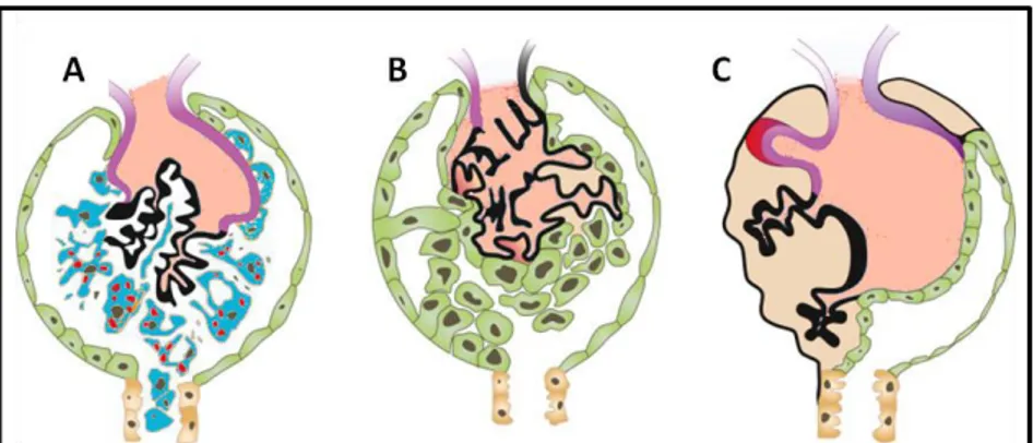
Figure 8. Parietal epithelial cells participate in the progression of glomerulosclerosis. (A) Severe injury leading to fulminant loss of podocytes (B) Activation and migration of PECs to the glomerular tuft through synechiae formed et ee the de uded GBM a d the Bo a s apsule (C) Late stages of glomerulosclerosis showing a monolayer epithelium of PECs covering the glomerular basement membrane. PECs further synthesize extracellular matrix that leads to complete sclerosis. Adapted from Nagata et al. 2016.
finding was replicated in another mouse model of acute podocyte depletion (Eng, Sunseri et al. 2015).
If the regeneration of podocytes from PECs is still a controversial subject, several lines of evidence have firmly established that an excessive proliferative response by PECs after glomerular injury can lead to the formation of glomerular lesions such as cellular or fibrous crescents and collapsing lesions in FSGS (Lasagni and Romagnani 2010). During injury, PECs can undergo phenotypic changes that have been described the ge e al te of activation (Shankland, Smeets et al. 2014). These include changes in morphology (larger cytoplasm and nucleus) as well as increased migration, proliferation and synthesis of extra-cellular matrix, and de novo expression of marker proteins, such as CD44 (a cell surface adhesion receptor) (Shankland, Smeets et al. 2014). In several mouse models of glomerular injury using genetic lineage tracing of PECs, and in human transplant biopsies, it has been shown that activated PECs can migrate to the glomerular tuft through adhesion sites formed between the tuft and the Bowman capsule, and produce extracellular matrix, contributing to the development of sclerosis (Appel, Kershaw et al. 2009, Smeets, Kuppe et al. 2011, Smeets, Stucker et al. 2014). Interestingly the presence of activated PECs may distinguish between the sclerosing lesions of FSGS and the absence of fibrosis in MCD, with the former showing invasion of the glomerular tuft by CD44 positive PECs (Smeets, Stucker et al. 2014). A very detailed study on human biopsies of several types of glomerular disease using multiple markers for PECs, activated PECs and podocytes has clearly defined that the earliest lesion is the formation of a cellular bridge between the Bo a s apsule a d the glo e ula tuft, fo ed PECs that deposit at i i the tuft, hi h is followed by the migration of PECs onto the tuft and the formation of fibrotic adhesions or early sclerotic lesions, and finally the formation of advanced sclerotic lesions, where a larger part of the tuft is covered by PECs that continue to deposit extracellular matrix (Kuppe, Grone et al. 2015) (Figure 8).
45 A recent study has shown that for effective podocyte regeneration in response to a glomerular injury, PECs activation has to be followed by differentiation and expression of podocyte markers, otherwise, the maintenance of activated PECs leads to glomerulosclerosis. In a mouse model of FSGS with genetic lineage tracing of PECs, these authors demonstrated that if activated PECs successfully differentiated into podocytes the disease remitted, while glomerulosclerosis occurred if these cells were not able to differentiate (Lasagni, Angelotti et al. 2015). Promisingly, enhancing the activity of endogenous retinoic acid, that controls differentiation in podocytes, with a glycogen synthase kinase3 inhibitor significantly increased the trans-differentiation of PECs into podocytes and increased disease remission (Lasagni, Angelotti et al. 2015). Currently several other molecules that target molecular pathways associated with PEC proliferation, activation and extracellular matrix production are being further studied in animal models (Miesen, Steenbergen et al. 2017).
CoRL and bone marrow derived stem cells
CoRL are vascular smooth cells that under normal conditions produce renin and are localized to the juxtaglomerular compartment, between afferent and efferent arterioles. These cells have been found to be able to lose their characteristic endocrine and contractile functions and differentiate into several adult cell types, including mesangial cells (Thoma 2014, Castellanos-Rivera, Pentz et al. 2015), pericytes (Castellanos-Rivera, Pentz et al. 2015, Pippin, Kaverina et al. 2015), vascular smooth cells (Castellanos-Rivera, Pentz et al. 2015, Pippin, Kaverina et al. 2015) , erythropoietin-producing cells, hematopoietic immune-like cells (Castellanos Rivera, Monteagudo et al. 2011), PECs and podocytes (Shankland, Pippin et al. 2014). In a rodent model of acute podocyte depletion, cell fate tracing of CoRL and in vivo time lapse imaging showed that these cells populate the glomeruli and differentiate in PECs and podocytes (Kaverina, Kadoya et al. 2017). The same has been shown in models of chronic podocyte depletion (Pippin, Kaverina et al. 2015).
Other sources of podocyte progenitors have been described such as the bone marrow-derived stem cell source, as evidenced by the presence of 7% of podocytes derived from a male recipient on a female kidney allograft (Becker, Hoerning et al. 2007). Moreover, injection of bone marrow-derived stem cells ameliorates adriamycin induced FSGS lesions (Uchida, Kushida et al. 2017). However, discrepant results have been found in other studies (Meyer-Schwesinger, Lange et al. 2011), arguing for the need of more intensive investigation in this field before considering bone marrow-derived stem cells as definite podocyte progenitors.
46
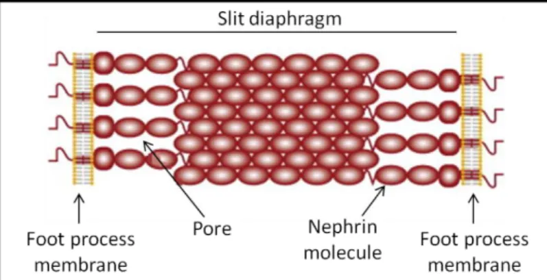
Figure 9. The slit diaphragm zipper-like structure. Nephrin molecules from neighboring foot processes interact at the center of the slit, forming a zipper-like structure with pores on both sides of the central density. Adapted from Patrakka et al. 2007.
The podocyte slit diaphragm, lipid rafts and actin cytoskeleton
The slit diaphragm
The slit diaphragm was first visualized by electron microscopy (EM) in 1955 (Yamada 1955) and it was only in 1974 that with better EM techniques the classic view of the slit diaphragm as a zipper-like structure was described (Rodewald and Karnovsky 1974, Karnovsky and Ryan 1975). In this model the slit diaphragm molecules from neighboring foot processes are thought to cross the extracellular space and are arranged in a dense midline, corresponding to the overlapping region of their extracellular domains (Figure 9). The pores between these strands, of about 12 nm, would be small enough to prevent the passage of albumin or higher molecular weight proteins. In the same year, the loss of the slit diaphragms and the process of foot process effacement in response to podocyte injury were also described (Robson, Giangiacomo et al. 1974). These findings provided a strong link between slit diaphragm loss, foot process effacement and proteinuric glomerular diseases.
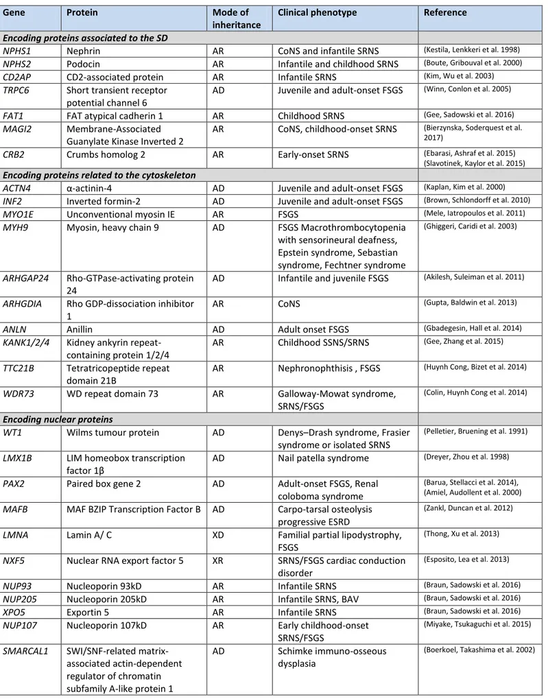
In recent decades genetic studies have contributed to a better understanding of the molecular composition of the slit diaphragm. In 1998, through a positional cloning approach, researchers identified the first gene known to be mutated in congenital cases of NS, NPHS1, encoding for the transmembrane Immunoglobulin superfamily protein nephrin (Kestila, Lenkkeri et al. 1998). In subsequent years nephrin was identified as the backbone molecule of the slit diaphragm filter (Ruotsalainen, Ljungberg et al. 1999, Wartiovaara, Ofverstedt et al. 2004). This was followed by the identification of NPHS2 (Boute, Gribouval et al. 2000), encoding podocin, a slit diaphragm associated protein. This is the most frequently mutated gene in familial cases of NS (Bouchireb, Boyer et al. 2014). In the following years other critical components of the slit diaphragm such as the Immunoglobulin superfamily nephrin like protein-1 (Neph1), as well as proteins linking the slit diaphragm to the actin cytoskeleton, were discovered.
47 Figure 10. New EM techniques suggest a novel slit diaphragm structure. The slit diaphragm is suggested to be a multilayered, bipartite protein scaffold. (A) High-pressure freezing and plastic electron tomography (PET) show several strands crossing the space between different FP. The narrower end of the slit is toward the GBM, while the broader end is to a ds the Bo a s spa e lue a o heads a k sho te st ands and red arrowheads correspond to longer strands). (B) Transversal view showing a quasiperiodical distribution of the slit layers (C) 3D reconstitution showing foot process bridging strands color-coded depending on their length (red 40 nm, attributed to nephrin, blue 25 nm, attributed to Neph1) (D) Transversal view. Adapted from Grahammer et al. 2016.
Very recently, development of different fixation and EM techniques, such as cryo-electrontomography and focused ion beam-scanning electron microscopy (FIB-SEM), permitted a review of the classical zipper-like structure of the slit diaphragm (Burghardt, Hochapfel et al. 2015, Grahammer, Wigge et al. 2016). Instead of a monolayer of nephrin and Neph1 that establish homo or heterophilic interactions, the slit diaphragm seems to have superposed layers of these transmembrane proteins. Both molecules seem to completely span the interpodocytary space and do not form homo or heteromeric interactions between them, contrary to the previous belief (Burghardt, Hochapfel et al. 2015, Grahammer, Wigge et al. 2016). Neph1 forms the bottom layers, close to the GBM, spanning a distance of 23 nm, nephrin molecules on the other hand form the upper layers and span a distance of around 45 nm. Both molecules are separated by a distance of 7 nm. (Figure 10). The absence of a well-defined pore structure with certain dimensions raises the question on how this structure contributes to the permselectivity of the GFB, a question that will most certainly be addressed in the coming years, and that should take into account other slit diaphragm proteins.
The slit diaphragm is a specialized type of cell junction that exhibits characteristics of both tight junctions, expressing proteins such as ZO-1, occludin and cingullin (Schnabel, Anderson et al. 1990, Fukasawa, Bornheimer et al. 2009), and of adherens junctions, expressing P-cadherin or , , catenin proteins (Reiser, Kriz et al. 2000, Lehtonen, Lehtonen et al. 2004). During podocyte development the slit diaphragm is not the first type of cellular junction to be formed. The first cellular junction is an apical tight junction (at the S stage) when podocyte precursors form a cobblestone-like epithelium (Ruotsalainen, Patrakka et al. 2000, Ichimura, Kakuta et al. 2017). Later in development these junctions migrate to the more baso-lateral aspect of the podocyte membrane (Ichimura, Kakuta et al. 2017) and become enriched with adherens junction proteins, nephrin and