HAL Id: tel-02977151
https://tel.archives-ouvertes.fr/tel-02977151
Submitted on 24 Oct 2020HAL is a multi-disciplinary open access archive for the deposit and dissemination of sci-entific research documents, whether they are pub-lished or not. The documents may come from teaching and research institutions in France or abroad, or from public or private research centers.
L’archive ouverte pluridisciplinaire HAL, est destinée au dépôt et à la diffusion de documents scientifiques de niveau recherche, publiés ou non, émanant des établissements d’enseignement et de recherche français ou étrangers, des laboratoires publics ou privés.
Rôles de l’autophagie dans l’homéostasie des cellules
souches intestinales
Coralie Trentesaux
To cite this version:
Coralie Trentesaux. Rôles de l’autophagie dans l’homéostasie des cellules souches intestinales. Hépatologie et Gastroentérologie. Université Paris Saclay (COmUE), 2018. Français. �NNT : 2018SACLS361�. �tel-02977151�
Rôles de l’autophagie dans
l'homéostasie des cellules
souches intestinales
Thèse de doctorat de l'Université Paris-Saclay
préparée à L’Université Paris Sud
École doctorale n°582 Cancérologie, Biologie, Médecine, Santé
Spécialité de doctorat: aspects moléculaires et cellulaires de la biologieThèse présentée et soutenue à Paris, le 23 octobre 2018, par
Coralie TRENTESAUX
Composition du Jury : Claude BOUCHEIX
DR, Institut André Lwoff (Inserm U936) Président
Danijela VIGNJEVIC
DR, Institut Curie (CNRS UMR 144) Rapporteur
Philippe JAY
DR, Institut de Génomique Fonctionnelle (CNRS UMR 5203, Inserm U1191) Rapporteur
Patrice CODOGNO
DR, Institut Necker Enfants-Malades (Inserm U1151, CNRS UMR 8253) Examinateur
Philippe SANSONETTI
DR, Institut Pasteur (Inserm U1202) Examinateur
Béatrice ROMAGNOLO
DR, Institut Cochin (Inserm U1016) Directeur de thèse
NNT
: 2018S
AC
LS
PhD M
an
uscr
ipt
Role of Autophagy in Intestinal
Stem Cell Homeostasis
Thesis presented for the degree of Doctor of Phylosophy
of the Université Paris-Saclay
prepared at the Université Paris Sud
Doctoral school n°582: Oncology, Biology, Medecine, Health
Thesis specialty: molecular and cellular aspects of biology
Thesis presented and defended in Paris, on the 23rd of october 2018, by
Coralie TRENTESAUX
Jury members: Claude BOUCHEIX
DR, Institut André Lwoff (Inserm U936) President
Danijela VIGNJEVIC
DR, Institut Curie (CNRS UMR 144) Reviewer
Philippe JAY
DR, Institut de Génomique Fonctionnelle (CNRS UMR 5203, Inserm U1191) Reviewer
Patrice CODOGNO
DR, Institut Necker Enfants-Malades (Inserm U1151, CNRS UMR 8253) Examiner
Philippe SANSONETTI
DR, Institut Pasteur (Inserm U1202) Examiner
Béatrice ROMAGNOLO
DR, Institut Cochin (Inserm U1016) Thesis Supervisor
NNT
: 2018S
AC
LS
“A painting is not thought out in advance. While it is being done, it changes as one’s thoughts change. And when it’s finished, it goes on changing, according to the state of mind of whoever is looking at it” –Pablo Picasso on Guernica
A
CKNOWLEDGEMENTS
Ce manuscrit représente pour moi l’occasion de mettre en valeur quatre belles années de recherche scientifique et d’apprentissage. Il représente également l’occasion de remercier toutes les personnes ayant participé à cette recherche et sans qui ce travail n’aurait pu être réalisé. Tout d’abord je souhaiterais remercier les membres de mon jury d’avoir accepté d’évaluer mon travail de thèse. Mes rapporteurs Danijela Vignjevic et Philippe Jay, pour l’attention qu’ils ont accordé à la relecture de mon manuscrit et leurs commentaires encourageants. Mes examinateurs : Patrice Codogno, pour tout ce que j’ai pu apprendre aussi bien scientifiquement qu’humainement lors de mon Master et des dernières années. Philippe Sansonetti, je suis heureuse d’avoir eu la chance d’assister aux discussions avec vous et votre équipe au cours de ces dernières années et d’avoir à nouveau cette chance lors de ma soutenance. Claude Boucheix, c’est avec grand plaisir que je ferais votre rencontre, merci d’avoir accepté de prendre le temps d’assister à ma soutenance et d’apporter une nouvelle perspective sur mon travail.Un énorme merci à Christine Perret, tout d’abord de m’avoir accueilli dans ton équipe mais aussi pour tout ton soutien, scientifique et autre, au cours de ces années. Tu es pour moi un véritable exemple, tu as toujours su me guider sans le rendre évident et te rendre abordable malgré tes connaissances impressionnantes.
Merci surtout à Béatrice Romagnolo, de m’avoir confié ce projet que j’ai adopté au cours des 4 dernières années mais qui est ton bébé depuis très longtemps. Je te remercie de ton encadrement, tu m’as soutenue et poussée lorsque j’en avais besoin et m’as aussi accordé ta confiance et la liberté qui m’ont permis d’avancer et prendre confiance en moi au cours de cette thèse. Merci aussi pour toutes les opportunités que tu m’as données : les congrès, les revues, l’encadrement d’étudiants, les collaborations et tous les conseils qui ont également rendu cette thèse une expérience enrichissante. Je continuerais à prendre exemple sur ta persévérance (même si je reste convaincue qu’au-delà d’un marathon, c‘est de la folie), ta capacité à te lancer dans de nouveaux domaines sans hésitation, et ta passion lorsque tu parles de science et de tes idées et folies.
Merci tout particulièrement à toutes les personnes de l’équipe intestin, petite au début mais en pleine croissance, et à toute l’équipe Perret !
Marie, je l’ai déjà dit, je pense que je ne retrouverai jamais une collègue comme toi. Merci d’avoir été aussi patiente avec moi et de m’avoir accordé une seconde chance (et toutes les suivantes) malgré ma très mauvaise première impression et toutes mes bêtises. Sans toi, je ne serais clairement pas allée très loin, tu as été indispensable tout au long de ce projet : tu étais dessus bien avant que j’arrive, tu m’as formée, tu as continué à gérer une énorme partie de ce projet en parallèle du tien et en plus de toute ton implication pour l’équipe, tu as toujours proposé ton aide même quand je n’osais pas demander, tu es sans doute la seule en qui je fais confiance sans hésiter pour les manips, et tu as souvent été la première personne que j’allais voir pour discuter
de manips et projets. J’ai aussi toujours pu compter sur toi pour me motiver quand il le fallait, ou pour partir en délire après une longue journée. En dehors de tout ça tu es une véritable amie, ta porte a toujours été ouverte que ce soit pour rigoler ou pour pleurer, tu as toujours su être juste et franche et c’est aussi pour ça que je demande beaucoup tes conseils. Il va falloir qu’on aille prendre beaucoup de verres pour que je te pardonne de m’abandonner en premier. J’ai hâte que ce soit ton tour pour que tout le monde (y compris toi même !!) se rende compte de ce dont tu es capable, et je suis sûre que tu trouveras la suite sans problème et qu’ils se rendront très vite compte aussi de tout ce que tu vaux. J’espère par-dessus tout te voir heureuse, bébé !
Julie, mes deux premières années n’auraient pas été pareil sans toi et sans tout ton travail. Il faut dire que j’ai perdu non seulement une aide indispensable mais aussi une source de rires et chansons et potins quand tu nous as quitté pour tes moutons !
Luana, je suis sûre que tu assureras la suite de ce projet qui me tient tant à cœur. Ça a été un vrai plaisir d’apprendre à te connaître ces derniers mois, je suis heureuse d’avoir encore quelques mois à tes côtés avant de devoir lâcher le bébé, je ne pouvais pas partir sans avoir eu le temps de perfectionner mon accent italien (pomodoro !).
Nadia, tu as été ma grande sœur de labo : je t’ai adorée dès le début même si tu me détestais et j’ai beaucoup pris exemple sur toi tout au long de me thèse. Grâce à toi, je me suis vite sentie intégrée et j’ai pu me faire de vrais amis à Cochin. Tu es quelqu’un de brillant, d’extrêmement gentil (même si tu te crois méchante), de bosseur, de droit dans ses baskets, tu as toujours su me reprendre quand il le fallait et j’ai toujours pu discuter ouvertement avec toi même quand on n’était pas du même avis (c’est mon esprit de contradiction ça…). Je crois que je risque de te copier jusqu’au bout, et si c’est le cas (cadanstabouche) j’espère qu’on se reverra bientôt !
Mathilde, je suis tellement heureuse que tu aies rejoint notre équipe et surtout qu’on ait pu former notre super trio ! Tu es brillante et rigoureuse, et je suis certaine que tous ceux qui auront la chance de travailler à tes côtés s’en rendront très vite compte et que tu auras une magnifique carrière après ta thèse. Merci d’avoir gentiment ri à toutes mes blagues de papa et subi mes pétages de cable réguliers et d’être toujours prête à m’aider quand j’ai des questions bêtes ou même juste des calculs à faire. Au-delà du côté scientifique et professionnel, la thèse nous aura permis de nous rencontrer et de devenir amies et je pense que je me souviendrai plus de ça que de tous les côtés « peineux » dans quelques années.
Sara et Shirley (le duo inséparable), je suis contente d’avoir pu partager ces 4 années de thèse avec vous du début jusqu’à la fin (± quelques mois), ça n’aurait pas été pareil sans tous les déjeuners et pauses café (très occasionnelles, bien sûr) ensembles. Sara, ça n’a pas été facile de te voir partir avant moi, mais tu es bien la preuve que de belles choses nous attendent. Shirley, à très bientôt ton tour !
Romain, heureusement que tu es là, sinon on manquerait sérieusement de testostérone !!! Mais plus sérieusement, ça a été un plaisir de travailler avec toi et d’apprendre à te connaitre ces deux dernières années, à toi de porter le flambeau des thésards intestins maintenant (oui, c’est très bizarre, mais bon, c’est dit) …
Clément, heureusement que tu es si discret vu toutes les fois où je débarque dans votre bureau en disant des conneries ! En tout cas je suis ravie de t’avoir eu dans les parages ces derniers mois, il y a certaines scènes que je ne suis pas prête d’oublier !
Pascalette, merci pour toute ton aide et tous tes conseils, tu es une source de connaissances incroyables sur la science et sur le monde, et tu es toujours disponible pour aider les autres, quelque chose dont j’ai beaucoup bénéficié au cours des 4 dernières années. Hélène, merci pour ta gentillesse qui m’a marqué dès mon premier lab meeting et pour tous tes conseils au cours des années. Benoit Terris, d’avoir à plusieurs occasions pris le temps de regarder nos phénotypes parfois étranges et de nous avoir pointé dans la bonne direction ou réconforté dans nos idées.
Merci également à tous les stagiaires qui sont passés par notre labo, qui m’ont permis d’avancer plus vite et qui m’ont appris à apprendre aux autres !
Merci à tous les membres des plateformes de l’institut que j’ai beaucoup embêté et sur qui j’ai beaucoup compté tout au long de ce projet. Je pense notamment à HistIM, CYBIO (en particulier Emmanuelle Maillard, Karine Bailly, et Muriel Andrieu), GENOM’IC (Sébastien Jacques, Angéline Duché, Franck Letourneur), IMAG’IC (Thomas Guilbert et Valérie), la plateforme de microscopie électronique (Alain Schmitt), et un énorme merci aux animaleries de Cochin (en particulier Nadia Boussetta, JB Houille, et Christophe Dez).
Roro et Mariangela (encore un duo inoubliable), je suis tellement heureuse d’avoir partagé chaque étape de nos thèses ensembles ! Que ce soit dans le bureau de Nadia, à la formation expérimentation animale (RIP pauvre petite souris), où quelque part entre l’harmonie et l’académie, vous êtes devenues de vraies amies et j’espère qu’on continuera à partager toutes les étapes à venir !
Un énorme merci à tous les collègues et les amis qui ont pu m’aider, me soutenir, me conseiller, partager des bières, ou tout simplement rendre ces 4 dernières une aussi belle expérience au quotidien. En particuliers aux gens de la Fac : Patricia, les Colnots, les Maires, les Desdouets, les Peysonnaux, les Vaulont/Violets, les filles de la laverie et bien sûr les agents spéciaux !
Un énorme merci aux amis de l’ENS, de m’avoir obligée à avoir une vie quand même en dehors du boulot, de m’avoir fait découvrir la culture française et de véritables chef d’œuvres cinématographiques, d’avoir écouté mes histoires de souris et de caca, et d’avoir partagé cette aventure avec moi !
Antonin, je ne pourrais jamais te remercier assez pour les bactéries compétentes que tu m’as filé en deuxième année. Sans toi, je n’aurais pas pu faire la figure 1d. C’est cool, merci.
Bon il faut dire que tu as aussi supporté mon stress, ma mauvaise humeur, mes nuits et weekends au labo, ma fatigue, mon stress, mon côté très légèrement psychopathe et un tout petit peu chiante, ma vaisselle sale et mon courrier qui trainent, mon stress, et tout ce qui venait avec ces six dernières années. Je suis sûre que tu serais capable de soutenir à ma place tellement tu as passé de temps à m’écouter parler de cette thèse. Et malgré le fait que tu vivais la même chose de ton côté, tu as su être l’homme le plus patient du monde et t’occuper de moi (et me faire plein de bons repas). Je ne pourrais jamais assez te remercier pour tout ce que tu fais pour moi, que ce soit me montrer comment transfecter des bactéries ou passer la soirée à regarder des séries et à rien faire ensemble. J’ai hâte de commencer notre prochaine aventure ensemble !
Finalement, je tiens à remercier ma famille, qui supportent mon stress et mon humeur depuis bien avant la thèse, et qui pourtant continuent à m’encourager et à m’offrir des peluches de souris. Et dire que j’aurais pu faire architecte ! (mercipapamamanpipitoto)
1
T
ABLE OF
C
ONTENTS
List of Figures ... 4 Abbreviations...
5 Summary in French... 11
I
NTRODUCTION...
13 Foreword ... 15C
HAPTER I: Physiopathology of the Intestinal Epithelium ... 171. Structure and Function of the Intestine ... 19
1.1. Overview of the adult digestive system & gastrointestinal tract ... 19
1.2. Structural organization of the intestine ... 21
1.3. The Intestinal Epithelium ... 22
1.3.1. Enterocytes ... 23 1.3.2. Goblet cells ... 24 1.3.3. Paneth cells ... 25 1.3.4. Enteroendocrine cells ... 25 1.3.5. Tuft cells ... 26 1.3.6. Microfold cells ... 27 1.3.7. Cup cells ... 27
2. Intestinal stem cells and their defining niche ... 29
2.1. Models and markers of intestinal stem cells... 29
2.1.1. Markers for crypt basal columnar cells ... 29
2.1.2. Markers for +4 stem cells ... 31
2.1.3. Progenitors as potential stem cells: plasticity in the intestinal crypt... 32
2.2. Signaling pathways regulating crypt homeostasis ... 33
2.2.1. WNT/β-catenin signaling ... 33
2.2.2. Notch signaling ... 36
2.2.3. Hedgehog and BMP signaling ... 37
2.3. Organoid culture and the experimental niche ... 38
2.4. The intestinal stem cell niche ... 39
2.4.1. The epithelial niche: Paneth cells and deep crypt secretory cells ... 39
2.4.2. The mesenchymal niche ... 42
2.4.3. The microbial niche ... 44
2.5. Regulation of intestinal stem cell integrity... 48
2.5.1. Crypt monoclonality and neutral drift ... 49
2.5.2. p53 and apoptosis ... 50
2.5.3. Reactive oxygen species ... 52
3. Colorectal cancer ... 55
3.1. Prevalence and risk factors ... 55
3.2. Tumorigenesis and progression ... 56
3.3. Types of colorectal cancer ... 57
3.4. Treatment ... 59
3.5. Intestinal stem cells and colorectal cancer ... 59
3.5.1. Intestinal stem cells as the founder cells of colorectal cancer ... 59
3.5.2. Colorectal cancer stem cells ... 60
2
C
HAPTER II: Autophagy ... 651. Mechanisms and regulation of autophagy ... 67
1.1. Overview of autophagy ... 67
1.1.1. Molecular machinery of autophagy ... 69
1.1.2. Selective autophagy ... 70
1.2. Regulation and functional consequences of autophagy... 72
1.2.1. Growth factors, nutrients, and energy levels: mTOR signaling ... 73
1.2.2. Apoptotic signals: BCL2 and p53 ... 75
1.2.3. Endoplasmic reticulum stress ... 76
1.2.4. Hypoxia ... 77
1.2.5. Oxidative stress ... 78
1.2.6. DNA damage ... 79
1.2.7. Infection, inflammation and immunity... 83
2. Autophagy in physiology and pathology ... 87
2.1. Autophagy in health and disease ... 87
2.1.1. Development ... 87
2.1.2. Metabolic disease ... 88
2.1.3. Neurodegeneration... 89
2.2. Autophagy in adult stem cells ... 89
2.3. Autophagy and aging ... 91
2.4. Autophagy in cancer... 92
2.4.1. Autophagy in pre-malignant cells... 92
2.4.2. Autophagy supports tumor growth and metabolism ... 93
2.4.3. Autophagy and resistance to therapy ... 96
2.4.4. Autophagy and cancer stem cells... 97
2.4.5. Autophagy and the tumor microenvironment ... 97
3. Autophagy and the intestinal epithelium ... 99
3.1. Autophagy in secretory cells of the intestinal epithelium ... 99
3.2. Xenophagy ...100
3.3. Autophagy, the microbiota and inflammation ...100
3.4. Autophagy in intestinal pathology ...102
3.4.1. Autophagy in Crohn’s disease ...102
3.4.2. Autophagy in colorectal cancer ...102
R
ESULTS...
1051. Research context ...107
2. Article ...109
3. Figures and legends...125
4. Supplementary figures and legends ...131
D
ISCUSSION...
1371. Autophagy as a protective stress response in intestinal stem cells ...139
1.1. Autophagy protects intestinal stem cells from oxidative stress ...139
1.2. Autophagy protects intestinal stem cells from DNA damage ...140
1.3. Autophagy protects intestinal stem cells from microenvironmental stress ...142
2. Other models of Atg depletion in the intestinal epithelium ...143
3. Replenishment of the autophagy-deficient intestinal stem cell pool ...144
3
5. Colon stem cells and autophagy ...146
6. Autophagy and tumor progression ...146
R
EFERENCES...
149A
PPENDIX...
1971. Review: Trentesaux et al. (2017). L’autophagie, l’homéostasie intestinale et ses pathologies [Contribution of autophagy to intestinal homeostasis and pathology]. 2. Book chapter : Trentesaux and Romagnolo. (2018). Intestinal stem cells and their
4
L
IST OF
F
IGURES
I
NTRODUCTIONC
HAPTER I: Physiopathology of the Intestinal EpitheliumFIGURE 1. The human digestive system ... 20
FIGURE 2. Organization of the intestinal wall and anatomy of the mucosa ... 21
FIGURE 3. Topography of the intestinal epithelium ... 24
FIGURE 4. The four predominant differentiated cell types of the intestinal epithelium ... 26
FIGURE 5. Establishment of Lgr5 as an intestinal stem cell marker ... 30
FIGURE 6. Intestinal organoid culture from isolated Lgr5+ ISC ... 30
FIGURE 7. Plasticity in the intestinal crypt after injury ... 33
FIGURE 8. Wnt/β-catenin signaling pathway in the intestine ... 34
FIGURE 9. β-catenin staining reveals active Wnt signaling ... 35
FIGURE 10. Notch signaling pathway in the intestine ... 37
FIGURE 11. Hedgehog and BMP signaling pathways ... 38
FIGURE 12. The Paneth cell niche is dispensable in vivo but essential for ISC function ex-vivo ... 41
FIGURE 13. Distribution and composition of the gut microbiota ... 45
FIGURE 14. Common Pattern Recognition Receptors and downstream signaling pathways ... 47
FIGURE 15. Neutral competition between intestinal stem cells ... 50
FIGURE 16. Response to irradiation in the small intestinal crypt ... 51
FIGURE 17. Reactive oxygen species production and scavenging ... 53
FIGURE 18. Incidence and mortality associated with the ten most common cancer types... 55
FIGURE 19. Classical sequence of genetic changes underlying the development of CRC ... 57
FIGURE 20. Classification of human colorectal cancer ... 59
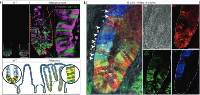
FIGURE 21. Demonstration of Lgr5+ ISC as the drivers of colorectal adenoma growth ... 61
C
HAPTER II: Autophagy FIGURE 22. Morphology of macroautophagic vacuoles ... 68FIGURE 23. Autophagic pathways in mammals ... 68
FIGURE 24. Schematic representation of (macro)autophagy ... 70
FIGURE 25. Examples of selective autophagy in mammals ... 71
FIGURE 26. mTOR-dependent regulation of autophagy ... 74
FIGURE 27. Coordinated stimulation of apoptosis and autophagy ... 76
FIGURE 28. Keap1-Nrf2 regulation by autophagy ... 79
FIGURE 29. Single-stranded and double-stranded DNA damage repair mechanisms ... 81
FIGURE 30. Autophagy regulation of double-stranded break repair ... 83
FIGURE 31. Roles of autophagy in physiology and pathology ... 88
FIGURE 32. Tumor-suppressive and tumor-promoting roles of autophagy ... 92
FIGURE 33. Effects of autophagy inhibition in mouse models of pancreatic cancer ... 95
FIGURE 34. 61 ongoing clinical trials on autophagy and cancer by cancer type ... 96
5
A
BBREVIATIONS
+4SC +4 label-retaining stem cells αSMA α smooth muscle actin
β-TrCP β-transducin repeats-containing proteins
4E-BP1 Inhibitor of the eukaryotic intiations factor 4E binding protein 1 4OHT 4-hydroxy-tamoxifen
5-FU 5-fluorouracil
53BP1 Tumor protein p53 binding protein 1 ALPI Intestinal alkaline phosphatase AMP Antimicrobial peptide
AMPK AMP-activated protein kinase
AOM Azoxymethane
APC Adenomatous polyposis coli ASCL2 Achaeate-scute homologue 2 ATB Antibiotics
ATF6 Activating transcription factor 6 ATG Autophagy-related gene
ATM Ataxia telangiectasia mutated ATM Ataxia telangiectasia mutated
ATOH1 Atonal BHLH transcription factor 1, Math1 in mice BAD Bcl2 antagonist of cell death
BCL-XL B cell lymphoma extra large BER Base-excision repair
BID BH3-interacting domain death agonist BiP Binding immunoglobulin protein BMP bone morphogenic protein BMPR1 type 1 BMP receptor BMPR2 type 2 BMP receptor BNIP3 Bcl2 interacting protein 3
BNIP3L BCL2 interacting protein 3 like, also called NIX CaMKII Calcium calmodulin kinase II
CBC Crypt basal columnar cells CHK Checkpoint kinase
CHOP c/EBP homologous protein
CIMP CpG island methylation phenotype CIN Chromosome instability
CK1α Casein kinase 1α
CMA Chaperone-mediated autophagy CMS Consensus molecular subtypes
CQ Chloroquine
CRC Colorectal cancer
CRCSC Colorectal cancer subtyping consortium DAMP Danger-associated molecular pattern DAPK1 Death-associated protein kinase 1 DCAMKL1 Doublecortin like kinase 1
6 DCS Deep crypt secretory
DDB2 DNA damage-binding protein 2 DDR DNA damage repair
DEPTOR DEP-domain containing mTOR-interacting protein
DKK Dickkopf
DLL Delta-like dMMR Deficient MMR
DNA-PK DNA-dependent protein kinase
DRAM Damage-regulated autophagy modulator DSB Double-stranded break
DSS Dextran Sulfate Sodiun DT Diphteria toxin
DTR Diphteria toxin receptor DUOX2 Dual oxidase 2
ECM Extra-cellular matrix EGF Epidermal growth factor EGFR EGF receptor
eIF2α Eukaryotic translation initiation factor 2α eIF4e Inhibitor of the eukaryotic intiations factor 4E ER endoplasmic reticulum
ERO1α ER oxidoreductase 1α
ESCRT Endosomal sorting complex required for transport FAP Famililal adenomatous polyposis
FIP200 Focal adhesion kinase family-interacting protein of 200 kDa FOXL1 Forkhead box L1, also known as Fkh6
FOXO Forkhead box family, class O FZD Frizzled
GABARAP Gamma-aminobutyric acid receptor-associated protein GALT Gut-associated lymphoid tissue
GAP GTPase-activating protein GSEA Gene set enrichment analysis GSK-3β Glycogen synthase 3β
HCQ Hydroxychloroquine HDAC Histone deacetylase HGF Hepatocyte growth factor HIF1 Hypoxia-inducible factor 1
HMGB1 High mobility group box 1, a chromatin-associated protein HNPCC Hereditary non-polyposis colon cancer, or Lynch syndrome HOPX Homeodomain-only protein
HP1α Heterochromatin protein 1α HR Homologous recombination HSC Hematopoietic stem cell
HSC70 Heat shock cognate 70 kDa protein IBD Inflammatory bowel disease IFN Interferon
IgA Immunoglobulin A IHH Indian Hedgehog
7 IL Interleukin
ILC Innate lymphoid cells
IMM Inner mitochondrial membrane IPA Ingenuity pathway analysis IR Ionizing radiation
IRE1α Inositol-requiring enzyme 1α
IRGM Interferon-inducible immunity-related GTPase family M member 1 ISC Intestinal stem cells
JNK c-Jun N-terminal kinases
KAP1 Krüppel associated box associated protein 1 KEAP1 Kelch-like ECH-associated protein 1
LAMP2 Lysosome-associated membrane protein 2 LC3 Microtubule-associated protein 1 light chain 3 LEF Lymphoid enhancer factor
LGR5 Leucine-rich repeat-containing G-protein-coupled transmembrane receptor 5 LIG4 Ligase 4
LIR LC3 interacting region LPR Lipoprotein-related protein LPS Lipopolysaccharide
LRIG1 Leucine rich repeats and immunoglobulin like domains 1 LRRK2 Leucine-rich repeat kinase 2
LTA Lipoteichoic acid M cells Microfold cells
MAMPs Microbe-associated molecular patterns MAPK Mitogen activated protein kinase MCL1 Myeloid cell leukemia sequence 1 MDP Muramyl dipeptide
MLH1 MutL homolog 1
mLTS8 Mammalian lethal with SEC13 protein 8 MMR Mismatch repair
MNV Murine norovirus MSH2 MutS homolog 2 MSH6 MutS homolog 6
MSI Microsatellite instability, MSI-H or -L for High or Low MSI MSS Microsatellite stable
mTOR Mammalian target of rapamycin
mTORC1 Mammalian target of rapamycin complex 1 MyD88 Myeloid differentiation primary response gene 88 MYH11 Myosin heavy chain 11
NAC N-acetyl Cysteine
NBR1 Neighbor of BRCA1 gene 1 NCID Notch intracellular domain NDP52 Nuclear dot protein 52 NER Nucleotide-excition repair NFκB Nuclear factor κB
NHEJ Non-homologous end joining NK Natural killer
8 NLR NOD-like receptor
NOX1 NADPH oxidase 1
NRF2 Nuclear factor erythroid 2 NSC Neural stem cells
OLFM4 Olfactomedin 4
OMM Outer mitochondrial membrane p70-S6K Ribosomal protein S6 kinase-1 PARP1 Poly (ADP-ribose) polymerase 1 PDGF Platelet-derived growth factor PE Phosphotidylethanolamine
PERK Protein kinase RNA-like ER kinase
PGC1α Peroxisome proliferator-activated receptor α coactivator 1α PI(3)P Phosphatidylinositol 3-phosphate
PI3K Phosphatidylinositol-4,5-bisphosphate 3-kinase PI3K Phosphoinositide 3 Kinase
PINK1 PTEN-induced kinase 1 PKR Protein kinase R POLD1 DNA polymerase δ POLE DNA polymerase ε
PolyIC Polyinosinic:polycytidylic acid
PPARγ Peroxisome proliferator-activated receptor γ PRAS40 Proline-riche Akt substrate of 40 kDa
PRR Pattern recognition receptor PTCH Patched
PTEN Phosphatase and tensin homolog
PTPN2 Protein tyrosine phosphatase, non-receptor type 2 RAB8a Ras-related proteins in brain 8a
RAG Ras-related GTP-binding protein RAPTOR Regulatory-associated protein of mTOR REG3 Regenerating islet-derived protein 3 REG4 Regenerating islet-derived 4
RHEB Ras homolog enriched in the brain RNF168 Ring finger protein 168
RNF3 Ring finger 3
RNF43 Ring finger protein 43 RNS Reactive nitrogen species ROS Reactive oxygens species
SCNA DNA somatic copy number alterations SHH Sonic Hedgehog
SLC Solute carriers
SMO Smoothened
SMOC2 SPARC related modular calcium binding 2
SNAP Soluble N-ethylmaleimide-sensitive factor attachment protein
SNARE Soluble N-ethylmaleimide-sensitive factor attachment protein receptors SOD Superoxide dismutase
9 SSA Sessile serrated adenomas
STAT3 Signal transduce and activator of transcription 3 TA Transit amplifying
TAX1BP1 Tax-1 binding protein 1 TCF T cell factor
TCGA The cancer genome altlas
TERT Telomerase reverse transcriptase TFEB Transcription factor EB
TGF Transforming growth factor
TIM Translocase of the inner mitochondrial membrane TLR Toll-like receptor
TNF Tumor necrosis factor
TOM Translocase of the outer mitochondrial membrane
TRIF Toll-IL-1 receptor domain-containing adapter-inducing interferon TRMP5 Transient receptor potential cation channel subfamily M member 5 TSA Traditional serrated adenomas
TSC Tuberous sclerosis complex
ULK Unc51-like autophagy activating kinase UPR Unfolded protein response
UVRAG UV radiation resistance-associated gene protein VEGFR Vascular endotherlial growth factor
VMP1 Vacuole membrane protein 1 VPS34 Vacuolar protein sorting 34 XBP1 X-box binding protein 1
XPC Xeroderma pirmentosum, complementation group C XRCC4 X-ray repair cross complementing 4
ZNRF3 Zinc and Ring Finger 3 ZNRF43 Zinc and ring finger 43
11
R
ESUME EN
F
RANÇAIS
Le renouvellement de l’épithélium intestinal repose sur la prolifération incessante de cellules souches intestinales (CSI). Le maintien de ces dernières est donc essentiel à l’homéostasie de l’épithélium intestinal mais aussi à sa régénération suite à des dommages. Ces CSI sont également considérées comme étant à l’origine de la transformation et l’initiation tumorale. L’étude des mécanismes impliqués dans la protection des CSI face à différents stress est donc essentielle pour mieux comprendre l’homéostasie et les pathologies intestinales. Notre équipe a précédemment pu démontrer, à la fois dans des échantillons de cancers colorectaux humains et dans un modèle murin prédisposé à développer des tumeurs intestinales, une induction de l’autophagie dans le tissu tumoral. L’autophagie est un mécanisme hautement conservé au cours de l’évolution et permettant la dégradation de différentes composantes cytoplasmiques par la voie des lysosomes. Ce mécanisme est impliqué dans de nombreux types de cancers ainsi que dans l’homéostasie de différentes populations de cellules souches adultes, telles que les cellules souches hématopoïétiques, les cellules souches neuronales, ou encore les cellules satellites. Grâce à des modèles murins permettant l’invalidation d’un gène clé du processus autophagique,
Atg7, spécifiquement dans l’épithélium intestinal, l’équipe a pu démontrer un rôle clé de ce
processus dans l’initiation et la croissance des tumeurs intestinales, ainsi que dans l’homéostasie microbienne de l’intestin. Suite à ces travaux, mes travaux de thèse visent à étudier le rôle de ce processus catabolique dans l’homéostasie des CSI. Pour ce faire, nous utilisons des modèles murins génétiquement modifiés et des cultures d’organoïdes afin d’étudier les effets de l’inhibition de l’autophagie dans l’homéostasie intestinale et en particulier dans les CSI.
Nos travaux indiquent que l’inhibition de l’autophagie par l’invalidation du gène Atg7 conduit à une activation de p53 et à une apoptose spécifique des CSI. De même, les cryptes invalidées pour
Atg7 ont une survie moindre que des cryptes contrôles en culture, ce qui souligne d’avantage un
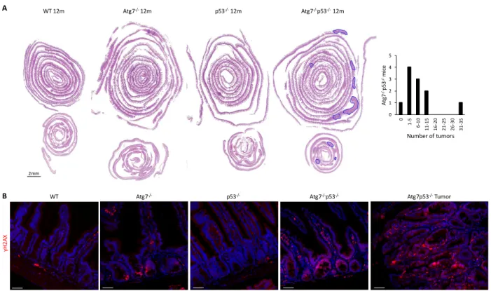
défaut de cellules souches. L’invalidation simultanée du gène Tp53 empêche la mort des CSI déficientes en autophagie. De plus, au long terme, ces souris doublement invalidées pour Atg7 et
Tp53 développent des tumeurs, contrairement aux souris invalidées uniquement pour les gènes Atg7 ou Tp53. Nous avons donc émis l’hypothèse que l’inhibition de l’autophagie sensibilisait les
CSI à l’apoptose suite à une accumulation de dommages cytotoxiques.
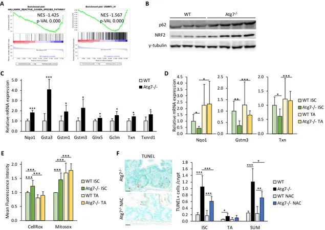
Par une analyse d’expression génique des CSI issues de cryptes contrôles et invalidées pour le gène Atg7, nous avons mis en évidence une altération des réponses associées au stress oxydant et à la réparation de l’ADN. Confirmant ces signatures, nous avons observé des dommages de l’ADN dans les cryptes déficientes en autophagie et un défaut de réparation de ces dommages suite à une irradiation. Nous observons également une accumulation d’espèces réactives de l’oxygène dans les CSI déficientes en autophagie associée à une atténuation de la réponse antioxidante médiée par NRF2. Des traitements antioxydants améliorent la survie des CSI invalidées pour Atg7 autant ex-vivo sur cultures d’organoides qu’in vivo. Ces expériences soutiennent l’implication des espèces réactives de l’oxygène accumulées suite à la perte de l’autophagie dans la mort des CSI.
12
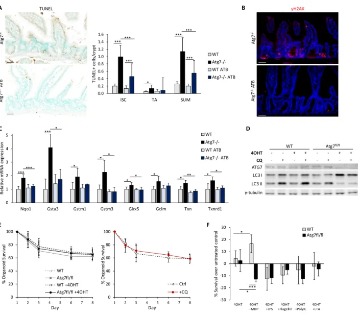
Par ailleurs, l’inhibition de l’autophagie induit des défauts de défense antimicrobienne, notamment au niveau de la sécrétion de peptides antimicrobiens par les cellules de Paneth, type sécrétoire retrouvé au fond des cryptes intestinales à proximité des CSI. Grâce à des traitements antibiotiques à large-spectre, nous avons observé un rôle important de la flore intestinale sur la survie des CSI déficientes en autophagie in vivo. Ce résultat est confirmé par la survie normale d’organoides inhibés pour l’autophagie seulement une fois en culture (et donc isolé du microenvironnement intestinal), mais cette survie est réduite en présence de certains signaux microbiens.
Nos travaux indiquent donc un rôle important de l’autophagie dans la protection et le maintien des CSI, de par son contrôle des espèces réactives de l’oxygène, du microenvironnement bactérien et des voies de réparation de l’ADN. Cependant, malgré la mort en continu des CSI déficientes en autophagie, un pool de CSI est maintenu en continu, ce qui permet le renouvellement normal de l’épithélium et souligne une plasticité importante au sein des cryptes épithéliales. L’ensemble de ces travaux suggèrent que l’autophagie pourrait être une cible thérapeutique prometteuse pour traiter les cancers colorectaux sans pour autant empêcher la fonction homéostatique de l’épithélium.
15
F
OREWORD
The work of my host team and Dr. ROMAGNOLO’s group focuses on the physiopathological renewal of the intestinal epithelium. For years, the group has investigated the molecular mechanisms involved in both the homeostatic regeneration of this highly proliferative epithelium and its deregulation in colorectal cancer. Among these, the WNT/β-catenin signaling pathway is both a crucial driver of proliferation and differentiation in the intestinal crypt and a critical target of oncogenic processes resulting in tumor development. Importantly, this pathway is essential for the maintenance and function of intestinal stem cells, which drive homeostatic proliferation in the epithelium and have also been implicated in tumor initiation and growth. Using both mouse models of intestinal tumorigenesis through WNT/β-catenin hyperactivation and human colorectal cancer samples, our group identified a crucial role of autophagy, a highly conserved cellular catabolic mechanism, in intestinal tumorigenesis. They further showed that blocking autophagy in the intestinal epithelium dramatically hindered tumor development and tumor growth, in part by affecting the gut’s microbiota and immunity. Interestingly, autophagy inhibition also affected the intestinal stem cell compartment of the non-tumoral epithelium. The work of my thesis aimed to understand the physiological functions of autophagy in the homeostatic intestinal epithelium and, more specifically, in intestinal stem cells.
This introduction will focus on the two principal aspects of my thesis work. First, the current knowledge on the intestinal epithelium at homeostasis and the rupture of this homeostasis in colorectal cancer will be reviewed. Particular attention will be paid to intestinal stem cells and the mechanisms regulating their maintenance, self-renewal, and proliferation. Second, we will explore the mechanisms and cellular functions of autophagy and its physiological impact. In both of these fields, major advancements have been made using invertebrate models such as
Drosophila melanogaster and Caenorhabditis elegans or, in the case of autophagy, in unicellular
eukaryotes like Saccharomyces cerevisiae. Nevertheless, throughout this introduction, we will focus primarily on mammalian systems and, whenever possible, on in vivo studies.
C
HAPTER
1:
P
HYSIOPATHOLOGY OF THE
19
I.
S
TRUCTURE
&
F
UNCTION OF THE
I
NTESTINE
1.1 OVERVIEW OF THE ADULT DIGESTIVE SYSTEM & GASTROINTESTINAL TRACT
The adult digestive system consists of the organs along the length of the digestive tube – the mouth, esophagus, stomach, small intestine, and large intestine (or colon) – as well as several accessory organs including the tongue, salivary glands, liver, pancreas and gallbladder [FIGURE 1]. Along the route of ingested food from the mouth to the anus, the digestive tube plays two principal functions: it allows the breakdown of food and absorption of nutrients (digestion), and it acts as a physical and chemical barrier between the outside luminal environment and the organism.
Digestion begins directly upon ingestion of food, in the mouth. The chewing and grinding action of the teeth (mastication) helped by the muscular action of the tongue allow the mechanical breakdown of food into smaller pieces. This increases the surface area accessible to the digestive enzymes in saliva, produced in the salivary glands. In addition to digestive enzymes, saliva also contains antimicrobial proteins, antifungal proteins and immunoglobulins, which act as early defense against microbes arriving in the gastrointestinal tract along with the ingested food. Saliva also softens the food, transforming it into a soft bolus, and facilitating its passage through the pharynx and into the esophagus upon swallowing.
The esophagus is a muscular tube carrying the bolus to the stomach through rhythmic contractions called peristalsis that will continue along the rest of the gastrointestinal tract. Two sphincters at the top and bottom of the esophagus open upon swallowing and prevent backflow from the stomach, respectively. Once in the stomach, the bolus is further broken down by the action of numerous digestive enzymes. Gastric acid (mainly hydrochloric acid) creates an acidic pH that both allows pepsin proteases to work and kills most ingested bacteria. The whole of the gastrointestinal tract, and particularly the stomach, is covered by mucus that lubricates the passage of digested food and protects the underlying, single-layered columnar epithelium. Along with the enzymatic digestion, mechanical churning helps transform the bolus into chyme.
It is at this stage, approximately one to two hours after ingestion, that the partially-digested food enters the small intestine through the pyloric sphincter. The majority of digestion and absorption occurs in the small intestine. The segment directly connected to the stomach is called the duodenum. Bile produced by the liver and stored in the gallbladder is released into the duodenum upon the arrival of the chyme to help break down fats. The pancreas also releases pancreatic juice rich in bicarbonate and digestive enzymes into the duodenum to neutralize the acidic chyme from the stomach and continue the digestion of carbohydrates, proteins and lipids into monosaccharides, amino acids and fatty acids, respectively. As nutrients are taken up by the organism, the muscular walls of the intestine help slowly pass the chyme along the relatively short duodenum and the other two segments of the small intestine: the jejunum and ileum. The intestinal wall contains many folds, which allow the intestine to stretch and contract as food passes along. To increase the surface area of absorption, the small intestinal epithelium consists
20
of a single layer of cells folded upon itself creating finger-like protrusions called villi. Moreover, the absorptive cells of the epithelial lining, called enterocytes, have densely packed microvilli on the luminal side to further increase surface area. These cells express additional digestive enzymes at their surface, as well as transporters that take up monosaccharides, amino acids, some vitamins and ions. Fatty acids, other vitamins and water, on the other hand, can diffuse passively across the epithelium and into the bloodstream. Once in the bloodstream, nutrients and other substances absorbed in the intestine are directly sent to be processed by the liver. The majority of nutrients are thus absorbed in the duodenum and jejunum. Along with the amount of available nutrients, the length of the epithelial villi decrease from the jejunum to the ileum [FIGURE 2]. Remaining water, salts, and undigested materials are passed on to the large intestine, or colon, through the ileocecal valve. The colon is subdivided into several segments; the first pouch is called the cecum, followed by the proximal and distal colon – subdivided in humans into the ascending, transverse, descending, and sigmoid colon – leading to the rectum. Salts and remaining water are absorbed while undigested materials are packed into feces to be excreted by contractions of the rectum. The colon epithelium lacks villi entirely, and is progressively smoother from the proximal to the distal end [FIGURE 2].
21
FIGURE 2. Organization of the intestinal wall and anatomy of the mucosa in
different segments of the mouse intestine (hematoxilin & eosin staining).
1.2 STRUCTURAL ORGANIZATION OF THE INTESTINE
The different elements that make up the intestinal wall work together to accomplish the two major functions of this organ in digestion and as a barrier.
The intestinal lumen is home to a large amount of microorganisms, altogether known as the gut microbiota, particularly present in the colon. Although technically an environmental factor, the notion of the gut microbiota as an essential symbiont for host development and physiology has become more and more accepted over the last decade. Bacteria dominate the microbiota, which also includes archea, viruses, fungi, and protozoa. Colonization of the gut at birth is important for the development of the intestinal mucosa, including the maturation of vasculature
(Stappenbeck et al., 2002; Reinhardt et al., 2012) and of the gut-associated lymphoid tissue (GALT) (Macpherson et al., 2000; Macpherson, 2004; Bouskra et al., 2008; Uematsu et al., 2008; Gaboriau-Routhiau et al., 2009; Sanos et al., 2009; Atarashi et al., 2011; Kawamoto et al., 2012; Olszak et al., 2012). In the adult intestine, the microbiota notably contributes to digestion as it helps break down undigested products (ie. polysaccharides into short-chain fatty acids). In addition to metabolizing otherwise indigestible nutrients, the microbiota plays an important role in metabolizing xenobiotics – including drugs or pollutants (Koppel et al., 2017). Lastly, the gut commensals, through competition for nutrients and production of virulence factors, protect their host against infection by opportunistic pathogens (Kamada et al., 2012).
Although a single-layered epithelium is advantageous in terms of absorption, it only provides a thin barrier between the luminal contents and the host tissues. The barrier function of the mucosa therefore depends on intracellular junctions, especially tight junctions, that seal neighboring epithelial cells together (Buckley and Turner, 2018). Furthermore, specialized cells
Mucosa Submucosa Muscularis mucosae Serosa Lumen Lamina Propria Mucus layer Crypt Villus Lymphatic vessel Artery Vein Smooth Muscle Lymphoid follicle Lacteal Arteriole Capillaries Venule Nerve Muscularis externa Epithelium Je junum Duo de num Ile um Dis tal Col on Pr oxi m al C olo n 250 µm
22
protect the epithelial lining, including mucus-producing goblet cells antimicrobial peptide-producing Paneth cells. As an additional protective measure, the lining of the intestine is continuously regenerated; in fact, the intestinal epithelium renews itself almost entirely every 4 to 5 days (Cheng and Leblond, 1974; Wright and Irwin, 1982) – faster than any other tissue. The regenerative capacity of the gut epithelium depends on a pool of actively dividing intestinal stem cells (ISC) and their highly proliferative transit amplifying (TA) progenitors, all located in epithelial pockets away from the lumen called crypts of Lieberkühn.
The intestinal epithelium is surrounded on the basal side by a connective mesenchyme called the lamina propria. The basal side of intestinal epithelial cells is in direct contact with a network of extra-cellular proteins that form the basement membrane, including type IV Collagen, Laminin and proteoglycans. Basement membrane components are produced both from the epithelium and mesenchymal cells in the lamina propria. Beneath the basement membrane, the mesenchyme comprises a complex extra-cellular matrix (ECM) composed of similar components including type I Collagen and Fibronectin. Embedded in this ECM are various mesenchymal cell types, including various fibroblast populations, myofibroblasts, a layer of smooth muscle called the muscularis mucosa which contributes to peristalsis, endothelial cells of the blood and lymphatic networks, pericytes, and enteric neurons [FIGURE 2]. In addition to these, the lamina propria houses various innate and adaptive immune cells and structures altogether comprising the GALT, capable of mounting an appropriate inflammatory or tolerogenic response to passing antigens. These include B cells (especially immunoglobulin A (IgA)-producing plasma cells), T cells (including pro-inflammatory Th1, Th2 and Th17 cells as well as anti-inflammatory regulatory T cells), neutrophils, natural killer (NK) cells, macrophages and dendritic cells. In addition to these, specialized subsets of T cells (intra-epithelial lymphocytes) and some dendritic cells are found between the intestinal epithelial cells, where they continuously sample luminal contents.
The submucosa contains blood vessels, lymphatic vessels, and nerves embedded in a matrix of fibres and collagen. Beneath those, two layers of smooth muscle, one circular and one longitudinal, also aid in peristalsis. Finally, the intestinal wall is surrounded by a loose layer of connective tissue called the serosa, and the whole of the intestine is held in place in the peritoneum by the mesentery.
1.3 THE INTESTINAL EPITHELIUM
The single-layered mammalian intestinal epithelium is folded upon itself creating finger-like protrusions towards the lumen called villi, beneath which lie small pockets towards the lamina propria called the crypts of Lieberkühn. The small intestinal crypts are almost entirely composed of proliferative cells, with the exception of Paneth cells, a secretory lineage first recognized for their ability to secrete antimicrobial peptides (AMP). The adult mouse intestine contains approximately 1.1 million crypts, with around two-thirds of the cells of each crypt dividing every twelve hours, producing over 300 million new cells each day (Gordon et al., 1992). This astounding regenerative capacity relies on three to sixteen (depending on the study) actively dividing ISC localized at the crypt bottom alongside Paneth cells (in the small intestine)
23
or related secretory cells (in the colon) (Bjerknes and Cheng, 1981a; Altmann, 1983; Potten and Loeffler, 1987; Sato et al., 2011a; Sasaki et al., 2016). ISC give rise both to new ISC and to TA progenitor cells that will rapidly proliferate and progressively differentiate as they migrate up the crypt. TA daughters will either be absorptive-lineage progenitors, giving rise to enterocytes, or secretory progenitors that will give rise to Paneth cells, goblet cells, or enteroendocrine cells
(Schmidt et al., 1985; Bjerknes and Cheng, 1999). In the small intestine, cells become terminally differentiated upon reaching the villus, and then continue their columnar migration to the tip of the villus, where they are expelled into the lumen and die in a process termed anoikis [FIGURE 3]. The entire process of proliferation, differentiation, and expulsion of intestinal epithelial cells takes only 4-5 days (Cheng and Leblond, 1974; Wright and Irwin, 1982).
The villus epithelium therefore contains exclusively differentiated cells, over 80 percent of which are absorptive enterocytes. In addition to these, the differentiated compartment of the epithelium contains the secretory cells mentioned above (with the exception of Paneth cells), as well as three rarer cell types: tuft cells (Gerbe and Jay, 2016), cup cells (Madara, 1982) and microfold (M) cells associated with lymphoid follicles called Peyer’s patches (Kraehenbuhl and Neutra, 2000). Each of these differentiated cell types will be discussed in more detail below. The colon has a flat surface with no villi; instead, the bottom third of colonic crypts contains proliferative cells while the upper crypt is differentiated with a considerably high ratio of goblet cells [FIGURE 3]. Paneth cells are notably absent in the colon.
1.3.1
E
NTEROCYTESEnterocytes are the predominant cell type of the intestinal epithelium, making up over 80% of epithelial cells. They are highly polarized absorptive cells characterized by their columnar shape and a brush border of microvilli at the apical pole that increases the surface area of absorption
[FIGURE 4]. On the surface of these microvilli, enterocytes express digestive enzymes that play a
role in the final breakdown of food particles before they are selectively taken up by the cell and transited to the basal pole. The uptake of nutrients can be active via specific transporters – as is the case for ions, sugars, amino acids, or water-soluble vitamins – or passive – as is the case for water, lipids, and lipid-soluble vitamins. Enterocytes also transport IgA from plasma cells in the lamina propria in the opposite direction; these are endocytosed on the basolateral surface and released into the intestinal lumen.
Differentiation into either the absorptive lineage or secretory lineage depends on NOTCH signaling in the crypt. Expression of the NOTCH target gene Hes1 is required for enterocyte differentiation, whereas it represses expression of NOTCH ligands and of ATOH1 (atonal BHLH transcription factor 1, Math1 in mice), both key to secretory lineage commitment (Yang et al., 2001; Stanger et al., 2005).
24
FIGURE 3. Topography of the intestinal epithelium in the small intestine (A) and colon (B).
Adapted from Barker, 2014.
1.3.2
G
OBLET CELLSGoblet cells, as previously noted, are secretory ligneage cells scattered throughout the epithelium that produce and secrete mucus to both facilitate movement of luminal contents and protect the epithelium. Contrary to enterocytes, these cells depend on ATOH1 expression early in their lineage commitment. They contain characteristic mucin-containing secretion granules in their apical cytoplasm [FIGURE 4] and release these granules into the lumen by exocytosis. The main component of the intestinal mucus layer is the mucin MUC2. Before secretion, mucins are highly glycosylated in the golgi apparatus of goblet cells, giving them the capacity to bind water and form a gel-like network once secreted (Johansson et al., 2011). Muc2-deficient mice have bacteria in direct contact with the epithelium as well as bacterial infiltration across the epithelium (Van der Sluis et al., 2006; Johansson et al., 2008). This results in inflammation and symptoms of inflammatory bowel disease (IBD). Interestingly, these mice also develop spontaneous intestinal adenomas (Velcich et al., 2002).
25
The proportion of mucin-producing goblet cells in the epithelium and the thickness of the mucus layer increases from the proximal small intestine (~4%) to the distal colon (~16%) (Cheng,
1974) in direct correlation with the density of the microbiota. In the small intestine, the mucus
layer is discontinuous but is supported by the secretion of antimicrobial peptides by Paneth cells. In the colon, two structurally distinct layers of mucus are found: the firm inner layer is considered to be relatively sterile (Johansson et al., 2008; Li et al., 2015a), while the thicker outer layer, made looser by proteolytic dispersion of mucin polymers, houses a distinct subset of bacterial species that can digest complex glycoproteins (Johansson et al., 2011; Li et al., 2015a), therefore creating a unique microbial niche (Li et al., 2015a).
Recent studies have also shown that goblet cells have the ability to endocytose luminal substances and transfer them to antigen-presenting cells in the lamina propria (McDole et al.,
2012).
1.3.3
P
ANETHC
ELLSPaneth cells are the major secretory cell type of the small intestinal crypt. Like goblet cells, differentiation of Paneth cells depends upon the expression of the ATOH1 transcription factor. Paneth cells, unlike the other progenitor lineages, terminally differentiate as they migrate down to the bottom of the crypt and remain at the crypt bottom throughout their uniquely long 6- to 8-week lifespan (Ireland et al., 2005). The position of a Paneth cell is therefore linked to its maturity, with the oldest and more mature Paneth cells found at the crypt base (Bjerknes and Cheng, 1981b). These cells have a characteristic pyramidal shape with prominent, large granules filling their cytoplasm on the apical side. They also have an extensive endoplasmic reticulum and Golgi network, in link with their secretory functions [FIGURE 4].
The distinguishing apical granules of Paneth cells were later shown to contain Defensins, Lysozyme, REG3 (regenerating islet-derived protein 3, REG3A in humans, REG3γ and REG3β in mice) and other AMPs, establishing the role of Paneth cells in innate imunity. Although Paneth cells constitutively express Defensins under the control of WNT signaling (van Es et al., 2005a; Andreu et al., 2008), production of other AMPs is up-regulated in response to microbial signals
(Mallow et al., 1996; Ayabe et al., 2000; Pütsep et al., 2000; Vaishnava et al., 2008) or pro-inflammatory cytokines like Interferon gamma (IFNγ) (Farin et al., 2014).These secreted AMPs not only protect the crypt epithelium from enteric pathogens (Vaishnava et al., 2008), but also directly affect the composition of the microbiota (Salzman et al., 2010).
Due to their strategic localization at the crypt bottom alongside ISC, a tight functional link between Paneth cells and ISC was proposed early on (Cheng and Leblond, 1974). Indeed, Paneth cells also play an important role in the ISC niche by producing essential trophic factors like WNT ligands, NOTCH ligands, and EGF (Gregorieff et al., 2005; Sato et al., 2011a). The role of Paneth cells in the ISC niche will be discussed in more detail in the following section.
1.3.4
E
NTEROENDOCRINEC
ELLSEnteroendocrine cells are secretory lineage cells specified by ATOH1 and Neurogenin-3 expression (Jenny, 2002). They secrete hormones like Serotonin (which notably affects
26
gastrointestinal motility) and the insulinotropic hormone Glucagon-like peptide-1. There exists up to 15 different subtypes of enteroendocrine cells, depending on their morphology and the hormones they produce. These represent about 1% of epithelial cells, dispersed throughout the differentiated tissue.
FIGURE 4. The four predominant differentiated cell types of the intestinal epithelium.
Adapted from Alberts et al., 2008.
1.3.5
T
UFTC
ELLSTuft cells were identified by electron microscopy based on their unique tubulovesicular system and apical bundle of microfilaments connected to a tuft of microvilli that are longer and thicker than those of enterocytes. In light of their morphology, tuft cells were thought to be secretory cells (Sato and Miyoshi, 1997) but whether they are derived from the secretory lineage specified by ATOH1 is still a matter of debate (Gerbe et al., 2011; Bjerknes et al., 2012). Despite their discovery more than 60 years ago (Jarvi and Keyrilainen, 1956; Rhodin and Dalhamn, 1956), their function was only very recently brought to light. The first identified function of tuft cells was the secretion of opioids (Kokrashvili et al., 2009; Gerbe et al., 2011). Then, in 2016, three independent groups published studies identifying tuft cells as essential to trigger a type 2 immune in response to parasitic infections in the gut (Gerbe et al., 2016; Howitt et al., 2016; Von
Moltke et al., 2016). Mechanistically, this involves signal transduction via the TRMP5 (Transient
receptor potential cation channel subfamily M member 5) cation channel and Interleukin (IL) -25 alarmin production (Gerbe et al., 2016; Howitt et al., 2016; Von Moltke et al., 2016). In the intestinal epithelium, tuft cells are much less frequent than the previous four differentiated cell types, as they make up only about 0.4% of epithelial cells. However, during infections with helminthes Nippostrongylus brasiliensis, Heligmosomoides polygyrus or the protozoa
Tritrichomonas muris, tuft cell numbers expand dramatically, as do goblet cell numbers and
epithelial cell cytokine production, leading to the recruitment of type 2 helper T cells and group 2 innate lymphoid cells (ILC) in the lamina propria. The loss of tuft cells or of their function (by deleting either the Trpm5 or Il25 genes) prevents each of these responses and delays the resolution of the infection. More recently, the tuft cell-ILC2 circuit was shown to be basaly
27
activated by commensal protozoan-derived metabolites as a way to prevent infection (Schneider
et al., 2018). Tuft cells therefore play a unique role in the context of parasite infection.
1.3.6
M
ICROFOLDC
ELLSM cells are specialized cells found on follicle-associated epithelium such as that found over Peyer’s patches. These cells owe their name to their characteristic apical membrane, which lacks microvilli and instead has a microfold topography (Kraehenbuhl and Neutra, 2000). M cells are believed to act as sentinels that take up antigens from the lumen and transport them to the underlying GALT. In line with this, their basolateral membrane is invaginated to form pockets that reduce the intracellular distance of antigen transport from the apical membrane and harbor infiltrating lymphocytes. In this way, M cells help the intestinal immune system mount either inflammatory or tolerogenic responses to foreign antigens. M cells are thought to differentiate from absorptive-lineage progenitors in response to stimuli from the underlying lymphoid tissue, namely NF-κB signaling (Knoop et al., 2009; Kanaya et al., 2018).
1.3.7
C
UPC
ELLSCup cells can be distinguished by light or electron microscopy due to their lighter cytoplasm and shorter brush border compared to neighboring enterocytes, creating the cup-like apical indentation that gave them their name (Madara, 1982). They are absent in the jejunum but relatively abundant (~6% of villus epithelial cells) in the ileum of rodents, although less so in primates. To date, the function of these cells remains unknown.
II.
I
NTESTINAL
S
TEM
C
ELLS AND THEIR
D
EFINING
N
ICHE
2.1 MODELS AND MARKERS OF INTESTINAL STEM CELLS
Although the localization of ISC at the crypt bottom has long been established, the identity of ISC has remained a subject of debate over the past decades. Two historical models of ISC have been originally proposed but direct evidence to illustrate their stemness was put forward only in the last decade. Pioneer work by Cheng, Bjerknes and Leblond using electron microscopy, 3
H-thymidine incorporation and clonal mutagenesis demonstrated the presence of undifferentiated proliferative cells capable of giving rise to the different intestinal epithelial lineages (multipotent) at positions 1-4 from the bottom of the intestinal crypt, which they called crypt basal columnar cells (CBC)(Cheng and CP., 1974; Bjerknes and Cheng, 1999, 2002). At the same time, works led by Potten’s group recognized cells at the fourth position from the crypt bottom (+4) as capable of long-term 3H-thymidine label retention (Potten et al., 1978), an established
property of tissue stem cells. Without specific molecular markers to isolate them at the time, it remained unclear whether the mitotically active CBC or the relatively quiescent +4 label-retaining cells (+4SC) ought to be considered as the bona fide ISC.
2.1.1
M
ARKERS FORC
RYPTB
ASALC
OLUMNAR CELLSIn 2007, the Clevers lab and collaborators identified LGR5 as a marker for CBC. Lgr5 encodes a leucine-rich repeat-containing G-protein-coupled transmembrane receptor first identified as a Wnt/β-catenin target gene. Although LGR5 immuno-labelling has proved difficult due to its low expression levels, the development of a mouse model in which an EGFP and a tamoxifen-inducible Cre recombinase cassette was knocked in at the Lgr5 locus (Lgr5-EGFP-ires-CreERT2)
has allowed characterization of Lgr5-expressing cells. In the adult small intestine, Lgr5+ cells are
located at the base of the crypt, intermingled with Paneth cells [FIGURE 5B]. They are rapidly cycling since within 24 hours almost all Lgr5+ cells of a crypt undergo mitosis. In the colon, Lgr5+
cells are also found at the crypt bottom interspersed with secretory cells [FIGURE 5F] but have slower cycling kinetics than in the small intestine. The generation of in vivo lineage tracing from Lgr5+ cells using an Lgr5-EGFP-ires-CreERT2/Rosa26lacZ mouse model indicated that Lgr5+ cells
transmit LacZ staining to their progeny and repopulate the entire epithelium during homeostasis [FIGURE 5]. Furthermore, Lgr5+ cells are long-lived and contribute to tissue renewal
over the entire lifetime of the mouse. Therefore, LGR5 is a reliable marker of CBC (Barker et al., 2007), and Lgr5-EGFP-ires-CreERT2 mice a valuable tool to understand their regulation. The
ultimate proof of Lgr5+ CBC as ISC is the capacity of single isolated Lgr5+ cells cultured in vitro in
matrigel to generate intestinal organoids that mimic the structure of the in vivo epithelium, with Lgr5+ cells found at the bottom of the proliferative crypt domains [FIGURE 6A-D]. Importantly,
these organoids contain the four major differentiated lineages of the intestinal epithelium
30
FIGURE 5. Establishment of Lgr5 as an intestinal stem cell marker (A) Generation of mice expressing EGFP and
creERT2 from a single bicistronic message by gene knock-in into the first exon of Lgr5. SA, splice acceptor; UTR, untranslated region. (B,F) Confocal GFP imaging counterstained with the red DNA dye ToPro-3 confirming that Lgr5 expression is restircted to the 6-8 slender cells at the crypt bottom in the small intestine
(B) and in the colon (F). (C-E) Histological analysis of LacZ activity in the small intestine 1 day after induction (C), 5 days after induction (D) and 60 days after induction (E). (G-I) LacZ activity in the colon 1 day
after induction (G), 5 days after induction (H) and 60 days after induction (I). Adapted from Barker et al., 2007.
FIGURE 6. Intestinal organoid culture from isolated Lgr5+ ISC sorted from Lgr5-EGFP-IRES-CreERT2 mice (A)
Two positive populations, GFPHigh and GFPLow, are discriminated from Lgr5-EGFP-IRES-CreERT2 intestines.
FSC, forward scatter. (B) Example of a single GFPHigh cell successfully growing into an organoid. Numbers
above the images are the days of growth. (C) 14 days after sorting, organoids from isolated GFPHigh cells have
Lgr5-GFP positive cells at the bottom of the crypt domains. (D) Organoids cultured with EdU (red) for 1h incorporate EdU only in the crypt domains. Counterstain, DAPI (blue). (E-H) Confocal image for villin (E, green, enterocytes), lysozyme (F, green, Paneth cells), Muc2 (G, red, goblet cells), and chromogranin A (H,
green, enteroendocrine cells). Adapted from Sato et al., 2009.
b c – 1 day d – 5 days
f
e – 60 days a
g – 1 day h – 5 days i – 60 days
a b c d e f g h
31
A CBC stem cell signature using gene expression and proteome profiling has been determined using FACS-purified Lgr5+ cells from the Lgr5-EGFP-ires-CreERT2 model. Indeed, comparison
between Lgr5+ CBC and their direct TA progeny sorted by different levels of EGFP (high in CBC
and low in their TA progeny) provided a comprehensive Lgr5+ ISC signature of approximately
500 genes (Muñoz et al., 2012). The signature contains many Wnt/β-catenin modulators and target genes such as Lgr5, Sox9, Ascl2 (achaeate-scute homologue 2), EphB2, Troy/Tnfrsf19,
Axin2, Znrf43 (zinc and ring finger 43) (Hao et al., 2012) and Rnf3 (ring finger 3) (Koo et al., 2012). This and subsequent Lgr5+ cell signatures (van der Flier et al., 2009a; Muñoz et al., 2012)
along with candidate-approach studies also provided several other verified CBC markers. Among the new markers identified, some have been confirmed as ISC markers by lineage tracing: Smoc2 (SPARC related modular calcium binding 2) (Muñoz et al., 2012), Musashi-1 (Potten et al., 2003),
Prominin-1 (Zhu et al., 2009) and notably Olfm4 (Olfactomedin 4), which is highly expressed in the small intestine compared to other mentioned CBC markers and therefore allows easier detection of CBC (van der Flier et al., 2009b; Schuijers et al., 2014). Ascl2, a basic helix-loop-helix transcription factor was identified as a master regulator of CBC maintenance. The conditional deletion of Ascl2 results in the loss of CBC and its overexpression promotes drastic expansion of the ISC compartment (van der Flier et al., 2009a; Reed et al., 2012; Schuijers et al., 2015).
2.1.2
M
ARKERS FOR+4
S
TEMC
ELLSThe first +4SC marker investigated by lineage tracing was BMI1, a polycomb-repressing complex member essential in hematopoietic and neural stem cell self-renewal. In intestinal homeostasis,
Bmi1 is expressed at the +4 position in a minority of small intestinal crypts (around 10%) and
particularly in the duodenum. By in vivo lineage tracing using a Bmi1-CreERT2/Rosa26lacZ
mouse model and organoid culture experiments, Bmi1+ cells were shown to self-renew and give
rise to the different epithelial lineages but with much slower cycling kinetics compared to Lrg5+
CBC (Sangiorgi and Capecchi, 2008; Yan et al., 2012). In addition, ablation of Bmi1+ cells using
targeted expression of diphteria toxin (DT) caused crypt loss consistent with a stem cell defect. Ablation of the Lgr5+ CBC using targeted expression of the DT receptor (DTR), on the other hand,
caused an increase of Bmi1+ cells which can regenerate the Lgr5+ stem cell population and
maintain intestinal homeostasis (Tian et al., 2011). Thus, these relatively quiescent +4SC, have been considered as reserve stem cells in case of damage to the active CBC stem cells. In support of the mobilization of the +4SC following injury, after high dose of radiation (up to 12 Gy), Lgr5+
cells are rapidly lost whereas Bmi1+ cells expand and allow epithelial recovery though
replenishment of Lgr5+ cells (Yan et al., 2012). In fact, BMI1 has been shown to play a role in
DNA damage response signaling; it may therefore play a direct role in the resistance of reserve stem cells to irradiation (Ismail et al., 2010; Ginjala et al., 2011; Pan et al., 2011). Nevertheless, the restoration of a pool of active Lgr5+ ISC pool by reserve stem cells appears to be an essential
step for epithelial repair following injury, as depletion of Lgr5-expressing cells during radiation-induced damage and subsequent repair is detrimental to the epithelium (Metcalfe et al., 2014). However, BMI1-expressing cells are only present in a small proportion of crypts in the proximal small intestine and are not responsible for the post-injury regeneration throughout the distal