Association of Intraocular Cataract Lens Replacement
With Circadian Rhythms, Cognitive Function,
and Sleep in Older Adults
Sarah L. Chellappa, MD, PhD; Vivien Bromundt, PhD; Sylvia Frey, PhD; Alexandra Steinemann, MD; Christina Schmidt, PhD; Torsten Schlote, PhD; David Goldblum, MD; Christian Cajochen, PhD
IMPORTANCECataract is associated with a progressive decline in light transmission due to the clouding and yellowing of the natural crystalline lens. While the downstream effects of aging lenses include long-term disruption of circadian rhythms, cognitive function, and sleep regulation, it remains unknown whether there is an association of intraocular cataract lens (IOLs) replacement with circadian rhythms, cognition, and sleep.
OBJECTIVETo test whether IOL replacement (blue blocking [BB] or ultraviolet [UV] only blocking) in older patients with previous cataract is associated with the beneficial light effects on the circadian system, cognition, and sleep regulation.
DESIGN, SETTING, AND PARTICIPANTS Cross-sectional study at the Centre for Chronobiology, University of Basel in Switzerland from February 2012 to April 2014, analyzed between June 2012 and September 2018. Sixteen healthy older controls and 13 patients with previous cataract and IOL replacement participated without medication and no medical and sleep comorbidities. EXPOSURES Three and a half hours of prior light control (dim-dark adaptation), followed by 2 hours of evening blue-enriched (6500 K) or non–blue-enriched light exposure (3000 K and 2500 K), 30 minutes in dim post–light exposure, 8 hours of sleep opportunity, and 2 hours of morning dim light following sleep.
MAIN OUTCOMES AND MEASURES Salivary melatonin, cognitive tests, and sleep structure and electroencephalographic activity to test the association of IOLs with markers of circadian rhythmicity, cognitive performance, and sleep regulation, respectively.
RESULTS The participants included 16 healthy older controls with a mean (standard error of the mean [SEM]) of 63.6 (5.6) years; 8 women and 13 patients with previous cataract (mean [SEM] age, 69.9 [5.2] years; 10 women); 5 patients had UV IOLs and 8 had BB IOLs. Patients with previous cataract and IOLs had an attenuated increase in melatonin levels during light exposure (mean [SEM] increase in the BB group: 23.3% [2.6%] and in the UV lens group: 19.1% [2.1%]) than controls (mean [SEM] increase, 48.8% [5.2%]) (difference between means, 27.7; 95% CI, 15.4%-41.7%; P < .001). Cognitive function, indexed by sustained attention performance, was improved in patients with UV lens (mean [SEM], 276.9 [11.1] milliseconds) compared with patients with BB lens (mean [SEM], 348.3 [17.8] milliseconds) (difference between means, 71.4; 95% CI, 29.5%-113.1%; P = .002) during light exposure and in the morning after sleep. Patients with UV lens had increased slow-wave sleep (mean [SEM] increase, 13% [3.4%]) compared with controls (mean [SEM] increase, 5.2% [0.8%]) (percentage of total sleep time; difference between means, 7.9; 95% CI, 2.4%-13.4%; P = .02) and frontal non–rapid eye movement slow-wave activity (0.75-4.5 Hz) during the first sleep cycle (mean [SEM], 79.9 [13.6] μV2
/Hz) compared with patients with BB lens (mean [SEM], 53.2 [10.7] μV2
/Hz) (difference between means, 26.7; 95% CI, 9.2-48.9; P = .03).
CONCLUSIONS AND RELEVANCE These in-laboratory empirical findings suggest that optimizing the spectral lens transmission in patients with previous cataract may minimize the adverse age-related effects on circadian rhythms, cognition, and sleep.
JAMA Ophthalmol. doi:10.1001/jamaophthalmol.2019.1406 Published online May 23, 2019.
Invited Commentary Supplemental content
Author Affiliations: Author affiliations are listed at the end of this article.
Corresponding authors: Sarah L. Chellappa, MD, PhD, Division of Sleep Medicine, Harvard Medical School, 221 Longwood Ave BL 039C, Boston, MA, 02115 (schellappa@bwh. harvard.edu); Christian Cajochen, PhD, Centre for Chronobiology, Psychiatric Hospital of the University of Basel, Wilhelm Kleinstrasse 27, CH-4002 Basel, Switzerland ([email protected]).
JAMA Ophthalmology |
Original Investigation
C
ataract is the most common source of vision loss in older adults and is the major worldwide cause of blindness in the general population.1Age-related cataract is as-sociated with a progressive decline in light transmission due to the clouding and yellowing of the natural crystalline lens,2 particularly in the shorter blue light wavelength.3The down-stream effects of aging lens include reduced photic input to the circadian clock, with subsequent long-term disruption of circadian rhythms, cognitive brain function, and sleep regulation.4-6However, it remains unknown whether modi-fying the photic input to the circadian clock, by optimizing the spectral characteristics of crystalline lens after cataract sur-gery, improves circadian, sleep, and cognitive symptoms in older adults. Here, we investigated whether there is an asso-ciation between intraocular cataract lens replacement (intra-ocular blue blocking [BB] or ultraviolet [UV] only blocking) with hallmarks of the circadian system, cognitive function, and sleep regulation.
Methods
Participants
The protocol, advertisements, screening questionnaires, and consent form were approved by the local ethical committee (EKBB/Ethikkommission beider Basel, Switzerland) and agreed with the Declaration of Helsinki.7
All participants provided writ-ten consent.
Our study sample included an extensive list of inclusion criteria: age, 55 to 80 years; body mass index (calculated as weight in kilograms divided by height in meters squared), 18 to 28 for women and 19 to 29 for men; bilateral cataract sur-gery for age-related cataract (4-8 weeks after intraocular lens [IOL] replacement of the second eye); new lens, 18 to 26 di-opters; willing and able to give informed consent; stable medi-cation for past 2 months and throughout the study duration (exception: medication in exclusion criteria); compliance with regular sleep-wake schedules (see Study Design section); no coexisting ocular pathologies, medical, or psychiatric conditions based on clinical history, biochemical blood screening tests and physical examination (eMethods in the
Supplement). Owing to our stringently controlled inclusion/
exclusion criteria list, approximately 1200 patients with pre-vious cataract were assessed to recruit 60 patients. Of these 60 patients, 44 were excluded owing to (1) inability to follow the mandatory regular sleep-wake schedule throughout the 3 weeks of the study duration; (2) inadequate sleep quality, as indexed by a Pittsburgh sleep quality index score more than 58; and (3) extreme morning or evening chronotype ratings.9 The control group was enrolled using the same inclusion/ exclusion criteria (except for the cataract-related study re-quirements). Sixteen participants were enrolled of approxi-mately 60 participants (similar dropout rate for the ambulatory segment as for the postcataract surgery patients).
Study Design
The study, including all experimental setups, ophthalmologi-cal examinations, participant enrollment, and study
proto-cols, was conducted from February 2012 to April 2014. Analy-sis began in June 2012 and ended in September 2018. The ambulatory segment of the study consisted of approximately 3 weeks of sleep-wake cycle regularity (from the week before the first in-laboratory protocol until the third in-laboratory pro-tocol). Participants were instructed to keep a regular sleep-wake schedule (bed times and sleep-wake-up times within 30 min-utes of self-selected target). Compliance was verified by wrist actigraphy (actiwatch L; Cambridge Neurotechnology). The 3 groups did not significantly differ in their ambulatory sleep and wake-up times, nor their sleep duration. No sleep assess-ment was performed prior to the bilateral post–cataract lens replacement surgery owing to potential sleep disorders and sleep complaints associated with cataract, which would have been deemed as exclusion criteria in our study protocol.
The in-laboratory protocol consisted of a withparticipant cross-sectional observational design with 3 in-laboratory segments, separated by a 1-week intervening pe-riod (Figure 1). Based on each participant’s ambulatory habitual sleep and wake-up times, each of the 3 study protocols started approximately 10 hours after usual wake-up time in the early evening (eg, 6PMfor a participant whose wake-up time was 8AM) and ended the next day, 2 hours after usual wake-up time (eg, 10AM). On arrival, participants remained in an indi-vidual laboratory room during each laboratory protocol to en-sure stringent environmental condition control (ie, no win-dows to avoid external environmental light). Participants underwent 1.5 hours of dim light (<8 lux), followed by 2 hours of dark adaptation (0 lux). The 3.5 hours of controlled prior light history exposure allowed us to adequately assess the 2 hours of evening light exposure thereafter, as non–image-forming ef-fects of light crucially rely on its specific properties, includ-ing photic history.10
Subsequently, light exposure was initi-ated for the next 2 hours. During this 2-hour episode, participants received light from either compact fluorescent light source with 6500 K or 2500 K or incandescent light source at 3000 K (eMethods and eFigure 1 in theSupplement). Light ex-posure was timed to occur during the evening given its well-established effects on all our primary and secondary out-come measures during that time of day.11-13Afterwards, participants remained awake for a 30-minute episode under
Key Points
QuestionWhat is the association of intraocular cataract lens replacement with circadian rhythms, cognitive function, and sleep?
FindingsIn this cross-sectional study of 13 patients with previous cataract and 16 healthy controls, intraocular cataract lens replacement significantly increased melatonin sensitivity to light by approximately 45%, ultraviolet lens improved cognitive function by approximately 70%, and sleep function by approximately 50% compared with blue-blocking lens.
MeaningThese in-laboratory empirical findings suggest that optimizing the spectral lens transmission in patients with cataract may be associated with better circadian, cognitive, and sleep function.
dim light conditions (<8 lux) before an 8-hour sleep opportu-nity timed to occur at each participant’s habitual sleep time (based on their ambulatory sleep-wake timings). The time in bed lasted 8 hours, and participants remained in complete dark-ness and on the bed throughout. On habitual wake-up time on the subsequent day, each participant remained in their indi-vidual room for 2 hours under dim light (<8 lux) to perform 4 assessments of their endogenous melatonin levels and 1 cog-nitive test battery. Each study protocol was performed 1 week apart, and the entire study protocol was identical, except for the 2-hour evening light setting that was either 6500 K, 3000 K, or 2500 K per study protocol. The treatment order (6500 K vs 2500 K vs 3000 K) was randomized before the first study protocol for each participant, and the treatment order was counterbalanced to avoid possible order effects of the light treatment with outcomes.
Primary and Secondary Outcome Measures
Our primary outcome measures were endogenous melatonin levels, a well-established marker of the circadian system14and sleep structure (slow-wave sleep), and non–rapid eye move-ment (NREM) sleep slow-wave electroencephalographic (EEG) activity, which are hallmarks of sleep homeostatic regulation.15 Our secondary outcome measure was cognitive performance, as indexed by sustained attention and working memory, which are cognitive domains exquisitely sensitive to the effects of sleep loss and circadian phase.11,16,17
Salivary melatonin col-lections were scheduled during wakefulness every 30 min-utes throughout each study protocol. Sleep EEG activity was recorded throughout the scheduled sleep opportunity with the Vitaport Ambulatory system (Vitaport-3 digital recorder TEMEC Instruments BV). Sleep stages were visually scored per 20-second epochs (Vitaport Paperless Sleep Scoring Software).18 Spectral analysis was conducted using fast Fourier transfor-mation (10% cosine 4-second window), which yielded a 0.25-Hz bin resolution. Electroencephalographic power spec-tra were calculated during NREM sleep and rapid eye move-ment (REM) sleep in the frequency range 0 Hz to 32 Hz. Here, we assessed frontal NREM slow-wave activity (0.75-4.5 Hz), a classic hallmark of sleep homeostatic process15and the
deri-vation most sensitive to light effects12(eMethods in the Supple-ment). The psychomotor vigilance task and the 0-back task, both of which related to attentional resources, and the 2-back, a working memory task, were administered during dark adaptation, light exposure, and the morning after sleep (approximately 1 hour after the start of dark, light, and wake-up time).
Statistical Analyses
Statistical analyses were performed with SAS, version 9.4 (SAS Institute). Melatonin and cognitive data were assessed with mixed-model analyses of variance (PROC MIXED) using within-participant factors light condition (6500 K, 3000 K, and 2500 K) and time and between-participant factor group (healthy con-trols, patients with UV lens, and patients with BB lens), as well as the 2-way interaction of light condition vs group, time vs group, and their 3-way interaction. Visually scored sleep stages were expressed as percentages of total sleep time and ana-lyzed with mixed-model analyses of variance (PROC MIXED) using within-participant factors light condition and group, and their interaction. Electroencephalographic power density in NREM sleep was analyzed for the frontal derivation (most sen-sitive to light12
and sleep homeostatic process effects15 ). For the dynamics of frontal slow-wave activity across sleep epi-sodes, NREM-REM sleep cycles were defined according to Fein-berg et al.19Thereafter, each sleep cycle was subdivided into 10 time intervals (percentiles) of equal length during NREM sleep and 4 time intervals during REM sleep. Data from the first NREM-REM sleep cycle were used for the mixed-model analy-sis of variance (PROC MIXED), using within-participant fac-tors light condition and percentiles and between-participant factor group, as well as the 2-way interaction of light condi-tion vs group, time vs group, and their 3-way interaccondi-tion. As the interaction of light condition with group was not signifi-cant for the primary and secondary outcome measures, we re-port the results of main effect group and interaction of group and time. All P values were based on Kenward-Rogers cor-rected df (significance level, 2-sided P < .05). Least squares means statement was used for post hoc comparisons, and the Tukey-Kramer test was used for post hoc corrections.20 Figure 1. Study Design
<2 lux 3000 K 6500 K 2500 K 0 lux Salivary melatonin Cognitive tests (PVT and n-BACK tests) Dim light 6 PM 7:30 PM 9:30 PM 11:30 PM 12 AM 8 AM 10 AM Dim light Dark adaptation Randomized light exposure PL Polysomnographic measures
Sleep
Within-participant cross-sectional observational study design. The study protocol comprised 3 in-laboratory light segments, separated by a 1-week intervening period (eg, 6PMfor a participant whose wake-up time was
habitually 8AM; for detailed explanation, see the Methods section). PL indicates post–light exposure (dim light, <8 lux); PVT, psychomotor vigilance task.
Results
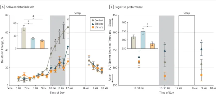
The study sample comprised 16 healthy older controls (age range, 55-80 years; mean [standard error of the mean (SEM)] age, 63.6 [5.6] years; 8 women) and 13 patients with previous cataract (age range, 55-80 years; mean [SEM] age, 69.9 [5.2] years; 10 women), whereby 5 patients had UV-only lens re-placement (mean [SEM] age, 70.8 [4] years; 4 women) and 8 patients had BB lens replacement (mean [SEM] age, 69.8 [6.2] years; 7 women) (Table). We first assessed whether there was an association of intraocular cataract lens replacement with circadian rhythms, as indexed by the acute suppression of en-dogenous melatonin levels by light exposure (primary out-come measure). Compared with healthy older individuals, pa-tients with previous cataract and lens replacement had an attenuated increase in melatonin levels during light expo-sure (mean [SEM] BB: 23.3% [2.6%] and UV lens: 19.1% [2.1%]) compared with controls (mean [SEM], 48.8% [5.2%]) (percent-age of change in melatonin levels from baseline levels; inter-action time vs group; difference between means, 27.7; 95% CI, 15.4%-41.7%; P < .001) (Figure 2A).
We then tested whether there was an association of intra-ocular cataract lens replacement with cognitive function (sec-ondary outcome measure). Cognitive function, as indexed by tasks tapping onto attentional resources (psychomotor vigi-lance task slowest reaction times), significantly improved sus-tained attention performance across time (interaction of time
vs group; difference between means, 71.4; 95% CI, 29.5%-113.1%; P=.002). Accordingly, patients with UV lens had faster reaction times during light exposure (mean [SEM], 276.9 [11.1] milliseconds) and in the morning after sleep (mean [SEM], 279.3 [8.9] milliseconds) compared with patients with BB lens re-placement (mean [SEM], 348.3 [17.8] milliseconds and 353 [18.8] milliseconds, respectively) (Figure 2B). Furthermore, we observed a significant improvement in performance to a 0-back task, which is a type of reaction-time task (main factor group; difference between means, 9.1; 95% CI, 0.2%-18%; P = .03) (eFigure 2A in theSupplement), such that patients with UV lens showed increased percentage of correct responses (mean [SEM], 98.1% [1.3%]), compared with those with BB lens (mean [SEM], 88.9% [3.3%]). Conversely, no significant effects were observed for the 2-back task, which is a more demanding work-ing memory task (eFigure 2B in theSupplement).
Lastly, we investigated whether there was an association of intraocular cataract lens replacement with sleep function, as indexed by NREM slow-wave sleep and frontal NREM sleep slow-wave activity. We observed that in the sleep opportu-nity after evening light exposure, patients with UV lens had increased slow-wave sleep (mean [SEM], 13% [3.4%]), compared with controls (mean [SEM], 5.2% [0.8%]) (percent-age of total sleep time; main effect, group; difference be-tween means, 7.9; 95% CI, 2.4%-13.4%; P = .02 multiple com-parison correction with Tukey-Kramer test) (Figure 3A and eTable in theSupplement). Furthermore, NREM slow-wave sleep associations were consistent among participants: 80%
Table. Participants Demographics, Sleep and Wake Times, and Ocular Characteristics
Characteristic
Healthy Older Controls (n = 16)
Patients With Previous Cataract and BB Lens (n = 8)
Patients With Previous Cataract and UV-Only Lens (n = 5)
Age, mean (SD), ya 63.6 (5.6) 69.8 (6.2) 70.8 (4)
Men, No. (%)b 8 (50) 4 (50) 1 (20)
Women, No. (%)b 8 (50) 4 (50) 4 (80)
Body mass index, mean (SD)c 23.4 (1.7) 24.8 (2.6) 24.3 (2.5)
White, No. (%) 16 (100) 8 (100) 5 (100)
Sleep times, mean (SEM, min) 11:19PM(6) 11:04PM(12) 10:57PM(8)
Wake times, mean (SEM, min) 7:11AM(10) 7:22AM(12) 7:12AM(7)
Sleep duration, h:min, (SEM, min) 6:55 (11) 6:58 (11) 7:05 (12)
Intraocular pressure, mean (SD) [range], mm Hg Right eye 15.1 (2.1) [11-19] 14.3 (2.1) [10-17] 15.5 (2.4) [13-20] Left eye 15.5 (2.2) [11-19] 14.3 (2.5) [10-17] 15.5 (3.1) [13-20] LOCIII grade Right eye NO = 2 NO = 4 NO = 4 NC = 2 NC = 4 NC = 4 CC = 0 CC = 1 CC = 1 PSC = 0 PSC = 1 PSC = 1 Left eye NO = 2 NO = 4 NO = 4 NC = 2 NC = 4 NC = 4 CC = 0 CC = 1 CC = 1 PSC = 0 PSC = 2 PSC = 2
BCVA, mean (SD), [range], logMAR
Right eye 0.93 (0.1) [0.6-1.0] 0.86 (0.2) [0.63-1.0] 0.92 (0.2) [0.63-1.0] Left eye 0.94 (0.2) [0.6-1.0] 0.94 (0.1) [0.8-1.0] 0.96 (0.1) [0.8-1.0]
Abbreviations: BB, blue-blocking; BCVA, best-corrected visual acuity; CC, cortical cataract; LOCIII, Lens Opacities Classification System III; NC, nuclear color; NO, nuclear opalescence; PSC, posterior subcapsular cataract (mean values); SEM, standard error of the mean; UV, ultraviolet.
aAnalysis of variance: P = .04. bAnalysis of variance: P = .03. cCalculated as weight in kilograms
divided by height in meters squared.
(4 of 5) of patients with UV lens, 12.5% (1 of 8) of patients with BB lens, and only 6.25% (1 of 16) controls had increased NREM slow-wave sleep (threshold set as >10% of increase in NREM slow-wave sleep; χ2= 11.4, P = .007; eFigure 3 in the Supple-ment). Furthermore, the dynamics of frontal NREM sleep slow-wave activity (0.75-4.5 Hz), a functional index of homeo-static sleep pressure,15
was higher during the first NREM-REM sleep cycle in patients with UV lens (mean [SEM], 79.9 [13.6] μV2/Hz) compared with patients with BB lens (mean [SEM], 53.2 [10.7] μV2/Hz) (analyses on first NREM-REM sleep cycle; interaction, time vs group; difference between means, 26.7; 95% CI, 9.2-48.9; P = .03; contrasts of interest: percen-tiles 5-8) (Figure 3B).
Discussion
Our in-laboratory empirical findings suggest an association of intraocular cataract lens replacement with key aspects of cir-cadian rhythms, cognitive performance, and sleep regula-tion. By using a cross-sectional observational study, we ob-served that replacing natural lenses with IOLs normalizes melatonin response to light, regardless of whether they were UV-only or BB lens, while UV lens improved sustained atten-tion performance and sleep funcatten-tion compared with BB lens. Our participants’ demographics indicate that the healthy older controls were significantly younger (ie, approximately 5 Figure 3. Sleep Structure and Sleep Electroencephalographic (EEG) Activity
150 120 60 90 30 18 12 6 0 NREM Slo w -W av e Sleep, T
otal Sleep Time, %
Group a Sleep structure A UV Lens BB Lens Control 00 1.5 3.0 4.5 6.0 6.5 F
rontal NREM Sleep Slo
w-W a v e Sleep (EE G Activit y = 0.75-4.5 Hz; μV 2/Hz)
Time Since Lights Off, h Sleep EEG activity
B
Control BB lens UV lens
Association of intraocular cataract lens replacement with sleep structure (slow-wave sleep) (A) and sleep EEG activity (slow-wave activity) (B). B, EEG activity = 0.75 Hz to 4.5 Hz; μV2/Hz. Data are reported as mean (standard error of the mean). BB indicates blue blocking; NREM, non–rapid eye movement; UV, ultraviolet.
a
P < .05 (see Results section
for more information). Figure 2. Melatonin Rhythms and Cognitive Performance
60 40 20 0 450 400 350 300 250 80 Melatonin Change, % Time of Day a a Saliva melatonin levels
A 8 AM 12 AM 11 PM 9 PM 7 PM 8 PM 10 PM 6 PM 5 PM 9 AM 10 AM Control BB lens UV lens Sleep a a a a a a a 60 30 0 PVT Slo w est R eaction Times, ms Time of Day a Cognitive performance B 8 AM 12 AM 10:30 PM 8:30 PM 9 AM 10 AM Sleep 400 300 350 250 Fa st e r Slo w er
Association of intraocular cataract lens replacement with circadian photosensitivity by saliva melatonin levels (0 indicates prelight levels) (A) and cognitive performance (psychomotor vigilance task performance) (B). Data are reported as mean (standard error of the mean). BB indicates blue blocking;
PVT, psychomotor vigilance task; UV, ultraviolet. aP < .05 (see Results section for more information).
years) than patients with BB and UV lens. Nonetheless, they had comparatively lower circadian photosensitivity and NREM sleep slow-wave sleep and frontal NREM slow-wave activity. Further-more, the 50%:50% men:women distribution in healthy controls and patients with BB lens was not observed in patients with UV lens (25%:75%). Although we were not powered to test for sex-dependent effects on light sensitivity, previous data indicate that men may show higher light sensitivity than women for cognitive performance and hallmarks of sleep regulation (slow-wave sleep and slow-wave activity).21Importantly, no ocular pathologies were observed across the groups (apart from the cataract in the clinical groups).
Circadian photoentrainment is predominantly initiated by stimulation of intrinsically photosensitive retinal ganglion cells containing the photopigment melanopsin by light, particularly in the blue range (450-490 nm).22With aging, the natural lens of the eye acquires a yellow-brownish discoloration because of accumulation of chromophores that absorb preferentially the short wavelength region of visible spectrum. Consequently, de-creased stimulation of melanopsin is expected with age, whereby not only the amount of light reaching the retina is dramatically reduced to about one-tenth that of younger adults,23
but the spec-trum of light transmission into the eye is also altered. This sce-nario is posited to worsen even further in patients with previous cataract and may impair their ability to entrain the circadian clock, with subsequent long-term disruption of the circadian system. Here, we show that optimizing the spectral lens trans-mission in patients after cataract lens replacement can restore circadian photosensitivity, as indexed by their increased acute melatonin suppression to evening light exposure.
We then showed an association between lens replacement in patients with previous cataract and cognitive performance, such that, during acute light exposure and in the morning sub-sequent to sleep, sustained attention performance is improved in patients with UV lens only. Healthy aging is accompanied by a decline in cognitive fitness, due to a combination of different factors, such as morphological age-related brain differences, and reduced non–image-forming responses to light.4Cataract and cognitive impairment are common age-related ailments, and sur-gical intervention in patients with cataract may improve some aspects of cognitive function, including reaction time tasks.24 While increasing ambient light levels for 1 week may improve cog-nitive impairment in older patients with dementia,25similar ef-fects in patients with previous cataract remain largely unknown. Here, we observed that IOL replacement in patients with previ-ous cataract may enhance the beneficial light effects on cogni-tive tasks associated with attentional resources. Recent functional magnetic resonance imaging data suggest a plasticity to light sen-sitivity in aging, such that after approximately 4 years of surgery, lens replacement did not significantly affect daytime light im-pact on cognitive brain function (associated with 0-back and 2-back task performance).26These results suggest that some as-pects of cognitive brain function may adapt to the progressive decrease in daytime light exposure in aging. Importantly, sleep ailments are common in older patients with cataract, who sub-jectively report difficulties initiating and/or maintaining sleep, and impoverished subjective sleep quality, which can be allevi-ated after IOL replacement cataract.27While different lens
re-placements exist, there is a scarcity of knowledge on how these different treatment strategies translate to objective hallmarks of sleep function. Currently, most evidence builds on epidemiologi-cal data using subjective sleep questionnaires and/or ambulatory actigraphy measures,27,28thus devoid of objective in-laboratory sleep measures. To our knowledge, no in-laboratory studies have been carried out to assess the association of cataract lens replace-ment with sleep physiology. Here, we show that slow-wave sleep and frontal NREM slow-wave activity, a functional index of ho-meostatic sleep pressure,15was higher in patients with replace-ment UV lens, suggesting a beneficial effect on their sleep physi-ology. Age-related changes in sleep29
include reduced total sleep time, slow-wave sleep, and frontal NREM slow-wave activity. Our data indicate that these age-related effects on homeostatic sleep regulation might be mitigated in patients with previous cataract and UV lens.
It is important to consider the potential visual and nonvisual benefits of different IOLs for patients with cataract. The devel-opment of BB IOLs was driven by concerns about blue light hazard30,31and for correcting the cyanopsia reported with early IOLs.32Furthermore, BB and UV lens may show similar contrast sensitivity, visual and glare acuity, and color perception under photopic conditions,33,34
even after 5 years of surgery.35
However, evidence also suggests that, while BB and UV lens have similar postoperative visual function, color perception may be improved in patients with UV lens compared with BB lens after 1, 3, and 6 months of surgery.36
Taken together, while our study shows that UV IOLs may benefit nonvisual function, the associations with visual function must also be considered.37
Strengths and Limitations
The key strengths of our study include stringently controlled in-laboratory study protocols that allow us to reliably assess the association of IOL cataract lens replacement with well-established objective hallmarks of circadian photosensitiv-ity, cognitive function, and sleep regulation. Furthermore, be-cause we used stringent exclusion criteria to minimize medical conditions, which might add variability to our outcome mea-sures, we could reliably test our study hypotheses. Thus, our empirical evidence provides a demonstration of a measur-able change in nonvisual function following an increasingly routine clinical intervention.
Despite the stringency of our inclusion/exclusion criteria and study protocols, ours is a laboratory empirical study with a limited sample size. Our results suggest the value of larger and longer-term clinical and observational studies to better un-derstand the circadian, cognitive and sleep-related outcomes that might differ by light transmission properties of IOLs. Therefore, caution should be made in extrapolating our find-ings to larger cataract populations.
Conclusions
Collectively, our stringently controlled laboratory data pro-vide epro-vidence for an association of intraocular cataract lens re-placement with the beneficial effects of light on the circadian photosensitivity (ie, greater melatonin suppression),
cogni-tive performance (sustained attention), and sleep stages (ie, slow-wave sleep) and EEG hallmarks (ie, EEG slow-wave activity) of homeostatic sleep regulation. Our data suggest that
optimizing the spectral lens transmission in cataract patients may mitigate some of the adverse circadian, cognitive, and sleep effects associated with aging cataract lens.
ARTICLE INFORMATION
Accepted for Publication: March 29, 2019. Published Online: May 23, 2019. doi:10.1001/jamaophthalmol.2019.1406 Author Affiliations: Medical Chronobiology Program, Division of Sleep and Circadian Disorders, Brigham and Women’s Hospital, Boston, Massachusetts (Chellappa); Division of Sleep Medicine, Harvard Medical School, Boston, Massachusetts (Chellappa);
Sleep-Wake-Epilepsy-Center, Department of Neurology, Inselspital, Bern University Hospital, Bern, Switzerland (Bromundt); Centre for Chronobiology, Psychiatric Hospital of the University of Basel, Transfaculty Research Platform Molecular and Cognitive Neurosciences, University of Basel, Basel, Switzerland (Frey, Cajochen); University Eye Clinic in Basel, University of Basel, Basel, Switzerland (Steinemann, Goldblum); GIGA-Research, Cyclotron Research Centre/In Vivo Imaging Unit, University of Liège, Liège, Belgium (Schmidt); Augenarztpraxis Klingentalstrasse, Basel, Switzerland (Schlote).
Author Contributions: Drs Chellappa and Cajochen had full access to all of the data in the study and take responsibility for the integrity of the data and the accuracy of the data analysis.
Concept and design: Chellappa, Bromundt, Frey,
Schlote, Goldblum, Cajochen.
Acquisition, analysis, or interpretation of data:
All authors.
Drafting of the manuscript: Chellappa. Critical revision of the manuscript for important intellectual content: All authors.
Statistical analysis: Chellappa, Cajochen. Obtained funding: Steinemann, Cajochen. Administrative, technical, or material support: Frey,
Goldblum, Cajochen.
Supervision: Goldblum, Cajochen.
Conflict of Interest Disclosures: Drs Cajochen, Bromundt, and Frey report grants from AXA Foundation and the Swiss Federal Office for Public Health during the conduct of the study. No other disclosures were reported.
Funding/Support: This study was financially supported by the AXA Foundation (https://www. axa-research.org/en/project/christian-cajochen) and by the Swiss Federal Office for Public Health (Consumer Protection Directorate, 11.007647). Role of the Funder/Sponsor: The funders had no role in the design and conduct of the study; collection, management, analysis, and interpretation of the data; preparation, review, or approval of the manuscript; and decision to submit the manuscript for publication. Additional Contributions: We thank research volunteers and the technical staff at the Center for Chronobiology, University of Basel. In particular, we thank Roland Steiner, Ing. HTL (University of Basel), for performing the transmission spectra of the lenses. Dr Steiner was not compensated for this work.
REFERENCES
1. Bourne RRA, Flaxman SR, Braithwaite T, et al; Vision Loss Expert Group. Magnitude, temporal trends, and projections of the global prevalence of blindness and distance and near vision impairment: a systematic review and meta-analysis. Lancet Glob
Health. 2017;5(9):e888-e897. doi: 10.1016/S2214-109X(17)30293-0
2. Pescosolido N, Barbato A, Giannotti R, Komaiha C, Lenarduzzi F. Age-related changes in the kinetics of human lenses: prevention of the cataract.Int J Ophthalmol. 2016;9(10):1506-1517.
3. Xu J, Pokorny J, Smith VC. Optical density of the human lens. J Opt Soc Am A Opt Image Sci Vis. 1997; 14(5):953-960. doi:10.1364/JOSAA.14.000953 4. Daneault V, Hébert M, Albouy G, et al. Aging reduces the stimulating effect of blue light on cognitive brain functions. Sleep. 2014;37(1):85-96. doi:10.5665/sleep.3314
5. Gabel V, Reichert CF, Maire M, et al Differential impact in young and older individuals of blue-enriched white light on circadian physiology and alertness during sustained wakefulness. Sci Rep. 2017;7(1):7620. doi:10.1038/s41598-017-07060-8 6. Mishima K, Okawa M, Shimizu T, Hishikawa Y. Diminished melatonin secretion in the elderly caused by insufficient environmental illumination.
J Clin Endocrinol Metab. 2001;86(1):129-134. 7. World Medical Association. World Medical Association Declaration of Helsinki: ethical principles for medical research involving human subjects. JAMA. 2013;310(20):2191-2194. doi:10. 1001/jama.2013.281053.
8. Buysse DJ, Reynolds CF III, Monk TH, Berman SR, Kupfer DJ. The Pittsburgh Sleep Quality Index: a new instrument for psychiatric practice and research. Psychiatry Res. 1989;28(2):193-213. doi:10.1016/0165-1781(89)90047-4
9. Roenneberg T, Wirz-Justice A, Merrow M. Life between clocks: daily temporal patterns of human chronotypes. J Biol Rhythms. 2003;18(1):80-90. doi:10.1177/0748730402239679
10. Chellappa SL, Ly JQ, Meyer C, et al. Photic memory for executive brain responses. Proc Natl
Acad Sci U S A. 2014;111(16):6087-6091. doi:10. 1073/pnas.1320005111
11. Cajochen C, Frey S, Anders D, et al. Evening exposure to a light-emitting diodes (LED)-backlit computer screen affects circadian physiology and cognitive performance. J Appl Physiol (1985). 2011; 110(5):1432-1438. doi:10.1152/japplphysiol.00165.2011 12. Chellappa SL, Steiner R, Oelhafen P, et al. Acute exposure to evening blue-enriched light impacts on human sleep. J Sleep Res. 2013;22(5):573-580. doi:10.1111/jsr.12050
13. Münch M, Kobialka S, Steiner R, Oelhafen P, Wirz-Justice A, Cajochen C. Wavelength-dependent effects of evening light exposure on sleep architecture and sleep EEG power density in men.
Am J Physiol Regul Integr Comp Physiol. 2006;290
(5):r1421-r1428. doi:10.1152/ajpregu.00478.2005 14. Skene DJ, Arendt J. Human circadian rhythms: physiological and therapeutic relevance of light and
melatonin. Ann Clin Biochem. 2006;43(Pt 5): 344-353. doi:10.1258/000456306778520142 15. Aeschbach D, Borbély AA. All-night dynamics of the human sleep EEG. J Sleep Res. 1993;2(2):70-81. doi:10.1111/j.1365-2869.1993.tb00065.x 16. Dinges DF, Pack F, Williams K, et al. Cumulative sleepiness, mood disturbance, and psychomotor vigilance performance decrements during a week of sleep restricted to 4-5 hours per night.Sleep. 1997; 20(4):267-277.
17. Graw P, Kräuchi K, Knoblauch V, Wirz-Justice A, Cajochen C. Circadian and wake-dependent modulation of fastest and slowest reaction times during the psychomotor vigilance task. Physiol Behav. 2004;80(5):695-701. doi:10.1016/j.physbeh.2003. 12.004
18. Rechtschaffen A, Kales A. A manual of standardized terminology, techniques and scoring system for sleep stages of human subjects. Bethesda,
MD: US Dept of Health, Education and Welfare, Public Health Service; 1968.
19. Feinberg I, Floyd TC. Systematic trends across the night in human sleep cycles. Psychophysiology. 1979;16(3):283-291. doi:10.1111/j.1469-8986.1979. tb02991.x
20. Ludbrook J. Multiple comparison procedures updated. Clin Exp Pharmacol Physiol. 1998;25(12): 1032-1037. doi:10.1111/j.1440-1681.1998.tb02179.x 21. Chellappa SL, Steiner R, Oelhafen P, Cajochen C. Sex differences in light sensitivity impact on brightness perception, vigilant attention and sleep in humans. Sci Rep. 2017;7(1):14215. doi:10.1038/ s41598-017-13973-1
22. Provencio I, Rodriguez IR, Jiang G, Hayes WP, Moreira EF, Rollag MD. A novel human opsin in the inner retina. J Neurosci. 2000;20(2):600-605. doi:10.1523/JNEUROSCI.20-02-00600.2000 23. Charman WN. Age, lens transmittance, and the possible effects of light on melatonin suppression.
Ophthalmic Physiol Opt. 2003;23(2):181-187. doi:10. 1046/j.1475-1313.2003.00105.x
24. Schmoll C, Tendo C, Aspinall P, Dhillon B. Reaction time as a measure of enhanced blue-light mediated cognitive function following cataract surgery. Br J Ophthalmol. 2011;95(12):1656-1659. doi:10.1136/bjophthalmol-2011-300677 25. Riemersma-van der Lek RF, Swaab DF, Twisk J, Hol EM, Hoogendijk WJ, Van Someren EJ. Effect of bright light and melatonin on cognitive and noncognitive function in elderly residents of group care facilities: a randomized controlled trial. JAMA. 2008;299(22):2642-2655. doi:10.1001/jama.299.22. 2642
26. Daneault V, Dumont M, Massé É, et al. Plasticity in the sensitivity to light in aging: decreased non-visual impact of light on cognitive brain activity in older individuals but no impact of lens replacement. Front Physiol. 2018;9(1557):1557. doi:10.3389/fphys.2018.01557
27. Kessel L, Siganos G, Jørgensen T, Larsen M. Sleep disturbances are related to decreased transmission of blue light to the retina caused by
lens yellowing. Sleep. 2011;34(9):1215-1219. doi:10. 5665/SLEEP.1242
28. Münch M, Ladaique M, Roemer S, Hashemi K, Kawasaki A. Melanopsin-mediated acute light responses measured in winter and in summer: seasonal variations in adults with and without cataracts. Front Neurol. 2017;8(464):464. doi:10. 3389/fneur.2017.00464
29. Dijk DJ, Duffy JF, Czeisler CA. Age-related increase in awakenings: impaired consolidation of nonREM sleep at all circadian phases. Sleep. 2001; 24(5):565-577. doi:10.1093/sleep/24.5.565 30. Ham WT Jr, Mueller HA, Sliney DH. Retinal sensitivity to damage from short wavelength light.
Nature. 1976;260(5547):153-155. doi:10.1038/ 260153a0
31. Noell WK. Possible mechanisms of
photoreceptor damage by light in mammalian eyes.
Vision Res. 1980;20(12):1163-1171. doi:10.1016/ 0042-6989(80)90055-3
32. Davison JA, Patel AS. Light normalizing intraocular lenses.Int Ophthalmol Clin. 2005;45(1): 55-106.
33. Neumaier-Ammerer B, Felke S, Hagen S, et al. Comparison of visual performance with blue light-filtering and ultraviolet light-filtering intraocular lenses. J Cataract Refract Surg. 2010;36 (12):2073-2079. doi:10.1016/j.jcrs.2010.06.069 34. Hayashi K, Hayashi H. Visual function in patients with yellow tinted intraocular lenses compared with vision in patients with non-tinted intraocular lenses. Br J Ophthalmol. 2006;90(8): 1019-1023. doi:10.1136/bjo.2006.090712
35. Kara-Junior N, Espindola RF, Gomes BA, Ventura B, Smadja D, Santhiago MR. Effects of blue light-filtering intraocular lenses on the macula, contrast sensitivity, and color vision after a long-term follow-up. J Cataract Refract Surg. 2011; 37(12):2115-2119. doi:10.1016/j.jcrs.2011.06.024 36. Schmack I, Schimpf M, Stolzenberg A, Conrad-Hengerer I, Hengerer FH, Dick HB. Visual quality assessment in patients with orange-tinted blue light-filtering and clear ultraviolet light-filtering intraocular lenses. J Cataract Refract Surg. 2012;38 (5):823-832. doi:10.1016/j.jcrs.2011.12.028 37. Cuthbertson FM, Peirson SN, Wulff K, Foster RG, Downes SM. Blue light-filtering intraocular lenses: review of potential benefits and side effects. J Cataract Refract Surg. 2009;35(7): 1281-1297. doi:10.1016/j.jcrs.2009.04.017