Université de Montréal
Thiel Embalmed Cadaveric Tissue
A Model for Surgical Simulation and Research
par
Andrei Odobescu M.D., C.M. Département de Chirurgie
Faculté de Médécine
Thèse présentée à la Faculté Médecine en vue de l’obtention du grade de PhD en
Sciences Biomédicales, option Générale
Mars, 2019
Résumé
Le Collège royal des médecins et chirurgiens du Canada met actuellement en place des curriculums basés sur les compétences, plutôt que sur le temps, dans toutes les spécialités médicales et chirurgicales. La transition devrait être complétée en 2022. Les programmes de formation en chirurgie plastique au Canada devront repenser leurs curriculums pour se plier aux directives nationales. La simulation est la pierre angulaire du modèle de formation des résidents basé sur les compétences puisqu'elle permet aux résidents d'apprendre et d'améliorer leurs compétences dans un contexte éthique, sécuritaire, et mesurable objectivement.
Un consensus récent des directeurs de programme canadiens en chirurgie plastique a nommé 154 procédures essentielles de bases que les résidents doivent maîtriser avant la fin de leur formation. Nous proposons l'utilisation du modèle cadavérique Thiel pour la simulation haute fidélité des procédures en plastie. Les spécimens Thiel ont déjà été introduits dans une multitude de spécialités, incluant la plastie pour la dissection de lambeaux et la réparation de tendons. Nous nous sommes concentrés sur l'évaluation des spécimens Thiel pour la maîtrise des anastomoses vasculaires, la réparation des nerfs périphériques, et la réparation des tendons fléchisseurs. Par ailleurs, nous avons développé des instruments d'évaluation pour chacun de ces domaines de simulation. Des trois instruments, nous avons validé les échelles d'évaluation des anastomoses vasculaires et nerveuses. Ces deux échelles ont démontré d'excellents degrés de fiabilité et de reproductibilité et sont bien corrélés avec le niveau de formation et d'expérience des sujets. Le modèle de réparation des tendons fléchisseurs a démontré un degré plus élevé de variaiblité inter-évaluateur, et, quoique prometteur, il n'a pas pu être complètement validé basé sur les données actuelles. De plus, nous avons utilisé les vaisseaux Thiel comme un modèle de recherche pour l'investigation de nouvelles techniques microvasculaires.
Notre expérience montre que les spécimens cadavériques Thiel sont un excellent modèle de simulation pour la chirurgie microvasculaire et la réparation des nerfs périphériques et des tendons fléchisseurs. Nous proposons des instruments d'évaluation pour assister à
l'implémentation de ces modèles de simulation dans les curriculums basés sur les compétences en chirurgie plastique.
Mots-clés : Thiel, simulation, chirurgie, chirurgie plastique, microchirurgie,
microvasculair, micro-neurorrhaphie, tenorrhaphie, reparation tendon, cadaver, technique de conservation, technique d'embaumement.
Abstract
The Royal College of Physicians and Surgeons is currently implementing a major shift from a time based to a competence based curriculum in all medical and surgical specialties. By 2022 the transition is to be complete. The plastic surgery training programs in Canada will have to rethink their curriculum in order to comply with the national directives. Simulation is a cornerstone of the competence based model of resident training as it not only allows residents to safely learn and hone their skill in a setting that is ethical and promotes patient safety, but it allows for objective evaluation of their performance.
A recent consensus statement from the Canadian plastic surgery program directors identified 154 essential core procedures for residents to master by the end of their training. We propose the use of the Thiel cadaveric model for high fidelity simulation of plastic surgery procedures. While Thiel cadaveric specimens have been proposed for use in a multitude of specialties, including in plastic surgery for flap dissection and tendon repair, we focused on evaluating the use of the Thiel embalmed specimens on three core procedures: microvascular anastomoses, peripheral nerve repair, and flexor tendon repair. In addition, we designed evaluation instruments for each of these three simulation areas to help grade performance and aid in the feedback/debriefing process. Of the three evaluation instruments, we successfully validated the microvascular evaluation and micro-neurorrhaphy evaluation scales. Both of these scales showed excellent degrees of reliability and reproducibility and correlated well with the level of training and self-declared experience of the subjects. The flexor tendon evaluation scale showed a higher degree of inter-rater variability and, while it shows promise with a larger cohort of participants and additional calibration, it could not be validated fully based on the available data. Additionally, we used the Thiel embalmed cadaveric vessels as a research model for the investigation of new microvascular techniques.
Our experience shows the Thiel cadaveric specimens to provide an excellent model for simulating microvascular, peripheral nerve and flexor tendon repairs. We propose evaluation instruments to assist in the implementation of these simulation models in a comprehensive, competence based curriculum in plastic surgery.
Keywords: Thiel, simulation, surgery, plastic surgery, microsurgery, microvascular,
micro-neurorrhaphy, tenorrhaphy, tendon repair, cadaver, preservation technique, embalming technique.
Table des matières
Introduction
0.1. The evolution of medical simulation…....……….…...….01
0.2. From time based to competence based surgical training………..03
0.3. Simulation models in surgery………...05
0.4. High and low fidelity simulation………..10
0.5. Simulation models in plastic surgery………...13
0.6. Thiel embalmed cadavers in medical simulation………...18
0.7. Study objectives……….…25
0.8. Thesis overview……….25
Chapter 1: Microvascular simulation 1.1. High fidelity microsurgical simulation: the Thiel model and evaluation instrument...….27
1.1.1. Introduction………28
1.1.2. Methods………..30
1.1.2.1. Thiel vessel specimens………...30
1.1.2.2. Study design……….31
1.1.2.3. Microvascular evaluation scale………..33
1.1.2.4. Statistical analysis………34
1.1.3. Results……….34
1.1.3.1. Reliability assessment……….……….34
1.1.3.2. Regression models………...34
1.1.4. Discussion………..….37
Chapter 2: Peripheral nerve simulation 2.1. Thiel cadaveric nerve tissue: a model for microsurgical simulation………...43
2.2. High fidelity microsurgical simulation: the Thiel cadaveric nerve model and evaluation evaluation instrument……….…….…..49
2.2.1. Introduction………50
2.2.2.1. Thiel nerve specimens……….………51
2.2.2.2. Study design………52
2.2.2.3. Micro-neurorrhaphy evaluation scale..……….………..54
2.2.2.4. Statistical analysis……….………..55
2.2.3. Results………56
2.2.3.1. Assessment of inter-rater reliability………..…….56
2.2.3.2. Cronbach’s alpha……….……….…………..57
2.2.3.3. Relationship between performance as measured by average micro-neurorrhaphy evaluation scale and resident characteristics – bivariate analyses……….……...58
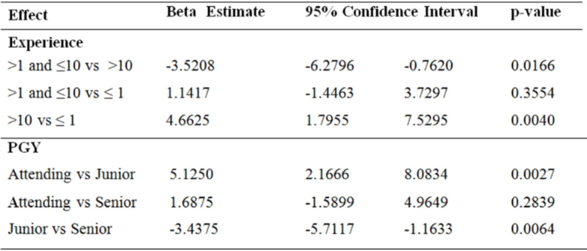
2.2.3.4. Relationship between performance as measured by average micro-neurorrhaphy evaluation scale and resident characteristics – Results from mixed modeling………...…...59
2.2.3.5. Subgroup analysis: plastic surgery residents………..……...….62
2.2.4. Discussion………..…...…64
Chapter 3: Tendon repair simulation 3.1. The Thiel cadaveric tendon simulation model and evaluation instrument……....……...70
3.1.1. Introduction………71
3.1.2. Methods………..72
3.1.2.1. Thiel tendon specimens………...72
3.1.2.2. Study design………72
3.1.2.3. Flexor tendon evaluation scale………..………..….…...73
3.1.2.4. Statistical analysis………....74
3.1.3. Results……….75
3.1.3.1. Assessment of inter-rater reliability………...………...75
3.1.3.2. Cronbach’s alpha……….…76
3.1.3.3. Relationship between performance as measured by the tendon evaluation scale and resident characteristics – bivariate analyses………...77
3.1.3.4. Relationship between performance as measured by tendon surgery evaluation scale and resident characteristics – results from mixed
modeling……….………...78
3.1.4. Discussion……….…..81
Chapter 4: Thiel research model 4.1. A new microsurgical research model using Thiel embalmed arteries and comparison of two suture techniques………...………....84
4.1.1.Introduction………...…...……..85
4.1.2. Materials and methods………...85
4.1.2.1. Model set-up………...86
4.1.2.2. Specimen preparation………...86
4.1.2.3. Surgical technique………..87
4.1.2.4. Measurement of outcomes……….89
4.1.3. Results………...90
4.1.3.1. Research model evaluation………90
4.1.3.2. Anastomotic leaks………..90
4.1.3.3. Anastomotic stricture……….91
4.1.3.4. Time to completion of anastomosis………...94
4.1.4. Discussion………...94
4.2. Horizontal mattress technique for the anastomosis of size mismatched vessels………100
4.2.1. Introduction………...101
4.2.2. Materials and Methods………..101
4.2.2.1. Specimen preparation………...101
4.2.2.2 Surgical technique……….102
4.2.2.3. Measurement of outcomes………104
4.2.3. Results………...105
4.2.4. Discussion………...107
Chapter 5: Discussions and conclusions 5.1. From time base to competence based resident training………...109
5.2. The versatility of the Thiel model: microvascular, nerve and tendon……….112
5.4. The implementation of simulation and the plastic surgery curriculum in Canada…....114 5.5. Future directions………115
Liste des tableaux
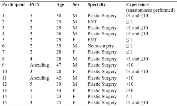
Table 1. Participant information ... 32
Table 2. Microvascular evaluation scale ... 33
Table 3. Multivariable linear mixed models ... 37
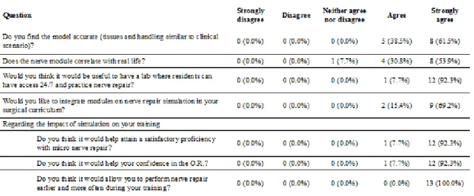
Table 4. Post-simulation survey ... 47
Table 5. Participant information. ... 53
Table 6. Micro-neurorrhaphy evaluation scale (MNES) ...55
Table 7. Intraclass correlations assessing inter-rater reliability of the micro-neurorrhaphy evaluation scale for all possible pairs among five evaluators. ... 57
Table 8. Correlation between PGY level and experience ... 61
Table 9. Participant information ... 73
Table 10. Flexor tendon evaluation scale (FTES) ... 74
Table 11. Intraclass correlations assessing inter-rater reliability of the tendon evaluation scale for all possible pairs among four evaluators ... 76
Liste des figures
Figure 1 ACS basic skills for PGY 1 and 2 in general surgery and plastic surgery. ... 8
Figure 3 Types of simulation available.. ... 10
Figure 4 Basic components of Thiel embalming solution.. ... 20
Figure 5 Stress-strain relationships in fresh frozen and Thiel embalmed cadavers... 24
Figure 6. Set-up used for microvascular training. ... 31
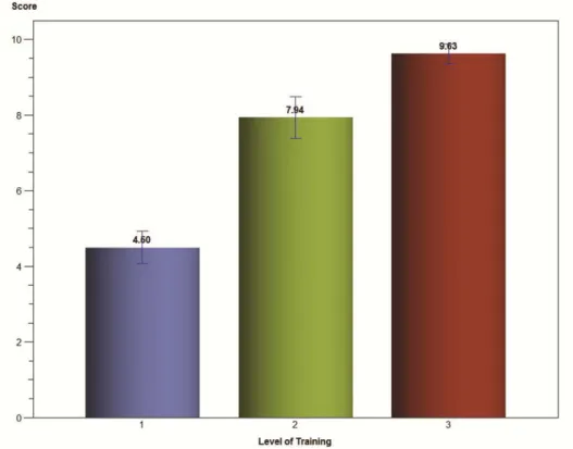
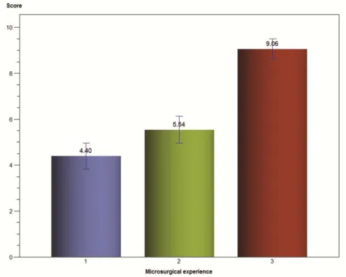
Figure 7. Level of training. ... 35
Figure 8. Microsurgical experience. ... 36
Figure 9. Histologic appearance of Thiel embalmed vessel. ... ..38
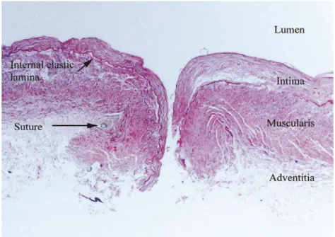
Figure 10. Thiel embalmed nerve for nerve repair simulation. ...46
Figure 11. Average MNES score for junior and senior residents ... 58
Figure 12. Average MNES score for three levels of resident experience. ... 59
Figure 13. Average MNES score vs. level of resident experience – plastic surgery residents only. ... 64
Figure 14. Differences in the distribution of the experience measured by self-reported number of procedures performed based upon all four raters between junior and senior plastic surgery residents. ... 77
Figure 15. Tendon evaluation score vs. breaking strength ... 79
Figure 16. Tendon evaluation score vs. program level ... 80
Figure 17. Tendon evaluation score vs. level of experience. ... 81
Figure 18. Research model setup with the visible operative field ... 87
Figure 19. Schematic representation of the horizontal mattress suture technique ... 88
Figure 20. Leakage for simple interrupted and horizontal mattress anastomoses. ... 91
Figure 21. Angiographic evaluation of the luminal stricture. ... 92
Figure 22. Light and scanning electron microscopy of the anastomotic sites ... 93
Figure 23. Illustration of the technique used for size-mismatched vessel anastomosis ... 103
Figure 24. Appearance of anastomosis showing a funnelled out smaller vessel that meets a cinched-down larger vessel ... 104
Figure 25. Scanning electron micrograph and hematoxylin and eosin staining of two size-mismatched vessels. ... 106
Liste des sigles
ABS: American Board of Surgery ACS: American College of Surgeons ALT: Anterolateral thigh flap
AO: Arbeitsgemeinschaft für Osteosynthesefragen (German for "Association for the Study of Internal Fixation")
CanMEDS: Physician competency framework implemented by the Royal College of Physicians and Surgeons of Canada
CBD: Competence by design CI: Confidence interval
DIEP: Deep inferior epigastric perforator flap ENT: Ear nose and throat
EPA: Entrustable professional activity FDS: Flexor digitorum superficialis FDP: Flexor digitorum profundus FTES: Flexor tendon evaluation scale GRS: Global rating scale
H&E: Hematoxylin and Eosin
HM: Horizontal mattress
ICC: Intra-class correlation coefficient MNES: Micro-neurorrhaphy evaluation scale
OSATS: Objective structured assessment of technical skills OSCE: Objective structured clinical exam
PACS: Picture archiving and communication system PGY: Postgraduate year
SAMS: Structured assessment of microsurgery skills SEM: Scanning electron microscope
SI: Simple interrupted
Three R: Replacement, reduction, and refinement
TRAM: Transverse rectus abdominis musculocutaneous flap
Liste des abréviations
Dr. : DoctorIn memory of Virgil Odobescu MD Ph.D., a great father and surgeon.
Remerciements
I would first like to thank Prof. Michel Alain Danino for his relentless support during my graduate studies, and throughout my surgical training. Prof. Danino is for me a mentor in the truest sense and the embodiment of academic plastic surgery. I am grateful for the relentless support of Prof. Manon Choiniere, president of my thesis committee, as well as the members of the committee: Dr. Mehdi Benkhadra, Dr. Issam Tanoubi, Dr. Joseph Bou-Merhi and Dr. Michel Carrier.
I am also grateful to Prof. Patrick Harris for not only teaching me hand surgery but also invaluable lessons in leadership and healthcare management. Prof. Harris is for me the embodiment of leadership in plastic surgery. I thank my research collaborators Sami Moubayed MD and Isak Goodwin MD for their support throughout this research endeavor and beyond. I am grateful to Prof. Deborah Dawson and Djamal Berbiche Ph.D. for their help with the statistical analysis for this research. Prof. Eugene Daniels of McGill University has kindly offered us access to the Thiel cadaveric model, and I would like to thank him for his kind support. They have all been instrumental in helping me complete this dissertation.
I am eternally grateful to my brother, Matei Odobescu, and my mother, Mihaela Odobescu for their unrelenting support throughout my medical and surgical training. Without them, I would not be where I am today. Their sacrifices and love eclipse any merit I may have in my education. I would also like to thank Felicia and Don Greer, who have taken my brother and me into their home and supported us through our higher education.
I owe a great debt of gratitude to my father, Virgil Odobescu MD Ph.D. and my grandfather, Eugen Lescovar MD, who introduced me to the wonderful world of their profession and inspired me to follow in their footsteps. I will always remember Prof. H Bruce Williams for introducing me to the wonderful world of Plastic and Reconstructive Surgery.
I am grateful to all my teachers, colleagues, friends and patients for helping me constantly better myself.
Introduction
0.1. The evolution of medical simulation
It is human nature to seek medicine when treating ailments of the body, much in the same way as it is human nature to turn to religion to seek comfort for the soul. We lay our faith and future in the hands of a physician with the expectation that, through their knowledge and skill, they will help us pass through our difficulties and make us well again. This, in essence, constitutes an extraordinary leap of faith, unparalleled in any other area of social interaction. This faith is balanced by society’s expectation from physicians to live by a moral code established by Hippocrates, by many considered the father of western medicine, more than two millennia ago and commonly referred to as the Hippocratic oath. As doctors, we carry the responsibility to honor and protect the confidence and faith our patients have, and always strive to do better for our patients. Yet if we look at what constituted the field of medicine at the time of Hippocrates, and compare it to what it is today, the change is nothing short of a miracle, giving us hope that we can accomplish far more in the future.
From times immemorial, medical teaching, like any other art or craft, has been passed on from one generation to another in the form of apprenticeship. Each member practicing the art would receive a certain wealth of knowledge from the teacher, crystalize and hopefully improve it and eventually pass it on to the next generation of learners. In the absence of any central regulatory body, either governmental or professional, this apprentice model must have led to a significant degree of variability in physician’s skill and knowledge. While the Hippocratic Oath served as a moral beacon for medical practitioners, it took many centuries for society to evaluate the role of medicine and set standards and rules to provide order to chaos. In North America, Abraham Flexner is credited for having catalyzed the reform of medical education through his 1910 report on medical education, commonly referred to as the Flexner Report (1-3). In short, Flexner describes the state of medical education in the United States and Canada, where at his time there were 155 medical schools in existence with no set standard. He
that the quality of the doctors should be high and more or less evenly distributed, such as to provide good care anywhere. As a solution to this problem, Flexner suggested a reform of the medical education system with strict admission standards, incorporate medical schools into larger universities placed in urban areas that could support the infrastructure and required patient basis. Since he considered inflation of medical doctors a sign of poor quality service, he suggested reducing the number of medical schools from 155 to 31, with the total number of graduates to echo the need of the population.
Medical simulation is somewhat of a new concept, and it was born out of the same need Flexner identified as crucial in the standardization of medical care: to offer high quality and safe medical care. Like any skill we learn in life, the surgical technique has a learning curve, sometimes steeper, sometimes less steep. In the same way, a child falls over and over again when learning how to walk, so medical students and residents “fall” when they first learn how to asses a patient, when they incise and dissect, when they handle tissue with instruments or start to suture. It is only natural and nobody is born an expert. Yet the difference between surgery and most other learning processes we encounter in life is that the patient is paying the bill for our mistakes. This is in a sense the collateral damage of medical education and patients are willing to accept it when seeking healthcare in a teaching hospital, provided they receive care from an experienced medical provider supervising the learner. For this reason, Arbogast and Rosen suggest that a revolution comparable in magnitude to Flexner’s report is now necessary; according to the authors, this is found in the form of simulation (2).
We often hear the dictum « see one, do one, teach one ». We probably all heard it many times during medical school, and we use it often ourselves, even though it implies a crude approach to skill learning and teaching. To take the example of arterial line placement: how do medical students learn to do an arterial line? They first see it done by a trainee senior to them, and then they pull themselves together and try to reproduce what they learned. One attempt, two, three, or more, eventually either the procedure succeeds or the trainee stops and hopes for a better day next time. How many tries is considered safe is subject to interpretation, and the trainee and teacher need to determine where the « do no harm » stops and the harm begins. Are there
possible permanent negative consequences the patient can sustain? Certainly! The radial artery can thrombose, which can sometimes lead to hand ischemia and even necrosis. The next question becomes: what ethical alternatives do we have to learn medical and surgical skills? The answer, in short, is simulation. In the case of learning the skill of placing arterial lines, mannequins have been developed offering trainees the possibility to climb their learning curve outside the clinical setting.
While these mannequin simulators have been implemented by most medical schools in the western hemisphere in the undergraduate medical curriculum, we still teach many of the more complex surgical skills directly on patients. As medical educators, we have the responsibility to develop adequate simulation models for our trainees such that they climb most of their learning curve in a patient free setting and only then apply what they have learned to the operating room. This is a moral responsibility, much like the implementation of ethics in medical research. The interest in medical simulation research over the last two decades is proof that we are witnesses to a shift in medical education towards simulation-based learning.
0.2. From time-based to competency-based surgical training
As mentioned above, there is an ethical reason to implement simulation in the medical curriculum. We have the moral obligation to do all in our power to provide the best medical care and do no harm, and by moving along the learning curve in a simulated setting we decrease the harm inflicted on the patient. It is in this context that we have to consider two competing models of education: the time-based and the competency-based models. While the current trend in North American postgraduate medical education is to shift towards the competency-based model, most curricular are still time-based. That means that trainees are expected to spend a set time in training, hoping that the required competence is directly related to the training period. Most surgical residencies vary from 5-7 years in length. A competency-based curriculum, on the other hand, has certain objectives that trainees need to meet in order to promote to the next level.
4
To understand the advantages of the competence-based over the time-based curriculum, we need to consider a simple scenario: Resident A and B are at the same postgraduate level and start on rotation in a microsurgical center. Resident A has practiced microsurgery in the lab, has acquired the necessary skill to perform safely and efficiently the microvascular anastomosis. Resident B has not had the same opportunity, and the mere thought of doing microsurgery causes, as expected from the unknown, a measure of distress. Resident A will assist his attending in microsurgery and at one point the latter will offer the instruments to his resident to perform the anastomosis, of course after asking if he had done so before. Since he had practiced, the answer is affirmative, with the caveat that it had been done in a simulated setting. Resident A takes the instruments and likely performs the procedure in a safe manner. Resident B is asked the same question, and after apprehension fueled by the negative answer, the attending gives over the instruments. Since microsurgical skill differs from general surgical skill, resident B is poorly positioned, executes the suture with difficulty and possibly injures the vessel, at which point their supervisor, in the interest of patient safety, takes over. At the end of the day, resident A will be allowed to do more microsurgery, while with the normal human apprehension of the attending, resident B will be a while before doing microsurgery. The skill gap between residents A and B quickly increases, with the former soon dissecting the entire flap and the latter following a more frustrating path. This scenario, while hypothetical, is certainly not far removed from the reality of today's surgical training models. Research has shown that now all trainees acquire the necessary skill by the end of their training (4). While the resident A will likely turn out to be very well trained, his or her proficiency does not excuse the lower level of skill and knowledge of resident B. This leads us back to Flexner’s principle that a good medical education has to provide a homogenous population of surgeons who will serve their patients well.
The Royal College of Physician and Surgeons of Canada has decided to move to a competency-based curriculum and all medical and surgical specialties. This program, named “Competence by Design” (CBD), is in full transition mode and is projected to be completed by 2022 (5). While the full meaning of CBD curriculum is still a matter of debate, simulation
will undoubtedly play a crucial role in the development and implementation of these types of curricula.
0.3. Simulation models in surgery
The focus of this thesis is on simulation as it pertains learning and improving surgical skills and reasoning. We look at simulation as an instrument for acquiring the necessary knowledge and skill to become or improve one’s level of proficiency in surgery. As such, this thesis is focused on postgraduate medical education, at the residency and fellowship levels. The other meaning of the term simulation in the field of surgery, pertaining to pre- or intra-operative modeling, while it aims to improve the outcome of a procedure, does not directly aim to improve the skill of the surgeon and as such it is left out of the spectrum of this work and analysis.
In order to define how simulation applies to surgery, a number of definitions have to be discussed. Rosen and colleagues, in their 2009 article on simulation in plastic surgery training and education, provide an excellent section on definitions and principles pertaining to simulation (6). A model is defined a « physical, mathematical, or logical representation of a system, entity, phenomenon, or process » (6). The Oxford dictionary provides five definitions of the noun model, of which only two could pertain to what a model is considered in surgery. A model is « a simplified description, especially a mathematical one, of a system or process, to assist calculations and predictions » or alternatively, « a three-dimensional representation of a person or thing or of a proposed structure, typically on a smaller scale than the original » (7). For the purpose of simulation in medical education, one could consider the following characteristics as necessary to represent a model: 1. simplified yet effective representation of a real life, 2. the state of which could be physical or virtual, and 3. can be used as a replacement for the reality it imitates.
Simulation refers to a « model implemented over time, from nanoseconds to centuries, displayed either in “real time” or faster or slower than real-time » (6). The Oxford Dictionaries define the verb to simulate as « imitate the appearance or character of » something else (8),
6
which seems to be a less opportune definition than that set forth by Rosen (6). For the purpose of medical education, we can consider the following characteristics for simulation: It represents a scenario that implements the model, for the purpose of improving skill, allowing a dissection of the performance into its basic steps and analysis for the purpose of feedback. Finally, a simulator is « a device that uses simulation to replace a real-world system or apparatus, allowing users to gain experience and to observe and interact with the simulation via realistic visual, auditory, or tactile cues » (6). According to the Oxford Dictionaries, a simulator is « a machine designed to provide a realistic imitation of the controls and operation of a vehicle, aircraft, or other complex system, used for training purposes » (9). For the purpose of medical education, one can look at a simulator as an instrument to confer a virtual reality model, realistic properties such as visual, acoustic or tactile cues. Since the subject of this thesis pertains to a physical model, a simulator is unnecessary.
There are a number of elements that need to align in order to allow effective simulation. The first is an adequate model. In surgery, this entails a substitute for the organ or body part that is as close to reality as possible. Once this prerogative is met, the trainee needs to perform a scenario that will allow them to practice the surgical technique in question in a manner most closely resembling a real operating room setting. The better the model and scenario, the higher the fidelity of the simulation. The last element in the loop is the evaluation of the performance followed by a debriefing, which has the objective of providing insight to the trainee on the adequacy of his gestures and the areas of improvement. This allows for correction of improperly performed gestures in subsequent simulation sessions with an eventual improvement in skill.
Dr. Satava, in a 2010 article on the emerging trends in surgical simulation, provides a thorough history of simulation in surgery and the state of the art at the time of publication (3). He speculates that the implementation of simulation in the surgical curriculum is driven by two major forces: the emergence of new technology and the social and political pressure for safer patient care. The new technologies in this context could be interpreted as new models, not necessarily virtual reality models. The curricula and assessment tools are another essential
element. The objective structured clinical exam, also known as OSCE, is one such instrument designed to assess the history and physical examination and has since been widely implemented in the medical curricula. On the psychomotor skills side, an instrument was developed by Resnick and colleagues entitled « objective structured assessment of technical skills », also known as OSATS, which has since been adapted to a number of specific areas, such as laparoscopy or microsurgery. Satava stresses the importance of benchmark assessment of skill, such as to allow a shift from time-based to competency-based learning (3). The principle of this is that in order to be considered proficient in performing a technique, one has to practice it, be evaluated and pass the benchmark test in order to be allowed to move on. Just having spent time seeing or doing the technique with no measure of success is not adequate in this day and age. This concept is starting to be introduced in the surgical education world, with the American Board of Surgery (ABS) requiring trainees to show proof, for example, of successful completion of the fundamentals of laparoscopic surgery simulation course before sitting for their surgery qualifying exam (2).
The American College of Surgeons (ACS) defined three phases that would span the entire spectrum of simulation as it pertains to surgery. The most basic is skills training, followed by procedure training and finally team training (3, 6). The ACS has defined twenty-one basic surgical skills that junior residents need to master. These are enumerated by Rosen et al and reproduced, with permission, in figure 1 (6). These skills are purely technical and require the trainee to familiarize and polish a set of fine motor skills that they will be able to integrate and refine (6). All these skills are undoubtedly necessary for the technical progress of any resident in surgery, regardless of subspecialty, however one has to realize that some essential skills necessary to the modern plastic surgeon are omitted on the list, and certainly far enough from these 21 skills that they cannot just be mastered by extrapolation of any of these. This topic will be further discussed in the following subchapter.
Figure 1. ACS basic skills for PGY 1 and 2 in general surgery and plastic
surgery. Reproduced with permission from Rosen et al Plast Reconstr Surg 123(2009):729-38
The procedure training builds on the base established by the skills training but adds to the complexity by combining cognitive learning in addition to the mechanical learning (6). One such example of procedure training is laparoscopic appendicectomy. The trainee needs to integrate the basic laparoscopic skills learned with knowledge of the normal and pathologic anatomy, the surgical sequence, possible pitfalls etc. In the field of plastic surgery, carpal tunnel release can be thought of as a prototypal procedure. As more than just fine motor skills need to be mastered, a model for procedure training exponentially increases in complexity, and consequently cost. Team training deals with communication and interprofessional skills
Skill GeneralSurgery Surgery BothPlastic
Advanced laparoscopy skills + Advanced tissue handling:
flaps, skin grafts +
Airway management + Anastomosis: hand-sewn gastrointestinal + Anastomosis: stapled gastrointestinal + Anastomosis: vascular +
Asepsis and instrument
identification + +
Basic laparoscopy skills + Catheterization, uretheral
and suprapubic +
Central line and arterial
line insertion +
Chest tube and thoracentesis +
Colonoscopy +
Introduction to inguinal
anatomy +
Knot tying + +
Laparotomy opening and
closure +
Principles of bone fixation
and casting +
Surgical biopsy + +
Suturing + +
Tissue handling, dissection, wound closure and
management + +
Upper endoscopy +
Wound management + +
*American College of Surgeons/Association of Program Directors in Surgery National Skills Curriculum specifies 21 basic surgical skills for postgraduate year 1 and postgraduate year 2 surgical trainees to master.
that are necessary for the good undertaking of any surgery, and as such is quite similar between surgical specialties (6).
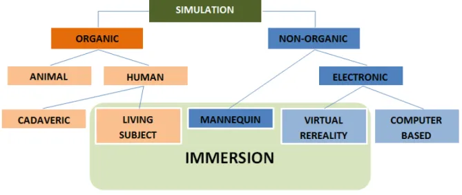
In his article on the emerging trends in medical simulation, Dr. Satava looks at simulation models as either of the traditional or emerging kind (3). Traditional models are physical models or animal parts, whereas the emerging models are mannequins, computer-based interactive programs or virtual reality models. The different simulation modalities are summarized in figure 2 as proposed by Dr. Chiniara (10).
Figure 2: Classification of simulation models. Adapted with modifications from Dr.
Chiniara.
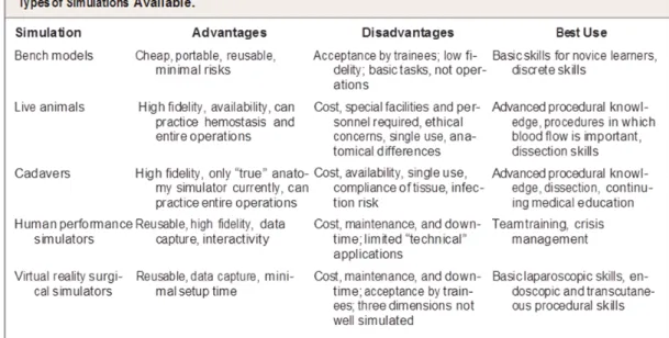
Reznick and MacRae, in their New England Journal of Medicine review article on teaching surgical skills, classify surgical simulators as bench models, live animals, cadavers, human performance simulators and virtual reality simulators, and give a list of advantages, disadvantages and best use for each (figure 3) (11). These five classes described by Reznick and MacRae cover the entire spectrum of current simulation, and we think can be further classified as either physical or virtual models. Bench, live animal, cadaveric and human performance simulators are physical simulators while virtual simulators are those that involve
10
as « the only true anatomy simulator » available, statement that is a testament to the irreplaceable contribution of cadaveric models in medical education. While much effort and expense have been geared towards virtual reality in an attempt to replace the cadavers with a safer, more available model that avoids the disadvantages of cadavers, there is no virtual reality simulator that is able to match the high fidelity of the cadaveric model.
Figure 3. Types of simulation available. Reproduced with permission from Reznick and
MacRae / NEJM 355 (2006) 2667, Copyright Massachusetts Medical Society.
0.4. High and low fidelity simulation
As Miller eloquently put it « simulation is not all or none, it is a matter of degree » (12). Simulation models and simulators can range from very basic, like a piece of latex glove for the teaching of microsurgical sutures, to very complex models, such as lifelike mannequins giving feedback in real time to the trainee or virtual reality models. This degree of complexity also corresponds with the associated cost, despite the fact that technology is becoming more affordable. For example, mannequins such as the SimMan® cost around 70,000$, to which one has to add the consumables and service fees. Yet does the level of complexity of a model, alone, give any indication of how good the model is? And what are the properties that make a
simulator good? How closely a model emulates reality is measured by a concept called « fidelity ».
Fidelity is synonymous with faithfulness and is defined as « the degree of exactness with which something is copied or reproduced » (13). There is generally a lack of information in the medical simulation literature regarding the meaning of fidelity. While most authors use the term « high fidelity » as a branding for their simulator, the careful definition of what constitutes fidelity is less well established in medicine. As defined by Maran and Galvin, « fidelity is the extent to which the appearance and behavior of the simulator ⁄ simulation match the appearance and behavior of the simulated system » (14). We assume that the more complex a model and simulator, the more life-like it will be, however, this does not necessarily hold true. A mannequin can serve as a model for suturing, give real-time feedback regarding discomfort and pain, yet by the limitation of the artificial materials it cannot, to date, reproduce the texture, elastic modulus or feedback that a cheaper model, such as animal parts can offer. We can consider that animal skin and subcutaneous tissue, by virtue of its architecture and tissue composition, are closer to human skin and therefore confer a higher degree of fidelity. Yet fidelity is a much more complex concept than that.
An excellent description of fidelity and how it can be further subdivided is found in the article of Maran and Galvin (14). According to the authors, the distinction between engineering and psychological fidelity has been initially described by Miller (12). While he does not use the word « fidelity » in his description, Miller talks about two components to simulation: an engineering component, which relates to the degree to which a physical model and its properties are copied, and the psychological part, which relates to how well the responses learned during simulation translate into appropriate actions in real life operations. It is the engineering part of his description which matches the dictionary description of the word fidelity and also the meaning of Maran and Galvin’s definition stated above; the psychological part, while considered the major aim of any simulator by Miller, is less well understood.
medical education world. One has to consider that the work of Miller predates by decades the development of medical simulation, and comes from the field of military aviation. We need to consider this when regarding the use of terms such as engineering and psychology in his description since simulation in aviation and/or military has other technical requirements and objectives. Since models in medicine do not have to be necessarily engineered, contrary to say aviation, we can refer to this property of the model and simulator as physical fidelity. Conversely, as it applies to surgery, it seems more opportune to functional fidelity rather than psychological fidelity.
Functional fidelity is considered more important than its physical counterpart because it is the actual aim of the training session. At the end of the training, what is important is the skill learned and how well it applies to the real world application. The degree of physical fidelity, if it does not modify the functional fidelity of the model, has in itself little-added value. Miller concludes that the training problem is to « provide stimuli so that responses learned to them will transfer from training to operations with little or no loss ». Yet if we look at where the money is spent in designing a simulator, the high cost is associated with improving the physical fidelity. Miller provides an accurate if rudimentary cost-benefit analysis of simulation. The author is of the opinion that as one increases the engineering fidelity, the training value increases proportionally at the beginning, followed by a flattening of the curve where there is little training value gained for each increment in physical or engineering fidelity. If this graph is now superposed on that of cost as a function of engineering fidelity, one can determine the point along the x-axis (engineering fidelity) optimizes the training value and cost. This point is described as the point of diminished return since any increase in fidelity from this point on would result in an exponential increase in cost with only a marginal increase in training value (12). For these reasons, the objective of creating a simulation model is maximizing the training value, and not creating the most lifelike environment.
A number of authors have suggested that for acquiring simple skills a simple model can be more than adequate, especially when the trainee has very little experience (11, 14). In complex tasks and when the trainee has a higher level of proficiency, it is beneficial that the
model is of high fidelity so as to avoid negative transfer (14). This increase in fidelity would reach higher and higher levels and one has to wonder when this increase in fidelity, based on the associated higher and higher costs mentioned in Miller’s cost-benefit analysis above, becomes prohibitive. One has to determine when the need for simulation becomes obsolete. Somebody who is already very proficient at laparoscopic surgery need not use a simulator for further improvement; at that point, their skill would be sufficient to justify operating on patients and with perpetual practice and performance of more and more complex cases, the skills of this already proficient surgeon would improve. There is one counterargument to this way of thinking, and that is the « deliberate practice » theory proposed by Ericsson (15) and described in detail in the debriefing section below.
Based on the definitions and theories on fidelity mentioned above, the question we need to ask is: What constitutes a high fidelity simulation model or simulator in medical education? If we consider the information provided by Reznick and MacRae in the table mentioned above (16), live animals, cadavers and human performance simulators are all labeled as « high fidelity ». If we look from the perspective set forth by Miller, the label of what is high or low fidelity is more related to the specific task, more in terms of functional than physical fidelity (12). A model or simulator could potentially be high fidelity for one task and low fidelity for another. For example, a mannequin simulator such as SimMan ® is of high fidelity for training crisis management skills, such as advanced cardiac life support, yet it fails to perform as well when it comes to performing a tracheostomy, for which the pig model is more adequate. Furthermore, the proficiency level of the trainee also factors into where along the spectrum of fidelity a simulator is placed. A well-trained microsurgeon wanting to hone their skill in order to acquire proficiency on very small vessels, such as lymphatics, will exert much more scrutiny than a resident who is learning the basics of microsurgery, for whom silastic tubing can be considered of high fidelity.
0.5. Simulation models in plastic surgery
model for microsurgical skill training (17). This systematic review, while focused on the microsurgical literature, suggests that while much has been published in recent years in simulation, these studies are often of questionable quality.
As described in figure 3 above, Reznick and MacRae make the difference between bench models, cadaveric, animal, human performance simulators and virtual reality simulators (11). While Rosen and colleagues focus on the virtual reality part of the simulation (6), it would be a gross oversight to forget the lower technology simulators which have been used and continue to occupy a strong role in the simulation arsenal pertaining to plastic surgery.
Bench models are the foundation of surgical skill training. The prototypal model is learning to do a hand tie. Medical students learn their knot tying yarn to chairs or doorknobs. Ethicon Inc (Johnson&Johnson, Somerville, NJ) produces a knot tying kit that in addition allows for placement of deep knots. More specific to plastic surgery, microvascular sutures are practiced on latex gloves or increasingly more complex artificial models that are currently commercially available. These rather basic models, under magnification, allow for basic microvascular skill practice, such as suture placement, correct manipulation of the needle and suture, knot tying. Silicone tube models permit the practice of a complete anastomosis including learning how to distribute the stitches around the circumference of the vessel. Diathermy cleaning pads, polyurethane and rubber cards have been validated for basic skill training (17). Since microsurgical skill is very different from the skills one learns in other surgical specialties, the most basic of skills in microsurgery can be efficiently learned using these cheap models despite the low physical fidelity.
Higher fidelity models are needed in order to have adequate tactile feedback, lifelike tissue handling and the possibility to verify the patency of the anastomosis. Most authors would agree that the current gold standard for microvascular anastomosis practice remains the rat femoral artery (18). However, a number of problems are associated with the use of animal models:
a) Rats are expensive and cumbersome to use, as this requires a facility to breed and keep the animals, as well as the infrastructure to anesthetize and afterward euthanize the rats. b) As training in such facilities requires assistance, this can only be done during working hours and thus conflicts arise with the clinical responsibilities of residents.
c) Rat vessels are smaller and more friable than human tissue, yielding thus a steeper learning curve than what would be necessary for the basic microsurgical training.
d) Due to the sacrifice of the animals at the end of the training session, the use of rats in microsurgery has met criticism in recent years from animal rights activists.
In keeping with the “three R” model of replacement, reduction and refinement described by Russel and Burch (19), other models for microvascular simulation have been described, including the use of animal parts, such as chicken or turkey wings, chicken thighs or rat tails, all of which can be used for dissection and anastomosis of vessels. Of these ex vivo animal models, chicken legs and porcine eye have been validated (17).
Regarding microneurorrhaphy bench training, the literature is quite scant. Precision, needle placement, and knot tying are similar to microvascular, and can, therefore, be practiced on latex gloves. However, there are no commercially available artificial models available. A piece of twine rolled up in saran wrap, as well as textile-covered rubber threads (20) have previously been described. Bench models for tendon repair are commercially available animal tendons that can be procured from butcher shops, or synthetic tendon substitutes, such as silicone fishing worms or other types of silicone and latex materials. Kamath and colleagues describe a model that permits the simulation of both flexor digitorum superficialis (FDS) and flexor digitorum profundus (FDP) repair, however, they fail to provide enough information as to how to construct this model (21). Higher fidelity than the silicone/rubber rods is animal tendons (22) as they provide both the feedback and tissue handling that is seen in the operating room. Rhodes and colleagues construct a « jig », as they refer to it, in order to provide placement of the pig tendon in a more anatomical position
A silicon rod can tear, but it will not fray or pull through the way a traumatized tendon or an improperly placed suture will do in real life.
The Arbeitsgemeinschaft für Osteosynthesefragen foundation, also know as the AO foundation has courses, ranging from basic to expert level, in which they teach principles of osteosynthesis. While a good number of these courses are targeting orthopedics, the craniomaxillofacial and hand courses are equally applicable to plastic surgery. These are one to two-day courses teaching theory as well as surgical skills and procedures. Wood models for performing and understanding lag-screw fixation, as well as osteosynthesis of several facial fractures, are explained and performed in the basic course. Trainees have the opportunity to familiarize themselves with the different instrument kits, plate molding, and reduction of fractures on sawbones models. These procedures are all performed under the supervision of craniofacial or hand surgeons respectively, who provide feedback and tips. While the physical fidelity of the sawbones model is limited, the functional fidelity of the overall model using medical grade titanium plates and instruments renders the simulation exercise of good value for the purpose of learning basic craniomaxillofacial or hand techniques. Can this be replaced by a virtual reality model? It is likely that a virtual model can provide a higher physical fidelity, however, the functional fidelity learned from manipulating the actual plates would be lost. Plate molding involves trial and error with action feedback.
Cadavers have been used in surgical training from the very beginning. Whether for acquiring a better understanding of the anatomy, or to simulate procedures, surgeons have turned to both fresh frozen and embalmed cadavers. In some areas of plastic surgery formal cadaver-based training models are used, such as for example in the case of flap courses. In these courses, under the direct supervision of experienced surgeons, the trainees learn anatomy and dissection techniques necessary for raising pedicled or free flaps. The dissection exercises are usually preceded by lectures, and immediate feedback and suggestions are given by the educators during the dissection exercise itself. The cadavers have historically been formaldehyde embalmed or fresh frozen, and more recently a novel method of embalmment has been proposed and tested by Prof. Thiel of Graz, Austria (23). This method maintains the
cadavers in a more lifelike state while reducing the decay and infectious risks associated with fresh frozen cadavers. In addition to flap dissection, Thiel's vessels have been used for microvascular surgery (24, 25), as well as tendon repair workshops (26). Other areas where cadaveric specimens have been used are specialized dissection courses, such as rhytidectomy course at the University of Texas, Southwestern in Dallas.
Animal models have been an invaluable tool for the practice of both flap dissection, as well as other more specific skills like microvascular anastomoses, and to lesser extent nerve repair. In microsurgery, the rat femoral artery model has been validated in two studies (17) and enjoys widespread popularity and is the closest model to a gold standard. Most major North American universities organize microsurgical training labs using rats that are euthanized at the end of the procedure. The rat sciatic nerve model is also one of the best available models for nerve repair (27). With the advent of supermicrosurgery and lymphatic surgery, the rat model has been used for the practice of lympho-venous anastomoses (28), which takes simulation to the very edge of its capabilities. Despite the high fidelity of the live rat model, a number of shortcomings exist that need to be weighed in. While these often are rats at the end of studies that would be euthanized anyway, and the euthanasia during anesthesia is an ethical way to dispose of these animals, the use of live animals in training sessions has received much criticism from animal rights activists and society at large. In addition, live animal models are expensive, require technician expertise for growing, anesthesia and euthanasia, and require specialized facilities for their use. Since the introduction of the “three R” principle (reduction, refinement, and replacement) by Russel and Burch in 1959, stricter rules regarding the use of animals in research and education have been implemented (29), and it has become the ethical responsibility of researchers and educators alike to find alternatives to animal use.
The fourth type of simulator, human performance simulators, do not seem to yet have a role in plastic surgery education, however, the virtual reality simulators have shown significant improvement in the last decade and appear to be gaining ground as an important instrument in plastic surgery training. As technology becomes more complex and more affordable, medical educators look at a virtual reality as the new revolution in medical training (3). Rosen et al, in
their review of the state of simulation and plastic surgery and the path forward, put much emphasis on the development of virtual reality models, to parallel the advances in abdominal surgery simulation (6). Arbogast and Rosen give a number of virtual reality examples, such as simulators for robotic surgery, cleft lip, and palate, latissimus dorsi myocutaneous flap dissection, and Dr. McCarthy’s Interactive Craniofacial Surgical Atlas (2). While these models cannot match the functional fidelity of some lower technology models, their exquisite level of physical fidelity make them an invaluable teaching tool. With further advances in technology, we can expect more from this branch of surgical simulation.
0.6. Thiel embalmed cadavers in medical simulation
Parallelling the technological boom of the 20th century, simulation has followed the same upward trend with a significant amount of research and development geared towards the development of hi-tech simulators, such as high-fidelity mannequins or virtual reality models. This high-tech high-fidelity simulation business has become very popular and the majority of authors consider this the holy grail of medical simulation. Perhaps it is, and time will tell. Yet we cannot ignore the basic principles of simulation established by Miller (12), which emphasize the functional rather than physical fidelity. And one cannot ignore the classical models for surgical simulation, of which the cadaveric model is considered by Reznick and MacRae the only true anatomic simulator in existence (11). Traditionally two cadaveric models existed: the fresh frozen cadaver which provides the highest level of fidelity to the live human tissue yet it is plagued by a quick decay and exposes the trainee to infectious risks. Formaldehyde embalmed cadavers can be preserved for an extended period of time and have a low infectious risk, however, the fixation of tissues by formaldehyde changes the color and texture of the tissues. Over the last twenty years, a new cadaveric model that combines the advantages of fresh frozen and formaldehyde cadavers has been developed and acquired fast acceptance in the anatomic community. This new model is the Thiel embalmed cadaveric model (23).
In a review article on the origins of formaldehyde tissue fixation, Fox et al give a history of formaldehyde synthesis and its introduction in the medical and biological sciences. Ferdinand
Blum is credited for the introduction of formaldehyde for the purpose of tissue fixation and anatomical labwork, including the embalming of whole cadavers (30). Because formaldehyde is very effective at preventing decomposition of the tissue and at the same time provides an antiseptic medium, it has remained the mainstay of tissue preservation to this date. The excellent tissue fixing capabilities of formaldehyde are however plagued by a number of problems. First, formaldehyde has a hardening effect on tissue, and dissection of embalmed tissue can often be cumbersome and at the very least not life-like. The colors of the tissues are also not preserved, due to the transformation of hemoglobin to methemoglobin. This change in pigmentation appears to be an effect of formic acid, the product of the oxidation reaction of formaldehyde (30). Lastly, depending on the concentration of the embalming solution, formaldehyde has a pungent, irritating smell (23), and can cause contact dermatitis (31). While it has not been conclusively shown that formaldehyde can be carcinogenic to humans, neither through animal or epidemiological studies, the health hazard posed by formaldehyde warrants, according to Pabst, the lowering of exposure to formaldehyde to minimal levels (31).
In 1992, Professor Walter Thiel from the University of Graz in Austria published a report of a new embalming solution which, contrary to formaldehyde, provided soft, life-like tissue (23). In his seminal work, he describes the evolution of embalming of whole cadavers from the introduction of formaldehyde by Blum. The few alternatives to formaldehyde which had been published in the literature had been tested by Prof. Thiel in his laboratory and found to provide soft, albeit inadequately preserved tissue. He describes in detail the development of a novel embalming technique based on Boric acid, ethylene glycol, ammonium nitrate and potassium nitrate. Over the course of two decades, from 1970 to 1991, Thiel had embalmed with this new technique 977 cadavers, perfecting the solution to the composition used in the present day (23). Professor Thiel has tested his embalming technique to prove the fixation properties as well as its antiseptic nature against some of the more concerning pathogens, such as Staphylococcus, Pseudomonas, and Mycobacterium. Its efficacy against fungi has also been proven. This was the first viable alternative to formaldehyde embalmment published in the literature.
In 2002, Prof. Thiel published a supplement to his original 1992 paper intended to give an update on the experience with the new embalmment method at his institution (32). The notable change introduced in 2002 is pertaining to the embalmment of the central nervous system. Since the Thiel method of embalming would not solidify the gray and white matter sufficiently, Prof. Thiel proposes in this supplement an initial intrathecal infusion of a formaldehyde-isopropyl alcohol followed by the « Thiel solution ». The other evolution of the method relates to the change of the mono-ethylene glycol for mono-propylene glycol in the embalming solution, the former being found to be irritating to tissues, and an increase in the concentration of formaldehyde. These changes finalized Prof. Thiel’s powerful contribution to the anatomy literature and are shown in figure 4.
Figure 4. Basic components of Thiel embalming solution. From K Wolff et al /
Microsurgery 28 (2008) 273-278, reprinted with permission.
Since its publication, the Thiel technique has been met with much enthusiasm by the anatomic and scientific community, and currently, laboratories around the world utilize this technique of embalmment. The lifelike properties of the embalmed cadavers have been used in a number of fields, from ultrasound, ultrasound-guided peripheral nerve block, arthroscopy, flap dissection and other surgical simulation procedures. At the McGill University simulation center, Thiel embalmed cadavers have been used for military emergency medical simulation, including
Basic components of Thiel solution
Hot tap water 100
Bor acid 3
(Mono-)Ethylenglycol 30
Ammoniumnitrate 20
Potassiumnitrate 5
Chlorkresol-solution with (Mono-)Ethylenglycol 10
4-Chlor-3-Methylphenol 1
surgical airway, thoracic tubes, vascular access etc. The potential for Thiel cadaveric utilization in surgery is almost limitless; almost any surgical procedure can be simulated with success on these cadaveric specimens that maintain both the color and supple nature of living tissue.
A number of studies have looked at the adequacy of Thiel cadavers in surgical dissection. Eisma et al designed a surgical workshop for thyroidectomy where they used both Thiel and formalin embalmed cadavers (33). They enrolled 8 trainees and 4 surgeons in the workshop and had them perform a thyroidectomy on both formalin and Thiel cadavers. Each trainee performed either the first or second half of the thyroidectomy on a Thiel cadaver, after which they were asked to evaluate the two experiences. The author asked the participants to evaluate the adequacy of the skin, muscle, fat, vessels as well as nerves, as well as a number of surgical parameters, such as raising the subplatysmal flap or retraction of tissues. While the study provides no information regarding the statistical significance of the findings, the authors claim that the Thiel specimens scored higher than formaldehyde embalmed specimens in all but 10 of the 180 pairs of scores. The difference was most marked in areas where supple tissues are necessary, such as subplatysmal flap dissection or tissue retraction. The authors conclude that Thiel embalmment is superior to formaldehyde for the purpose of simulating thyroid surgery and that these findings could extend to other surgical areas as well.
Wolff and colleagues, in an article published in the Journal of Microsurgery in 2008, introduced the use of Thiel cadavers in the field of flap dissection and microvascular simulation (24). The authors report their experience with the use of Thiel cadavers in their flap dissection courses, which they consider an ideal model combining the tissue characteristics of fresh-frozen cadavers with the conservation of the formalin embalmment. They provide a vivid description of the different tissues encountered during the dissection and compare them to what would be expected during intra-operative flap dissection. The authors note that with the Thiel embalming technique, the epidermis, as well as the nails and the body hair peeled off, leaving a smooth and slightly oily surface. While they found the skin to be slightly firmer than in the living body, this was not considered an impediment. The quality of the
subcutaneous fat, fascia, and muscle was reported as being of good quality, with the exception of the muscles of the back which were found soft, possibly due to the sustained pressure. The authors further continue their description of the vascular pedicles, which they consider suitable for dissection with properties similar to those found in the clinical setting. At the microscopic level, the arteries were found to be thicker, with the three layers identifiable, while the veins were found to be thinner walled and collapsible, with the valvular system preserved. During the exercise of the microvascular anastomosis, the vessels were found to offer normal tissue resistance and permit suture and knot placement. With respect to nerve tissue, this was found to be less well preserved than its vascular counterpart, with a weaker structure and less compact bundles. We note that Wolff et al describe having used the embalming solution which Professor Thiel upgraded in 2002, as described above, particularly because the preservation of the central nervous system tissues with the original solution was suboptimal (32).
While tissue handling has been reported to be lifelike (23, 24), with excellent pliability and elasticity of the tissues superior to that obtained with formaldehyde embalmment (33), one has to wonder if, at a microscopic or molecular level the properties of the tissues have been affected. Wolff et al have commented on the integrity of the vessel walls in Thiel cadavers, as well as the three layers being identifiable under magnification (24). The only report of Thiel embalmment on the histologic appearance of arteries has been published by our team (34) and is described in chapter 4 of this thesis. The suitability of Thiel embalmed nerves for simulation of peripheral nerve surgery has not previously been evaluated, neither has it been shown how Thiel preservation affects the histologic preservation of the nerves and their fascicles. The only data on Thiel embalmment of nerves comes from Prof Thiel’s 2002 paper, where he made changes to the embalming solution with the purpose of improving the quality of central nervous system fixation (32), and from Wolff et al, who from a gross anatomic perspective considered the quality of the peripheral nervous tissue to be inferior to that of the vessels (24). To date, there are two studies in the literature that looked at the effects of Thiel embalmment on tendons. Benkhadra et al looked at the effects of Thiel embalmment on the microarchitecture of muscle and tendon tissues (35). Their biopsies were obtained from the
biceps muscle and the brachioradialis tendon and stained with Masson’s trichrome, Sirius red and Ramon y Cajal. They compared their findings with similar samples obtained from fresh frozen cadavers and formalin preserved cadavers. When looking at the muscle biopsies, the authors found a very noticeable fragmentation of the muscle fibers with a minced appearance which did not, however, affect the alignment of the fibers. The collagen fibers within the muscle interstitium were found in continuity. This fragmentation of the muscle was not seen in either fresh frozen or formalin embalmed cadaveric tissues. The authors postulated that this muscle fragmentation with preserved alignment potentially explained the exquisite suppleness and flexibility of the Thiel embalmed cadavers. When looking at the microarchitecture of the tendons, Benkhadra concluded that histologically Thiel embalmed tendons are very similar to fresh frozen specimens.
On the other hand, Fessel et al, based on evidence from a previous biomechanical study that found a decreased elastic modulus in Thiel embalmed bone when compared to fresh-frozen controls, postulate that Thiel embalmment reduces the elastic modulus and failure stress of Thiel cadaveric tendons (36). The authors found that in cadaveric FDP tendons, the fresh frozen specimens had a significantly higher median ultimate stress than Thiel embalmed specimens (60MPa vs 38MPa, p0.048), while the failure strain and tangential elastic modulus showed a trend towards superiority of the fresh frozen specimens without reaching statistical significance. When looking at rat tendon fascicles, both stiffness and failure force of the fresh frozen specimens were superior to the Thiel exposed counterparts, with p < 0.05. The stress-strain and force stress-strain relationships of the cadaveric and rat specimens respectively can be visualized in figure 5. The authors conclude that Thiel embalmed cadaveric tendons do not faithfully reflect the biomechanical properties of living tendon substance, for which the gold standard for biomechanical research remains fresh frozen tissue. However, while cautioning against the use of Thiel specimens in biomechanical studies, the authors suggest that Thiel embalmed tissue could be used in preliminary studies as long as one is aware of the biomechanical differences this model entails.