Université de Montréal
The plant ovule omics: an integrative approach for
pollen
−pistil interactions and pollen tube guidance studies
in solanaceous species
par Yang Liu
Département de sciences biologiques, Institut de recherche en biologie végétale Faculté des Arts et des Sciences
Thèse présentée à la Faculté des Arts et des Sciences en vue de l’obtention du grade de doctorat
en biologie végétale
Octobre, 2015
Université de Montréal
Faculté des études supérieures et postdoctorales
Cette thèse intitulée:
The plant ovule omics: an integrative approach for
pollen
−pistil interactions and pollen tube guidance studies
in solanaceous species
Présentée par: Yang Liu
a été évaluée par un jury composé des personnes suivantes :
David Morse, président-rapporteur Daniel P. Matton, directeur de recherche
Simon Joly, membre du jury Alice Cheung, examinatrice externe
Résumé
Chez les plantes à fleurs, l’ovaire est l’organe reproducteur femelle et il interagit de façon importante avec les gamètes mâles durant la croissance, le guidage, la réception et la rupture du tube pollinique ainsi que la fusion des gamètes. Le processus débute lorsque de nombreux gènes de l’ovule sont activés à longue distance lors de la réception du pollen sur le stigmate. Afin d’explorer les signaux provenant de l’ovule ayant un impact important sur les interactions pollen–pistil, particulièrement les molécules sécrétées impliquées dans la signalisation espèce-spécifique, l’expression génique des ovules sous forme d’ARNm ainsi et la sécrétion protéique ont été étudiées chez Solanum chacoense, une espèce diploïde de pomme de terre sauvage. S. chacoense a subi beaucoup d’hybridation interspécifique avec d’autres espèces sympatriques de solanacées, facilitant ainsi grandement l’étude des interactions pollen–ovule de façon espèce-spécifique ainsi que leur évolution. Dans ce projet, des ovules provenant de trois conditions différentes ont été comparés: des ovules matures de type sauvage, des ovules légèrement immatures, récoltés deux jours avant l’anthèse et des ovules provenant du mutant frk1 pour lesquels le sac embryonnaire est absent. Un séquençage d’ARN à haut débit a d’abord été effectué sur les ovules de type sauvage de S. chacoense afin de générer un assemblage de référence comprenant 33852 séquences codantes. D’autres séquençages ont été effectués sur les trois conditions d’ovules et sur les feuilles afin de faire une analyse d’expression différentielle des gènes. En comparaison avec les ovules de type sauvage, 818 gènes sont réprimés dans les ovules du mutant frk1. Un sous-groupe de 284 gènes, étaient également sous-exprimés dans les ovules légèrement immatures, suggérant un rôle spécifique dans les stades tardifs de la maturation du sac embryonnaire (stade de développent FG6 à FG7) ainsi que du guidage du tube pollinique, puisque ni les ovules du mutant frk1 ni ceux légèrement immatures ne sont capables d’attirer les tubes polliniques lors d’essais de croissance semi in vivo. De plus, 21% de ces gènes sont des peptides riches en cystéines (CRPs). En utilisant un transcriptome assemblé de novo provenant de deux proches parents de S. chacoense, S. gandarillasii et S. tarijense, une analyse d’orthologie a été effectuée sur ces CRPs, révélant une grande variabilité et une évolution rapide chez les solanacées. De nouveaux motifs de cystéine uniques à cette famille ont également été découverts. En comparant avec des études similaires chez Arabidopsis, le sac embryonnaire de S. chacoense montre un transcriptome fortement divergent, particulièrement en en ce qui a trait à la catégorisation fonctionnelle des gènes et de la similarité entre les gènes orthologues. De plus,
même si la glycosylation n’est pas requise lors du guidage mycropylaire du tube pollinique chez Arabidopsis, Torenia ou le maïs, des extraits d’ovules glycosylés de S. chacoense sont capables d’augmenter la capacité de guidage de 18%. Cette étude est donc la première à montrer une corrélation entre glycosylation et le guidage du tube pollinique par l’ovule. En complément à l’approche transcriptomique, une approche protéomique portant sur les protéine sécrétées par l’ovule (le secrétome) a été utilisée afin d’identifier des protéines impliquées dans l’interaction entre ovule et tube pollinique. Des exsudats d’ovules matures (capables d’attirer le tube pollinique) et d’ovules immatures (incapables d’attirer le tube pollinique) ont été récoltés en utilisant une nouvelle méthode d’extraction par gravité permettant de réduire efficacement les contaminants cytosoliques à moins de 1% de l’échantillon. Un total de 305 protéines sécrétées par les ovules (OSPs) ont été identifiées par spectrométrie de masse, parmi lesquelles 58% étaient spécifiques aux ovules lorsque comparées avec des données de protéines sécrétées par des tissus végétatifs. De plus, la sécrétion de 128 OSPs est augmentée dans les ovules matures par rapport aux ovules immatures. Ces 128 protéines sont donc considérées en tant que candidates potentiellement impliquées dans la maturation tardive de l’ovule et dans le guidage du tube pollinique. Cette étude a également montré que la maturation du sac embryonnaire du stade FG6 au stade FG7 influence le niveau de sécrétion de 44% du sécrétome total de l’ovule. De façon surprenante, la grande majorité (83%) de ces protéines n’est pas régulée au niveau de l’ARN, soulignant ainsi l’importance de cette approche dans l’étude du guidage du tube pollinique comme complément essentiel aux études transcriptomiques. Parmi tous les signaux sécrétés par l’ovule et reliés au guidage, obtenus à partir des approches transcriptomiques et protéomiques décrites ci-haut, nous avons spécifiquement évalué l’implication des CRPs dans le guidage du tube pollinique par l’ovule chez S. chacoense, vu l’implication de ce type de protéine dans les interactions pollen-pistil et le guidage du tube pollinique chez d’autres espèces. Au total, 28 CRPs étaient présentes dans les ovules capables d’attirer le tube pollinique tout en étant absentes dans les ovules incapables de l’attirer, et ce, soit au niveau de l’ARNm et/ou au niveau du sécrétome. De celles-ci, 17 CRPs ont été exprimées dans un système bactérien et purifiées en quantité suffisante pour tester le guidage. Alors que des exsudats d’ovules ont été utilisés avec succès pour attirer par chimiotactisme le tube pollinique, les candidats exprimés dans les bactéries n’ont quant à eux pas été capables d’attirer les tubes polliniques. Comme l’utilisation de systèmes d’expression hétérologue eucaryote peut permettre un meilleur repliement et une plus grande activité des protéines, les candidats restants seront de nouveau exprimés, cette fois
dans un système de levure ainsi que dans un système végétal pour produire les peptides sécrétés. Ceux-ci seront ensuite utilisés lors d’essais fonctionnels pour évaluer leur capacité à guider les tubes polliniques et ainsi isoler les attractants chimiques responsable du guidage du tube pollinique chez les solanacées comme S. chacoense.
Mots-clés: Interactions pollen–pistil, ovule, sac embryonnaire, maturation, guidage du tube
pollinique, chimio-attractants, peptide riche en cystéine, RNA-seq, spectrométrie de masse, sécrétome, analyse d’expression différentielle
Abstract
In flowering plants, the ovary is the female reproductive organ that interacts extensively with the male gametophyte during pollen tube (PT) growth, guidance, reception, discharge and gamete fusion. The process begins when numerous ovule-expressed genes are activated when pollen lands on the stigma. To explore the ovular signals that have a great impact on successful pollen– pistil interactions, especially the secreted molecules that mediate species-specific signaling events, ovule mRNA expression and protein secretion profiles were studied in Solanum chacoense, a wild diploid potato species. Solanum chacoense has undergone extensive interspecific hybridization with sympatric solanaceous species that greatly facilitates the study of species-specific pollen–ovule interactions and evolution. In this project, three ovule conditions were studied: wild-type mature ovules, slightly immature ovules at two days before anthesis (2DBA), and frk1 mutant ovules that lack an embryo sac (ES). RNA-seq was performed on S. chacoense ovules to provide a scaffold assembly comprising 33852 CDS-containing sequences, then to provide read counts for differential gene expression analyses on three ovule conditions as well as on leaf. Compared to wild-type ovules, 818 genes were downregulated in frk1 ovules. A subset of 284 genes was concurrently under-expressed in 2DBA ovules, suggestive of their specific involvement in late stages of ES maturation (female gametophyte (FG), FG6 to FG7 developmental stage), as well as in PT guidance processes, as neither frk1 nor 2DBA ovules attract semi in vivo-grown PTs. Of these 284, 21% encoded cysteine-rich peptides (CRPs). Using de novo assembled ovule transcriptomes of two close relatives, S. gandarillasii and S. tarijense, an orthology survey was conducted on these CRPs, revealing their highly polymorphic nature among species and rapid evolution. Interestingly, novel cysteine motifs unique to this family were also uncovered. As compared to parallel studies in Arabidopsis, S. chacoense was found to possess a highly divergent ES transcriptome, in terms of both functional categories and individual ortholog similarities. Although glycosylation is not required for micropylar guidance cues to attract PTs in Arabidopsis, Torenia or maize, glycosylated ovule extracts from S. chacoense showed enhanced PT guidance competency by 18%. This is the first time a positive regulation between glycosylation and ovular PT guidance has been observed. As a complement to the transcriptomic approach, a proteomic approach using secreted proteins from the ovule (secretome) was employed to identify proteins involved in pollen–pistil interactions. Ovule exudates were collected from mature ovules (PT attracting) and immature ovules at 2DBA (PT
nonattracting), using a novel tissue free-gravity extraction method (tf-GEM), which efficiently reduced the cytosolic contamination to less than 1%. Through mass spectrometry analyses, a total of 305 ovule-secreted proteins (OSPs) were identified, of which 58% were considered ovule-specific when compared to secretome studies conducted in other plant tissues. The secretion of 128 OSPs was upregulated in mature ovules vs. immature ovules. These OSPs were considered as candidate proteins involved in late ovule maturation and PT guidance. This study demonstrated that the ES maturation from FG6 to FG7 stages influenced the secretion status of 44% of ovule secretome. Surprisingly, the majority (83%) of these proteins were not regulated at the RNA level, vindicating this novel approach in the study of PT guidance as a robust complement to transcriptomic studies. Among all identified guidance-related ovular signals from the transcriptomic and proteomic approaches described above, we focused on the evaluation of the involvement of CRPs in ovular PT guidance of S. chacoense, due to the implication of various CRPs in pollen–pistil interactions and, especially, in PT guidance. A total of 28 CRPs were present in PT attracting ovules while being low or absent in nonattracting ovules, at the mRNA and/or protein secretion levels. Of these, 17 CRPs were expressed in bacteria and purified in sufficient amount for PT guidance assays. However, while ovule exudates were shown to induce PT chemotropism in the bead assay, refolded candidates did not show guidance competency. Since the use of eukaryotic protein expression systems might lead to better refolding and higher protein activity, the remaining candidates will be expressed in both yeast and plant-based expression systems and tested for their ability to attract PTs in a semi in-vivo assay, in order to lead us toward the isolation of PT guidance chemoattractants in solanaceous species like S. chacoense.
Keywords: pollen–pistil interactions, ovule, embryo sac, maturation, pollen tube guidance,
chemoattractants, cysteine-rich peptide, RNA-seq, mass spectrometry, secretome, differential gene expression analysis
Table of Contents
Abstract ... i
Table of Contents ... vi
List of Tables ... x
List of Figures ... xi
List of Abbreviations ... xiii
Acknowledgements ... xvi
Chapters 1. Introduction ... 17
1.1 Plant Reproduction ... 17
1.1.1 Female Gametophyte Development ... 18
1.1.2 Male Gametophyte Development ... 22
1.2 The Pollen Tube (PT) Path In the Pistil of S. chacoense ... 24
1.3 Why Studying PT Guidance In S. chacoense? ... 27
1.3.1 The Significance of PT Guidance In Plant Reproduction ... 27
1.3.2 PT Guidance In S. chacoense and Species-Specific Pollen−Pistil Interactions ... 27
1.4 PT Guidance In the Style Is Governed By Sporophytic Tissues ... 30
1.4.1 Sporophytic Guidance Down the Stigma ... 30
1.4.2 Sporophytic Guidance From Stigma to Ovary ... 33
1.4.3 Sporophytic Guidance To the Micropylar Region ... 33
1.5 PT Guidance In the Ovary Is Controlled By Both Gametophytic and Sporophytic Tissues ... 35
1.5.1 Long-Distance Guidance Is Governed by the Ovule ... 36
1.5.2 Funicular Guidance Is Under the Influence of Ovular Sporophytic Tissues ... 36
1.5.3 Micropylar Guidance Is Under the Influence of the FG ... 37
1.5.3.1 Synergid Cells Are the Sources of Micropylar PT Guidance Attractants ... 37
1.5.3.2 Identified Micropylar PT Attractants ... 38
1.5.3.3 Characteristics of Micropylar Attractants ... 40
1.5.3.4 Key Genes Affecting Micropylar Guidance ... 41
1.5.3.5 Micropylar Guidance Is Orchestrated By the Egg Cell and the Central Cell ... 42
1.5.4 PTs Slow Down for Fine-Tuning Before Targeting the Micropyle ... 44
1.7 Objectives ... 47
2. Gene expression during late stages of embryo sac (ES) development: a critical building block for successful pollen−pistil interactions ... 49
2.1 Introduction ... 51
2.2 Material and Methods ... 53
2.2.1 RNA Purification and Sequencing ... 53
2.2.2 Sequence Assembly and Curation ... 53
2.2.3 Gene Expression Analysis ... 54
2.2.4 Aniline Blue Staining ... 54
2.2.5 Scanning Electron Microscopy ... 55
2.2.6 Protein Extraction ... 55
2.2.7 semi in vivo PT Guidance Assay ... 55
2.2.8 in silico Predictions ... 56
2.2.9 Estimates of Diversifying Positive Selection ... 57
2.2.10 Accession numbers ... 57
2.3 Results ... 57
2.3.1 Data Curation ... 57
2.3.2 Validation of the Assembly Through Known Ovule-Expressed Genes ... 58
2.3.3 Gene Expression Validation ... 58
2.3.4 Functional Annotation of S. chacoense Ovule Transcriptome ... 59
2.3.5 Identification of ES-dependent genes ... 60
2.3.6 Developmentally Regulated Genes During ES Transition From FG6 to FG7 Stage ... 62
2.3.7 Solanaceous Species Encode Highly Diversified Cysteine-Rich Peptides (CRPs) ... 64
2.3.8 Divergent Molecules Are Recruited From the ES of S. chacoense and A. thaliana To Accomplish Reproductive Processes ... 67
2.3.9 The Glycoprotein Fraction of Total Ovule Extracts Enhances PT Attraction Efficiency70 2.4 Discussion ... 73
2.4.1 All Features In One Hybrid Assembly: Long Reads, Coverage, Optimized Accuracy and Extracted Consensus From Contig Groups to Facilitate High Throughput Bioinformatic Analysis ... 73 2.4.2 Solanaceae/Brassicaceae Split Resulted In Highly Divergent Reproductive Biology . 74
2.4.3 Glycoprotein Enrichment Enhances PT Attraction ... 76
2.5 Supplementary Data ... 77
2.6 Acknowledgments ... 83
3. The Plant Ovule Secretome: A Different View Toward Pollen−Pistil Interactions ... 84
3.1 Introduction ... 87
3.2 Material and Methods ... 90
3.2.1 Plant Materials and Growth Conditions ... 90
3.2.2 semi in vivo PT Guidance Assay for S. chacoense ... 90
3.2.3 Scanning Electron Microscopy ... 91
3.2.4 Collection of Ovule Exudates ... 91
3.2.5 Total Protein Extraction ... 92
3.2.6 Enzymatic Assay ... 92
3.2.7 SDS-PAGE and Immunoblot Analysis ... 93
3.2.8 RNA Sequencing and de novo Assembly ... 93
3.2.9 Quantification of RNA Expression ... 93
3.2.10 Mass Spectrometry ... 94
3.2.11 Protein Identification and Quantification ... 94
3.2.12 in silico Predictions ... 95
3.2.13 Estimates of Diversifying Positive Selection ... 95
3.3 Results ... 96
3.3.1 PTs Are Attracted By Mature Ovules ... 96
3.3.2 Secretome Protein Isolation Through A Modified Tissue-Free Gravity-Extraction Method (tf-GEM) ... 97
3.3.3 Purity Assessment ... 98
3.3.4 Curation of Transcriptomic Data Drastically Increased Coverage for Protein Identification ... 99
3.3.5 Ovule Secretome Annotation ... 100
3.3.6 Specificity of the Ovule Secretome ... 101
3.3.7 Label-Free Quantification Revealed Differentially Secreted Proteins Between Mature and Immature Ovules ... 103
3.4 Discussion ... 105
3.4.2 A Long-Distance Guidance semi in vivo Assay for PT Attraction ... 106
3.4.3 The Ovule Secretome Defines a Microenvironment for Pollen−Pistil Interactions Before Fertilization ... 106
3.4.4 ES Developmental Stage Influences Secretion Status of 44% of the Ovule Secretome111 3.5 Conclusion ... 112
3.6 Supplementary Data ... 113
3.7 Acknowledgments ... 116
4. Selection, Purification and Functional Assays of Candidate Proteins Involved In Ovular PT Guidance In S. chacoense ... 117
4.1 Introduction ... 119
4.2 Material and Methods ... 121
4.2.1 RT-PCR ... 121
4.2.2 Transient Expression by Agro-Infiltration of Nicotiana benthamiana Leaves ... 121
4.2.3 Protein Extraction and Western Blot ... 122
4.2.4 Expression and Purification of Recombinant Peptides ... 122
4.2.5 Bead Assay ... 124
4.3 Results ... 124
4.3.1 Candidate Selection Strategy ... 124
4.3.2 RT-PCR Confirmation of Candidate Genes Expression In Attracting Ovules with Low or Absent RNA Levels In Nonattracting Ovules ... 127
4.3.3 Candidate Protein Purification ... 127
4.3.4 Guidance Competency of Candidate Proteins ... 130
4.4 Discussion ... 134
4.4.1 Expression of Candidates In A Biologically Active Conformation For Functional Assay ... 135
4.4.2 Optimization of PT Growth Medium For the Bead Assay ... 136
4.5 Supplementary Data ... 138
4.6 Acknowledgments ... 139
5. Conclusion and Perspectives ... 140
List of Tables
Table 2.1 Statistics of two-choice PT attraction assay ... 71 Table 3.1 Assessment of TPI enzymatic activity in whole ovules extracts and ovule exudates from S. chacoense ... 99 Table 4.1 Summary of candidate genes for ovular PT guidance in S. chacoense ... 126 Table 4.2 Summary of candidate protein expression and purification ... 130
List of Figures
Figure 1.1 Synchronized development of ovule and the FG exemplified in Arabidopsis ... 19
Figure 1.2 Illustration of pollen development in Arabidopsis ... 23
Figure 1.3 The PT pathway in Arabidopsis ... 24
Figure 1.4 In vitro PT attraction assay showing PT reorientation over time (A,B) and corresponding quantitation method (C) ... 31
Figure 1.5 SEM images of wild-type Arabidopsis pollens on (A) wild-type stigmas and (B) plantacyanin over-expression stigmas OXP12 ... 32
Figure 1.6 Schematic depiction of PT growth in the S. chacoense pistil ... 35
Figure 1.7 Localization of AtLURE1 peptides in the ovule of A. thaliana ... 40
Figure 1.8 Diagram of different assay systems to evaluate PT attraction.. ... 45
Figure 2.1 RT-PCR validation of gene expression levels derived from RNA-seq data ... 59
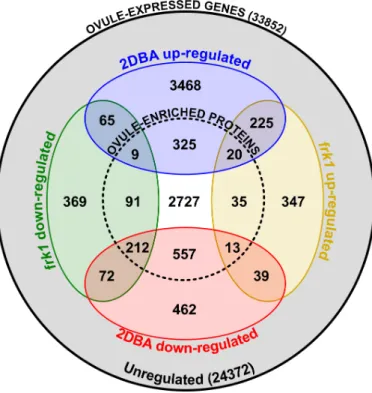
Figure 2.2 Euler diagram showing overlaps between gene sets differentially regulated in frk1, 2DBA ovules and leaf, as compared to anthesis ovules ... 61
Figure 2.3 PT guidance in wild-type and frk1 plants ... 64
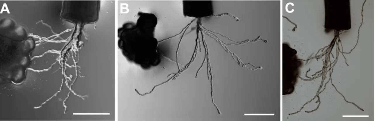
Figure 2.4 Chemotropism of S. chacoense PTs toward (A) S. chacoense ovule clusters (B) S. tarijense ovule clusters and (C) S. gandarillasii ovule clusters ... 66
Figure 2.5 Classification of ovule-enriched CRP subgroups in ovule transcriptome of S. chacoense and their orthologs in S. gandarillasii and S. tarijensi ... 67
Figure 2.6 Glycoprotein fraction from total ovule extracts attracts PTs in the two-choice assay system ... 72
Figure 3.1 Schematic depiction of PT growth in the S. chacoense pistil ... 89
Figure 3.2 Chemotropism of PTs toward (A) anthesis ovule clusters and (B) immature ovule clusters (2DBA) in S. chacoense ... 96
Figure 3.3 Tissue-free GEM system workflow for ovule secretome isolation ... 98
Figure 3.4 Ovule exudates’ purity assessment ... 99
Figure 3.5 Diagram illustrating the number of oOSPs and nOSPs in the ovule secretome as compared to GSPs obtained from PlantSecKB database and SEPs collected from lily, olive, and tobacco stigma exudates ... 102
Figure 3.7 Fold-change correlations between protein secretion abundance and gene expression
during the 2DBA to anthesis transition ... 105
Figure 4.1 RT-PCR validation of 18 candidate genes ... 127
Figure 4.2 Protein purification of ScCRP5.1 by FPLC ... 129
Figure 4.3 Validation of candidate gene expression in N. benthamiana leaves ... 131
Figure 4.4 Chemotropism of semi in vivo grown PTs toward ovule exudates collected from flowers at anthesis stage in S. chacoense ... 133
Figure 4.5 Purified candidates did not induce chemotactic behavior of semi in vivo grown PTs in the bead assay ... 134
List of Abbreviations
2DBA: two days before anthesis AGL: agamous-likeAGP: arabinogalactan protein
BCIP: 5-bromo-4-chloro-3-indolyl-phosphate BME: β‑mercaptoethanol
CCG: central cell guidance CDS: coding sequence
CNGC: cyclic nucleotide-gated ion channel COA: coatlicue
CPM: counts per million CRP: cysteine-rich peptide
cTPI: cytosolic triosephosphate isomerase CV: coefficient of variation
DEFL: defensin-like
DIF1: determinant infertile1
DUF784: domain of unknown function 784 EC1: egg cell 1
ECA1: early culture abundant 1
EDTA: ethylenediaminetetraacetic acid ES: embryo sac
ESF1: embryo surrounding factor 1 EST: expressed sequence tag FDR: false discovery rate FG: female gametophyte
FPLC: fast protein liquid chromatography FRK1: fertilization related kinase 1 FW: fresh weight
GABA-T: γ-amino butyric acid transaminase Gnd·HCl: guanidine hydrochloride
GO: gene ontology
GSP: generally secreted protein HAP: hours after pollination HRP: horseradish peroxidase HSP: heat shock protein IFR: isoflavone reductase
IPTG: isopropyl β-D-1-thiogalactopyranoside KS: Kolmogorov-Smirnov
LC: liquid chromatography LTP: lipid transfer protein
MAPKKK/MEKK: mitogen-activated protein kinase kinase kinase MMC: megaspore mother cell
MS: mass spectrometry NBT: nitro-blue tetrazolium ORF: open reading frame
OSP: ovule-secreted protein; nOSP: non-specific OSP; oOSP: ovule-specific OSP PELP III: class III pistil-specific extensin-like protein
PlantSecKB: plant secretome and subcellular proteome knowledgeBase PM: Pollen Mitosis
PR: pathogenesis-related PSK: phytosulfokine
PT: pollen tube
RALF: rapid alkalinization factor RNase: ribonuclease
ROS: reactive oxygen species
SCA: stigma/stylar cysteine-rich adhesion SCR: S-locus cysteine-rich ligand
SEM: scanning electron microscopy SEP: stigma exudate protein
siRNA: small interfering RNA SP: signal peptide
SPL/NZZ: sporocyteless/nozzle SRK: S-locus receptor kinase
tf-GEM: tissue-free gravity-extraction method TPST: tyrosylprotein transferase
TTS: transmitting-tissue specific UTR: untranslated regions
VIC: vacuum-infiltration centrifugation Zm: zea mays
Acknowledgements
First and foremost I would like to thank my dearest friends in IRBV to make me feel “one of us” and to feel that I belong here!
Heartfelt thanks to my Ph. D. supervisor, Daniel, who encouraged and supported me to go on trainings, meetings and exchange programs during my study, who invited Dr. Zahra Agharbaoui to train me for transient expression when I was stuck with that experiment, and who was dedicated to help me through difficulties in life and allowed me to grow up in this lab.
Sincere thanks to David for his positive comments on my very first class in Canada and showed me how to do research. Sincere thanks to dear Mario, who brought me THE magic chocolate to ease my tension during predoc exam!
I would like to thank my fellow Mattoneurs Audrey, Caroline, Fangwen, Valentin, Rachid for all your help in and out of the lab. I enjoy every moment spent with Audrey and Caro, for our chitchat in the cubical! I thank Fangwen, who spared no efforts in helping me through protein purification. I thank Valentin to collaborate with me on the two articles and nicely provided such a helping hand. I appreciate the company of Mathieu, Jonathan and Steve. I thank them for dropping by our lab and sharing the simple joys from every small progress during my PhD. I especially thank Mathieu for reading through my thesis and giving me so many comments! I also thank Jonathan for his corrections on the Chapter IV of my thesis. Equally important, I would like to thank Afsaneh, who took me as her family, and thank you for your precious coffee time! I also want to acknowledge all who helped me with my experiments, including Minako, who corrected my course application, Firas and Youssef, who gave suggestion for the experiments on pollen tubes, and Louise, who helped me so cheerfully with SEM imaging.
I would like to thank China Scholarship Council to have this excellent program to support me this far to Canada, with the reference 2007220003.
I would like to, finally, thank my beloved parents for tolerating my absence during my study and thank my husband for accompanying me here so I’m not alone!
1. Introduction
1.1 Plant Reproduction
Plants reproduce both asexually and sexually. In asexual reproduction mode, offspring arise from a single organism, without the fusion of gametes. Plants reproduced in this way are genetically identical to their “mother”. One type of asexual reproduction is vegetative reproduction, where offspring develop from a part of the parental tissues. Such case can be seen in tulips, where tulip bulbs are re-used year after year; or in commercial strawberries, which are reproduced by stolons or “clippings” from specific parts of the plants, which develop roots independently and create a new individual. Another type of asexual reproduction involves apomictic seed formation (Koltunow 1993), without the combination of genetic materials from both parents, as seen in dandelion reproduction. Asexual reproduction offers great advantages for farmers to grow plants of superior economical features rapidly. However, pathogens can also be introduced in asexually reproducing plants due to their lower genotypic diversity and consequently a weaker potential for rapid adaptation in case of environmental changes or pathogen attacks. In contrast, sexual reproduction requires the fusion of the male and female gametes to generate offspring genetically different from both parents, an important feature for species adaptation to new environment in plant evolution. In sexually reproducing angiosperms, the life cycles alternate between a diploid sporophyte generation and a haploid gametophyte generation, which is realized by double fertilization.
Our laboratory studies plant sexual reproduction, using a solanaceous model species, the wild diploid potato species Solanum chacoense. It is closely related to agronomically important species such as Solanum tuberosum (potato), Solanum lycopersicum (tomato), Solanum capsicum (pepper), Solanum melongena (eggplant) and Nicotiana tabacum (tobacco). To date, the potato, tomato, pepper and Solanum pennellii (wild tomato) genomes have been reported (Potato Genome Consortium 2011, Tomato Genome Consortium 2012, Kim, Park et al. 2014, Bolger, Scossa et al. 2014). Expressed sequence tag (EST) library from various other solanaceous species are also available from the Sol Genomics Network (Fernandez-Pozo, Menda et al. 2015) and the Potato Genomics
Resource Database known as the Spud DB (Hirsch, Hamilton et al. 2014). These databases facilitate the search of S. chacoense orthologs across several different species and thus make it possible to study species-specific interactions in reproductive processes. This introduction will take us on the courtship journey of the pollen and the pistil, with a special focus on the topic of pollen tube (PT) guidance, which involves different maternal tissues of the pistil in order to elicit chemotropic behavior of the PTs. Because of the stage-specific guidance cues along the PT path, the PTs can: 1) travel down the stigma, 2) navigate through the intercellular space of the transmitting tract, 3) emerge from the transmitting cells, 4) migrate up the funiculus and 5) target the micropyle of the ovule, with great precision (Palanivelu and Tsukamoto 2012).
At the end, PTs deliver the two sperm cells to the embryo sac (ES) to accomplish double fertilization, where one sperm cell fuses with the egg cell to form a zygote that develops into an embryo and the other sperm cell fuses with the central cell to form a triploid nucleus that will later develops into the endosperm (Hamamura, Nagahara et al. 2012). As the essence of this key process in flowering plants, the development of both female and male gametophyte will be described first.
1.1.1 Female Gametophyte Development
An ovule starts as a finger-like protrusion (nucellus) from the placenta during stage 9 of floral development (Smyth, Bowman et al. 1990). The hypodermal cell at the tip of the nucellus differentiates into a megaspore mother cell (MMC). A small RNA pathway ensures the initiation of a unique MMC at this stage through ARGONAUTE 9 (AGO 9). AGO protein is known to bind to microRNAs or small-interfering RNAs (siRNAs) and direct the cleavage of endogenous mRNA (Baumberger and Baulcombe 2005). It is expressed in the companion somatic cells of the female gametes, which restricts the specification of additional MMC in a non-cell-autonomous manner (Olmedo-Monfil, Duran-Figueroa et al. 2010).
As shown in Figure 1.1, the MMC undergoes meiosis to give rise to four haploid megaspores, where only one survives as the functional megaspore and develops into the
female gametophyte (FG). Here the functional megaspore undergoes three rounds of mitosis, resulting in a syncytial ES containing eight nuclei without cellularisation. This stage is followed by nuclei positioning and cell specification, in which the phytohormone auxin is believed to play a crucial role (Pagnussat, Alandete-Saez et al. 2009, Lituiev, Krohn et al. 2013).
Figure 1.1 Synchronized development of ovule and the FG in Arabidopsis. A. The development of an ovule initiates from the protrusion of the nucellus from the placental tissue. The integuments initiate concomitantly with megasporogenesis at stage 11. As megagametogenesis progresses, the integuments elongate and continue to enclose the developing FG during mid- and late-stage 12. At stage 13, the flower is at anthesis stage. The integuments almost completely enclose the mature ES, leaving only a trail for PT entry, the micropyle. B. FG development. During megasporogenesis, a MMC undergoes two successive meiotic divisions to form a tetrad, of which only one megaspore persists as the functional megaspore (FG1). Then it undergoes three rounds of mitosis to form syncytial ES containing eight-nuclei (FG5), followed by nucleus positioning and cell differentiation (FG6). At maturity (FG7), the ES contains two synergids, one egg cell and one central cell, whereas three antipodal cells located at the chalazal end of the ES have undergone degeneration (adapted from (Chevalier, Loubert-Hudon et al. 2011)).
Recently, the role of auxin in ES cell positioning and differentiation has been highly debated. While the Sundaresan lab (Davis, California) observed an auxin gradient along the micropyle-chalaza axis in the ES of Arabidopsis, with the highest concentration at the
micropylar end to specify the cell fate of each gamete (Pagnussat, Alandete-Saez et al. 2009), the Grossniklaus lab (Zurich, Switzerland) has suggested otherwise (Lituiev, Krohn et al. 2013). Their mathematical modeling indicates only shallow auxin gradients can be maintained in the FG, which is not sufficient to establish proper auxin patterning. This model is supported by microscopic observation of auxin activity in confined area of the nucellar tissues adjacent to the ES, with the highest concentration close to the micropylar end. This observation was made in both maize and Arabidopsis, using the same auxin-sensitive reporter DR5 (Ottenschlager, Wolff et al. 2003), and in addition, a novel degron-based reporter system, which exploits the auxin-dependent degeneration of Aux/IAA proteins by monitoring degron-GFP levels in the ovule (Lituiev, Krohn et al. 2013). Despite the discrepancies of the localization of an auxin gradient, these two groups consistently suggest an important role for auxin in cell fate specification, either through an intrinsic auxin gradient in the ES (Pagnussat, Alandete-Saez et al. 2009) or via an exterior polarized auxin activity from micropyle (highest) to chalaza (lowest) in the nucellar tissues in a non-cell-autonomous way (Lituiev, Krohn et al. 2013).
Following cell fate specification induced by an increasing auxin concentration along the micropyle-chalaza axis of the ovule, cellularised gametes acquire their own identity to form two synergid cells guarding the entrance of the ES, an egg cell, two polar nuclei that are about to fuse into a central cell and three antipodal cells at the chalazal end.
In S. chacoense, antipodal cells degenerate at maturity, while they may persist during fertilization and early endosperm development in Arabidopsis as recently observed (Song, Yuan et al. 2014), or alternatively proliferate into a cluster of cells in maize or other grasses. A mature FG consists of only three cell types in S. chacoense, two synergids, an egg and a central cell displayed along the micropyle-chalaza axis (Yang, Shi et al. 2010, Drews and Koltunow 2011).
The synergids are largely involved in guiding PTs toward the micropyle, as demonstrated by the identification of synergid-expressed transcription factor MYB98 involved in filiform apparatus formation and PT guidance in Arabidopsis (Kasahara, Portereiko et al. 2005), and by the identification of synergid-expressed LURE-type peptides as micropylar
chemoattractants in Torenia fournieri (Okuda, Tsutsui et al. 2009, Kanaoka, Kawano et al. 2011) and Arabidopsis (Takeuchi and Higashiyama 2012).
The egg cell is flanked by two synergid cells and is located at the end of the micropyle in the ES. It receives one sperm cell to form the embryo during fertilization. The second fertilization is conducted by the central cell, which receives another sperm cell, leading to endosperm development. However, the function of the central cell is not restricted to fertilization and initiation of endosperm alone (Liu, Yan et al. 2010). Anatomically, the central cell occupies most of the volume of the ES. It can communicate with adjacent cells (synergids, egg, and antipodals), as facilitated by the discontinuous cell wall structure at the junction of the central cell and other cell types. This makes the central cell a signaling hub of the ES, as evidenced by its ability to influence PT attraction, probably via interaction with the synergids as seen in micropylar guidance mutant ccg (central cell guidance) (Chen, Li et al. 2007), and its ability to determine the lifespan of adjacent antipodal cells via central cell-expressed SYCO gene in Arabidopsis (Kagi, Baumann et al. 2010).
The development and function of the antipodal cells remains unclear. Since auxin signaling is present in the antipodal cells in maize (Lituiev, Krohn et al. 2013), a positive correlation has been recently established between auxin signaling and antipodal proliferation (Chettoor and Evans 2015), one more leap forward toward unveiling the role of the mysterious “accessory cells”.
Concurrent with the FG development, the integuments differentiate from L1 cell layer at the chalazal end of the primordium (Figure 1.1). Different from S. chacoense, the Arabidopsis ovule has also an outer integument, which arises from the L2/L3 cell layers (Chevalier, Batoux et al. 2005). The integuments grow and engulf the nucellus, thus separating the funiculus (a stalk that connects the ovule to the placental tissues) from the ES. Finally, when the integuments meet up at the other side of the ES, they form an opening (a path), i.e. the micropyle, for PT entry during double fertilization.
1.1.2 Male Gametophyte Development
The male gametophyte (pollen grain) forms in the anther. Male gametophyte development includes two phases, microsporogenesis and microgametogenesis, as reviewed recently (Hafidh, Fíla et al. 2015).
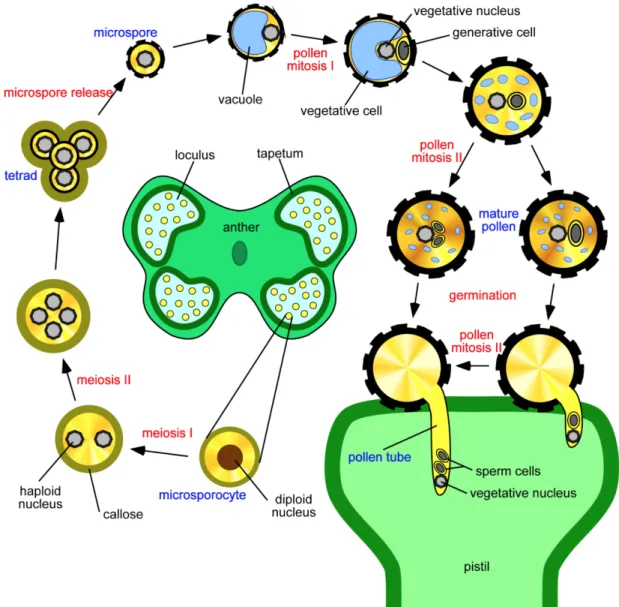
During microsporogenesis, a pollen microspore mother cell undergoes two meiotic divisions to produce haploid microspores in tetrads, separated by callose walls (Figure 1.2). Then distinct microspores are released. As microgametogenesis initiates, a vacuole develops and pushes the microspore nucleus against the cell wall (Yamamoto, Nishimura et al. 2003). The microspore then undergoes Pollen Mitosis I (PMI), producing a larger vegetative nucleus (haploid) and a smaller generative nucleus (haploid). Later the small generative nucleus is engulfed by the vegetative cell membrane to form a “cell-within-a-cell” structure. Finally, the vegetative cell will develop into a tube cell. The generative cell will undergo Pollen Mitosis II (PMII) to give rise to twin sperms. In Arabidopsis, PMII takes place before pollen grains leave the anther (Sanders, Bui et al. 1999), while in the case of S. chacoense, PMII takes place during PT germination (Boavida, Becker et al. 2005, O'Brien, Gray-Mitsumune et al. 2007).
Figure 1.2 Illustration of pollen development in Arabidopsis. The pollen of flowering plants is produced within the anther. Pollen development comprises two successive phases, microsporogenesis and microgametogenesis. Microsporogenesis initiates when the diploid microspore mother cell undergoes two meiotic divisions to give rise to four microspores tethered by the callose wall (β-1, 3-glucan). The callose then separates the tetrad into individual microspores. In each microspore, the outer pollen wall exine develops. During microgametogenesis, a vacuole forms that takes a large volume of the cytosol. By pushing the nucleus to a peripheral position, the pollen undergoes asymmetric cell division (mitosis I), producing a larger vegetative nucleus and a smaller generative cell. The former develops into a tube cell while the latter undergoes PMII to produce two sperm cells. In Arabidopsis, tricellular pollen is shed, while in S. chacoense, the bicellular pollen is shed and PMII takes place during PT germination. This picture is adapted from laboratory of pollen biology website (http://lbp.ueb.cas.cz/field_res.htm).
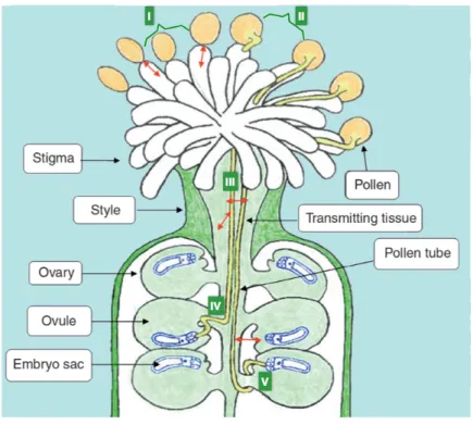
1.2 The PT Path In the Pistil of S. chacoense
In general, the PT path is partitioned into five distinct phases prior to the double fertilization (Figure 1.3) (Swanson, Edlund et al. 2004).
Figure 1.3 The PT pathway in Arabidopsis. In phase I, pollen grain adheres to and hydrates on the stigma. In phase II, pollen grain germinates and penetrates the papilla cell wall of the stigma. In phase III, PT enters the transmitting tract of the style. In phase IV, PT exits the transmitting tract and approaches the ovules. In phase V, PT targets the micropyle of the ovule for fertilization (adapted from (Feijó 2010)).
In phase I, S. chacoense pollen grains adhere to and hydrate on the wet stigma. Pollen hydration depends on the ability to sense a water gradient at the site of the contact zone between the pollen and the stigma, which is conferred through a lipid matrix derived from either the pollen coat or the stigma, which later determines the polarity of PT germination (Lush, Spurck et al. 2000). The hydrated pollen grain then extends a PT towards the aqueous interface.
In phase II, germinating PTs penetrate the papilla cell wall of the stigma. Once in the solid style of S. chacoense, PTs enter phase III, where they navigate through the
intercellular matrix of the transmitting tract. Growing through the style is considered vital for PTs to acquire the competency to respond to guidance cues emitted from the FG to accomplish fertilization (Palanivelu and Preuss 2006).
Okuda and colleagues (Okuda, Suzuki et al. 2013) reported that only when a PT elongates in the style for a sufficient length and for a sufficient time, can it acquire the ability to react to and be attracted to LURE attractants secreted from the synergid cells of the ovule in Torenia fournieri. In this experiment, Torenia PTs not only have to travel through 15 mm of a cut style (out of a total length of ~18 mm), they also need to grow an additional 7 h on the medium after exiting the cut end of the style to acquire this competency. Consistently, Qin and colleagues (Qin, Leydon et al. 2009) demonstrated that PT penetration of the stigma and style elicits a distinct gene expression profile, as opposed to that in pollen grains or in vitro grown PTs. In this comparison, the functional categories that associated with signal transduction, PT growth and transcription were over-represented in semi in vivo PTs (grow through the style), which have provided the molecular basis for this competency control.
Interestingly, a parallel can be drawn between PT-transmitting tract cell-interactions and spermatozoa-oviduct interactions in mammals, where in both cases the male gametes need to travel through a female reproductive tract to become capacitated. Studies conducted in human and mice showed that only 10% of the sperm populations are chemotactically responsive to the female reproductive organ (Eisenbach and Giojalas 2006). Moreover, the gene expression profile of the epithelial cells of the oviduct changes significantly upon spermatozoa arrival (Holt and Fazeli 2010), which is quite similar to the observation made in plants during PT elongation in phase III (Qin, Leydon et al. 2009).
In phase IV, the PTs exit the transmitting tract from the ovarian end of the style and start approaching the ovules. In S. chacoense, PT path splits along the ovary septa and merges with the placental epithelium, as described previously in the related solanaceous species Solanum lycopersicum (Webb and Williams 1988) and Nicotiana alata (Cornish, Pettitt
et al. 1987). At the final stage (stage V), PTs target the micropylar opening of each ovule to accomplish double fertilization (Hamamura, Nagahara et al. 2012).
However, between PTs entry to the ES and the onset of double fertilization, PTs go through two sequential stages, i.e. growth arrest and pollen discharge. First, PT growth arrest is achieved via interaction with the filiform apparatus-localized receptor-like kinase, feronia (fer) in the ovule (Escobar-Restrepo, Huck et al. 2007). Acting in the fer-mediated PT reception pathway, two endoplasmic reticulum-localized proteins TURAN and EVAN that are expressed both in pollen grains and ovules, mediate PT reception as their mutants displayed similar PT overgrowth phenotype reminiscent of the fer phenotype (Lindner, Kessler et al. 2015). Despite the fact that TURAN and EVAN were shown to affect pollen development and PT integrity, the fer-phenotype is traced back to a defect in the FG since no mutant PTs were formed in reciprocal crosses with wild-type plants (Lindner, Kessler et al. 2015). TURAN and EVAN encode proteins involved in the N-glycosylation pathway, suggestive of a “dual recognition system” in PT reception, where both the protein backbone and the glycosyl residues on the surface of the male and FG are required to be recognized for successful PT reception.
Following PT growth arrest, fer-dependent accumulation of reactive oxygen species (ROS) at the entrance of the FG induces PT rupture (Duan, Kita et al. 2014), where PTs discharge their content to fuse with the egg cell and the central cell.
Live cell imaging of double fertilization reveals three distinct sperm cell behavioral steps during PT discharge in Arabidopsis. Initially, the PT bursts to deliver two sperm cells to the ES within seconds. Sperm cells remain in a stationary phase for minutes before the final gamete fusion for double fertilization. Interestingly, the two sperm cells are functionally equivalent to fertilize the central cell or the egg cell (Hamamura, Saito et al. 2011).
In maize, PT discharge is mediated by ZmES4 (Zea mays embryo sac 4), a defensin-like (DEFL) CRP derived from the synergid cells, via the opening of potassium channel on the PTs. Fusion of the two female gametes with the sperm cells is found mediated by EC1 (egg cell 1) peptides. At this moment, PTs have finished their journey in the pistil.
Embryogenesis is initiated to switch the life cycle of a plant into the sporophyte generation.
1.3 Why Studying PT Guidance In S. chacoense?
1.3.1 The Significance of PT Guidance In Plant Reproduction
In general, PT guidance occurs all along the PT path in the pistil, which allows PTs to travel down the stigma, exit the transmitting tract of the style, and enter the micropyle to achieve double fertilization. These path finding events require intimate cell signaling between the PTs and various female tissues, including stigmatic papilla cells, stylar transmitting tract cells, nucellar cells and finally the female gametes (Palanivelu and Tsukamoto 2012), which makes PT guidance an excellent model to study cell-cell communication, and which will allow different guidance mechanisms to be revealed. Taking another view, a main goal of plant sexual reproduction, especially for crop plants, is to overcome breeding barriers so that the gene pool of one species can be expanded, allowing biodiversity and “new” species to be raised. Breeding barriers can be derived from a large number of prezygotic and/or postzygotic isolation. By studying gametophytic PT guidance, Márton and coworkers (Marton, Fastner et al. 2012) used genetic engineering to express maize micropylar guidance attractant ZmEA1 in the synergid cells of Arabidopsis thaliana. The proper secretion of ZmEA1 peptide enabled Arabidopsis ovules to guide maize PTs to enter the micropylar opening of the ovule, which is the first step to overcome the reproductive barrier between unrelated plant families, at the level of gametophytic guidance. The barrier induced by PT guidance represents only a small part of complex, multi-mechanism breeding barriers in plant. Yet, by studying PT guidance, an increased hybrid vigor arising from crossbreeding of much divergent families or species can be anticipated.
1.3.2 PT Guidance In S. chacoense and Species-Specific Pollen−Pistil
Interactions
PT guidance has been extensively studied in the model species Arabidopsis and maize, from which the genome had been annotated. Alternatively, Torenia (Linderniaceae) was
chosen as a model for its ovule anatomy since its ES protrudes from the ovule, enabling live cell imaging of PT attraction to the synergid cells directly under light microscopy (Higashiyama, Kuroiwa et al. 1998), and the destruction of individual protruding cells by laser cell ablation (Higashiyama, Yabe et al. 2001). This anatomical advantage also allowed cDNA sequencing from individually isolated synergid cells, thus providing the molecular basis for dissecting the complex network of PT–ovule interaction (Okuda, Tsutsui et al. 2009).
In this project, S. chacoense was chosen as our model species in the study of pollen−pistil interaction and PT guidance. Solanum chacoense belongs to the Solanaceae family, one of the most important families to human beings for its great value as food sources (potato and tomato), drugs (tobacco) and ornamentals (petunia) (Knapp, Bohs et al. 2004). Solanum chacoense is native to South America and is one of the most widely distributed wild potato species. It occurs sympatrically with various other Solanum species, thus allowing large opportunities for natural hybridization. However, the number of hybrids found so far is still limited, compared to the total number of species in this family (3000-4000 species for the Solanaceae, almost half for genus Solanum) (Knapp, Bohs et al. 2004). We thus hypothesize that species-specific attracting molecules may be produced by different solanaceous species to direct PT guidance. Such species-specific molecules could lead to reproductive isolation for sympatrically localized Solanum species.
In fact, such species-specific interactions are common in pollen–pistil interactions (Swanson, Edlund et al. 2004). For example, pollen adhesion to the stigma in Arabidopsis (dry stigma) depends on the exine wall component sporopollenin, a highly stable mixed polymer containing long-chain fatty acids and phenolics (Thom, Grote et al. 1998). Detergent wash assays demonstrated that Arabidopsis pollen barely binds to the stigma of monocot plants, while they exhibit significant binding to some dicot plants from various genera, such as Arabidopsis, Brassica, Platanus, Ambrosia, etc. More specifically, Arabidopsis pollens bind to an Arabidopsis stigma with greater affinity than to its close relative Brassica campestris (Zinkl, Zwiebel et al. 1999), suggestive of a species-specificity in pollen-stigma interaction according to their taxonomic distance. Different functional groups may exist in sporopollenin polymers from different plant taxa,
providing the basis for this selective pollen–pistil interaction (Domínguez, Mercado et al. 1999).
Likewise, studies on the self-incompatibility mechanism during early pollen–pistil interaction revealed that different families have adopted different mechanisms to reject self-pollen. In the Solanaceae, Scrophulariaceae and Rosaceae, the pistil produces S-RNases to degrade self-PT RNA in order to stop self-PT in the style (McClure and Franklin-Tong 2006). In Papaveraceae, the S-haplotype specific recognition between stigmatic PrsS (Papaver rhoeas stigma S) (Foote, Ride et al. 1994) and the pollen expressed PrpS (Papaver rhoeas pollen S) (Wheeler, de Graaf et al. 2009) triggers a Ca2+ increase and downstream responses to reject self-pollen. Different from the above-mentioned mechanisms where pollen rejection is determined by the haploid pollen haplotype, pollen rejection in Brassicaceae is determined by the diploid anther S-haplotype. In the Brassicaceae, the male determinant is S-locus cysteine-rich ligand (SCR/SP11) (Takayama, Shiba et al. 2000) and the female determinant is S-locus receptor kinase (SRK) (Stein, Howlett et al. 1991, Goring and Rothstein 1992). The two determinants co-evolve rapidly and are highly polymorphic between species (Boggs, Dwyer et al. 2009).
Along the PT path in the pistil, species-specificity can be found in various other phases, such as in PT guidance, reception and discharge. First, species-specific PT attraction has been demonstrated in Torenia (Kanaoka, Kawano et al. 2011), Arabidopsis (Takeuchi and Higashiyama 2012) and maize (Marton, Fastner et al. 2012), where the chemoattractants displayed a much compromised attraction efficiency toward PTs of a different species. Second, during PT reception, interspecific crosses using pollens from Arabidopsis lyrata and Cardamine flexuosa on Arabidopsis thaliana stigmas resulted in a PT overgrowth phenotype, emphasizing the importance of species-specific recognition in PT reception (Escobar-Restrepo, Huck et al. 2007). Third, in PT discharge, ZmES4, a defensin like (DEFL) protein, mediates PT burst in a species-specific manner in maize (Amien, Kliwer et al. 2010).
So far, several guidance cues have been uncovered from both the sporophytic (stigma, style, ovule integuments) and the gametophytic tissues of the pistil to guide directional PT growth in Arabidopsis, lily, maize and Torenia. However, PT guidance in solanaceous species is largely unknown. By using S. chacoense as our model species, and taking advantage of nine other Solanum species currently grown in the greenhouse, including S. bulbocastanum, S. commersonii, S. microdontum, S. pinnatisectum, S. tuberosum, S. tarijense, S. gandarillasii, S. boliviense, and S. megistacrolobum, we expect to study these species-specific signaling molecules involved in PT guidance and explore their possible involvement in creating a breeding barrier and thus in driving their speciation and evolution.
1.4 PT Guidance In the Style Is Governed By Sporophytic
Tissues
Before reaching the ES to undergo double fertilization, PTs experience numerous intimate contacts with the pistil sporophytic tissues, including the stigma, the transmitting tract and its extracellular matrix in the style, as well as the nucellus and integuments in the ovary. This takes place through dynamic vesicle exocytosis and endocytosis and intensive signaling at the clear zone (the extreme tip of the tube) (Zonia and Munnik 2008). The sporophytic tissues synthesize and secrete molecules, which not only promote a robust tube growth, but also assist the PTs in growth directionality from the stigma straight to the ovarian end of the pistil. This stage is termed sporophytic guidance. So far, chemotactic proteins involved in sporophytic guidance have been identified from the stigma, style and ovary, respectively.
1.4.1 Sporophytic Guidance Down the Stigma
In lily, stigma exudates attract in vitro-grown PTs, as shown below (Figure 1.4). By use of biochemical methods including cation exchange, gel filtration and HPLC, plantacyanin (9.9 kDa), a member of the blue copper protein family, was identified from the active fraction and named chemocyanin. In vitro bioassay indicates that chemocyanin induces PT chemotropism (Kim, Mollet et al. 2003). This attraction is enhanced by the presence
of two other molecules, including a 9.0 kDa SCA (stigma/stylar cysteine-rich adhesion) (Park, Jauh et al. 2000) and a larger stylar pectic polysaccharide (Mollet, Park et al. 2000).
Positively charged SCA interacts with negatively charged pectin in the transmitting tract of the pistil to create an active extracellular matrix for the PT to attach. SCA is a lipid transfer-like protein secreted from both the pollen and the transmitting tract tissues. It contains eight cysteine residues (Park, Jauh et al. 2000) and is part of the DEFL group. Therefore, chemocyanin is general considered to be involved in adhesion-mediated PT guidance.
Figure 1.4 in vitro PT attraction assay showing PT reorientation over time (A and B) and corresponding quantitation method (C) in lily. A, PT chemotropism was induced after application of stigma exudates (1.5 µg/µl) in the well 40 min before time zero. At this time point, individual pollen grains were placed around the wells. Germinated PTs were manually oriented away from the well before the time course imaging. B, no chemotropism was induced by water control in the well. C, determination of a positive chemotropism marked by PT reorientation of at least 90°. Bar = 2 mm. (Adapted from (Kim, Mollet et al. 2003)).
In Arabidopsis, only one gene encodes plantacyanin, which shares 86.8% sequence similarity to lily chemocyanin at the amino acid level (Kim, Mollet et al. 2003). This cell wall protein is secreted to the extracellular matrix of the transmitting tract and is found in a gradient of increasing concentration from the stigma to the ovary and the mature ES (Dong, Kim et al. 2005). Over-expression of plantacyanin caused wild-type PTs to dwell on the stigma and to turn around without penetrating the papillar cells, as shown in Figure 1.5. An increased plantacyanin concentration was detected in the stigma of overexpression lines, leading to a disrupted PT guidance. The role of Arabidopsis plantacyanin reinforced the chemotropic effect of lily chemocyanin and also confirmed the contribution of sporophytic factors to PT guidance.
Figure 1.5 Scanning Electron Microscopy (SEM) images of wild-type Arabidopsis pollens on (A) wild-type stigmas and (B) plantacyanin over-expression stigmas OXP12 Dotted line indicates the trajectory of PT path after it penetrates the papilla cell wall. The arrow in B indicates that the PT made many turns on the papilla cell, displaying aberrant tube growth on the stigma. P, papilla cell; Po, pollen grain. Bar = 20 µm. (Adapted from (Dong, Kim et al. 2005)).
1.4.2 Sporophytic Guidance From Stigma to Ovary
The transmitting tissue-specific (TTS) glycoprotein, was identified in the extracellular matrix of the style in tobacco (Wang, Wu et al. 1993). TTS was shown to stimulate PT growth in vitro, while transgenic plants expressing significantly lower levels of TTS mRNA and protein exhibited a reduced PT growth rate. Besides its role as a growth stimulant, TTS was also shown to attract PTs in a semi in vivo assay (Cheung, Wang et al. 1995). TTS belongs to the large arabinogalactan protein (AGP) family. The glycosylation status of TTS is required for PT guidance in the style. PTs that grew through the style were attracted by native TTS but not by a chemically deglycosylated version (Cheung, Wang et al. 1995). In the pistil, TTS works as a vehicle that carries sugar moieties, adheres to PT surface, becomes incorporated into the PT cell wall and finally deglycosylated by the PTs. The glycosylation level of TTS increases along the style, with lower levels of glycosylation in the stigma and higher level in the lower part of the pistil, as indicated by the molecular weight of TTS on a SDS-PAGE gel (Wu, Wang et al. 1995). Concurrently, the increasing level of TTS glycosylation is also associated with an increasing level of TTS protein acidity, which may contribute to PT guidance from the stigma to the ovary.
Characterization of tobacco TTS homolog in Nicotiana alata (NaTTS) revealed that they share similar features as a PT stimulant and attractant (Wu, Wong et al. 2000). In addition, NaTTS proteins are part of a protein family, with various degrees of sugar modification on the protein backbone. This variability significantly affects the function of TTS since purified low-molecular weight NaTTS proteins did not show any activity in stimulating PT growth or in PT guidance (Sommer-Knudsen, Lush et al. 1998).
1.4.3 Sporophytic Guidance To the Micropylar Region
Gamma-amino butyric acid (γ-amino butyric acid, GABA) is a signaling factor that plays critical roles in neural development of mammals (Pontes, Zhang et al. 2013). In plants, GABA stimulates PT growth (Palanivelu, Brass et al. 2003). A proper amount of GABA stimulates tube growth in vitro, while higher concentrations are inhibitory. GABA levels are regulated by POP2 (pollen pistil interaction 2) in Arabidopsis, which encodes a
transaminase that degrades GABA. By examination of the abundance of GABA in the pistil, an increasing GABA gradient was detected from the stigma to the inner integument, with the highest GABA level in the micropylar region of the ovule. This is likely due to limiting amount of POP2 proteins in high GABA cells, since anti-POP2 antibodies showed staining complementary to the GABA localization in the ovule. Since the GABA gradient coincides with the direction of PT growth, GABA has been considered as a sporophytic factor that guides PTs toward the micropyle region of the ovule.
POP2 is ubiquitously expressed in both the PT and the pistil, and interestingly, only when both were knocked down could a guidance defect be detected. Such defects included PT growth arrest or random growth on the surface of the ovule (Palanivelu, Brass et al. 2003). The pop2 mutant displays PT elongation defects in the transmitting tract (Renault, El Amrani et al. 2011), which is likely to be associated with modulation of putative Ca2+-permeable channels on the plasma membrane of the PTs by GABA, as shown in tobacco (Yu, Zou et al. 2014).
To sum up, when PTs navigate through the pistil to approach the ES, they are guided by different attracting molecules. On the stigma, PT directional growth is influenced by chemocyanin, a small basic protein in the plant cell wall that belongs to the phytocyanin family of blue copper proteins (Kim, Mollet et al. 2003). Next, when PTs have travelled further into the style, guidance is taken over by TTS, a heavily glycosylated AGP, which becomes deglycosylated as PTs elongate toward the ovarian end of the style (Cheung, Wang et al. 1995, Wu, Wang et al. 1995). Meanwhile, a non-protein amino acid, GABA, may be involved in guiding the PTs toward the micropylar region of the ovule, where highest GABA levels are detected.
The distinct nature of different guidance cues isolated so far suggests that different guidance mechanisms may act. Along the PT path, a new mechanism is likely to take over when PTs migrate into its effective range. With the presence of each new guidance cue, PTs are likely to break free from the previous attraction and be precisely guided under the new influence, until they achieve double fertilization.
1.5 PT Guidance In the Ovary Is Controlled By Both
Gametophytic and Sporophytic Tissues
Before PTs exit the bottom-end of the style, sporophytic stylar tissues navigate all the PTs toward the ovary. Once inside the ovary, a precise one-to-one guidance is achieved between the PT and the ovule, a stage described as ovular PT guidance (Higashiyama and Takeuchi 2015). Multi-steps control checkpoints are known to act in ovular guidance, as described in Figure 1.6.
Figure 1.6 Schematic depiction of PT growth in S. chacoense pistil. A. Longitudinal section of the pistil. Dark green indicates the PT pathway, a continuous cell tract originating from the stigma surface, extending through the style down to the placental epithelium, where it connects to the ovules. At the mature stage of an ovule, the ES consists of an egg cell, two synergid cells and a central cell, embedded in one layer of integument in S. chacoense. B. Close-up of an ovary transverse section. PT typically approaches the micropyle of an ovule through three guidance steps. Step 1 (long-distance guidance), PT emerges from the intercellular space of the transmitting tract to the placental surface; step 2 (funicular guidance), the PT climbs up the funiculus; and step 3 (micropylar guidance), PT navigates toward and into the micropyle. Noticeably, the funiculus in S. chacoense has a much shorter stalk compared to Arabidopsis, which largely reduces the distance between the micropyle and the placental surface, possibly rendering PTs that emerge onto the placental epidermis immediately competent for micropylar guidance (Liu, Joly et al. 2015).
1.5.1 Long-Distance Guidance Is Governed by the Ovule
A long-distance guidance mechanism was first suggested years ago to describe ovular signals that direct PTs to emerge from the intercellular space of the transmitting tract cells toward the ovules, preferentially at the upper part of the ovary (Hulskamp, Schneitz et al. 1995). It was observed that in wild-type Arabidopsis ovules, 40% of the PTs emerged from the transmitting tract at the position of the first ovule. In the bel1 and 54D12 mutants (Ray, Robinson-Beers et al. 1994), which are impaired in ES development, this preferentiality is compromised, but a slight preference was still observed. However, in the ES-devoid mutants sin1 and 47H4 (Ray, Lang et al. 1996), PT showed equal probability to emerge anywhere along the entire length of the transmitting tract. This shows there is an ovule-derived long-distance guidance (transmitting tract toward ovules), as opposed to micropylar short-distance guidance. Long-distance guidance is supported by the transmitting tract-trapped phenotype observed in the Arabidopsis chx21/chx23 double mutant. CHX21 and CHX23 are redundant cation/proton exchangers expressed in the PT. Mutant chx21/chx23 PTs do not respond to ovular cues and remain in the transmitting tract (Lu, Chanroj et al. 2011).
1.5.2 Funicular Guidance Is Under the Influence of Ovular Sporophytes
The second step involves funicular guidance, where PTs adhere to and migrate up the funiculus, a stalk-like structure that attaches the ovule to the placenta. The source of funicular guidance cues can be derived from the funiculus itself, as seen in single ovule semi in vivo assays, where ovules without a funiculus attracted PTs at an efficiency reduced from 54 to 35% (Palanivelu and Preuss 2006). However, little is known about this signal, due to the difficulty in obtaining sufficient amount of funiculi from the ovules without tissue injury. Alternatively, some evidence suggests that funicular guidance may be associated with ovule sporophytic tissues in general, including the funiculus, the integuments and the nucelluar tissues (Lausser, Kliwer et al. 2010), as observed in the sporophytic mutants pop2 (Palanivelu, Brass et al. 2003), siz1-2 (Ling, Zhang et al. 2012) and pdil2-1 (Wang, Boavida et al. 2008). In these mutants, a portion of PTs loses their way soon after exiting the transmitting tract tissue and does not migrate up the funiculus.