Université de Montréal
Molecular Basis of Native Inward Rectifier Currents:
Role of Kir2 Subunits
par
Gernot Schram
Département de Médecine Faculté de Médecine
Thèse présentée à la Faculté des études supérieures en vue de l’obtention du grade de Philosophiae Doctor (Ph.D.)
en Sciences Biomedicales
Aout 2005
(r•
\L
o
Université
(JI’
de Montréal
Direction des bibliothèques
AVIS
L’auteur a autorisé l’Université de Montréal à reproduire et diffuser, en totalité ou en partie, par quelque moyen que ce soit et sur quelque support que ce soit, et exclusivement à des fins non lucratives d’enseignement et de recherche, des copies de ce mémoire ou de cette thèse.
L’auteur et les coauteurs le cas échéant conservent la propriété du droit d’auteur et des droits moraux qui protègent ce document. Ni la thèse ou le mémoire, ni des extraits substantiels de ce document, ne doivent être imprimés ou autrement reproduits sans l’autorisation de l’auteur.
Afin de se conformer à la Loi canadienne sur la protection des renseignements personnels, quelques formulaires secondaires, coordonnées ou signatures intégrée-s-au--texte ont-pu--être-enLevésde-ce-document.-Bier que cela ait pu affecter la pagination, il n’y a aucun contenu manquant.
NOTICE
The author of this thesis or dissertation has granted a nonexclusive license allowing Université de Montréal to reproduce and publish the document, in part or in whole, and in any format, solely for noncommercial educational and research purposes.
The author and co-authors if applicable retain copyright ownership and moral rights in this document. Neither the whole thesis or dissertation, nor substantial extracts from it, may be printed or otherwise reproduced without the authoc’s permission.
In comptiance with the Canadian Privacy Act some supporting forms, contact information or signatures may have been removed from the document. While this may affect the document page count, it does flot represent any loss of content from the document.
Cette thèse intitulée
Molecular basis of native inward rectifier currents: Role ofKir2 subunits
présentée par Gernot Schram
a été évaluée par un jury composé des personnes suivantes:
Dr. Bruce G. Allen, président-rapporteur Dr. Stanley Nattel, directeur de recherche
Dr. Lucie Parent, membre du jury Dr. Alvin Shrier, examinateur externe
Résumé
Les canaux potassiques à rectification entrante (Kir) sont d’importants régulateurs de l’excitabilité cellulaire. Dans le système nerveux central, ils contrôlent l’excitabilité, participent à la signalisation électrique et à la transmission d’information. Leur dysfonction est associée à l’épilepsie et aux paralysies périodiques. Au niveau cardiaque, le canal potassique à rectification entrante ‘KI
détermine le potentiel de repos membranaire et contribue à la phase 3 du potentiel d’action. Sa dysfonction est responsable d’arythmies chez les sujets atteints du syndrome d’Andersen. Au niveau vasculaire, ‘Ki détermine le potentiel de repos
membranaire, régule l’influx de Ca2 et la signalisation intracellulaire dépendante du
2+ Ca.
Quatre membres de la sous-famille Kir 2 (Kir2.1-4) contribuent au courant ‘K!
endogène. Cependant, la contribution de chacune de ces sous-unités au courant ‘KI
natif est inconnue. Les propriétés biophysiques des sous unités homomériques Kir2 ne reproduisent pas les propriétés du courant ‘KI cardiaque. De plus, l’expression
relative de Kir2 au niveau ARNm et protéique ne permet pas d’expliquer plusieurs des ses caractéristiques.
Par l’utilisation de méthodes électrophysiologiques et biochimiques, nous avons étudié l’association des sous-unités Kir2.4 et Kir2.l et montré qu’elles s’assemblaient pour former un hétérornère Kir2.Ï/Kir2.4 fonctionnel possédant des propriétés distinctes des sous-unités Kir2. 1 ou Kir2.4 exprimées seules. Ce fut une des premières démonstrations de l’assemblage hétérométrique des sous-unités Kir2 et de sa contribution au phénotype du courant potassique à rectification entrante endogène. L’assemblage des sous-unités Kir2.l/Kir2.4 pourrait être physiologiquement important, particulièrement au niveau du cerveau où les deux sous-unités sont co-exprimées.
Nous avons par la suite comparé les propriétés de bloc par le Ba2 des canaux homo- et hétéromères Kir2 exprimés dans des oocytes de Xenopus avec celles du courant ‘K! endogène humain. Nous avons trouvé que le bloc par le Ba2 des
hétéromères Kir2.x/Kir2.y n’était pas la simple résultante de la somme des réponses individuelles des sous-unités et que les propriétés du bloc par le Ba2 de ‘KI étaient
p]us proches de celles des sous-unités co-injectées que de celles des sous unités exprimées individuellement. Ces résultats suggèrent la possibilité qu’une portion de
‘K] endogène puisse résulter de ces assemb]ages hétéromériques.
Des stratégies électrophysiologiques et biochimiques additionnelles ont été utilisées pour déterminer la composition des cellules endothéliales aortiques humaines (CEAHs) en sous-unités Kir2. Nous avons démontré que les CEAHs exprimaient les quatre types de sous-unités Kir2 mais que seules Kir2.2 et Kir2. 1 étaient fonctionnellement importantes, Kir2.2 apparaissant être le déterminant principal de la conduction Kir dans ces cellules.
Nous concluons que les canaux hétéromères Kir2 pourraient représenter un mécanisme important dans la détermination de la diversité des canaux potassiques à rectification entrante fonctionnellement distincts et que l’importance relative des différentes sous-unités Kir 2 pourrait varier selon le tissu.
Mots-clés: Kir2.l, Kir2.2, Kir2.3, Kir2.4, canaux potassiques, ‘KI courants à
rectification entrante, hétéromères, cerveau, coeur, endothélium aolïique, distribution différentielle, canaux ioniques, expression fonctionnelle
111
Abstract
Inwai-d rectifier potassium (Kir) channels are important regulators of cellular excitability. Central neiwous system Kir channels participate in electrical excitability, electrical signal]ing and information processing. Their dysfunction is associated with epilepsy and paralysis. Cardiac inward rectifier current Ii sets the resting membrane potential and contributes to phase 3 cardiac repolarization. Dysfunction of cardiac ‘KI
causes arrhythmias in Andersen’s syndrome. Vascular inward rectifier currents determine the resting membrane potential, regulating Ca2 influx and Ca2 dependent intracellular signalling. Four members of the Kir2 subfarnily (Kir2.1-4) contribute to the background inward rectifier ‘Ki. Their relative contribution to native Kir culTents
is unknown. Homomeric Kir2 subunit assembly fails to reproduce cardiac ‘KI and the
relative expression of Kir2 mRNA and protein cannot explain a variety of ‘KI
features. Using electrophysiological and biochemical rnethods, we studied co assembly of Kir2.4 and Kir2.1, finding that Kir2.1 and Kir2.4 co-assemble to form functional Kir2.l/Kir2.4 heteromers with properties distinct from homomeric channels formed by either subunit aJonc. This was one of the first demonstrations that heteromeric Kir2 subunit assembly can occur and contribute to physiological inward rectifier phenotype. Kir2.l/2.4 assembly miglit be physiologically important in regions of the brain, where both subunits are co-expressed.
We then compared properties of Ba2 block of homo- and heteromeric Kir2 channels in Xenopus oocytes to human cardiac ‘KJ. We found that Ba2 block of
heteromeric Kir2.x/Kir2.y subunits is not the simple sum of individual subunit responses and that Ba2 blocking properties of ‘KI are more sirnilar to co-injected
subunits than to each subunit expressed alone. These results suggest that a substantial proportion of native‘K! may resuit from Kir2 heteromultirner formation.
Complementary electrophysiological and biochemical strategies were used to determine Kir2-subunit composition of human aortic endothelial ceils (HAECs). We found that HAECs express alI four types of Kir2 subunits, but that only Kir2.2 and Kir2.1 are functionally relevant, with Kir2.2 appearing to be the primary determinant of Kir conductance.
We conclude that heteromeric Kir2 channels may represent an important mechanism to achieve diversity of functionally distinct inward rectifier channels and that the relative importance of different Kir2 subunits varies among tissues.
Keywords: Kir2.1, Kir2.2, Kir2.3, Kir2.4, potassium channels, ‘KI, inward rectifier
current, heterornultimer, brain, heart, aortic endothelium, differential distribution, ion channels, functional expression
V
Table of contents
Résumé î Abstract iii Table of contents V List of tab]es xvList of figures xvi
List of acronyms and abbreviations xix
Acknowledgements xxvii
Contributions of authors xxx
Related studies not included in this thesis xxxii
PART ONE: INTRODUCTION AND REVIEW 0f THE
LITERATURE 1
Chapter I: Introduction to Cellular Electrophysiology 2
I-1 The Action Potential in Neurons 7
I-2 The Cardiac Action Potential 12
I-3 Electrogenesis in Vascular Endothelial Ceils 20
Chapter II: Introduction to Potassium Channels 25
Chapter IiI: Structure-function Relationship of Potassium Channels 32
III-1 Introduction 33
III-2 K Channel Structure is Modular 34
111-2.1 The Ion-Conduction Pore 34
111-2.2.1 RCK-Dornains of Ligand-Gated
r
Channels .39 111-2.2.2 The Voltage Sensor: Coupling Electrical Workto Channel Gating 40
111-2.2.2.1 Voltage-Sensor Structure: KvAP and
Kvl.2 40
111-2.2.2.2 How do Gating Charges cross the
Membrane9 44
111-2.2.2.3 Mechanical Coupling of Voltage
Sensor Movements to the Pore 45 111-2.2.3 The TI Domain ofVoltage-Gated K Channels 46 111-2.2.3.1 Crystal Structure 46
111-2.2.3.2 Functional Role 46
111-2.2.4 The Cytoplasmic Pore of Inward Rectifier
Channels 47
111-2.2.4.1 Structure 47
111-2.2.4.2 Potential Functional Role 48 III-3 Structural Basis of Ion Channel Function 54
111-3.1 Basis of Ion Permeation 54
111-3.2 Mechanisrn of High Conduction Rate 57 111-3.3 Current Concepts of Ion Selectivity 61
III-3 .4 K Channel Gating 63
111-3.4.1 StructuraI Changes during Activation and
Deactivation 63
III-3 .4.2 Filter distortion might underlie C-type
vii
Chapter IV: Voltage-Gated Potassium Channels .69
W-1 The Delayed Rectifier Current ‘K 70
IV-1.1 Biophysical Properties 70
W-l.2 Pharrnacology 71
IV-1 .3 Functional relevance of‘K 72
IV-1.3.1 RoleofCardiaclK 72
W-1.3.2 Role of IK-like Currents in Endothelial Celis 74
W-1.4 Molecular Basis of‘K 74
IV-1.4.1 MinK and KvLQT1 underlie ‘Ks 74
W-l.4.2 HERG and MiRP: The Molecular Basis of‘Ki7 75
IV-1.4.3 Molecular Basis of Physiological Function in
the Heart 76
W-1.4.4 Role of IK-like Currents in the Central Nervous
System 76
W-2 The Transient Outward Current ‘to 78
W-2.1 Pharmacology and Biophysical Properties 78 W-2.2 Functional Role of A-type CulTents 79
W-2.2.1 Role of Cardiac 79
W-2.2.2 Role of A-Type Currents in Endothelial Celis 80
IV-2.3 Molecular Basis of A-type currents 81
W-2.3.1 Transmembrane Alpha-subunits 81
W-2.3.2 Auxiliary Subunits 82
IV-2.3.2.1 Kv13-Subunits 82
W-2.3.2.2 K channel interacting Protein (KCh1P) 83 IV-2.3.2.3 K channel Associated Peptide (KChAP) 83 IV-2.3.2.4 MinK Related Peptide (MiRP) 84
IV-2.3.3 Molecular Basis of Cardiac‘10 Heterogeneity.$4 W-2.3.4 Molecular Basis of A-Type Currents in the CNS $5
Chapter V: Inward Rectifier Potassium K- (Kir-) Channels 91
V-1 Introduction 92
V-2 The Cardiac Inward Recifier Current‘KI 95
V-2. 1 Biophysical Properties 95
V-2.2 Pharrnacology 97
V-2.2. I Relevance and Models of Barium block 97 V-2.3 Functional Role 0fcardiac‘Ki 100
V-2.3 Clinical Significance of‘KI Dysfunction 100
V-2.4 MolecularBasis of‘KI 103
V-2.4. 1 Regional Heterogeneity of Cardiac Kir2 Subunit
Expression 103
V-2.4.2 Functional Role of Kir2 Subunits in the Heart 106 V-2.4.3 Potential Role of Kir2 Subunits in the Brain 10$ V-2.4.3 Role of Kir2 Currents in Endothelial Ceils 114 V-2.4.4 Barium block of Kir2 channels 117
V-2.4.4. 1 Molecular determinants of Kir2 Ba2
block 119
V-3 The G-protein activated cardiac inward rectifier‘KACh 12$
V-3.1 The G-protein Signalling Pathway 12$ V-3.2 Direct Activation of‘KACh by fry-subunits 130
V-3.3 Biophysical Properties 131
V-3 .4 Pharmacological Properties 132
V-3.5 Role of Cardiac‘KACh 132
V-3.6 Role of1KACIi in Disease 133
ix
V-3.7.Ï Rote ofKir3 Subunits in the Heart .134 V-3.7.2 Role of Kir3 Subunits in the CNS 135 V-4 The ATP-sensitive Inward Rectifier Cuiient ‘KATP 138
V-4. I Biophysical and Pharmacological properties 139 V-4.2 Functional Ro]e ofKATP channels 139
V-4.2. 1 Regulation of Pancreatic Insulin Secretion 139
V-4.2.2 Role of Cardiac ‘KATP 140
V-4.2.3 Role ofKATPin the CNS 141
V-4.2.4 Role of VascularlKATp 141
V-4.3 Molecular Basis ofKATP channels 142
V-4.3.1 Molecular Basis ofKATP channel assemb]y 142
V-4.3.2 MolecularBasis of KATpchannel Regulation 144 V-4.3.3 Molecular Basis ofKATPchannel PharmacoÏogy 145
V-4.3.4 Molecular Tdentity of KATP 146
V-5 Role of other Inward Rectifier Channels 148
Chapter VI: Potential Role 0f Heteromultimer Formation in K channels 151 VI-1 Structural Determinants of Kv-channel Assernbly 152 VI-2 Physiological Role of Kv-channel Heterotetramers 153 VI-3 Structural Determinants of Kir Channel Assembly 153 VI-4 Potential Physiological Significance of Heteromeric Kir Channel
Complexes 154
PART TWO: ORIGINAL CONTRIBUTIONS .160
Chapter VIII: Kir2.4 and Kir2.1 K channel sub units co-assemble: A
potential new contributor to inward rectifier current heterogeneity 161
VIII-1 Abstract VIII-2 Introduction VIII-3 Methods VIII-3. 1 163 164 166 Construction of Dominant Negative (dn) Kir2.4 and
Kir2.1 Constructs 166
Co-Precipitation ofKir2.1 and Kir2.4 166
Construction of Kir2.4-FLAG 167
Cos-7 Ceil Maintenance and Transfection 167
His-Pulldown 16$
Western Blotting 16$
Constmction of Kir2. 1 -Kir2.4 Tandem 169
Electrophysiology 171
Data analysis 171
172 Co-expression of Kir2. 1 or 2.4 Channels with
Dominant Negative Subunits 172
Kir2.1-His6 I Kir2.4-Flag Co-Precipitation 172 Properties of Co-Expressed Kir2. 1 and Kir2.4 and of a
CovalentÏy linked Kir2.Ï/2.4 Tandem Construct 174 177 Heteromultimer Formation in Inward Rectifier Channels ....177
Novelty and Potential Significance I $0
Potential Limitations 181 183 VIII-3 .2 VIII-3.3 VIII-3 .4 VIII-3.5 VIII-3 .6 VIII-3.7 VIII-3.$ VIII-3 .9 VIII-4 Resuits VIII-4. I VIII-4.2 VIII-4.3 VIII-5 Discussion VIII-5. 1 VIII-5.2 VI 11-5.3 VIII-6 References
xi
VIII-7 Acknowledgments.190
VIII-8 Figures and Figure Legends 191
Chapter IX: Barium block of Kir2 and human cardiac inward rectifier currents: Evidence for subunit-heteromeric contribution to native
currents 199 IX-1 Abstract IX-2 Introduction IX-3 Methods IX-3.1 201 202 203 Functional Expression of Cloned Inward Rectifier
Subunits in Xenopus Oocytes 203
IX-3.2 Ceil Isolation 204
IX-3.3 Whole-Cell Patch-Clamp Recordings 204
IX-3.4 Statistical Analysis 205
IX-4 Results 206
IX-4. 1 Concentration and Time Dependence of Ba2 Block of
Homomeric Kir2 Subunits and Cardiac‘K 206
IX-4.2 Concentration and Time Dependence of Ba2tBlock of
Co-Injected Kir2 Subunits and Cardiac‘K! 208
IX-4.3 Comparison of Ba2 Blocking Properties of Homomeric
and Co-Injected Kir2 Subunits with Cardiac‘K! 20$
IX-5 Discussion 210
IX-5.1 Importance and MolecularBasis of‘K! 210 IX-5.2 Potential Role of Kir2 Heteromultimers 211
IX-5.3 Potential Limitations 212
IX-6 Conclusions 213
IX-7 Acknowledgements 214
IX-8 References 215
IX-10 Figures and Figure Legends .219
Chapter X: Functional expression of Kir2.x in human aortic endothelial
ceils: The dominant role of Kir2.2 229
X-l Abstract 231
X-2 Introduction 232
X-3 Materials and methods 234
X-3.1 Celi Culture 234
X-3.2 Electrophysiology 234
X-3.3 RNA Isolation and RT-PCR 235
X-3.4 Quantitative Real-Time PCR (QRT-PCR) 235
X-3.5 Immunoblotting 236
X-3.6 Construction and functional Assessment of Dominant
Negative (dn) Kir2.x Constructs 237
X-3.7 Transfection 237
X-3.$ Data Analysis 237
X-4 Resuits 239
X-4. I Membrane Conductance in HAECs is dominated by
strong Inwardly-Rectifying K Current 239
X-4.2 HAEC Kir2.x mRNA Expression 240
X-4.3 Relative Abundance of HAEC Kir 2.x mRNA by
quantitative Real Time PCR 240
X-4.4 Kir2.x Protein Expression in HAECs 241
X-4.5 Sensitivity of endothelial liwardly-Rectifying K CuiTent
to Ba2 Block and to pH 242
X-4.6 Distribution of Kir Unitary Conductance 243 X-4.7 Inhibition of endogenous Inwardly-Rectifying K
Current by dn-Kir2.x 243
xlii
X-5.1 Consideration of the System.245
X-5.2 Molecular Diversity of Kir2-based Native Currents 246 X-5.3 Potential Significance of our Findings 24$
X-6 Acknowledgements 249
X-7 References 250
X-$ Tables 256
X-9 Figures and Figure legends 257
PART III: GENERAL DISCUSSION AND CONCLUSION 272
Chapter XI: Discussion of resuits 273
XI-1 Major Novel Contributions and Significance in Context of Literature ...274 XI- 1.1 Kir2. 1 and Kir2.4 forrnHeteromeric Channels with
Distinct Properties 275
XI-1.1.1 Primary Novelty and Resuits Reported in this
Study 275
XI- 1.1.2 Discussion of Resuits in Context of the
Literature 275
XI-l.l.3 Potential Scientific Significance ofResults
Reported 277
XI-1.2 Comparison of Barium Block of Cardiac ‘K! with Homo
and Heteromeric Kir2 Channel Complexes 27$ XI-1.2.1 Primary Novelty and Resuits RepoiÏed in this
Study 278
XI-1.2.2 Potential Significance of Resuits in Context of
the Literature 279
XI-1.3 Kir2 Channels in HAECs 281
XI-1.3.1 Primary Novelty and Results Reported in this
XI-1.3.2 Potential Significance of Resuits in Context of
the Literature 282
XI-2 Potential Limitations 282
XI-3 Future Directions 285
XI-4 Conclusions 28$
xv
List
of Tables
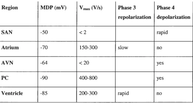
Table I: Actionpotential characteristics in different cardiac regions 1$
Table II: Potassium channel genes, ancillary subunits and tissue distribution 27 Table III: Sumrnary of voltage-gated channels and resulting currents 90
Table IV: Inward recitifier single channel conductances in different tissues
and species 97
Table V: Relative concentration of Kir2 rnRNA in human atrium and
ventricle 105
Table VI: Relative concentration of Kir2.1 and Kir2.3 protein in canine
atrium and ventricle 105
Table VII: Distribution of individual Kir2 subunits in different brain regions. 110
Table VIII: Effect of amino acid point mutations on Kir2.1 Ba2 block 122
Table IX: Ba2 sensitivity of Kir2.1-4 cloned from various tissues and
species 127
Table X: Potency and tirne-course of Ba2 block of various constructs 21$
Table XI: Molecular sequence and expected length of RT-PCR products for the different human Kir2.1, Kir2.2, Kir2.3, and Kir2.4 primers 256
List of Figures
Figure 1: Schematic diagram of action potential recordings from the squid
giantaxon $
Figure 2: Action potential of the squid axon and underlying ionic
conductances 10
Figure 3: Schernatic diagram of AP properties in different regions of the
heart 12
Figure 4: Representative cardiac action potential waveforms 14 Figure 5: AP properties of canine ventricular epicardium, rnidmyocardium
and endocardium 19
Figure 6: Two rnechanisrns of Ca2 entry into endothelial celis 24
Figure 7: Phylogenetic tree of Kv (6TM) channels 30 Figure 8: Phylogenetic tree of Kir (2TM) channels 31 Figure 9: Phylogenetic tree of K2P (4TM) channels 31 Figure 10: Ribbon presentation of the KcsA Kt channel 3$ Figure 11: Pore and voltage sensors of the Kvl.2 channel 43 Figure 12: Sequence alignrnent and size-exciusion chrornatograms of Kir3. 1,
Kir2.1, and KirBacl.1 with secondary structure elernents noted 51 Figure 13: Overall architecture of inwardly rectifying K (Kir) channels 53 Figure 14: Permeant ion binding sites in the KcsA K channel 55 Figure 15: Ion occupancy in the selectivity filte 60 Figure 16: Gating in the selectivity filter of the KcsA K Channel 67 Figure 17: lnward rectification of steady state current in a Staifish egg 93 Figure 18: Inward rectifier K current (‘K) in hurnan atrial and ventricular
myocytes 96
Figure 19: Visualization of barium in the KcsA K channel by x-ray crystallography and in a Ca2tactivated K channel by analysis of
xvii
Figure 20: ‘K urrent-voltage relationship and action potential in the
ventricular celi 102
Figure 21: Quantification of Kir2 mRNA in hurnan atrium and ventricle 104 Figure 22: AP phenotype is determined by‘K] density 107
Figure 23: Current-voltage relationships in non-stiinulated bovine endothelial
ceils 116
Figure 24: Ba2 block of Kir2. 1 118
figure 25: Comparison of Kir2.1-4 amino acid sequences 123 Figure 26: Schematic representation of the action of RGS protein 129 Figure 27: Effects of dn-Kir2.1 on Kir2.1 and Kir2.4 currents in Xenopus
oocytes 191
figure 2$: Effects of dn-Kir2.4 on Kir2.1 and Kir2.4 currents in Xenopus
oocytes 192
Figure 29: Western blots of celi lysates from Cos-7 ceils transfected with either Kir2.1-His6, Kir2.4-Flag or both 193 Figure 30: Ba2 block of Kir2.1, Kir2.4, Kir2.1-Kir2.4 tandem and co-injected
Kir2.1 and Kir2.4, original recordings 194 Figure 31: Ba2 block of Kir2.1, Kir2.4, Kir2.1-Kir2.4 tandem and co-injected
Kir2.1 and Kir2.4, mean ± s.e.m. current-voltage relations 195 Figure 32: Comparison of Ba2 block of Kir2. 1, Kir2.4, Kir2. 1 -Kir2.4 tandem
and co-expressed Kir2.1 and Kir2.4. Dose-response curves and
IC5OTFP 197
figure 33: Ba2 block of Kir2.1, Kir.2.2, Kir2.3 and cardiac ‘K]. original
current recordings 219
figure 34: Ba block of Kir2.1, Kir.2.2, Kir2.3 and cardiac ‘K], Mean±s.e.m.
current-voltage relations 220
Figure 35: Ba2 block of Kir2.x/Kir2.y heterorners and cardiac ‘KI original
Figure 36: Ba2 block of Kir2.x/Kir2.y heterorners and cardiac ‘K1
mean±s.e.m current-voltage relations 222
Figure 37: Kinetics of Ba2 block of Kir2.Ï, Kir.2.2, Kir2.3 and cardiac ‘K! 223
Figure 38: Kinetics of Ba2 block of Kir2.x/Kir2.y heterorners and cardiac‘K]. 225
Figure 39: Comparison of rnean±s.e.m. concentration-response curves based on end-pulse block at each concentration at a test potential of -120
mV 227
Figure 40: Basic properties of the inward rectifier K current (IK) in human
aortic endothelial ceils (HAECs) 257
Figure 41: Comparison of‘K in HAECs and in freshly isolated or 10w-passage
cultured porcine aortic endothelial ceils (PAECs) 259 Figure 42: RT-PCR analysis of mRNA encoding different types of Kir2
channel subunits in HAECs 260
Figure 43: Quantification of Kir2.x rnRNA level in HAECs using quantitative
real-time PCR (QRT-PCR) 261
Figure 44: Protein expression of Kir2.x in HAECs 263 Figure 45: Ba2 block and pH sensitivity of the‘K in HAECs 265
Figure 46: Distribution of single-channel conductances of endogenous Kir
channels in HAECs 267
Figure 47: Suppression of Kir2.x cttrrent by dorninant-negative (dn-) Kir2.x
constructs 269
xix
List of acronyms and abbreviatfons
°C: Degrees Celsius
[B a2J: Internai (cytosolic) B a2 concentration [Ba2J0: External (extraceliular) Ba2 concentration [KJ: K concentration
[K]0: Extracellular K concentration 2TM: two transmembrane domains 4TM: four transmembrane domains 4-AP: 4-Aminopyridine
5-HTIA: 5-hydroxytryptamine lA
6TM: six transmembrane domains Al receptor: Adenosine receptor A: Atrium
Â:
Angstriirn AA: Amino acidAAA: Sequence of three alanine residues; used to replace the GYG motif in the selectivity filter to create a dominant negative channel subunit
ABC: ATP-binding cassette (protein superfamiiy) ACh: Acetylchoiine
ADP: Adenosine diphosphate AF: Atriai fibrillation
Ag: Silver ion
AGS: Activators of G-protein signaliing AP: Action potentiai
APD: Action potential duration
ARNm: Acide ribonucléotique méssager ATP: Adenosine triphosphate
AV: Atrioventricular
AVN: Atrioventricular node Ba2: Barium
BAEC: Bovine artery endothelial celi
BCE: Bovine corneal endothelial ceil BK channel: Ca2 activated K channel Ca2: Calcium
CaM: Calmodulin ‘+ CCE: Capacitative Ca entry
cDNA: Complementary Deoxyribonucleic acid cRNA: Complementary Ribonucleic acid CCPA: 2-chloro N6-cyclopentyl adenosine Cd2: Cadmium
CEAHs: Céllules endothéliales aortiques humaines CHO ceils: Chinese hamster ovary ceils
Cf: Chloride cm: Centimetre
CNG channel: Cyclic nucleotide-gated channel CNS: Central nervous system
Co : Cobalt
COS ceils: Monkey celi lines derived from the CV-1 celI une by transformation with a replication origin defective mutant of SV4O VIRUS, which codes for wïld type large T antigen
CPPX: Dipeptidyl-peptidase-like protein CRAC: Ca2-release activated Ca2 channel Cs: Caesium
CTX: Charybdotoxin
D2 receptor: Dopamine receptor DAD: delayed after depolarization DAG: 1,2-diacylglycerol
DMEM: Dulbecco’s Modifled Eagle Medium
DDS: 4,4’-Diisothiocyanostilbene-2,2’-Disulfonic Acid Dn: Dominant negative
Dn-Kir: Dominant negative Kir2 subunit DNA: Deoxyribonucleic acid
DTX: Dendrotoxin E. cou: Escherichia cou
xxi
EA 1: Episodic ataxia type 1 EAD: Early after depolarization Eag: ether-à-go-go
EC: Endothelial celi ECG: Electrocardiogram
EDHF: Endothelium-derived hyperpolarizing factor
EGTA: ethylene glycol bis(2-aminoethyl ether)-N,N,N’N’-tetraacetic acid EGM-2: Endothelium growth medium-2
EM: Membrane potential
ENa: Sodium equilibrium potential Endo: Endocardium
Epi: Epicardium
ER: Endoplasmatic reticulum ERG: ether-related gene
ERP: Effective refractory period
EPR: Electron paramagnetic resonance spectroscopy EK: Equilibrium potential for potassium
FBS: Fetal bovine serum
FEP: Free energy perturbation (computations)
Flag sequence: ei ght amino-acid sequence (Asp-Tyr-Lys-Asp-Asp-Asp-Asp-Lys) GABAB: ‘y-aminobutyric acid B
GAP: GTPase accelerating protein GDP: Guanosine diphosphate GFP: Green fluorescent protein gK: Potassium conductance gNA: Sodium conductance
GPCR: G-protein coupled receptor GST: Gluthatione S-transferase GTP: Guanosine triphosphate
H5-segment: putative pore forming segment HAEC: Hurnan aortic endothelial celi HCEC: 1-lurnan capillary endothelial ceil HEK: Human embryonic kidney ceils
HCN: Hyperpolarization activated cyciic nucleotide-gated channel HEPES: 4-(2-hydroxyethyl)- 1 -piperazineethanesulfonic acid HERG: Human ether-related gene
HUGO: Human Genome Organization
HUVEC: Human umbilicai vein endothelial celi 1C50: Haif-maximai inhibitory concentration
‘Ca, L: L-type calcium current
‘CA, T: T-type calcium current
if: Funny cunent, aka pacemaker current
‘K: Inward rectifier current.
‘K: Deiayed rectifier potassium cuiTent.
JKACh: Acetyl choline-acti vated potassium current.
IKATP:ATP-sensitive potassium current
IKNDP: Vascular smooth muscle ccli ATP-dependent K-current
‘Kr: Delayed rectifier potassium cuiTent, rapid component ‘Ks:Delayed rectifier potassium current, slow component
jKur. Ultrarapid delayed rectifier current
jNa Inward sodium current IPC: Iscliemic preconditioning IPE celis: Iris pigment epitheliai ceils IsK: Synonym for minK
TUPHAR: International Union of Pharmacoiogy
ito: Transient outward current
h,f: Transient outward current with fast inactivation kinetics ‘to.: Transient outward current with slow inactivation kinetics
1P3: Inositol-1,4,5-trisphosphate K: Potassium
KAh: Acetyicholine-activated K channel
KATP: ATP inhibited K channel
KChIP2: Potassium Channei Interacting Protein 2 KCI: Potassium chloride
KCO: K channel openers
xxiii
Kd: Dissociation constant kDa: Kilo Dalton
KG-channel: G-protein activated K channel Kir: Inward rectifier potassium channel KirBac: Eukaryotic Kir channel
KNDP: Vascular smooth muscle cdl ATP dependent K channel
KO: Knockout
Kv: Voltage dependent potassium channel
KvAP: Voltage-gated potassium channel from Aeropyrum pernix KvLQTI: Pore forming subunit of the ‘Ks channel
LA: Left atrium
LB medium: Luria-Beiïani medium LC: Locus coeruleus
LC-CoA: Long chain CoA
LQT syndrome: Long QT syndrome LV: Left ventricle
mA: Milli ampère
M-cell: Midmyocardial celi
Ml-2 segments: Transmembrane segments one and two of 2TM K channels M2 receptor: Mu scarinic receptor
MD: Molecular dynamïcs
MDP: Maximum diastolic potential mM: Millimol
MinK: Minimal potassium channel MiRP1: MinK Related Peptide 1 mRNA: Messenger Ribonucleic acid rns: Milliseconds
mS: Millisiemens
MthK: Calcium-gated K channel from Methanobacterium thermoautotrophicuin
mV: Milli volts
MW: Molecular weight Na: Sodium
NBF: Nucleotide binding fold NCX: Na/Ca2 exchanger NDP: Nucleoside diphosphate NO: Nitric oxide
NOS: Nitric oxide synthase
NSC: Non-selective cation channel
P-region: Channel region containing putative pore forming segment, synonym to H5 segment
P2X4: Purinergic Ïigand-gated receptor channel complex PAEC: Porcine aortic endothelial celi
PAF: Platelet activating factor PC: Purkinje celis
PCR: Polymerase Chain Reaction
PECAM-1: EC-specific antiplatelet-endothelial celi adhesion molecule 1 PGI2: Prostacyclin PW2: Phosphatidylinositol-4,5-bisphosphate PIP3: Phosphatidyl-3,4,5-trisphosphate PKA: Phosphokinase A PLC: Phospholipase C PMSF: Phenylmethylsulphonylfluoride
PSD-95: Postsynaptic density protein of 95 kDa PTX: Pertussis toxin
QRT-PCR: Quantitative real-time PCR
QTc interval: Corrected QT interval in the ECG
r-eag: Rat ether-à-go-go RA: Right atrium
RACC: Receptor activated cation channel Rb: Rubidium ion
RCK: Regulator of Ktconductance
RIPA buffer: Modified radioimmunoprecipitation buffer RGS (protein): Regulators of G-protein signalling (protein) RMP: Resting membrane potential
xxv
RPE celis: Retinai pigmentai epitheiial ceiis RT: Reverse Transcription
RT-PCR: Reverse transcriptase-pol yrnerase chain reaction RV: Right ventricle
Si-6 segment: Transmembrane segments 1-6 of 6TM K channels SAN: Sinoatriai node
S.E.M.: Standard error of the mean Slo: Large conductance K channel SNr: Substantia nigra pars reticulata SOC: Store operated channel
Sr2: Strontium ion
SUR: Sulfonylurea receptor
TI domain: Amino terminal assembiy domain
T 1/2: Time for 50% of steady-state time-dependent block TASK: Twik-related acid-sensitive K channel
TdP: Torsade de pointes TEA: Tetra ethyl ammonium
TFPI: Tissue factor pathway inhibitor TM: Transmembrane segment
TMD: Transmembrane dornain TP: Testpotential
tPA: Tissue plasminogen activator TREK: Twik-related K channel
TRPC: Transient receptor potential channei
TRPM: Transient receptor potential channel of the melastatin famiiy TVGYG: K channel key sequence
TWIK: Tandem of p domains in a weak inward rectifier K channel V: Ventricle
V1/2: Voltage at which half maximal activation or inactivation occurs
VF: Ventricular fibrillation
Vmax: Maximum phase O upstroke slope of the action potential VRAC: Voiume regulated anion channei
vWf: von Willebrand factor WT: Wild type
xxvii
Acknowledgements
1 would like to thank Dr. Stanley Nattel for his exceptional support during my time in his laboratory. I greatly appreciate lis scientific excellence as well as his qualities as a hurnan being. Without lis help the achievement of many goals would have taken mudli longer if flot been impossible.
Amongst the staff at the Montreal Heart Institute, I am particulary indebted to Dr. Bruce Allen, Dr. Zhiguo Wang and Dr. Terence Hebert, who have substantially contributed to my success in the laboratory. Dr. Bruce Allen lias provided indispensable help for my experiments involving western blots of Cavl.2 channels. He lias been of great help as the president of my PhD jury and lis meticulous work on the manuscript helped to eliminate many mistakes. Dr. Wang bas introduced me to molecular biology and the two-electrode voltage-clamp technique. His recording setup lias always been available for me. Dr. Hebert has always been supportive in solving any kind of scientific problems, theoretical or practical.
Dr Lucie Parent lias helped me tremdendously with scientific discussions regarding experiments on the structural determinants of Ba2 block of Kir2.4 dhannels. Her participation in the committee of my pre-doctoral examination started my interest in structure-function related researdli which lias neyer ceased since. Her thourougli reading of the first manuscript of this thesis resulted in countless changes that have greatly contributed to improving its quality.
When I started my researdli activities in Dr. Nattel’s laboratory, Nathalie Ethier and Dr. Manjula Weerapura guided me through the many pitfalls of molecular biology, the handling of Xenopus oocytes and the two-electrode voltage clamp technique. I will neyer forget their essential help in times when I needed it the most. In this context I would also like to thank Dr. Hans Rindt and Kathy Schreiber, who provided both professional and personal support in the beginning.
A very special thank you goes to Maya Mamarbadhi. Maya lias neyer hesitated to help me. She was aiways approachable when I lad questions. Whenever I
ran out of reagents, she would lend them to me. She was always there to lend me a hand when the deadiine for an experirnent approached too quickly.
During my time in Dr. Nattel’s laboratory, I saw many people corne and go. Only few of them became friends. I would like to thank Dr. Marc Pourrier for lis exceptional lielp during ail these years, both professionally and in personal life. Working with him lias aiways been a pleasure owing to bis exceptional reliability and great sense of humour. His lielp witli the Frencli version of the abstract of the final version of the manuscript was indispensable. Dr. Stephen Ziclia lias spent considerable effort in helping me with western blot experiments. 11e provided substantial help in a project that unfortunately neyer rnatured to publication. Dr. Joachim Ehrlich spent many late niglits with me in the laboratory, recording HERG and minKJKvLQT1 currents. 11e became a dear friend who unfortunately had to return to Germany after the completion of bis research fellowship. Danici Herrera shares my passion for structure-function relationships of K channels. It is due to lis efforts that my final project can be continued and bas finally supplied us with exciting resuits. Dr. Ricardo Caballero did flot spend a long time with us in Montreal. I would like to thank him for lis friendship and scientific collaboration.
During the last five years I lad the opportunity to coliaborate with many people who I wouid like to thank for letting me participate in their projects as weÏl as their contribution to mine. In particular, I wish to express my gratitude to Dr. frena Levitan at the University of Pennsyivania and Yun Fang, University of Pennsylvania, for giving me the opportunity to coilaborate on the molecular basis of inward rectifier currents in a biologicai system I wouid have otherwise flot had access to.
No experiment can succeed without proper preparation. Chantai St Cyr bas impressed me with ber exceptional reliability and accuracy. Xiao Fan Yang spent many evenings helping me with the injection of Xenopiis oocytes. Evelyn Landry wouid neyer leave the iaboratory if there was one final request from any of us and prepared anything on the last minute. Chantal Maltais and Nathalie L’Heureux have aiways been heipful.
xxix
No article would have been published and no poster presented without the excellent work of France Thériault, Diane Campeau, Daniela Giuliani, Nadine Vespoli, Luce Bégin and the audiovisual departrnent at the Montreal Heart Institute.
For financial support, I sincerely want to thank the Ernst and Berta Grimmke Foundation, DUsseldorf, Gerrnany who first believed in me and granted me funding for rny research work in Montreal. I also wish to thank the Canadian Institutes of Health Research (CIHR) and Aventis Pharma for fellowship support. During my research, T have received substantial financial support from the University of Montreal, which awarded me a differential scholarship waiver whule I was flot a permanent resident of Canada, a “bourse d’excellence” and two “bourse de redaction”. I feel honoured to have been awarded these scholarships. At the University of Montreal, I would like to thank Dr Daniel Lajeunesse for giving rapid attention to any arising problems during the course ofmy thesis subrnission.
Due to considerable experimental delays of a project that was intended to be included in this thesis, writing had been significantly delayed and finally coincided with the beginning of my residency training in Internai Medicine. I would like to thank Dr Richard Gauthier, Dr Geneviève Grégoire, Dr Guy Lalonde, Madame Monelle Beaulieu, Madame Maryse Campion, Dr Sandra Bernardin, and Dr Douglas Fish for their support and flexibility needed to finish my PhD training.
Last not least, I would like to thank Lena Rivard for ail ber support and patience during the writing of this thesis and ber help with the rnanuscript and the French version of the abstract. She has always been there for me, in bappy times and in times of frustration.
I am sincerely grateful to my parents, who accepted my decision to engage in several more years of financial hardship without any objection. I could have neyer realized this project without their substantial moral and financial support.
Finally, I would like to thank my grandrnother for ber faith and for ber love. This thesis is dedicated to ber memory.
Contributions of authors
1- Schram G, Melnyk P, Pourrïer M, Wang Z, Nattel S, Kir2.4 and Kir2.l K channel subunits co-assemble: A potential new contributor to inward rectifier current heterogeneity, J Physiol, 544 (Pt2): 337 — 349, 2002
I designed the project and techniques to generate the chimeric Kir2.l-2.4 construct and Kir2.4-FLAG. I perforrned most of the molecular biology experirnents (generation of Kir2.1-2.4 concatemer, FLAG-tagging of Kir2.4 and creation of dn Kir2.4). Techniques used include PCR, DNA cloning, mutation and amplification. For heterologous expression in Xenopus oocytes, techniques used were preparation of cRNA, isolation and preparation of Xenopus oocytes, cRNA injection and recording of heteroïogously expressed currents using the two-electrode voltage clarnp technique. Ail electrophysiological experiments were performed exclusively by me. The rnanuscript of the article was written entirely by myseif and revised with the help of Dr. Nattel, who contributed largely to the discussion.
2- Schram G, Pourrier M, Wang Z, Nattel S., Barium block of Kir2 and human cardiac inward rectifier currents: evidence for subunit-heteromeric contribution to native currents, Cardiovascular Research, 59: 328-338, 2003
I designed the study and perforrned ail experimental work inc]uding molecular biology and electrophysiological experiments. Techniques used for oocyte experiments include DNA handiing (DNA amplification using E. cou, restriction enzyme digestions etc.), generation of cRNA, isolation and preparation of Xenopus
oocytes and cRNA injection as well as two-electrode voltage clamp experirnents. For patch clarnp experiments, hurnan tissue was obtained by right ventricular biopsy and single myocytes were prepared using standard enzyme digestion protocols. Currents were recorded using the patch clamp technique. I perforrned ail statistical analysis, wrote the manuscript and prepared ail figures.
xxxi
3- Fang Y., Schram G., Rornanenko V., Shi C., Conti, L., Vandenberg CA., Peter F. Davies PF., Nattel S., Levitan 1., Functional expression of Kir2.x in human aortic endothelial celis: the dominant role of Kir2.2, Ani J PÏiysioÏ CeÏl PhvsioÏ. 289: C1134-1 144, 2005
I was approached by Dr Levitan who asked me to collaborate with ber team on a project evaluating the molecular composition of native inward rectifier K currents in hurnan aortic endothelial ceils (HAECs). I agreed and participated in the development of the experimental approach together with Dr. Levitan, Yun Fang (the first author) and Dr. Nattel. Based on our experience with dominant negative (dn-) Kir2 subunits (Schram et aï., 2002a) and the previous demonstration that these constructs provide a helpful tool to evaluate the role of Kir2 subunits in native Kir current (Zobel et aÏ., 2003) we decided to use a dominant negative approach to determine the functional role of various Kir2 subunits in native HAEC Kir current. I generated dn-Kir2.2, dn-Kir2.3 and dn-Kir2.4 constmcts and subcloned dn-Kir2.1-4 from their respective cloning vectors into the bicistronic mammalian ceIl expression vector pIRES. Techniques used include PCR, DNA cloning, site directed mutagenesis and DNA amplification. To test the functionality of these dominant negative constructs, I co-expressed them with their respective Kir2.x-WT channels in Xenopus oocytes. Techniques applied include cRNA preparation, isolation of Xenopus oocytes, cRNA injection and the two-electrode voltage-clamp technique. I analyzed ah the data obtained in the oocyte experiments and prepared the figure for our publication.
During the course of the project, I participated in the review of data obtained. At the writing stage, I co-wrote sections of the manuscript and participated in its corrections. During the revision process, I pefformed additionah experiments (dn Kir2.x/Kir2.x-WT co-expression), wrote additional sections of the manuscript, added one figure and revised the manuscript.
The dominant negative approach, which was designed by me, was recognized by the reviewers as one of two key experiments in our project to demonstrate the main objective of this article. Due to my extensive contribution to the project, both Dr. Levitan and Yun Fang, the first author, offered me the second author position.
Related studies not included in this thesis
Substantial effort had been invested in a publication reviewing the “Differential distribution of cardiac ion channel expression as a basis for regional specialization in electrical function” (Schram et aï., 2002b) which due to its nature can flot be included in this thesis. This article has therefore been integrated in the introduction to the extent possible.
Due to its thematic restriction, the data presented in this thesis unfortunately under-represents the work done over the course of my PhD. For that reason I would like to mention that at the time of writing I have published four peer reviewed articles in which I am the first author (Schram et aï., 2002a; Schram et aï., 2002b; Schram et ai., 2003; Schram et aÏ., 2004) and one non-peer reviewed article, three articles in which I am the second author (Fang et aï., 2005; Shiroshita-Takeshita et al., 2004; Pourrier et aï., 2003a) (one of which is presented in this thesis) and one article in which I am the last author (Nattel et al., 2001). Important contributions to the work of co-workers in our laboratory resulted in five co-authorship publications (Wang et al., 2001; Han et aï., 2002; Shinagawa et al., 2003; Caballero et aï., 2003; Pourrier et aï., 2004). A last first authorship project examining structural determinants of voltage dependence and sensitivity of Ba2 block of Kir2.1 and Kir2.4 channels delayed the writing of this thesis and finally could not be cornpleted in time due to recurring technical difficulties. This project is now close to publication. Preliminary data has been published in abstract form (Schram et aï., 2005; Herrera et al. 2005; Herrea et al. 2006) and presented at the Annual Meeting of the Biophysical Society in Long Beach, California, 2005, the Meeting of the Canadian Cardiological Association in Montreal, 2005 and the Annual Meeting of the Biophysical Society in Sait Lake City, Utah, 2006. Presentations at scientific reunions of the University of Montreal were awarded prizes.
xxxiii
for inygrandmother
Forin)’ parents
PART ONE:
INTRODUCTION AND REVIEW 0F
THE LITERATURE
CHAPTER I: INTRODUCTION TO
CELLULAR ELECTROPHYSIOLOGY
Practically every process in the living being depends on electrical signalling. Charged particles, ions, cross the ceil membrane through highly specialized transmembraneous passages, so called ion channels. At the end of the I $th century,
Sidney Ringer demonstrated that a frog heart needs to be perfused with a solution containing potassium, sodium and calcium ions in a certain stoichiometry in order to keep on beating. A decade later, the “membrane breakdown” hypothesis of Julius Bernstein (Bernstein, 1902; Bemstein, 1912) suggested that the negative resting membrane potential of nerve and muscle ceils is determined by a membrane selectively permeable to potassium (Kj ions. During excitation, positively charged ions would move into the ceil owing to their higher concentration in the extracellular solution than in the cytoplasm, causing a transient loss of the negative resting membrane potential. 1952, Hodgkin and Huxley showed that membrane currents in the giant squid axon were dependent upon membrane permeability changes to Na and K in a time and voltage dependent manner, implying a feedback mechanism between membrane voltage and transmembraneous ion movement (Hodgkin & Huxley, 1952). Thus, ion channels control the membrane potential and in return their state, open or closed, is controlled by the membrane potential.
During the twentieth century, many cellular roles were discovered for various anions and cations. It is now firrnly established that electrical signalling of nerve and
+ +
muscle cells depends on the transmembraneous movement of Na , K , Ca and Cl ions through highly specialized ion channels. Complex interactions between inward and outward ionic currents result in transient membrane depolarization and subsequent repolarization. The ensemble of these changes in membrane potential is called action potential (AP) and represents the means by which excitable cells transfer information over large distances in short time intervals. A variety of non excitable cells including blood ceils, cells of the immune system, osteoblasts, fibroblasts and vascular endothelial cells, depend on the proper functioning of ion channels in ceil signalling. In vascular endothelial cells (ECs), a complex interplay between many different types of ion channels regulates intracellular Ca2 levels, which is crucial to many physiological functions of ECs (Nilius & Droogmans,
4
2001). However, current knowledge of the precise role of ion channels in various tissues is stiil very limited.
Potassium channels p]ay a key role in regulation of cellular excitability. K channels conduct K ions across the celi membrane down their electrochemical gradient achieving flow rates of 10$ ions per second whule stili maintaining perfect fidelity for K (MacKinnon, 2003). Their function is essential for the regular automatic beating of the heart, proper functioning of the nervous system, memory and rnood, secretion of hormones and enzymes and many other important physiological functions. K channels set the resting membrane potential, keep fast action potentials short, terminate periods of intense activity, time the interspike intervals during repetitive firing, and generally lower the effectiveness of excitatory inputs to a celi when open (Hille, 2001). Potassium channels are targets of rnany antiarrhythmic drugs and are responsible for the pro-arrhythrnic actions of various antiarrhythmic and non-cardiac drugs (Mitcheson et aÏ., 2000). In the heart, regional and transmural differences in K channel expression and density contributes to regional specialization in electrical function (Schram et aï., 2002b). Malfunctions of ion channels have been implicated in the pathogenesis of a growing number of diseases termed “channelopathies” (Abraham et aÏ., 1999; Marban, 2002; Kullmann, 2002). Dysfunctions of Kt channels underlie diseases of the brain such as epilepsy and episodic ataxia. In the heart, Kt channel dysfunction is the cause of various forrns of the genetic and acquired long QT syndrome (LQTÏ, LQT2, LQT5-7) (Splawski et aï., 2000; Plaster et aÏ., 2001) inducing potentially lethal cardiac alThythmias. In Andersen’s syndrome, dysfunction of inward rectifier K channels is associated with cardiac arrhythmias, periodic paralysis and dysmorphic bone features (Plaster et aï., 2001). K channel dysfunction in the ear, the kidney, muscles and the pancreas is associated with deafness, hypertension, myokyrnia and periodic paralysis and hyperinsulinaemic hypoinsulinemia, respectively (Shieli et aÏ., 2000; Bichet et cil., 2003).
A great variety of K currents has been discovered in various tissues and species. Pharmacological and biophysical properties of these currents have been studied with the help of the patch-clamp technique. However, such properties cou]d
characterization of specific K currents difficult. Application of channel blocking compounds, specific ionic constituents of recording solutions and distinct recording protocols were used to minimize potential confounding factors. Nevertheless, it could not be excluded that such procedures also changed the properties of the current under investigation.
In 1987, the first voltage-gated K channel subunit was cloned from the shaker locus of DrosophiÏa melanogaster (Tempel et aï., 1987; Kamb et aï., 1987; Pongs et aï., 198$) and characterized in Xenopus oocytes (Timpe et aï., 1988). One year later, three more genes encoding subunits of voltage-gated K channels were cloned and designated Shah, Shaw and Shal (Butier et aï., 1989). The following years saw an alrnost exponential discovery of new potassium channel clones and regulatory (beta-) subunits. In 199$, Roderick MacKinnon presented the crystal structure of a voltage gated K channel from Streptomyces ïividans, KcsA, allowing important insight into
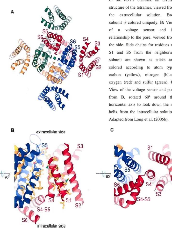
the structure-function relationship of K channels at the atomic level (Doyle et aï., 199$). A series of publications following the crystallization of KcsA explained the structural basis of K selectivity, ion conductance, channel inactivation and mechanisms of pharmacological channel blockade on the atornic level (Morais-Cabral et aï., 2001; Zhou et al., 2001b; Zhou et aï., 2001a; Roux & MacKinnon, 1999). Whereas KcsA showed a K channel in the closed state, the crystallization of MthK from Methanobacterium thermoautotrophicum captured a potassium channel in its open state and allowed important insight into basic mechanisms underlying channel opening and closure (Jiang et aï., 2002b; Jiang et aï., 2002c). The crystallization study of the cytoplasmic pore of Kir3.1 (Nishida & MacKinnon, 2002), the entire eukaryotic inward rectifier KirBac (Kuo et aï., 2003) and finally the cytoplasmic region of Kir2.1 (Pegan et aï., 2005) illustrated the structural basis of inward rectification. Most recently, the crystal structure of the voltage gated shaker channel Kv 1.2 (Long et aï., 2005a) and its S4 voltage sensor (Long et aï., 2005b) clarified the local environment of the voltage-sensor, its relation to the ion-conducting pore and coupling of voltage-sensing and channel gating. Computational studies such as
6
molecular dynamics simulations provided insight to gating movements on a nanosecond timescale (Sansom et aï., 2002; Roux, 2005).
The first part of this thesis introduces the basics of electrogenesis in different
biological systems, focusing on the central nervous system, the heart and vascular endothelia] ce]]s. A detailed section on the current knowledge of K channel structure and the structural basis of their functioning is followed by a section on voltage-gated and inward rectifier K channels. This part details the molecular basis, biophysical and pharmacological properties as well as the functional role of major Ktcurrents in different organ systems.
The second part of this thesis consists of original publications. The common theme of the articles presented here is the contribution of distinct Kir2 subunits in homo- or heteromeric assembly to native inward rectifier K-current in different tissues.
The third part states the major nove] scientific contributions presented in this thesis and discusses their scientific significance and potential limitations in the context of current literature as well as potential future directions.
I-1 The Action Potential
in
Neurons
Action potentials (APs) are pulse-]ike electricai messages that are rapidly propagated along a specific conduction system. Historicaliy, APs and their underlying ionic mechanisms were first investigated in the squid giant axon (Figure 1). Axonal APs are very short, ]asting only a few milliseconds, and propagate electrically in an ail-or-none Jike fashion. APs encode time and frequency but not amplitude or duration of a signai. Axonai APs depend practicaliy solely on K and Na conductances. At rest, the membrane potentiai of the axon is negative with typicai vaiues between -40 and -95 mV (Hiiie, 2001). When an eiectrical stirnuius sucli as an AP reaches the membrane, it depolarizes. Na channeis open and Na ions rush into the ceii, further depoiarizing the membrane until the membrane potential reaches values above O mV, an event termed “overshoot”. Na channels subsequently inactivate and deiayed rectifier K channeis activate to conduct K ions out of the ceii causing the membrane potentiai to return to its previous negative value. The transient overshoot depolarizes the next stretch of ceii membrane and so the AP travels aiong the axon to reach its effector site.
Larger axons differ from smalier axons in that they are covered by myelin, an electrically insulating layer of guai ceils (Schwann ceils). The myelin sheath is interrupted by the so-caiied “nodes of Ranvier”, axonai sections where there is no myelin sheath and the density of Na and K channels is higher than in unrnyeiinated axons (Hiiie, 2001). In myeiinated axons, ionic current can only fiow at these interruptions. This causes the axonal AP to jumps from node to node, thereby boosting the signal and speeding up its rate of transmission.
8
lime
after shock (ms)
Figure 1: Schematic diagram of action potential recordings from the squid giant axon.
A. Schematic diagram of squid axon with stirnulator and two recording microelectrodes a and b.
B. Action potential rneasured by electrodes a and b. Adapted from DeÏ CastilÏo & Moore (1959) in Hille (2001).
A
B
Stimulus
ii5Or
-50,
—ïSquid
a.xon
f û0
05
1.0
15
Figure 2A illustrates current recorded from a squid axon after depolarization from a negative holding potential to a test potential of O mV. Upon depolarization, a large transient inward current is seen that reverses- to- a large maintained outward cuITent. Figure 2B shows tirne course of Na and K conductances (gNa and g)
during a depolarizing voltage step from a holding potential of -65 rnV to a test potential of -9 mV. Conductances were calculated using the Hodgkin & Huxley formula defining ionic conductances:
Na Na
‘K
E-Eh
As shown in Figure 2B, middle panel, the sodium conductance increases rapidly and decreases slowly after membrane depolarization, indicating that the transient inward current shown in Figure 2A is a Na current that activates upon depolarization and subsequently inactivates. The slowly activating and non inactivating outward current shown in Figure 2A corresponds to a K current, as can be seen in Figure 2B, lower panel. Figure 2A shows an original current recording whereas Figure 2B displays calculated conductances under conditions different from those of the experiment shown in A. The purpose of panel B is not to show actual components of the cuiTent shown in panel A but simply to illustrate how the two different ionic components contribute to the native current. Therefore, the time course of currents shown in panels A and B is different.
10
1r
o
— A. —65mV
O
mV
Squid axon
-i
o
4
B.6
8
10
12
Ti.me after start of test pulse tms)
Squid —65 mV — mV 20 It) 20 O 4 6 10
Figure 2: Action potential of the squid axon and underlying ionic conductances. A. Depolarization of the squid axon from a holding potential from -65 mV w a test
potential of O mV at 3.8°C elicits a large, biphasic current. Adapted from Hille (2001).
B. Calculated time course of Na and K conductances in the squid axon during membrane depolarization from -65 mV to -9 rnV at a temperature of 8.5°C. Adapted from Hodgkin (195$) in Hille (2001).
I-2 The Cardiac Action Potential
12
The cardiac AP originates in the sinoatrial node (SAN). Propagation through the atria causes atrial contraction. The AP traverses the atrioventricular node (AVN) and then travels through the HIS-Puikinje system to reach the ventricles, where it triggers the contraction of ventricular muscle ceils. In order to ensure appropriate timing of cardiac contraction, cardiac action potentials vary between different cardiac regions reflecting an adaptation to its specific function. Figure 3 illustrates cardiac APs recorded from different cardiac regions.
A.
B.
figure 3: Schematic diagram of AP properties in different regions of the heart.
SAN
RA
AV node
Atrium
-60mVHis-Purkinje
1 sec. -$0mVIn contrast to axonal APs, the cardiac action potential is determined by a complex interpi ay between multiple depol arizing inward and repol arizing outward culTents. A hallrnark property is its long duration. Whereas the action potential of neurons lasts only a few miiiiseconds, the cardiac action potential can last up to several hundred milliseconds.
Figure 4 iliustrates relative current contribution to representative atrial and ventricular action potentials. Per definition, upward deflections denote outward currents and downward deflections inward currents. Native ionic current will be discussed in detail in chapter two. Five phases of the cardiac action potential can be distinguished. Phase O is the initial upstroke during membrane depolarization, mainiy due to inward Na current. following the rapid upstroke, the transient outward potassium current, ‘to, shifts the membrane potential to a more negative potential (phase 1) at which depolarizing, inward L-type (JCa,L) and T-type (JCa,T) calcium currents activate, initiating phase two, which is also referred to as the plateau phase. At the same time, the repolarizing K currents ‘I(Ur (ultrarapid delayed rectifier potassium current) and ‘KACh (acetylcholine activated potassium current) conduct
potassium out of the ceil. It has to be noted that ion channel RNA and protein expression differs between species and currents described here may flot be expressed in ail tissues. Outward deiayed rectifier currents activate in late phase 2 and are active throughout the late phase 3 until the membrane has reached its negative resting potential. ‘KI is the main K-conductance in diastolic phase 4. It becomes active in
late phase 3 where its outward component contributes to phase 3 repolarization (see also Figure 20). During phase 4, ‘KI is the main current governing the resting
membrane potential. Spontaneous diastolic depolarization in pacemaker ceils during phase 4 is rnainly carried by the depolarizing inward pacemaker current Ii-, conducted by Na and K ions, but in certain tissues also by the K current ‘KACh and the
outward component of the inward rectifier current ‘KI. As indicated in the upper two
panels of Figure 4, atrial APs have faster initiai repolarization, less positive plateau potentials, a shorter plateau phase and slower phase 3 repolarization.
Atrium LL 14 Ventricle 40 -0 -40 —80 1 o > E o u ‘Na w Ic ;? Lfl 3 4 w Q w
o
o
o
o
‘NCX ‘to ‘Kjr ‘Kr ‘Ks ‘K1 u LLo
wo
0 500o
o
500Figure 4: Representative cardiac action potential waveforms.
Top panels show APs obtained from atrial (left) and ventricular (right) myocytes. The five phases of the AP are labelled. The rate of change of the AP is directly proportional to the sum of the underlying transmembraneous ion currents (lower panels). Inward currents depolarize the membrane, whereas outward currents contribute to repolarization. Compared with an atrial AP, the ventricular AP typically has a longer duration, higher plateau potential (phase 2), and a more negative resting membrane potential (phase 4). The presence of an ultra-rapid delayed rectifier potassium (K) current (IKur) in atrial myocytes contributes to the lower plateau phase in the atrial AP. Greater inward rectifier K current (IKI) in ventricular ceils provides a faster phase 3 repolarization and a more negative resting membrane potential (phase 4). Adapted from Ouditet aI. (2004).
16
The sinoatrial node is the primary pacemaker of the heart. Sinoatrial APs have a relatively positive maximum diastolic potential (MDP) of 50 mV, a small phase O upstroke velocity (Vinax, < 2 VIs) (Bleeker et aÏ., 1980) and prorninent phase 4 depolarization to ensure SAN pacemaker predominance over distally located cardiac regions (Schram et aï., 2002b). ElectricaÏ coupling of the SAN to the atrium is complex and is designed to drive the large atrial muscle mass while protecting the SAN from hyperpolarizing atrial stimuli (Coppen et al., 1999).
Isolated atrial myocytes have a MDP of around -70 mV (Yue et aÏ., 1997; Li et aÏ., 2001), siower phase 3 repolarization and almost no spontaneous depolarization (Schram et aï., 2002b). Spatial heterogeneity of atrial APs and APD within and between the atria (Hogan & Davis, 1968; Spach et aÏ., 1989; Feng et aÏ., 1998; Fareh et aÏ., 1998) plays an important mie in atrial re-entrant arrhythmias, mostly due to shorter APD and ERP in the LA free wall than in the RA (Li et al., 2001). Rapid AP conduction follows muscle fibre orientation (Spach, 1995) to the AV node.
APs from intact AVN have a MDP of approximately -64 mV, small amplitudes and slow phase O upstrokes (Hoffman et al., 1959). AVN ceils have low excitability and postrepolarization responsiveness (Merideth et al., 196$) and display pacemaking activity (Munk et al., 1996; Watanabe & Dreifus, 1968). Phase 4 depolarization results in takeoff potentials of at -60 mV, andVmax is <20 V/s (Billette, 1987). However, atrial electrotonic influences suppress spontaneous activity in AVN cells (Kirchhofet al., 198$).
The His-Purkinje system directs the AP to the ventricles. Hence, Purkinje celis (PC) are specialized for rapid conduction. MDPs of PCs are very negative at around -90 rnV and Viax is high (—400-$00 V/s) (Davis & Temte, 1969; Kus & Sasyniuk, 1975; Carmeliet, 1977; Nattel & Quantz, 198$). APD is relatively long at slow rates (Carmeliet, 1977; Nattel & Quantz, 1988) and there is prominent spontaneous phase 4 depolarization (Callewaert et aL, 1984). PCs can generate dmg induced eariy afterdepolarizations (EADs) that trigger torsade de pointes arrhythrnias (El Sherifet cil., 1996; Asano et aÏ., 1997).
Ventricular myocytes have a MDP of -85 mV and a relatively positive plateau of about +10 to 20 mV. Phase 3 repolarization is rapid. Similarly to atrial muscle ceils, there is no significant phase 4 depolarization (Davis & Temte, 1969; Kus & Sasyniuk, 1975; Litovsky & Antzelevitch, 1989). Prorninent regional and transmural heterogeneity of ventricular APs is welJ established. In rat ventricle, APD is sliortest at the apex and longest in the septum (Gornez et aÏ., 1997). In canine ventricular myocardium, APs recorded from epicardium have a smaller overshoot, a more prominent phase I followed by a notch (“spike and dome”) and a briefer APD than endocardial celis, resulting in the T-wave in the ECG. MDP and Vrnax, however, are sirnilar in celis from botli regions (Litovsky & Antzelevitch, 1988). A distinct celi population lias been found in the midmyocardium and designated M-cells (Sicouri & Antzelevitch, 1991; Drouin et aÏ., 1995). M-cell APDs increase at slow heart rates and have a larger Vinax (P—300 Vis) than endo- or epicardial ceils (Sicouri & Antzelevitch, 1991). RV M-cell APs have a smaller upstroke, a deeper notch and a shorter duration than LV M-celI APs (Volders et aÏ., 1999). Transmural APD heterogeneity lias been implicated in the generation of torsade de pointes ventricular arrliythmias (Antzelevitch & Sicouri, 1994), especially under conditions that cause prolongation of APD sucli as APD prolonging drugs, hypokalernia or slow heart rates (Sicouri & Antzelevitcli, 1993). Figure 5 compares action potentials of ventricular epicardium, midmyocardium and endocardium.
1$
Region MDP (mV) Vmax (Vis) Phase 3 Phase 4
repolarization depolarization SAN -50 <2 rapid Atrium -70 150-300 slow no AVN -64 < 20 yes PC -90 400-$00 yes Ventricle -85 200-300 rapid no
Prominent spike and dome
Longer APD than Endo or Epi
Longer APD than Epi
Figure 5: AP properties of canine ventricular epicardium, midmyocardiurn and endocardium.
A: Epicardium (Epi), B: midrnyocardium (M region), C: Endocardium (Endo). Adapted from Schrarn et aï. (2002c). APs are reproduced from Sicouri & Antzelevitch, 1991.
A
o—
EpiB
o—
M regionC
0— Endo 50 mV No pacemaker activity 200msec20
I-3 Electrogenesis
in
Vascular Endotheliat Cetis
The mechanisms of electrical signalling in non-excitable ceils, such as vascular endothelial cells (ECs) are less understood. A multitude of different ion channels is found in endothelial ceils, rnany of which have flot been well characterised and whose molecular basis is yet unknown (Nilius et aï., 1997; Nilius & Droogmans, 2001).
Endothelial ceils provide the pathway for oxygen delivery from blood to tissue. Control of permeability to metabolites, macromolecules and gas exchange ai-e crucial for the proper functioning of celi rnetabolism (Born et aï., 199$). Modulation of vascular tone regulates blood pressure (Cannon, 1998). Endothelial celis are involved in control of blood coagulation, wound healing and angiogenesis (Shimokawa & Takeshita, 1995; Luscher, 1994; Born et aï., 199$). In cardiovasculai disease, the endothelium plays a key role in vascular pathology and as a target for pharrnacotherapy (Angus, 1996; Garcia-Palmieri, 1997; Born & Schwartz, 1997; Tan et aI., 2004; Deedwania, 2000). Endothelial ceil functions depend on both constitutive EC properties and on active responses (“on demand”) of ECs to chemical, mechanical or neuronal stimuli originating from the blood, tissue and coupling to neighboring ECs or adhering blood ceils. Responses such as the ielease of vasoactive substances by the endothelium depend upon fluctuations in their membrane potential and in the intracellular Ca2 concentration. Hyperpolarization of the membrane potential provides the main driving foi-ce for entry of extracellular Ca (Nilius et al., 1997; Nilius & Droogmans, 2001). The role of ion channels in the transduction of these signais is stilf controversial but it appears that they are involved in short-term EC responses in the i-ange of seconds to minutes. Roles of ion channels have been determined for release of pro- and anticoagulants, growth factors, and regulators of vasotonus such as nitric oxide (NO), prostacyclin (PG1), platelet activating factor (PAF), endothelium-derived hyperpolarizing factor (EDHF), release of von Willebrand factor (vWF), tissue plasminogen activator (tPA), the anticoagulant tissue factor pathway inhibitor (TFPI) and protein S (Nilius & Droogmans, 2001). Ion channels ai-e involved in regulation of vascular permeability and angiogenesis.