AVIS
Ce document a été numérisé par la Division de la gestion des documents et des archives de l’Université de Montréal.
L’auteur a autorisé l’Université de Montréal à reproduire et diffuser, en totalité ou en partie, par quelque moyen que ce soit et sur quelque support que ce soit, et exclusivement à des fins non lucratives d’enseignement et de recherche, des copies de ce mémoire ou de cette thèse.
L’auteur et les coauteurs le cas échéant conservent la propriété du droit d’auteur et des droits moraux qui protègent ce document. Ni la thèse ou le mémoire, ni des extraits substantiels de ce document, ne doivent être imprimés ou autrement reproduits sans l’autorisation de l’auteur.
Afin de se conformer à la Loi canadienne sur la protection des renseignements personnels, quelques formulaires secondaires, coordonnées ou signatures intégrées au texte ont pu être enlevés de ce document. Bien que cela ait pu affecter la pagination, il n’y a aucun contenu manquant.
NOTICE
This document was digitized by the Records Management & Archives Division of Université de Montréal.
The author of this thesis or dissertation has granted a nonexclusive license allowing Université de Montréal to reproduce and publish the document, in part or in whole, and in any format, solely for noncommercial educational and research purposes.
The author and co-authors if applicable retain copyright ownership and moral rights in this document. Neither the whole thesis or dissertation, nor substantial extracts from it, may be printed or otherwise reproduced without the author’s permission.
In compliance with the Canadian Privacy Act some supporting forms, contact information or signatures may have been removed from the document. While this may affect the document page count, it does not represent any loss of content from the document.
Investigating the neural substrates mediating
visuomotor adaptation;
From beginner to expert
par Raby Bouras
Département de Psychologie, Université de Montréal Faculté d'Art et Science
Thèse présentée à la Faculté des études supérieures en vue de l'obtention du grade de Doctorat
en Neuropsychologie option Recherche/Intervention
Décembre, 2007
© Raby Bouras, 2007
Cette thèse intitulée:
Investigating the neural substrates mediating visuomotor adaptation;
From beginner to expert
présentée par : Raby Bouras
a été évaluée par un jury composé des personnes suivantes:
Franco Leporé, président-rapporteur Julien Doyon, directeur de recherche Sylvie Belleville, membre du jury Virginia Penhune, examinateur externe
[Ta~er le Aê~ , représentant du doyen de la FES
Résumé
L'objectif général de cette thèse était d'explorer et d'identifier les substrats anatomiques impliqués dans les différents stades de l'apprentissage d'une adaptation visuomotrice à l'aide de l'imagerie par résonance magnétique fonctionnelle (lRMf). Deux expériences ont été réalisées pour atteindre ce but. La première étude avait pour objectif l'investigation du stade automatique de l'exécution de l'habileté motrice, tandis que la deuxième expérience a exploré la rétention à long terme de cet apprentissage. Ces expériences ont utilisé des sujets normaux, et ont fait usage d'une tâche de poursuite visuelle requerrant l'utilisation d'une manette de jeux. Les résultats ont révélé qu'une réorganisation fonctionnelle des régions anatomiques a eu lieux au travers des phases d'apprentissages, et que ces changements ont suivi le model d'apprentissage moteur proposé par Doyon et collaborateur (2002, 2003, 2005). En effet, les résultats de notre série d'expériences ont mis en évidence l'implication des circuits cortico-cerebelleux (CC) et cortico-striatal (CS) au stage initial d'apprentissage, tandis que seul le circuit CC était impliqué dans le stade lent, la rétention, et l'automatisation de l'habileté motrice. De plus, nos résultats ont aussi mis en évidence l'importance de considérer et de contrôle la stratégie d'exécution utilisée par les sujets pour compléter la tâche et lors de l'interprétation des résultats de l'imagerie cérébrale.
Mots-clés: Apprentissage moteur, Adaptation visuomotrice, IRMf, Rétention, et Automatisation.
Abstract
The general objective of this thesis was to explore and describe the underlying brain circuits involved at different leaming phases of a visuomotor adaptation skill through the use of functional magnetic resonance imaging (fMRI). Two experiments were designed to achieve this goal. The first study had for objective to investigate the automatic execution stage of leaming, whereas the second experiment investigated the long-term retention of procedural memory. AU experiments involved normal subjects, and employed a visuaUy-guided adaptation skill requiring subjects to use a joystick to complete the task. The results reveal that functional neural reorganisations take place throughout the leaming phases, and these changes foUow Doyon and colleagues' (2002, 2003, 2005) model of motor leaming. In fact, the results of our series of experiments revealed that both the cortico-cerebellar (CC) and the cortico-striatal (CS) systems play an important role during the early leaming stage of motor leaming, while only the CC circuit plays a pre-eminent role during the later leaming stage, and is the only system implicated in the recall and automatic execution of the visuomotor adaptation skill. What' s more, our results also suggest that the execution strategy used by subjects to complete the task has important consequences on the subcortical regions recruited for task completion, and is an important factor to consider when interpreting functional imaging data.
Keywords : Motor skillleaming, Visuomotor adaptation, fMRI, Retention, and Automatisation.
Table of Contents
Résumé ... iii
Abstract ... iv
List of Tables ... viii
List of Figures ... ix
Chapter 1. Introduction ... 14
Chapter 2. The experimentaJ study of motor adaptation ... 19
2.1. Defining and differentiating between motor skills ... 19
2.2. The neuroimaging of motor adaptations ... 23
2.3. The discrepant factors in the imaging studies ... 33
2.3.1. Different imaging technologies ... ; ... 33
2.3.2. Different regions of interest ... 34
2.3.3. Different methodological factors ... 34
2.3.4. Different experimental tasks ... 36
2.3.5. Different stages oflearning ... 36
2.3.6. Different cognitive factors ... 38
2.4. Conclusion ... 39
Chapter 3. Automaticity revi ewed ... 41
3.1. What is automaticity? ... 41
3.2. Different kinds of automaticity ... 43
3.3. The dual-task paradigm ... 45
3.4. Conclusion ... 46
Chapter 4. Doyon and colleagues' model.. ... 48
Chapter 5. The thesis' objectives and hypotheses ... 54
5.1. Experiment 1: Automatisation of a visuomotor adaptation skill ... 55
5.2. Experiment 2: Retention of a consolidated visuomotor adaptation skill ... 56
Chapter 6. Experiment 1: DifferentiaI contributions of the cortico-cerebellar and cortico-striatal circuits in the automatisation of a motor adaptation skiIl ... 57
6.1. Abstract ... 57
6.2. Introduction ... 58
6.3. . Materials and methods ... 59
6.3.1. Subjects ... 59
6.3.2. Materials ... 60
6.3.3. Experimental Tasks ... 60
6.3.4. Procedure ... 63
6.3.5. fMRI data analysis ... 63
6.4. Results ... 65
6.4.1. Behavioural resuIts: Training sessions ... 65
6.5. Behavioural results: Scanning sessions ... 67
6.6. Imaging results ... 67
6.7. Discussion ... 69
6.7.1. Conclusion ... 73
6.8. Acknowledgments ... 74
6.9. Specific contributions by authors ... 74
Reference List ... 75
Chapter 7. Experiment 2: The long-term retenti on of a visuomotor adaptation skill: a fRMI study 86 7.1. Abstract ... 86
7.2. Introduction ... 86
7.3. Materials & methods ... 88
7.3.1. Subjects ... 88
7.3.2. Materials ... 88
7.3.3. Task Description ... 89
7.3.4. Procedure ... 90
7.3.5. fMRI data analysis ... 91
7.4.1. Behavioural results ... 92
7.4.2. Imaging results ... 93
7.5. Discussion ... 94
7.6. Acknowledgments ... 97
7.7. Specifie contributions by authors ... 97
Reference List ... 99
Chapter 8. General conclusion ... 105
8.1. Thesis overview ... 105
8.2. Overview of the experimental results and other recent experimental data ... 106
8.3. A critical review of our work ... 113
List of Tables
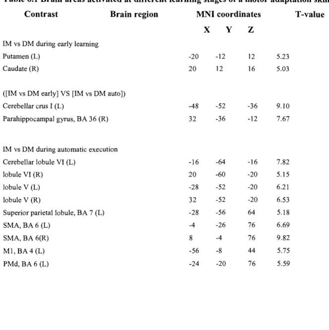
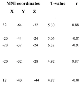
Table 6.1 Activation peaks are given using the MNI coordinates. AlI coordinates are significant as defined by puncorrected < 0.001. Abbreviations; L: left, R: right, PMd:
dorsal premotor cortex, SMA: supplementary motor area, Ml: primary motor cortex, BA: Broadman's area ... 79 Table 6.2 Activation peaks are given using the MNI coordinates. AlI coordinates are significant as defined by puncorrected < 0.001. Abbreviations; lYW: between; r:
correlation coefficient; PI: precision index, SI: speed index ... 80 Table 7.1 The coordinates are given in MNI coordinates. AlI coordinates are significant as defined by puncorrected < 0.005. Abbreviations; L: left, R: right, PMd: dorsal premotor
List of Figures
Figure 1.1 A taxonomy of memory and associated brain structures adapted from Squire &
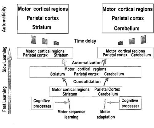
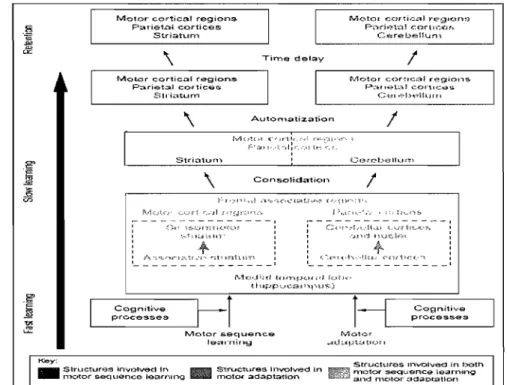
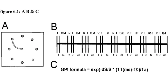
Knowlton (1995) ... 16 Figure 4.1. Model presented by Doyon and Ungerleide (2002) describing the cerebral plasticity within the cortico-cerebellar and cortico-striatal circuits during the course of leaming. Adapted from Neuropsychology of Memory, 3rd Edition ... 49 Figure 4.2. Revised model presented by Doyon and Underleider (2002) describing the cerebral plasticity within the cortico-cerebellar and cortico-striatal circuits during the course ofleaming. Adapted from Current.Opinion in Neurobiology(15), page 164 .. 53 Figure 6.1 (A) Visual interface of the Eight Target Tracking task (ETT). At the beginning of every trial, a starting point, represented by a white circle (0.75 cm in diameter), appeared in the middle of the computer screen. The cursor, a cross-shaped figure, appeared superimposed on top of the starting point. At the same time, the target represented by a red circle (1.5 cm in diameter), appeared 10 cm from the starting point. Appearing simultaneously was a line (0.5 cm in thickness) indicating the ellipse-shaped trajectory the subjects had to follow (radius of 2.5 cm) to reach the target with the cursor. (B) Timeline describing a possible configuration for one of the ten runs in one ofthe two scanning sessions. Subjects completed 2 tasks in these runs:
DM stands for eight target tracking in direct mode, lM stands for eight target tracking in indirect mode, 1 stands for instructions, and R stands for rest. Total duration of a run was 315 seconds. Each bloc was composed of 8 trials, and each trial was separated by a pseudo-randomly varied interval ranging from 500 to 1500 msec. This is known as a jittered design, and has the advantage of combining both the block and event-related run configurations. (C) Formula used to calculate the global performance index (GPI) on ETT trials. The GPI was calculated by combining the precision (dS/S) and speed performances ([TT-TO]/Ta) on the ETT. In the formulas
exp is the value given to the trial (l for successful and 0 for failed), the dS represents the differential surface area between the actual path followed to reach a target and the
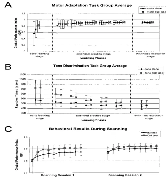
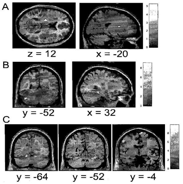
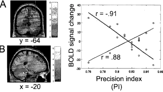
optimal path of the curved line, S is defined as the differential surface area between the optimal path of the curved line and a straight line between the starting point and the target center, TT constitutes the total time, T 0 the minimal time needed (500 ms), and Ta the time allowed (2,900 ms) to reach the target. ... 81 Figure 6.2 Average group performance across-subjects (mean ± SD) on (A) the visual-tracking task in the inversed mode (lM) executed alone (blue diamonds), and in the dual-task condition (red squares); (B) the Tone Discrimination Task (TDT) completed alone (blue diamonds), and in the dual-task condition (red squares); and (C) the visual-tracking task in the inversed mode (lM, blue diamonds) and direct mode (DM, red squares) measured during the two scanning sessions. In both figures A & B, every data point corresponds to the subjects' performance during a bloc of trials, whereas in figure C, each data point corresponds to the subjects' performance during an fMRI run ... 82 Figure 6.3 Statistical parametric maps ofbrain activity during motor adaptation. (A) Brain regions activated during the early learning phase (lM vs MD in the early stage). (B) Results of the contrast ([lM vs DM] early stage vs [lM vs DM] automatic stage) revealing the brain structures involved in the automatisation process. (C) Brain regions activated during the automatic execution of the task (lM vs MD in the automatic stage). AlI contrast images were obtained for subject individually and then used in the second level random-effects analyses as calculated by a one-sample t-test model, a statistical threshold of P < 0.001 uncorrected was considered to show significant activations. Color bars code for the value of the t statistic associated with each voxel. Right si de of the image corresponds to the right side of the brain ... 83 Figure 6.4 Between-subjects regression analyses coupling the subjects' average precision index (PI) and the BOLD signal measured during the automatic execution stage of the motor adaptation skill. (A) Blue crosshair: right cerebellar crus l (32 -64 -32). The scatter plot shows that the brain response at this coordinate was positively correlated with precision (blue; r = .88). (B) Red crosshair: left cerebellar lobule IV (20 32
-24). The scatter plot shows that the brain response at this coordinate was negatively correlated with precision (red; r = -.91). Note that a negative correlation was obtained
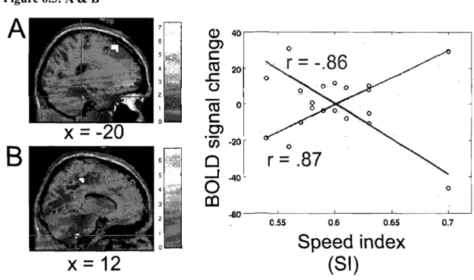
for the left lobule V (-20 -44 -24, r= -.87), but that it is not plotted on the graph ... 84 Figure 6.5 Between~subjects regression analyses coupling the average subjects' speed index (SI) and the BOLD signal measured during the automatic execution stage of the motor adaptation skill. (A) Blue crosshair: left cerebellar lobule IV (-20 -32 -28). The scatter plot shows that the brain response at this coordinate was positively correlated with speed (blue; r .87). (B) Red crosshair: right cerebellar lobule IX (12 -40 -44). The scatter plot shows that the brain response at this coordinate was negatively correlated with speed (red; r = -.86) ... 85
Figure 7.1 Average group performance (across-subjects mean ± SD) on the visual-tracking task in inversed mode (lM, blue diamonds), and in direct mode (DM, red squares). Every point corresponds to the average of trials completed during a scanning mn, and the axe represents the three days of experimentation: Days 1 and 2 completed during the previous study, and the present retention experiment (Day 3) ... 1 03 Figure 7.2 Statistical parametric maps of brain activity representing long-term retenti on of
the visuomotor adaptation task. (A) Brain regions identified following the contrast subtracting the direct mode (DM) from the inversed mode (lM) during the delayed recall of the learned task (lM VS MD on Day3). (B) Brain regions identified following the contrast subtracting the direct mode (DM) from the inversed mode (lM) during the delayed recall on Day3 and the same contrast on fMRI data acquired on Day2 ([lM vs DM] Day3 VS [lM vs DM] Day2). AlI contrast images were obtained for every subject individually and then used in the second level random-effects analyses as calculated by a one-sample t-test model, a statistical threshold ofP < 0.005 was considered to show significant activation. Col or bars code the value of the t
If
the brain was simple enough for us ta understand, then we would be tao simple ta understand il.Acknowledgements
It is with immense pleasure that 1 regard the past six years spent completing my graduate degree at the University of Montreal, a journey rippled with challenges, sucees ses and turning points. The lessons 1 have learned throughout these past years have paved the way towards a greater understanding of the exciting world that is science, but also of my self. This journey, however, would have been impossible without the understanding and support of the many gifted professors, researchers, and staff members that have composed my environment, and that took time to care and share their knowledge with me. 1 am indebted to my thesis supervisor, Dr. Julien Doyon, for introducing me to the field of brain imaging, and grateful for his patience and moral support throughout my studies. His knowledge and know-how have served me well, and will continue to serve me throughout my career. 1 would also like to thank the rest of the professorial body, inc1uding Drs. Franco Leporé and Maryse Lassonde for their attention and interest in my progress, and researchers like Dr. Pascal Belin and Dr. Jorge Harmony who were friendly enough to tolerate my numerous office intrusions, and answer my never-ending stream of questions. One cannot forget Maria Sanchez and the late Susanne Lamothe, colleagues and friends who have made my stay at the University pleasant and generally more enjoyable. Finally, this thesis would have been impossible to complete if 1 did not have the technical support of a number of extremely talented group of engineers and research technicians such as Boalem Mensour, Vo An Nguyen, Yves Roy, Stéphane Denis, and Anne Bellio.
Finally, a heartfelt thank you to my friends and family, to whom 1 will forever be grateful. Their enduring fortitude, compassion and patience served me well during these years. They were present in my moments of desperation and agitation, and now in this moment of blissful peace. Thank you again for everything you have done for me, and 1 only wish that 1 can someday return your generosity.
Although the study of human memory dates back to Plato's work five centuries before this era, the subject matter was mostly limited to philosophical writings and introspective probing like the kind practiced by Sigmund Freud.
It was only in the 1930's that a resourceful researcher, by the name of Karl Lashley, began a systematic quest for the exact location of memory in the brain (Viney & Brett King, 1998). His approach was fairly simple, he taught rats and monkeys a variety of tasks, then destroyed a part of their brain. He reasoned that if the animaIs could not remember after the lesion, then he must have found the place where memories reside. After countless experiments, Lashley's results lead him to conc1ude that nothing short of near complete destruction of the cortex caused the animaIs to forget their tasks. Tired and frustrated, he conc1uded that the biological study of memory was impossible.
He was proven wrong less than 25 years later when, in 1953, twenty-seven year old Henry M. entered the hospital for radical brain surgery that was to cure his epilepsy. Living with debilitating epileptic seizures, H.M. was hopeful that the procedure would change his life for the better. Instead, it trapped him in a mental time warp where television is always a new invention and Truman is forever president of the United States of America. This devastating si de effect made H.M. the most studied individual in the history of brain exploration and revolutionised the field of memory research. Following the resection of a large portion ofhis temporal lobes, H.M. was cured ofhis seizures, but was left with severe anterograde amnesia. Impressively however, H.M. did preserve sorne mnemonic abilities. For instance, he was able to hold sorne information in storage for very short periods oftime (short-term and working memory), and he could still leam various motor skills (Milner, 2005). Such observations of HM's case have lead to sorne of the seminal findings about memory. SpecificaIly, it was shown that the hippocampus is required for the formations of explicit long-term memories, but not for the short-term recall ofthese memories, nor for the acquisition of various motor skills. More importantly, H.M.'s case study had vividly
illustrated that there is a biological basis for memory, and that it lS possible to use biological techniques to study it.
Since then, a plethora of studies have given rise to a number of models suggesting that memory is not a single entity, but rather a heterogeneous phenomenon that can be broken down into different systems, each of which is sub-served by a distinct neural network (Cohen & Squire, 1980; Schacter, 1987; Squire, 1982; Tulving, 1985). Although aIl these researchers agreed that memory research should be understood as the study of various systems ofmemories, they disagreed on the classification scheme and terminology
that should be used to describe and define aIl its components. Sorne researchers believe that a distinction should be made between "explicit" and "implicit" memories (Schacter, 1992a; Schacter, 1992b), "cognitive memories" and "habits" (Mishkin, Malamut &
Bachevalier, 1984), "procedural" and "declarative" memories (Cohen, Eichenbaum, Deacedo & Corkin, 1985), or between "declarative" and "non-declarative" memories (Squire, 1992; Squire, Knowlton & Musen, 1993). Other models propose the existence of not two, but several distinct classes of memory: "semantic memories", "episodic memories", "procedural memories", "perceptual representation systems", and "working memory" (Schacter & Tulving 1994).
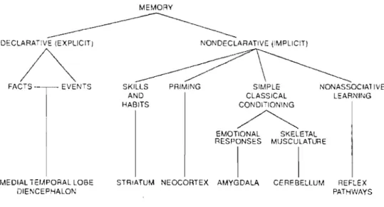
Despite these differences, the model proposed by Squire and colleagues (1992, 1993) is probably the conceptual framework that has gained the most recognition in neuropsychological research over the last decade. Part of its success is that in addition to dividing memories in two broad classes (declarative and non-declarative), their model goes a step further in decomposing memories into smaller subtypes (Fig. 1.1). Another reason for its success lies in the fact that this model is based on a wide variety of evidence, ranging from animal research to investigations in both normal and pathological human populations. A considerable amount of data now supports the existence of such functionally and neuroanatomically dissociable subsystems. This model, as weIl as the motor skill under investigation in this thesis, will be reviewed in sorne detail in the following lines.
DECLARATiVE (EXPLICIT)
A
FACTS EVENTS
MEDIAL TEfAPORAL LOBE DlENCEPHALON MEMORY NONDECLARATIVE (IMPliCIT) SKILLS AND HABITS
PRIMiNG SIMPLE NONASSOCIAllVE CLASSICAL LEARNli"G CONDI'IONING
~
EMOTIONAL SKELETAl RESPONSES MUSCULATURE 1 1STRIATuM NEOCORTEX AMYGDALA CERE8ElLUM REFLEX PATHWAYS
Figure 1.1 A taxonomy ofmemory and associated brain structures adapted from Squire & Knowlton (1995).
Briefly, Squire and colleagues propose that declarative memories are made up of facts and events that can be expressed in words. As such, two types of declarative memories have been distinguished: semantic memories, which refer to factual information that a group of people can share (e.g. who is the prime minister of Canada?), and episodic
memories, which refer to autobiographical information for events (e.g. where you were on September Il th 2001 ?). The acquisition of these types of memories is rapid, as it often takes only a brief exposure for them to be acquired, and their expression is conscious and explicit, meaning that they require conscious thought to be leamed or expressed (Cohen &
Squire, 1980). In contrast, Squire & Knowlton (1995) define non-declarative memories as skills or habits that are acquired gradually with practice (priming and taste aversion conditioning are exceptions since they can be acquired after a single trial). In general, however, non-declarative memories are leamed slowly and often require many repetitions over several training sessions to develop (Fitts, 1964). The acquisition of such abilities can be explicit and/or purely impIicit, as they do not require conscious thought to be leamed or expressed (Squire, 1992). The non-declarative memory system is comprised of four . subtypes; skills and habits, priming, simple classical conditioning and non associative
leaming. Of particular importance for this work is the acquisition of ski1ls. Skililearning refers to the leaming of motor, cognitive or perceptual skills, whereby performance on a given task improves with repetition, such that after a critical amount of training, highly skilled performance levels can be attained (Mishkin, Malamut & Bachevalier, 1984; Squire
& Knowlton, 1995; Squire & Zola, 1996).
As 1 will present in greater detail further, researchers differentiate between three general types of skills: cognitive, perceptual and motor skills. Motor skills themselves are subdivided into two smaller categories: sequence learning and motor adaptation. The work completed in this thesis has concentrated on studying a particular class of motor adaptation, namely visuomotor adaptation. Briefly defined, visuomotor adaptation skills refer to a set of abilities that allow an individual to change motor commands in response to alterations in sensory feedback. These motor capacities are essential to complete several everyday tasks, and actively contribute in such tasks as walking, driving, using a computer mouse, etc. In fact, their presence in our everyday functioning is ubiquitous, and researchers' interest in the leaming of such skills is justifiable.
However, although the cerebral structures and circuits involved in the leaming of declarative memories are weIl known (Meunier, Bachevalier, Mishkin & Murray, 1993; Squire, 1992; Squire, Knowlton & Musen, 1993), an analogous circuitry for motor skill leaming is only beginning to be defined. Based on animal and human work, several brain structures, including the basal ganglia, cerebeIlum, and motor cortical regions of the frontal lobe are thought to be critical for the acquisition and/or retention of skilled motor behaviours (e.g. Bloedel, 1992; Nezafat, Shadmehr & Holcomb, 200la; Della-Maggiore &
McIntosh, 2005; Krebs et a1., 1998; Doyon et a1., 2002a; Ungerleider, Doyon & Kami, 2002; Doyon et al., 2004; Doyon & Benali, 2005). The exact nature of their involvement in motor skill leaming, however, is far from being weIl understood. In fact, contradictory findings conceming the involvement of these structures in skill learning is quite common (Garavan, Kelley, Rosen, Rao & Stein, 2000). The general objective of the present thesis is
thus to use modern neuroimaging tools to contribute to our CUITent understanding of the memory system underlying motor skill learning. Based on the recent models presented by Doyon and collaborators (2002, 2003, 2005) that attempts to predict the involvement of the different brain regions during the learning of motor skills, we derived a-priory hypotheses concerning the automatisation and long-term retenti on of a visuomotor adaptation that we set out to test.
Chapter 2.
The experimental study of motor
adaptation
2.1.
Defining and differentiating between motor skills
As was previously stated, our understanding of the memory system in the early part of the 1990s was of two broad categories of learning exists (Le dec1arative and non-declarative), and that these two categories were further subdivided into sm aIl er subcategories. The specific subcategory of interest in this thesis was referred to as skills and habits. It is generally agreed that three broad types of skill can be learned (cognitive,
perceptual and motor) (Mishkin & Murray, 1994; Squire & Knowlton, 1995; Squire &
Zola, 1996). Their acquisition is usually measured by a graduaI reduction in reaction time, decrease in number of errors, and/or a reduction in the number of trials needed to reach successful completion criterion.
Cognitive skill learning can be defined as the process by which the procedures and strategies relevant to the performance of a task demanding mental operations come to be
combined and used effectively following repeated practice (Ouellet, Beauchamp, Owen &
Doyon, 2004). The learning and use of mathematics and arithmetic is a good example of cognitive skilllearning in everyday life. On the other hand, visual-perceptual skills involve the ability to accurately interpret and give meaning to what is seen. A number of specific skills fall into this category. They include; vi suaI discrimination -or the ability to distinguish one visual pattern from one another; and visual closure -or the ability to perceive a whole pattern when shown only parts of that pattern (R.Clay Reid, 1999).
The skill that has attracted most attention, and prompted the largest body of the research, including the experiments in this thesis, has been motor learning. The fact that motor learning has generated a great deal of investigations and deliberations is equitable to
the amount of activities in everyday life that require the graduaI acquisition of motor skills. Simply brushing our teeth necessitates the co-articulation of arm, hand and finger movements into specific and smoothly executed sequences of action. Motor ski111eaming can be operationally defined as the process by which movements, either produced al one or in a sequence, come to be performed effort1ess1y through repeated practice (Willingham, 1998). As one can readily recognise, this operation al definition is rather vague and can encompass a wide variety of tasks that are quintessentiaHy different. For instance, although knitting and playing consol games can be regarded as tasks that require the graduai acquisition of motor skills, they are essentially very different in nature. Just as memory research has invariably lead us to consider memory as a wide architecture of complex and distinguishable subtypes, so has the specific study of motor skills directed investigators to dissociate various forms of motor skills subtypes. Although varying in their complexity and nature, investigators agree to distinguish between two general categories of motor skill tasks: motor sequences and motor adaptation tasks.
Knitting is considered a motor sequence tasks because it requires the incremental acquisition of movements into a well-executed behaviour. To study the neural substrates mediating our agility to leam motor sequences, investigators have used a number of different experimental procedures. Researchers have tested subjects as they leamed to repeat sequences of fingers or limb movements (Kami et al., 1995; Doyon et al., 2002), to move a pen through a eut-out maze by trial and error (Van Mier, Tempel, Perlmutter, Raichle & Petersen, 1998), and ev en while subjects knit (Doyon, pers comm).
On the other hand, consol games are considered motor adaptation tasks because they require subjects to map new representations between the various motor commands and the sensory feedback involved in the tasks' execution (Klassen, Tong & Flanagan, 2005). In order to study motor adaptation leaming, researchers have employed a number of different paradigms. For instance; tasks requiring subjects to maintain contact between a metal stylus and a small target located on a disk that can be adjusted to rotate at different
velocities (rotor pursuit task) (Maquet, Schwartz, Passingham & Frith, 2003; Smith &
Smith, 2003), or asking subjects to draw figures through the reflection of a mirror
(mirror-drawing task) (Gabrieli, Stebbins, Singh, Willingham & Goetz, 1997), or to adapt to
changes in the relationship between the movements of a joystick and those of a cursor on a
screen (tracking task) (Della-Maggiore & McIntosh, 2005; Contreras-Vidal & Kerick,
2004; Krakauer, Ghez & Ghilardi, 2005; Graydon, Friston, Thomas, Brooks & Menon,
2005), or even to adapt to changes created by a force field applied to a robotic arm when
pointing to visual targets (force field adaptation task) (Diedrichsen, Hashambhoy, Rane &
Shadmehr, 2005; Smith, Brandt & Shadmehr, 2000; Smith & Shadmehr, 2005; Shadmehr
& Wise, 2005).
Learning from the errors of our predecessors, we can now recognize that comparing the results obtained while subjects learn to manipulate a joystick in a novel movement relationship to that of subjects learning a sequence of finger movements, is probably not very valid and clearly should be avoided. Ghilardi and his colleagues (2000) have argued that the different kinematic features and performance criteria of these tasks should discourage investigators from comparing the patterns of brain activity measured through one task to the other (Ghilardi et al., 2000). As these authors have pointed out, the simple act of reaching for an object requîres the learning of both the sensorimotor representatîons of external space and of internaI models of the dynamic properties of the musculoskeletal system. This kind of learning is believed to occur without the conscious awareness on the part of the subjects since they cannot describe the individual feedback events, the precise sequence of motor responses or the nature of the learned behaviour. On the other hand, subjects are generally aware of the specific responses during the learning of ordered sequences ofrequired movements (Ghilardi et al., 2000).
Just as these researchers have suggested, we now know that different patterns of brain activations follow the acquisition of these different type of motor learning. Furthermore, recent evidence has also suggested that motor adaptation themselves should
be further subdivided into two distinct categories (kinetic vs. kinematic adaptation), and that each form of motor adaptation constitute distinct processes that may require the use of
separate neural substrates (Shadmehr & Wise, 2005).
This idea has stemmed from work completed by Krakauer and collaborators (1999) where in they elaborated a simple yet elegant series of experiments in which it was hypothesised that, should the processes underlying their acquisition be distinct, learning a novel dynamics adaptation should not interfere with the consolidation of a previously leamed kinematic transformation. In addition, they hypothesised that, if distinct, these
pro cesses should be learned in paraUel (Krakauer, Ghilardi & Ghez, 1999). In hne with
their hypotheses, they observed that the leaming of novel dynamics do es not interfere with the consolidation of a newly learned kinematic transformation, whereas the leaming of another kinematic or dynamic interferes with the consolidation of a previously leamed transformations of the same type. They also conc1uded that novel kinematic and dynamic transformations can be leamed in paraUel, supporting the idea that their acquisition is
independent (Krakauer, Ghilardi & Ghez, 1999). One can therefore predict that these
,different forms of motor adaptation would involve separate regions of the motor system. Functional imaging data are consistent with a separation in the systems that mediate the different motor adaptations. In fact, kinematic leaming has been associated with activations in posterior parietal areas , whose inputs are predominantly visual, whereas Kinetic adaptation has been associated with activations in the anterior regions of the parietal cortex
(Stickgold, 2005; Walker & Stickgold, 2005).
In a more recent imaging study, Diedrichsen et aL (2005) investigated the neural responses evoked by these two adaptation tasks using fMRI and concluded that kinetic and kinematic transformations are not performed in two anatomical separate areas but rather in
one continuous, overlapping cascade (Diedrichsen, Hashambhoy, Rane & Shadmehr,
the relevance of closely considering the type of motor adaptation tasks when interpreting imaging data, and when comparing our results to previous findings ofbrain imaging.
Although important in the context of skill leaming, the acquisition of motor sequences constitutes, in itself, a vast domain of research which exceeds the aim of this thesis. For a more complete review on the matter, review the work of such authors as Ashe, Lungu, Basford & Lu (2006). Moreover, motor skillleaming has been empirically studied for more then 70 years to date, and over 15 400 studies have been completed on this specifie subject since 1935. These research endeavours include animal experiments in rodents and non-human primates, as well as research efforts in healthy hum ans and humans suffering from a range of debilitating conditions such as strokes and neurodegenerative diseases. More recently, the emergence of new imaging technologies have allowed researchers to expand this search and investigate the in-vivo implications of different brain structures in healthy humans. Considering the sc ale and magnitude of this research effort in the domain of motor skill leaming, the short review that follows will focus on the contribution of modem imaging techniques to our state of knowledge regarding motor adaptations.
2.2.
The neuroimaging of motor adaptations
A brief Pubmed review of articles published since 1990 reveals that over 4000 imaging studies concemed with motor adaptations have been completed. AIl of these studies have suggested the implication of a number of brain structures believed to be critical for the leaming and the execution of the motor adaptations. Not surprisingly, most of these studies have reported very different results, some were even contradictory. The objective of the following sections is to review some of the seminal studies in the field of motor adaptation leaming, to list and explain the contradictory findings, and discuss the important ]essons we keep from them.
lt is a well known fact that people demonstrate an impressive ability to acquire an almost unlimited repertoire of complex motor skills. The skills of musicians and athletes are good examples of such incredible learning feats. However, in the early 1990s, little was known about the neural systems that are required for motor control and task execution. Although the involvement of the cortical motor areas, the cerebellum and the striatum were derived from medical knowledge (i.e. stroke and neurodegenerative diseases), their implication and that of other brains structures was not weIl understood.
Grafton and colleagues (1992) completed sorne of the very first work specifically aimed at identifying the functional anatomy of the initial stages of motor skill acquisition (Grafton et al., 1992). Their study was designed to distinguish activations associated with the execution of the visuomotor task from the longitudinal changes associated with learning of the skill. These authors scanned six healthy subjects using positron ernission tomography (PET) while they learned to perform a rotor pursuit task with their dominant right hand. The experiment was carried out during a single scanning session in which six scan runs were completed. Between each of these runs, a short practice period was given so as to accelerate learning of the pur suit performance. Grafton and colleagues (1992) reported that motor execution was associated with the activation of a widely distributed set of cerebral areas that included the left and right primary motor cortices and supplementary motor areas (SMA), the left putamen, globus pallidus and substancia nigra, the middle and left parasaggital zones of the cerebellum, as well as bilaterally within the visual systems of the occipital lobes (Grafton et al., 1992). As the subjects' performance became smooth and continuous, and learning of the task had improved significantly, the authors measured increases in relative cerebral blood flow in only three regions: left SMA, left motor cortex and left thalamus. They concluded that early learning of the visuomotor task occurs within this small subset of the neural network where the behaviour is actualised (Grafton et al., 1992). Although the authors found it interesting that no longitudinal changes of activity were measured in the cerebellum and in the motor cortex during the learning of the
visuomotor skill, they conclude that their implication may be relate to the consolidation of skill following additional practice.
The cerebellum's role during the leaming of a visuomotor adaptation tasks was specifically investigated by Plament and his colleagues a few years later (Plament, Ellermann, Kim, Ugurbil & Ebner, 1996). These authors used functional magnetic resonance imaging (fMRI) to study the changes in cerebellar activation that occur during the acquisition of a pointing task. In this study, the experimenters scanned fourteen right handed healthy subjects while they used a joystick to superimpose a cursor onto a visual target. Two variations of this visuomotor adaptation task could be performed: 1) while the joystick and cursor movement were reversed (reversed paradigm), and 2) while the joystick and cursor relationship changed randomly for every trial (random paradigm). As such, this random condition kept subjects from making any significant gains in their leaming, and therefore subjects remained in the early leaming stages. The experiment was carried out during a single scanning session in which four scan runs were completed. No practice period was given between the scanning runs. Imaging the cerebellum only, the authors of this study observed a clear relationship between the activation in the cerebellum and the leaming of the motor skill. They reported the cerebellum's involvement was highest during the entire random paradigm and during the early leaming stages of the reversed paradigm. Inversely, cerebellar activation decreased when the subjects leamed to perform the reverse paradigm more efficiently and smoothly (Plament, Ellermann, Kim, U gurbil & Ebner, 1996). As such, the authors reported a negative correlation between the cerebellum's involvement and amount of leaming on the visuomotor adaptation task; as leaming progressed, the cerebellum became less involved in the task's execution. In line with this statement, the authors reported that repeated practice on the random condition paradigm did not produce improvements in performance and cerebellar activity remained high. Plament and his colleagues (1996) concluded that their results were consistent with the role of the cerebellum in error detection and correction during tasks in which there is a need to remap
sensory and mot or information (i.e. visuomotor adaptation) (Flament, Ellermann, Kim, Ugurbil & Ebner, 1996).
A pnsm-adaptation task was used to investigate visuomotor adaptation by Clower and his colleagues in 1996 (Clower et al., 1996). In their expenment, seven right handed healthy subjects wore goggles over each eye that created a visual displacement field of 17°. The subjects' task was to reach a visual target presented on a touch screen using their right index finger while viewing the distorted visual image. Using PET to investigate the underlying brain structures involved in the acquisition ofthis task, subjects were scanned in one session composed of several runs. During these scanning runs, subjects completed one of three different conditions of the above mentioned task. In four of these runs, subjects were asked to complete the experimental task described above. In another four runs, subjects were asked to complete a control condition in which the target's location was randomly displaced to either the left or the right while the subject was in mid-reach. In the final condition, subjects were simply asked to passively view the visual targets without making any reaching movements. These researchers reported that the net effect of the adaptation process was associated to selective activations limited to the left posterior parietal cortex (Clower et al., 1996). Surprisingly, no activation in other regions were identified as being involved in the adaptation process per se, as activations in other cerebral areas were cancelled out by their control procedures. The authors therefore argued that these other areas were probably implicated in the error correction that typically accompanies prism adaptation, a mechanism that could be anatomically and functionally distinct from the coordinative remapping between the visual and proprioceptive representations (Clower et a1., 1996).
In a series of experiments conducted by Shadmehr and Brashers-Krug in 1997, it was suggested that the formation of hum an long-term memory for mot or skills proceeds through functional stages that are anatomically distinguishable (Shadmehr & Brashers-Krug, 1997). These authors employed a robotic manipulandum that produces a force field
to the ann holding the handle. The subjects' task was to grip the handle of the robot and try to make reaching movements in order to move a cursor presented on a computer screen to attain a target. In effect, the subjects had to adapt to the task by compensating the forces produced by the robot. These authors gathered evidence that argues for a distinct change in the state of resistance of a motor memory within a few hours after its acquisition. In fact, their data suggested that the ability to learn a second task (similar to the first) dependent on the time elapsed since the learning of the first. They therefore argued that it is possible that neural basis of motor memory changes after its acquisition (Shadmehr & Brashers-Krug, 1997).
A few months later, Shadmehr and colleagues (1997) used positron emission tomography (PET) to investigate the neural correlates of early, late and delayed recall of the same force field adaptation task (Shadmehr & Holcomb, 1997). In their study, 16 healthy subjects were asked to execute rapid reaching movements to a series of targets while holding the handle of a robot that produces a force field. Their experiment was carried out on a single day and was divided into two sessions separated by a 5.5 hour period. Shadmehr et al. (1997) measured significant increase in activity in the right thalamus, medial occipital gyrus and dorsal prefrontal cortex during the early stages of learning. No significant differences where observed in brain activations as subjects progressed to the late learning stage. However, when subjects were required to recall the newly learned skill 5.5 hours later, these researchers observed a shift from the pre frontal cortical regions to the premotor, posterior parietal, and the anterior cerebellar cortex (Shadmehr & Holcomb, 1997). They interpret this shift in brain region activation as specific to the recall of an established motor skill, and conc1ude that there is a change in the neural representation of the internaI model that accompanies the passage of time.
A study undertaken by Krebs and colleagues (1998) had a similar goal, and used PET with the same force field adaptation task to investigate the early and late phases of adaptation learning (Krebs et al., 1998). They scanned 8 healthy subjects in a single
session that lasted only a few hours. These investigators observed a very different pattern of results as their subjects progressed from the early to the late stage of learning. In fact, the early learning stage was associated with increased activity in the right striatum and right parietal area, as weIl as in the left parietal and primary sensory cortex, whereas the late learning stage resulted in increased activity in the left motor and premotor cortex, as weIl as in the right cerebellar cortex (Krebs et al., 1998). Although these researchers identified different cortical and subcortical regions than those reported by Shadmehr and colleagues (1997), their conclusions are similar in that they suggest a shift in neural structures that accompanies the progressive stages of motor learning.
In 1998, Inoue and colleagues designed an imaging study to examine where in the human brain vi suai feedback of hand movement is processed and utilised to permit the accurate pointing required in visuomotor adaptation (Inoue et al., 1998). This team of researchers use PET to measure the regional cerebral blood flow in nine right handed healthy subjects as they completed two different version of a visually guided reaching task. In one version of the task, subjects had to point to the target with their right index finger while their right hand was visible to them (with visual feedback). In the other version, the same task was completed, but the subjects' hand was not made visible to them (without visual feedback). According to the authors, both conditions yield increased activity in the supramarginal cortex, the premotor cortex and the posterior cingulated cortex of the left hemisphere, as weIl as in the right caudate nucleus, thalamus and cerebellum. Interestingly, however, the authors report identifiable fields of activation within these regions that are specific to the visual feedback condition (Inoue et al., 1998). According to Inoue et al. (1998), these patterns of activity suggest that specific regions within a larger network may play important roi es in integrating visual feedback from hand movements and execution of right hand pointing (Inoue et al., 1998).
In an effort to better understand the cerebellum's role in the acquisition and maintenance of a visuomotor adaptation task, Imamizu and colleagues (2000) used fMRI to
try to identify reglOns within the cerebellum that are specifically involved in the maintenance and storage of the internaI model representing the motor task (Imamizu et al., 2000). These experimenters scanned ten healthy right handed subjects in six scanning runs in which they completed two versions of a tracking task using a computer mouse. In between these scanning runs, subjects completed a practice sessions on the tasks to accelerate learning and improve performance to an almost asymptotic level. In the visuomotor adaptation version of the task, the relationship between the mouse's movement and those of the cursor it controlled inc1uded a rotational transformation of 120°, while the second version was a control task in which the computer mouse normally controlled the cursor. As a result, the authors observed two types of activations in the cerebellum. One was spread out over wide areas of the cerebellum and was proportional to the error signal that guides the acquisition of internaI models during learning. The other was confined to the area near the posterior fissure and remained after learning, when the error levels had reduced and were equalised (lmamizu et al., 2000). According to Imamizu and his collaborators (2000), their findings are poof that the cerebellum in not simply involved in the early phases of learning, but that specific sites within the cerebellum are involved in the creation and st orage of an internaI model representing the altered relationship between the cursor and mouse movements (Imamizu et al., 2000).
In 2001, Nezafat et al. also used PET and the same robot arm as the one used by Shadmehr et al. (1997) and Krebs et al. (1998) to investigate the learning and delayed recall of the adaptation skill. These researchers asked 8 subjects to complete 3 scanning sessions that were each separated by periods of two weeks, and reported on the involvement of the cerebellum during this period. Their results demonstrated an inverse relationship between the posterior regions of the right cerebellum and ipsilateral deep cerebellar nuc1ei (DCN). As learning progressed during the first session, decreased activity measured in the cerebellar cortex was accompanied by increased activity in the DCN. Across time, and with improvement in performance, the same negative correlation between regions was
measured, and the strength of the latter significantly increased during the four-week period (Nezafat, Shadmehr & Holcomb, 2001).
Building on the results reported in their 2000 study, Imamizu and his colleagues (2003) investigated the cerebellum in more detail to determine if it could inc1ude a modular organisation for internaI models (Imamizu, Kuroda, Miyauchi, Yoshioka & Kawato, 2003). As such, Imamizu et al. (2003) asked whether or not the use of two separate tools could produce different patterns of activation within the cerebellum. Through the use of fMRI, the authors scanned seven healthy subjects while they performed three version of the same pointing task they employed in 2000. Two of the versions were identical to the on es used in 2000 (rotational mouse and control mouse), but the third computer mouse task did not create a rotational transformation, but rather a speed adaptation (velocity mouse). In this version of the task, the speed of the cursor's movements was determined by the mouse's position at the beginning of the trial. Following an extensive training period that was intended to make subjects proficient enough on both tasks so that they may easily switch between the two, subjects underwent four scanning runs: the rotated mouse followed by the control task, and the velocity mouse followed by the control task. Their results indicated that the two different tools were spatially segregated within the cerebellum (Imamizu, Kuroda, Miyauchi, Yoshioka & Kawato, 2003). In fact, activations resulting from the use of the rotational mouse were more anterior and lateral to those resulting from the use of the velocity mouse, which were more posterior and medial (Imamizu, Kuroda, Miyauchi, Yoshioka & Kawato, 2003). The authors do, however, bring up the difficulties in controlling and analysing the different kinetic components of the tasks, and conc1ude by stating that although these trends were common to aIl subjects, their precise location differed among them.
Similar to the work of Imamizu and colleagues (2003) with regard the multiplicity of internaI models, and building on the psychophysical data accumulated throughout the better part of the 1990s, Krakauer and his colleagues (2004) investigated the possibility of
separate anatomical substrates for the processing and storage of directional and extent errors needed for adapting to rotational and gain transformation. According to these authors, these two types of adaptation tasks are fundamentally different, and should therefore involve distinct functional and anatomical substrates (Krakauer et al., 2004). Furthermore, according to their hypothesis, activations measured in these substrates should change with the progressive leaming from the rapid to the slow stages. To investigate their hypothesis, the authors used a reaching task in which subject manipulated a joystick in order to move a cursor displayed on a computer screen to a visual target in synchrony with a tempo. The adaptations were produced by changing the gain between the cursor and joystick movements (gain adaptation), or the direction of the cursor movement relative to the direction of the joystick movements (rotational adaptation). Leaming on these tasks was manipulated by randomly altering the gain and rotation within blocs of trials. That is, when the changes are manipulated randomly, no leaming is made and subjects should remain within the fast leaming phase (Krakauer et al., 2004). Using PET and twelve right handed healthy subjects, Krakauer and his colleagues partly confirmed their theory. Firstly, the authors reporte~ the regions activated in rotation adaptation were principally cortical for both rapid and slow leaming phases. As such, activations were measured in the right posterior parietal cortex, right ventral premotor cortex and in the left lateral cerebellum during the slow leaming phases. The fast leaming of a rotation adaptation only revealed activation in the supplementary premotor area. In contrast, they found that the rapid phase of gain leaming involves subcortical components; left medial cerebellum and bilateral putamen. No significant activation changes were measured outside of the areas with increased leaming (Krakauer et al., 2004). Based on their imaging results, the authors come to two conclusions: 1) that the time course of rotation adaptation is paralleled by a frontoparietal shi ft in activated cortical regions, and 2) early gain adaptation involves only subcortical structures, which they suggest reflects a more automatic process of contextual recalibration of a scaling factor (Krakauer et al., 2004).
In 2004, Floyer-Lea and Matthews scanned fifteen subjects using fMRI to characterise the changes in brain activity that take place between early visuomotor learning and greater automaticity on the task (Floyer-Lea & Matthews, 2004). Unlike previous research exploring visuomotor adaptations, these experimenters used a task in which subjects had to visually track a moving target by varying the isometric force applied to a pressure plate held in the right hand. Also unlike previous work, this research tried to move beyond the early and late learning phases, and explored the changes in brain networks that accompany the later automatic execution stage. Their experiment was carried out in a single scanning session in which subjects completed ten scanning runs. The attainment of automaticity was verified in a separate experiment following the scanning session and used a dual task paradigm to validate the subjects' performance levels. The authors identified two distinct and time-dependent patterns of functional changes in the brain associated to the automatisation process. According to Floyer-Lea and Matthews (2004), the initial stage of learning, which was more attentionally demanding, was associated with the greatest relative activity in widely distributed cortical regions including the prefrontal, bilateral sensorimotor and parietal cortices (Floyer-Lea & Matthews, 2004). Activity at this stage was also measured in the caudate and ipsilateral cerebellar hemisphere. As learning progressed, the activity in these regions decreased, and activity increases were measured in subcortical motor regions including that of the cerebellar dentate, thalamus and putamen. These researchers interpreted their data by stating that the early performance gain in visuomotor adaptation rely strongly on prefrontal-caudate interactions, however as the task becomes automatic, activity increases in a subcortical circuit involving the cerebellum and the basal ganglia (Floyer-Lea & Matthews, 2004).
More recently, Della-Maggiore and her colleagues (2005) also used PET to investigate the time course of changes in brain activity and functional connectivity associated with the early and slow learning phase of a task that required a rotational transformation (Della-Maggiore & McIntosh, 2005). These researchers used a reaching
task that required their twenty healthy subjects to adapt to distorted visual feedback similar to a mirror image. This study took seven days to complete, and subjects were scanned on the second and last day in order to monitor the brain plasticity mediating the early and slow learning stages. Early learning on the adaptation skiU was associated with greater activity in bilateral dorso- and ventrolateral prefrontal cortices, frontal eye field, and the human homologue of area MT. As adaptation proceeded, however, the improvement in performance was associated with greater activity in the left sensorimotor cortex, bilateral anterior cerebellar regions, left cingulated, right putamen and middle temporal gyrus.
As we can see, although aU of these studies focussed on the acquisition of a motor adaptation skill learning, and aU studies dealt with the importance of specific brain regions during learning, their conclusions differ greatly. What are the reasons for such discrepancies? Can the differing pattern of results be explained away on methodological ground? The following section will be devoted to addressing this last question and enumerating sorne of these important experimental factors.
2.3.
The discrepant factors in the imaging studies
2.3.1. Different imaging technologies
The first and most obvious discrepancy between the above reviewed studies is the imaging technology used to investigate the underlying brain structures. In fact, although fMRI and PET imaging are based on the increase in blood flow to the local vasculature that accompanies neural activity in the brain, they measure activity in different manners. The source of the fMRI signal cornes from the local reduction in deoxyhemoglobin that foUows neural activity in a brain regions. It is this relative reduction in deoxyhemoglobin as compared to the oxyhemoglobin that is measured and analysed (Fox & Raichle, 1985). On the other hand, PET technology measures the decay of a short-lived radioactive tracer isotope after it has been injected into the bloodstream of a living subject. As such, PET
rneasures the flow of this tracer through the blood stream into the brain areas that are more active during the task, whereas the fMRI measures the difference between deoxyhemoglobin and oxyhemoglobin in the activated region. Another factor to consider is the discrepancies within studies using PET technology; the different investigators used assorted tracers to monitor and measure regional cerebral blood flow. For instance, Krebs and colleagues asked subjects to inhale the tracer 15 O-C02 30, while Nezafat and Della-Maggiore's groups used a bolus injection of mCi H2015. Since radioactive tracers are designed to examine different aspects of brain functions, it follows that this divergence in PET methodology may have lead the researchers to measure dissimilar brain functions.
2.3.2. Different regions of interest
The second factor of importance is the regions visuaHsed and investigated by the research teams. For instance, while researchers like Shadmher et al. (1997), Inoue et al. (1998), Floyer-Lea et al. (2004) and Della-Maggiore et al. (2005) looked at activity in the entire brain, other researchers like Flament et al. (1996), Krebs et al. (1998) and Imamizu et al. (2000, 2003) focussed on cerebellum's involvement in the learning process. One can readily understand the consequences of such a difference on the investigation ofbrain areas involved in the process.
2.3.3. Different methodological factors
Yet another factor to consider when interpreting the discrepancies between studies is the different methodological techniques used to analyse the data. It was previously thought that one of the most important limitations of modern neuroimaging is that the results are greatly underdetermined by the data, and that any particular finding is open to a number of interpretations. Going further, sorne authors have suggested that every data point can be disputed as being either real or artefactual (Poldrack, 2000; Ashe, Lungu, Basford & Lu, 2006). Although this may appear to be an extreme viewpoint, the fact
remains that a number of data analysis approaches have been applied to identify plastic changes in neuroimaging data, and relatively little is known about their comparative virtues. Because leaming the adaptation task includes components associated to visual perception, force production, attention and error reduction processes, subtracting the adaptation condition to the rest condition cannot reveal leaming related activity per se. For instance, Although Shadmehr (1997) and Krebs (1998) used contrast analyses to interpret their data, they each used a different control task in the subtraction. Shadmehr and colleagues (1997) contrasted the adaptation condition to another adaptation condition (random field condition) that could not be leamed. On the other hand, Krebs and his colleagues (1998) contrasted the motor adaptation task with a condition in which subjects completed the robot arm task while it did not pro duce any force field. It therefore follows that they each removed different condition-related activations from their data. For their part, Nezafat (2001) and Della-Maggiore (2005) employed a combination of contrast and parametric designs, and both were also interested in observing changes in the strength of functional connectivity between brain regions. These differences limit the interpretations and the conclusions researchers can come to conceming a region's role in the leaming process, and therefore greatly undermines the consistency of the results reported in the literature.
Another methodological factor that we need to consider is the experiments' time line. In fact, because the studies described above varied in their leaming time-line, the amount of sleep that subjects had is also likely to have varied across these studies. This may be an important factor since we now know that sleep is an important variable to consider when studying leaming and it's consolidation (Stickgold, 2005; Walker & Stickgold, 2005). Interestingly however, recent data acquired in our laboratory confirms the importance of sleep in the consolidation of a motor sequence task, but the data also suggests that sleep has little effect in the consolidation of a visuomotor adaptation (Morin, pers comm).
2.3.4. Different experimental tasks
The fourth factor that can cause important discrepancies in the results of the different studies deals with the motor adaptation tasks investigated. In the studies reviewed above, we counted several different motor tasks that required subjects to adapt their movement to various kinds of distorted feedback. Sorne teams of researchers used reaching tasks in which the vi suaI feedback was distorted through a rotational transformation (Flament, Ellermann, Kim, Ugurbil & Ebner, 1996; Inoue. et al., 1998; Imamizu et al., 2000), through a gain in movement of the manipulated cursor (Imamizu, Kuroda, Miyauchi, Y oshioka &
Kawato, 2003), or in its speed (Krakauer et al., 2004). Other tasks measure subjects' adaptation to miscalibration of dynamics in which subjects have to adapt to a change in force (Shadmehr & Brashers-Krug, 1997; Krebs et al., 1998; Nezafat, Shadmehr &
Holcomb, 2001). The differences in the nature of these tasks is very important to consider since research has shown that not only is there a difference between kinetic and kinematic types of adaptation (Ghilardi et al., 2000), but that different kinds of kinematic adaptation also pro duce anatomically distinct patterns of brain activation (Imamizu, Kuroda, Miyauchi, Y oshioka & Kawato, 2003). Other researchers have also suggested that visuomotor adaptation mechanisms engaged during perceptual recalibration (e.g. prism adaptation tasks) differ from those employed during visuomotor skill acquisition (e.g. pointing task with distorted visual feedback) (Clower & Boussaoud, 2000). Such a statement cornes from findings that indicate that prism adaptation paradigms produce a shift in the entire visual field, including the targets, an may also engage recalibration of the visual system with respect to neck or trunk position (Ingram et al., 2000).
2.3.5. Different stages of learning
The amount of practice that subjects received prior to each stage of learning is the fifth point of contention. Even if we only consider the studies that investigated the earlier stages of acquisition, important differences exist in the way they defined and measured the