Université de Montréal
PKB mediates Insulin-Like Growth Factor 1-induced
phosphorylation and nuclear export of Histone Deacetylase 5
via NADPH Oxidase 4 activation in vascular smooth muscle
cells
Par: Paulina Pietruczuk
Programme des Sciences Biomédicales Faculté de Médecine
Mémoire présenté à la Faculté des études supérieures en vue de l’obtention du grade de Maitrise en Sciences (M.Sc.) en Sciences Biomédicales option Générale
Août 2016
Université de Montréal
Faculté des études supérieures et postdoctorales
Ce mémoire intitulé:
PKB mediates Insulin-Like Growth Factor 1-induced
phosphorylation and nuclear export of Histone Deacetylase 5
via NADPH Oxidase 4 activation in vascular smooth muscle
cells
Présentée par: Paulina Pietruczuk
Évalué par un jury composé des personnes suivantes : Dr. Ryszard Grygorczyk, président-rapporteur Dr. Ashok K. Srivastava, directeur de recherche
Résumé
L’Insulin-like growth factor-1 (IGF-1) est un peptide vasoconstricteur qui joue un rôle proéminent dans la progression des maladies cardiovasculaires, grâce à la génération d'espèces réactives de l'oxygène (ERO), ainsi que par l'hyperactivation des voies de signalisation qui promeuvent la croissance et l'expression aberrante des gènes. Les histones désacétylases (HDACs), par leur aptitude à modifier le statut d'acétylation des résidus lysine dans les protéines d'histones et non-histones, régulent la transcription des gènes. Des études récentes ont montré qu'une activation accrue des HDACs, notamment HDAC5, est associée à des troubles vasculaires tels que l'athérosclérose. Cependant, le rôle de l'IGF-1 dans l'activation des HDACs par phosphorylation demeure mal caractérisé. Donc, dans les études présentes, on a examiné l’effet de l’IGF-1 sur la phosphorylation de HDAC5 dans les cellules musculaires lisses vasculaires (CMLV) de type A10 et on a identifié les voies de signalisation impliquées dans ce processus. Le traitement des CMLV avec l’IGF-1 a augmenté la phosphorylation de HDAC5 sur la serine 498 de manière temps- et dose-dépendante. La pré-incubation des cellules avec l’AG1024, un inhibiteur pharmacologique du récepteur membranaire à l’IGF-1, a significativement atténué la phosphorylation d’HDAC5 induite par l'IGF-1. Par contre, le prétraitement avec l’AG1478, un inhibiteur du récepteur du facteur de croissance épidermique, n'a pas eu d'effet significatif sur les niveaux de phosphorylation d’HDAC5. En outre, le blocage pharmacologique de la voie MAP kinase avec divers inhibiteurs (PD98059, UO126, SP600125 et SB203580) n’a pas abrogé la phosphorylation d’HDAC5, cependant les inhibiteurs de la voie protéine kinase B (PKB)/PI3-K, SC-66 et wortmannine respectivement, ont complètement aboli la phosphorylation d’HDAC5 induite par l’IGF-1. Ces données ont été confirmées par des expériences d’immunofluorescence et de silençage génique de PKB par interférence d’ARN. En outre, le prétraitement des cellules avec le diphénylèneiodonium (DPI) et l’apocynine, deux inhibiteurs de la NAD(P)H oxydase, ainsi que l'antioxydant N-acétyl-cystéine (NAC), a atténué la phosphorylation de HDAC5 et de PKB par l’IGF-1. De plus, le silençage génique de Nox4, la principale NAD(P)H oxydase des CMLV, a atténué la phosphorylation d’HDAC5 induite par l’IGF-1. De plus, l’utilisation des techniques d’extraction nucléaire a indiqué que le SC-66 et le DPI inhibent l’exportation nucléaire de HDAC5 induit par IGF-1. En résumé, ces données suggèrent que l'IGF-1 induit la phosphorylation et l’exportation nucléaire de HDAC5 de façon ERO et PKB-dépendante dans les CMLV.
Abstract
Insulin-like growth factor 1 (IGF-1), a potent mitogenic and vasoactive factor, has been shown to play a role in the development of cardiovascular diseases. This occurs through the generation of reactive oxygen species (ROS) as well as through the hyperactivation of mitogenic and growth promoting signaling pathways and the subsequent alteration in gene expression. Histone deacetylases (HDACs), by their ability to modify the acetylation status of the lysine residues in histone and non-histone proteins, regulate gene transcription. Recent studies have demonstrated that a heightened activation of HDACs, notably HDAC5, is associated with vascular disorders such as atherosclerosis. However, a role of IGF-1 in HDAC phosphorylation and activation has not been investigated. Therefore, in the present studies, we examined the effect of IGF-1 on the phosphorylation of HDAC5 in vascular smooth muscle cells (VSMCs) and identified the signaling pathways involved in this process. Treatment of A10 VSMCs with IGF-1 enhanced the phosphorylation of HDAC5 at serine 498 in a time and dose-dependent fashion. Pretreatment of cells with AG1024, a selective pharmacological inhibitor of IGF-1R, significantly inhibited IGF-1-induced HDAC5 phosphorylation in A10 VSMCs whereas AG1478, a selective inhibitor of epidermal growth factor receptor (EGFR), did not have an inhibitory effect on the levels of phospho-HDAC5. Pharmacological blockade of the MAPK pathway with PD98059, UO126, SP600125 and SB203580 had no effect on HDAC5 phosphorylation, whereas inhibitors of the PI3K/ PKB pathways, wortmannin and SC-66 respectively, almost completely attenuated IGF-1-induced HDAC5 phosphorylation. These findings were confirmed by immunofluorescence localization of phospho-HDAC5 and by siRNA-induced silencing of PKB. In addition, pretreatment of A10 VSMCs with Diphenyleneiodonium (DPI) and apocynin, two NAD(P)H oxidase inhibitors, as well as the antioxidant N-Acetyl-Cysteine (NAC), resulted in an attenuation of IGF-1-induced HDAC5 and PKB phosphorylation. Furthermore, siRNA-induced silencing of Nox4, the main NADPH oxidase expressed in VSMC, inhibited IGF-1 induced HDAC5 phosphorylation. Moreover, IGF-1-induced phosphorylation of HDAC5 resulted in its nuclear export, which was reversed by blockade of PKB by SC-66 or NAD(P)H oxidase inhibition by DPI. In summary, these data demonstrate that IGF-1 induces the phosphorylation and nuclear export of HDAC5 in a Nox4-derived ROS- and PKB-dependent fashion in VSMCs.
Keywords: IGF-1, HDAC5, PKB, PI3-K, MAPK, ROS, VSMC
Table of contents
Résumé ……….... i
Abstract ……….. ii
Table of contents ……….. iii
List of figures ……… v
List of abbreviations and acronyms ………..vii
Acknowledgements………. x
CHAPTER 1 ……….. 1
INTRODUCTION ………. 2
1. General properties of IGFs ……… 3
1.1 Structural features of IGFs ………. 3
1.1A Structural features of IGF-1……… 3
1.1B Structural features of IGF-2 ……….. 5
1.2 Regulation of IGF-1 levels ………. 6
1.2A Regulation of IGF-1 at the level of synthesis ………6
1.2B Regulation of IGF-1at the post-translational stage ………7
1.2C Regulation of IGF-1 through IGFBP interaction ……….. 8
2. Functional features of IGF-1 ……… 9
2.1 IGF-1 receptor ………...9
2.2 IGF-2 receptor ……… 12
2.3 IGF-1 signaling ……….. 12
2.3A IGF-1 signaling and MAPK cascade ……….. 13
2.3B IGF-1 signaling and PKB pathway ………. 14
2.3C IGF-1 signaling and ROS generation ……….. 15
3. IGF-1 actions in the vasculature ……….17
3.1 Protective actions of IGF-1 in the vasculature ………...18
3.2 IGF-1 and vascular pathophysiology ……….19
3.2A IGF-1 and atherosclerosis ……….. 20
3.2B IGF-1 and hypertension ………. 21
4. IGF-1 and histone modifications ………21 iii
4.1 General overview of histone modifications ……….. 22
4.2 HDAC system and classes ……… 23
4.3 HDAC localization ……… 24
4.4 HDAC structure ……… 25
4.5 HDAC expression and regulation ………..27
4.5.1 Class I HDACs ………. 27
4.5.2 Class II HDACs ……….28
4.5.2a Class IIa HDACs ……….28
4.5.2b Class IIb HDACs ………. 29
4.5.3 Class III HDACs ………29
4.5.4 Class IV HDACs ………30
4.6 HDAC activation and signaling in vascular pathophysiology ………...31
4.6A Vasoactive peptide-induced HDAC activation and signaling ………31
4.6B Growth factor-induced HDAC activation and signaling ………33
4.6C Prostanoids and β-adrenergic agonists-induced HDAC activation and signaling..35
4.7 Involvement of HDACs in cardiovascular pathophysiology ……….35
4.7A Hypertension ………..36
4.7B Atherosclerosis ………...36
5. Goal of this study ………....38
CHAPTER 2: ARTICLE ………. 39
Abstract ……… 42
Introduction ………. 43
Materials and Methods ………. 44
Results ……….. 48
Discussion ……….52
Acknowledgements ……….. 55
Reference List ………...56
Figure Legends ………..58
CHAPTER 3: GENERAL DISCUSSION ………85
CHAPTER4: CONCLUSION ………. 89
REFERENCES ……… 91 iv
List of figures and tables
CHAPTER 1 - INTRODUCTION
Figure 1: Schematic illustration of the IGF-1 mRNA transcript and protein structure Figure 2: IGF-1R mRNA structure, processing and protein structure
Figure 3: Schematic representation of IGF-1 signaling pathways Figure 4: Domain organization of human HDACs
Figure 5: Illustration depicting CaMKII-dependent nuclear export of HDAC4 and HDAC5 Table 1: Table depicting HDAC classes and localization
CHAPTER 2
– ARTICLE
Figure 1: IGF-1 stimulates HDAC5 phosphorylation in A10 VSMCs in a time- and dose-dependent fashion
Figure 2: IGF-1-induced HDAC5 phosphorylation is attenuated by AG1024 (IGF-1R-PTK inhibitor) and not by AG1478 (EGFR TK inhibitor) in A10 VSMCs
Figure 3: Pharmacological inhibitors of MAP Kinases (PD98059 (MEK1/2 inhibitor), UO126 (MEK1/2 inhibitor), SP600125 (JNK inhibitor) and SB203580 (p38 MAPK inhibitor)) did not inhibit IGF-1-induced HDAC5 phosphorylation in A10 VSMCs Figure 4: Attenuation of IGF-1-induced HDAC5 phosphorylation by pharmacological inhibitors wortmannin (PI3-K inhibitor) and SC-66 (PKB inhibitor) in A10 VSMCs Figure 5: siRNA-induced silencing of PKB inhibits IGF-1-induced HDAC5
phosphorylation in A10 VSMCs
Figure 6: Nuclear and cytoplasmic fractionation experiments illustrate that IGF-1-induced nuclear export of HDAC5 is mediated by PKB
Figure 7: ROS inhibitors diphenyleneiodonium (DPI), apocynin and N-Acetyl-Cysteine (NAC) inhibited IGF-1-induced HDAC5 and PKB phosphorylation in A10 VSMCs
Figure 8: siRNA-induced silencing of Nox4 inhibits IGF-1-induced HDAC5 phosphorylation in A10 VSMCs
Figure 9: Nuclear and cytoplasmic fractionation experiments illustrate that IGF-1-induced nuclear export of HDAC5 is ROS dependent
Figure 10: Schematic model of the involvement of PKB and ROS signaling in IGF-1-induced HDAC5 phosphorylation and nuclear export in A10 VSMC
List of abbreviations and acronyms
ALS acid labile subunit
AMPK AMP-activated protein kinase AngII angiotensin II
ATF2 activating transcription factor 2 Ca2+ calcium
CaMK Ca2+/calmodulin-dependent protein kinase cAMP cyclic adenosine monophosphate
CVD cardiovascular disease EC endothelial cell
EGFR epidermal growth factor receptor eNOS endothelial nitric oxide synthase ER endoplasmic reticulum
ERK1/2 extracellular signal-regulated kinase ½ ET-1 endothelin 1
FOXO forkhead box O protein GDP guanosine diphosphate GH growth hormone
GPCR G-protein coupled receptor
Grb-2 growth factor receptor-binding protein 2 GSK-3 glycogen synthase kinase 3
GTP guanosine-5’-triphosphate HAT histone acetyltransferase HDAC histone deacetylases HSP heat shock protein
IGF-1/IGF-2 insulin-like growth factor 1/ insulin-like growth factor 2
IGF-1R/IGF-2R insulin-like growth factor type 1 receptor/ insulin-like growth factor type 2 receptor
IGFBP 1-6 insulin-like growth factor binding proteins 1-6 IR insulin receptor
IRS insulin receptor substrate JNK 1/2/3 c-Jun N-terminal kinase 1/2/3
JAK/STAT Janus kinase/signal transducer and activator of transcription KLF4 Kruppel-like factor 4
LDL low-density lipoprotein
MAPK mitogen activated protein kinase Mef2 myocyte enhancer factor-2
MEK mitogen extracellular regulated kinase MMP matrix metalloproteinase
mTOR mammalian target of rapamycin
NAD+ nicotinamide adenine dinucleotide Nox NADPH oxidase
NO nitric oxide
PDGF platelet-derived growth factor
PDGFR platelet-derived growth factor receptor PDK1/2 phosphoinositide-dependent kinase 1 and 2 PH domain pleckstrin homology domain
PI3-K phosphatidylinositol 3-kinase
PIP2 phosphatidylinositol 4,5-bisphosphate PIP3 phosphatidylinositol (3,4,5) – triphosphate PKA protein kinase A
PKB protein kinase B PKC protein kinase C PKD protein kinase D PTK protein tyrosine kinase
R-PTK receptor protein tyrosine kinase ROS reactive oxygen species
SERCA sarco/endoplasmic reticulum Ca2+-ATPase
SH2 src homology 2 SIRT sirtuin
SOS son of sevenless SRF serum response factor
STIM1 stromal interaction molecule 1 T3 triiodothyronine
TF transcription factor UTR untranslated region
VSMC vascular smooth muscle cell
Acknowledgements
I would firstly like to thank my supervisor, Dr. Ashok K. Srivastava, for his patience, guidance, continued encouragement and support throughout the entirety of this research project. It has been an absolute privilege to work under his supervision. His timely advice, his kind and patient instructions, and his commitment towards his students are exemplary of true mentorship. I honestly couldn’t have asked for a better, friendlier or more knowledgeable supervisor!
I would also like to thank my lab mates Viktoria and Estelle for their continued guidance, expertise and camaraderie. They taught me every technique I know, offered countless hours of unconditional help and happily answered all of my questions (and I ask A LOT of questions). I would also like to thank my other lab mates Vincent and Lu for their support, stimulating discussions and for creating such a friendly work environment. I am very fortunate to have had the opportunity to work with such hard working, intelligent, friendly people for the past two years.
Last but not least, I would like to express special thanks to my Mom and Dad, my boyfriend Pete and my brother Alex for their unconditional love, encouragement and support in all of my endeavors. I couldn’t have completed this degree without any of you and I am very grateful to have you in my life!
Chapter 1
Introduction
According to the World Health Organization (WHO), cardiovascular diseases (CVDs) are the leading causes of death worldwide, with an estimated 17.5 million CVD-related deaths in 2012 (1). CVDs are associated with vascular remodeling characterized by increased vascular resistance (2). Abnormal migration, growth and proliferation of vascular smooth muscle cells (VSMCs) have been implicated in vascular remodeling and vascular diseases such as atherosclerosis, hypertension, intimal hyperplasia and restenosis (3, 4, 5). Vasoactive peptides and growth factors, such as insulin-like growth factor 1 (IGF-1), have been suggested to play a key role in vascular remodeling by promoting VSMC proliferation, migration and hypertrophy (6, 7). Studies have suggested that IGF-1, a potent mitogenic and vasoactive factor (8), contributes to the aberrant phenotype of VSMCs through the hyperactivation of growth promoting signaling pathways and the subsequent alteration in gene expression (9). Histone deacetylases (HDACs), by their ability to modify the acetylation status of lysine residues in histone and non-histone proteins, have been suggested to be involved in epigenetic regulation of gene transcription (10) and recent studies have suggested that HDACs may be involved in vascular remodelling events leading to CVDs via their effects on genes implicated in cell growth, proliferation and migration (11). Epigenetic modifications, such as acetylation, have emerged as important mechanisms that modulate gene expression without changing the DNA sequence and thus provide rapid and reversible regulation of many genes (12). The following sections provide a brief overview on the structure and function of IGFs and HDACs, as well IGF-1’s mechanism of action, with particular interest to its role in the cardiovascular system.
1. General properties of IGFs
Insulin-like growth factors (IGFs) belong to a family of polypeptide hormones, also called somatomedins (13). Prior to this nomenclature, IGFs were first termed sulfation factors because of their ability to stimulate the sulfation of proteoglycans that are present in cartilage (14). To date, the IGF system consists of IGF-1 and IGF-2, two cell surface receptors (IGF1R and IGF2R) and IGFBP-1 to IGFBP-6, six high affinity IGF-binding proteins, as well as IGFBP degrading enzymes. The IGF system has been shown to be involved in the regulation of important cellular and physiological processes, including development, growth, cell regulation and metabolism (15).
1.1 Structural features of IGFs
As previously mentioned, there exist two IGFs, IGF-1 and IGF-2, and their structural features can be examined at the gene as well as the protein level.
1.1A Structural features of IGF-1
The IGF-1 gene is located on human chromosome 12 (16) and the gene sequence contains six exons and five introns spanning 80kb of chromosomal DNA (17). Exons 3 and 4 encode a portion of the N-terminal signal sequence, the complete sequence of the mature protein and a portion of the C terminus. Exons 1, 2, 5 or 6 can be alternatively spliced leading to four splice variants or mRNA subtypes containing differences in the 5’ and 3’ untranslated regions (UTR) (17). The IGF-1 gene sequence has been shown to be highly conserved amongst species (18). Two promoter regions in the IGF-1 gene sequence have been identified to date and are
termed P1 and P2. P1 and P2 are located upstream of exon 1 and exon 2, respectively, and do not contain TATA and CAAT elements (19).
The IGF-1 protein is a single chain polypeptide composed of 70 amino acid residues having a molecular weight of 7.6 kDa (20). The IGF-1 sequence is known for a number of different species and is shown to be highly conserved (18). It has a 70% sequence homology with IGF-2 and a 50% sequence homology with proinsulin. IGF-1 protein is comprised of 4 domains, with domain A and B having the most homology with insulin (60 to 70% similarity) (20). The A domain, which is made up of 21 residues, contains alpha helix 2 and alpha helix 3 joined by a loop. IGF-1’s B domain consists of 29 residues and contains an extended N-terminal coil followed by a tight turn and a central alpha helix 1, forming a hydrophobic core (20, 21). The C and D domains, 12 and 8 amino acids respectively, have no homology with proinsulin and form the carboxy terminus of the protein (22). The three dimensional structure of IGF-1, through nuclear magnetic resonance (NMR) and X-ray crystallography methods, has been reported by several groups (20, 22). Figure 1 depicts the IGF-1 mRNA transcript and protein structure.
Figure 1: Schematic illustration of the IGF-1 mRNA transcript and protein structure. The mRNA transcript possesses a 5’UTR and leader sequence derived primarily from exons 1 or 2 depending on the mRNA subtype. A 3’UTR is derived from exons 5 and 6 and is also dependent on mRNA subtype. Exons 3 and 4 encode B, C, A, D and E domains which are translated into pre-protein. Proteolytic cleavage of the C-terminal E domain results in the formation of a mature protein. The protein contains three disulfide bridges (17, 20-22).
1.1B Structural features of IGF-2
The IGF-2 gene is found on chromosome 11. IGF-2 preprohormone consists of 180 amino acids and contains a carboxy-terminal peptide of 89 amino acids and a signal peptide of 24 amino acids, both of which are cleaved following translation to yield a 67-amino acid plasma protein (23).
IGF-2 is a single chain polypeptide hormone composed of 67 amino acids with a molecular weight of 7.4kDa (22). Like IGF-1, IGF-2 contains A, B, C and D domains. The A domain is comprised of 21 residues, an alpha helix 2 (Glu44-Phe48) and an alpha helix 3 (Ala54-Tyr59). Its B domain has 29 residues and a central alpha helix 1 (Gly10-Val20). IGF-2’s C and D domains have the same properties as IGF-1’s C and D domains (21, 22).
1.2 Regulation of IGF-1 levels
During prenatal and postnatal growth and development, serum IGF-1 levels vary from 20ng/mL in fetal stages to 200 ng/mL after birth (24). IGF-1 concentration can be regulated in a number of ways at different functional levels. Namely, at the level of IGF-1 synthesis, at the post-translational stage, and by interactions with insulin-like growth factor-binding proteins (IGFBPs).
1.2A Regulation of IGF-1 at the level of synthesis
IGF-1 is predominantly synthesized in the liver and its plasma concentration is regulated by liver growth hormone (GH) depending on nutritional status. Plasma IGF-1 levels are
maintained at a normal level through a negative-feedback mechanism that suppresses GH synthesis in the pituitary gland when IGF-1 levels are high (25). In addition to synthesis in the liver, IGF-1 can also be synthesized in many peripheral tissues, including skeletal muscle, bone, and cartilage, enabling it to act locally as an autocrine/paracrine factor or more distantly as an endocrine factor (26). Furthermore, IGF-1 has also been shown to be synthesized and secreted by vascular cells. Studies indicate the existence of three IGF-1 mRNA transcripts localized in the smooth muscle layer of the adult rat aorta and two IGF-1 mRNA transcripts in aortic endothelial cells (27). 6
Three IGF-1 transcription factors (TFs) have been identified, CAAT-box/enhancer binding protein (C/EBP), hepatocyte nuclear factor-1 (HNF-1) and HNF-3, and they bind to six binding sites within the P1 (expressed in all tissues) and P2 (expressed primarily in the liver) promoter regions (28). Once transcription is initiated at these promoter regions, a pre-pro-IGF-1 precursor protein is formed, which contains a signal peptide, a signal cleavage site, the mature IGF-1, a pro-protein convertase cleavage site and a C-terminal E-peptide extension (29). During translation, which occurs in the endoplasmic reticulum (ER), the N-terminal signal sequence is cleaved and the pro-IGF-1 polypeptide (mature IGF-1 and E-peptide) is formed.
A number of factors have been found to regulate the bioactive levels of IGF-1. For example, insulin has been shown to increase liver IGF-1 transcription in vivo (30) and in primary hepatocyte cultures (31). GH also has been found to increase IGF-1 mRNA in primary hepatocytes (32) and in the liver (in vivo) (33). Furthermore, glucagon (34) and the thyroid derived hormone, triiodothyronine (T3) (35), have been shown to increase IGF-1 mRNA in primary hepatocytes. In addition to these hormones, genetic factors and age are also important determinants in controlling the variability in IGF-1 levels (25).
1.2B Regulation of IGF-1 at the post-translational stage
Prior to secretion, the pro-IGF-1 polypeptide can undergo various post-translational modifications. It can undergo intracellular protease cleavage, which disconnects the mature IGF-1 protein from the E-peptide. Additionally, it can be secreted without cleavage or it may be subjected to E-peptide N-glycosylation and then be secreted. Thus, there are three resulting forms of IGF-1: mature IGF-1, glycosylated-pro-IGF-1 and nonglycosylated-pro-IGF-1 (36).
1.2C Regulation of IGF-1 through IGFBP interaction
Following synthesis and release, IGF-1 can be found in a free form (2%) or bound to IGFBPs (98%) (37). IGFBPs, which regulate the level of free IGFs in circulation, are composed of a family of six proteins (IGFBP-1-6) that bind to IGFs with high affinity and specificity and a family of IGFBP-related proteins (IGFBP-rPs), which are structurally similar to the IGFBPs but bind IGFs with much lower affinity (38).
IGFBPs were initially discovered while purifying IGF-1 from serum (39). To date, all six members of the IGFBP family have been cloned and their complete sequences have been elucidated (40). IGFBPs have a highly conserved structure consisting of three domains: an N-terminal and a C N-terminal domain, which are both cysteine rich, and a highly variable central domain. The N- and C-terminal domains mediate IGF-binding, whereas the central domain differs between all IGFBPs and serves as a hinge between the N- and C-terminal domains (41). Post-translational IGFBP modifications, such as proteolysis, phosphorylation and glycosylation, occur in the central domain and have the ability to alter IGF-binding affinity (42).
IGFBP-3 is the most common IGF binding protein in the blood stream followed by IGFBP-2, which is produced in the liver. Most of the circulating IGF-1 and IGF-2 are associated with a high molecular weight complex (~150 kDa) consisting of IGFBP-3 and the acid labile subunit (ALS) (43). Following separation from the ternary complex, the binary complexes of IGFBP-IGF are removed from the circulation and cross the endothelium to reach their target tissues and to interact with cell surface receptors (43). In addition to transporting IGFs, IGFBPs can protect IGFs from degradation, thus prolonging their half-life. Furthermore, IGFBPs can act
as promoters by promoting IGF interaction with their receptor, as well as inhibitors by sequestering IGF from its receptor, as the IGFBPs have a higher affinity for the IGFs than the receptors (44).
IGFBP action is controlled by IGFBP proteases, which belong to a superfamily of proteases with high-affinity for IGFBPs. These proteases are key modulators of the levels of IGFBPs and ultimately regulate the bioactivity and downstream actions of IGFs (45). Matrix metalloproteinases (MMPs), which degrade several extracellular matrix molecules including collagens, elastins and proteoglycans, are also known to be active against IGFBPs (46).
2. Functional features of IGF-1
To carry out their biological actions, including regulation of development, growth and metabolism, IGFs interact with IGF receptors. IGF-1 exerts its physiological actions through its interaction with IGF-1R, a high-affinity receptor specific to IGF-1. IGF-1 has also been shown to bind to IGF-2R with low affinity, though IGF-2 is the high affinity ligand for IGF-2R (21). Additionally, there is high homology between IGF-1R and the insulin receptor and activation of these receptors triggers similar downstream signaling pathways. Insulin and IGF-1 demonstrate high affinity binding with their specific receptors, however insulin can also bind and activate IGF-1R. Therefore, though IGF-1 is the main activator of IGF-1R, insulin may potentially contribute to its activation as well (47).
Human IGF-1R is a ubiquitously expressed product of a single-copy gene located on chromosome 15 (47). In terms of its structure, IGF-1R is a heterotetramer consisting of two extracellular ligand binding α-subunits that are linked by disulfide bonds to each other and to two transmembrane β-subunits that contain intrinsic tyrosine kinase activity, which is believed to be essential for most of the receptor’s biological effects (47).
IGF-1R is first synthesized as a single polypeptide chain, known as the IGF-1 preproreceptor, and is then cleaved post-translationally to form a proreceptor. The proreceptor is then glycosylated, folded and dimerized by calnexin and calreticulin (48), two chaperones, and finally it is transported to the Golgi apparatus to yield the mature IGF-1 receptor. Mature IGF-1R bears strong similarities in biochemical structure to the insulin receptor (IR) (49). IGF-1R is composed of three domains: an N-terminal extracellular hormone-binding domain, a transmembrane region, and a C-terminal intracellular domain with intrinsic kinase activity and regulatory residues (50). IGF-1R’s α-subunit and 194 residues of the β-subunit are situated in the N-terminal and this region includes 11 potential N-linked glycosylation sites, as well as two homologous leucine-rich domains separated by a cysteine-rich region (51). The rest of the β-subunit is composed of the transmembrane region and the C-terminal intracellular domain (52). The C-terminal intracellular domain consists of a tyrosine kinase catalytic domain that is flanked by two regulatory regions, which contribute to IGF-1R activation. The first regulatory region is a juxtamembrane site, which is involved in the docking of insulin receptor substrates (IRS) and Src homology collagen (Shc) and the internalization of the IGF-1R (53). The second regulatory region is a 108-residue carboxyterminal tail that contains phosphotyrosine binding sites (54). Figure 2 depicts IGF-1R’s mRNA structure, its processing and its protein structure. 10
Figure 2: IGF-1R mRNA structure, processing and protein structure. IGF-1R’s functional domains are indicated above corresponding to numbered exons. Upon translation, the prepeptide is processed by means of glycosylation, proteolytic cleavage and addition of disulphide linkages. The α and β subunits are assembled and expressed on the cell membrane. The α subunit is exclusively extracellular and the β subunit is primarily intracellular, but it also contains a transmembrane region and a small extracellular region (47, 50-52).
Once IGF-1 activates IGF-1R, its physiological effects are exerted through multiple signaling cascades, such as the mitogen activated protein kinase (MAPK) and the phosphatidyl-inositol-3-kinase/protein kinase B (PI3-K/PKB) cascades (47, 55, 56). The activation of these pathways is implicated in many cellular processes including VSMC growth, proliferation, migration and survival (57, 58). In terms of the specific events leading to IGF-1R activation, after IGF-1 binds to IGF-1R’s extracellular α-subunit, the receptor undergoes a
conformational change, which activates the tyrosine kinase activity of the β-subunit. This then leads to the transautophosphorylation of several tyrosine residues in the regulatory domain of the β-subunit of IGF-1R (Tyr 1131, 1135 and 1136), resulting in increased receptor tyrosine kinase activity (59). IGF-1R’s activated protein tyrosine kinase activity then phosphorylates several cytosolic substrates, which include IRS 1-4 and the adaptor protein Shc. These phosphorylated cytosolic substrates then serve as docking sites that bind with Src homology 2 (SH2) domain-containing proteins, such as growth factor receptor-binding protein 2 (Grb-2), the guanine nucleotide exchange factor son of sevenless (SOS) and the p85 regulatory subunit of PI3-K (60).
2.2. IGF-2 receptor
Unlike IGF-1R, IGF-2R, which is also called mannose-6-phosphate receptor, is a cell-surface receptor with no protein tyrosine kinase activity. IGF-2R is made up of 4 structural domains: an amino-terminal signal sequence domain, an extracytoplasmic domain, a transmembrane region and a carboxyterminal tail (61). IGF-2R functions in clearing IGF-2 from the cell surface by internalization and lysosomal degradation, thereby decreasing IGF-2’s mitogenic effects (62). Furthermore, it has been shown that IGF-2R can exist in a cleaved form in circulation, therefore enabling it to act as an IGFBP for IGF-2 protein (62).
2.3. IGF-1 signaling
As previously mentioned, activation of IGF-1R by IGF-1 can activate multiple signaling pathways including the MAPK and the PI3-K/PKB cascades. The activation of these signaling
pathways induces different biological actions of IGF-1, including cell growth, migration, and survival. In addition to activating growth promoting signaling pathways, IGF-1 has also been shown to mediate its downstream effects through reactive oxygen species (ROS), namely superoxide and hydrogen peroxide, generation (15, 63, 64).
2.3A IGF-1 signaling and MAPK cascade
One of the signaling cascades activated by IGF-1 is the MAPK pathway. MAPKs, a group of serine-threonine kinases, are classically associated with cell growth, proliferation, differentiation and apoptosis, which are all key processes involved in VSMC remodelling (61). Five members of the MAPK family have been described in mammalian cells: extracellular signal regulated kinases 1 and 2 (ERK1/2), Jun N-terminal kinase 1, 2 and 3 (JNK1/2/3), p38 MAPK, ERK5, and ERK7 (65). These MAPKs all follow a similar system of sequential activation by several upstream signaling components, in which a stimulus activates a MAPKKK, which will then phosphorylate and activate a MAPKK, which will at last phosphorylate Thr and Tyr residues in the activation loop of the final effector, MAPK, leading to its activation and hence cellular response. For example, ERK1/2 activation by IGF-1 is initiated through the recruitment of the Grb-2/SOS complex to phosphorylated IRS or Shc. Subsequently, guanosine diphosphate (GDP) is exchanged for guanosine triphosphate (GTP), leading to the activation of Ras, a GTP-binding protein. This process initiates sequential phosphorylation by recruiting the serine-threonine kinase Raf to the membrane. This then leads to the activation of MAP kinase kinase 1 and 2 (MEK1 and MEK2) and ERK1/2 by phosphorylating threonine and tyrosine residues within a conserved Thr-Glu-Tyr motif in their activation loop (66, 67). Activation of ERK1/2 can lead to the phosphorylation of serine and threonine residues on downstream cytosolic targets of
ERK1/2. ERK1/2, along with other MAPK family members, can also be translocated from the cytosol to the nucleus where they can phosphorylate and activate several transcription factors, such as SRF, ATF2 and Jun, which are implicated in the activation of genes involved in cell growth and differentiation (68).
2.3B IGF-1 signaling and PKB pathway
Another well-known pathway that is triggered by IGF-1R activation in the vascular system is the PI3-K/PKB cascade. Similarly to the MAPK cascade, the PI3-K/PKB pathway plays a pivotal role in cell migration, differentiation, proliferation and survival (15).
PI3-Ks are grouped into three classes based on their substrate specificity, molecular structure and mode of regulation. Class I PI3-Ks generate phosphatidylinositol 3 phosphate (PI(3)P), phosphatidylinositol 3,4-bisphosphate (PI(3,4)P2) and phosphatidylinositol 3,4,5-triphosphate (PI(3,4,5)P3) and are activated by both R-PTK as well as G-protein coupled receptors (GPCR). Class II PI3-Ks generate PI(3)P and PI(3,4)P2, and possess a lipid binding domain, whereas, class III PI3-Ks only generate PI(3,4,5)P3 (69). Class I PI3-Ks are further subdivided into class IA and IB PI3-Ks and are heterodimeric proteins composed of a catalytic and regulatory subunit. Class IA proteins consist of a 110 kDa (p110) catalytic subunit and an associated 85 kDa (p85) regulatory subunit. Class IA has three isoforms of the catalytic subunit, p110α, p110β and p110δ, and several isoforms of the regulatory subunit, p85α, p55α, p50α, p85β and p55γ. In contrast to class IA, class IB has only one catalytic member, p110γ, and one form of the regulatory subunit, p101. The class IA PI3-Ks are activated by R-PTK, while class IB is activated by GPCR (70).
The p85 regulatory subunit of PI3-K, which is an SH2 domain-containing protein, interacts with phosphorylated IRS and stimulates the activation of p110 (69, 70). Activated PI3-K catalyzes the conversion of phosphatidylinositol 4,5-bisphosphate (PI(4,5)P2) to phosphatidylinositol 3,4,5-triphosphate (PI(3,4,5)P3), a key signaling molecule and important secondary messenger. PI(3,4,5)P3 recruits cytosolic proteins with pleckstrin-homology (PH) domains such as the serine/threonine kinase PKB and phosphoinositide-dependent kinase 1 and 2 (PDK1/2) (71).
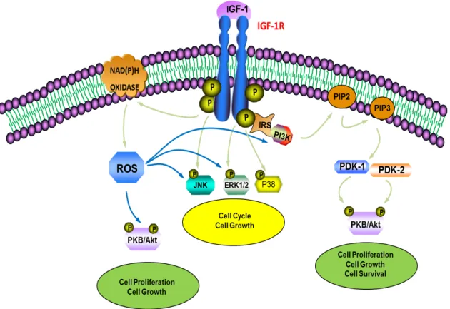
Several downstream targets of PI3-K have been documented, the most studied being PKB, also known as Akt (a product of akt proto-oncogene). PKB is a 57 kDa serine-threonine kinase with three identified isoforms in the mammalian system, PKBα/Akt1, PKBβ/Akt2 and PKBγ/Akt3 (72). All isoforms possess an N-terminal pleckstrin homology (PH) domain that binds to phospholipids generated by PI3-K, a central catalytic kinase domain with specificity for serine or threonine residues on substrate proteins, and a C-terminal regulatory domain containing a hydrophobic motif (73). Full activation of PKB is a sequential two-step process where the lipid products generated from PI3-K are recognized by the PH domain of PKB, translocating it to the plasma membrane, where it then gets phosphorylated on Thr308 by PDK-1 and on Ser473 by mTORC2 for its complete activation (74). PKB has numerous downstream targets, such as mammalian target of rapamycin (mTOR) (72), glycogen synthase kinase-3 (GSK-3) (75), and caspase-9 (76). Figure 3 illustrates IGF-1-mediated activation of the MAPK and PKB cascades.
Figure 3: Schematic representation of IGF-1 signaling pathways. IGF-1 functions as a high-affinity ligand for IGF-1R in the cellular membrane, leading to autophosphorylation of IGF-1R and recruitment of the IRS adaptor proteins. The interaction of IGF-1R with the IRS proteins induces the activation of PI3K. PI3K converts PIP2 into the lipid second messenger PIP3. PIP3 then activates PDK1 and PDK2, which go on to phosphorylate PKB at threonine 308 (Thr308) and serine 473 (Ser473), respectively. Activated PKB regulates downstream signaling molecules leading to cell proliferation and growth. In parallel, the activation of the MAPK pathway leads to increased cell survival, growth and cell cycle progression. Additionally, IGF-1R activation can lead to increased ROS generation via NAD(P)H oxidase activation, triggering the PKB and MAPK pathways (15, 61, 70, 71).
2.3C IGF-1 signaling and ROS generation
As indicated in Figure 3, in addition to activating growth promoting signaling pathways, IGF-1 has also been shown to mediate its downstream effects through ROS generation (15). ROS are very small, rapidly diffusible, highly reactive molecules that take part in physiological
reactions as well as signal transduction, but excessive synthesis of these molecules can overcome their antioxidant mechanisms and generate deleterious effects, often resulting in the development of CVDs (77). This is in part due to their physicochemical properties which allow them to disrupt biological macromolecules such as lipids, DNA, carbohydrates and proteins. Hydrogen radicals (OH˙), superoxide anions (O2˙-), hydrogen peroxide (H2O2), reactive nitrogen
radicals and its derivative peroxynitrite (ONOO-) are amongst the most important ROS. They are generated both enzymatically and non-enzymatically by nearly every cell type including VSMCs and endothelial cells (77). Studies have illustrated that the major source of ROS in VSMCs is NADPH oxidase (Nox), a flavoprotein (63, 64). It has been demonstrated that, in rat aortic VSMCs, the main components of the NADPH complex are the membrane-bound subunits of Nox and p22phox as well as Rac1, a small Rho-GTPase (63). A range of mediators detected in vascular diseases, including IGF-1, have been shown to modify Nox activity, leading to an increase in ROS production (63). Nox4 and Nox1 are the predominantly expressed isoforms of Nox in rat aortic VSMCs, however studies indicate that IGF-1 regulates the expression of Nox4 at a post-transcriptional level and, conversely, has little effect on the expression of Nox1 in VSMCs (63, 64). Inhibition of Nox4 expression by siRNA technology inhibits IGF-1-induced ROS generation in VSMCs. IGF-1-induced ROS generation has been shown to play an important role in VSMC proliferation and migration via MMPs, notably MMP-2 and -9 (63).
3. IGF-1 actions in the vasculature
IGF-1 exerts various actions in the vasculature. Numerous studies support the notion that IGF-1 is a potent mitogen that plays a permissive role in vascular dysfunction due to its pro-atherogenic properties, which contribute to VSMC proliferation and migration as well as neointimal formation (78, 79). Furthermore, IGF-1 has also been shown to promote chemotactic macrophage migration (80) and cell adherent molecule expression leading to the development of CVDs, such as atherosclerosis (58). On the other hand, IGF-1 has also been suggested to have a protective role in the vasculature.
3.1 Protective actions of IGF-1 in the vasculature
Evidence suggests that IGF-1 serves in endothelial repair and regulation of vascular tone under pathological conditions. It has been shown that IGF-1 induces vasorelaxation through the upregulation of nitric oxide (NO) production by stimulating inducible NO synthase (iNOS) in VSMC as well as through the activation of endothelial NO synthase (eNOS) in endothelial cells (ECs) (81). In fact, a study involving IGF-1 supplementation to diabetic patients, characterized by impaired vascular tone, resulted in improved vasodilatory responses mainly by increasing eNOS activity (58). Furthermore, studies using a pharmacological blocker of NO synthase (N-nitro-L-arginine methyl ester), suggest that IGF-1 acts on vascular tone by stimulating endothelial NO generation (82, 83). Moreover, studies using ECs have demonstrated that IGF-1 mediates NO production in an eNOS-dependent manner; this process also depends on the activation of the PI3-K/PKB pathway involving Ser1177 phosphorylation (58, 84).
In addition to its effects on vascular tone, high levels of IGF-1 in the intimal layer could potentially act as a potent survival factor preventing apoptosis of VSMCs, which assists in maintaining the structural integrity of the fibrous cap. This VSMC accumulation may in turn act as a vascular repair mechanism. Therefore, this protective role of IGF-1 may be beneficial for stabilization of the atherosclerotic plaque in the early stage of atherosclerosis (85).
Furthermore, IGF-1 has also been shown to exert anti-inflammatory and antioxidant properties by modulating cytokine responses and by upregulating eNOS activity. Using an ApoE-null mouse model, it was shown that IGF-1 infusion to the aorta reduced macrophage infiltration within atherosclerotic lesions, decreased aortic expression of pro-inflammatory cytokines and suppressed vascular superoxide levels via an increase in eNOS activity and NO bioavailability (86, 87). IGF-1R deficiency in the macrophages of ApoE-null mice was also shown to be associated with accelerated atherosclerosis (88). Furthermore, an in vitro study using cultured human aortic endothelial cells also found that IGF-1 has potent endothelial antioxidant effects. This effect is largely mediated through the upregulation of glutathione peroxidase-1 (GPX-1) expression and activity (89).
3.2. IGF-1 and vascular pathophysiology
In response to vascular injury, VSMCs downregulate the genes necessary for smooth muscle contractile function, such as α-smooth muscle actin and smooth muscle myosin heavy chain, they increase their proliferation rate and migration capacity, and they actively secrete matrix proteins (90). An upregulation of genes associated with the proliferative state, such as
KLF4 and PDGFR (91, 92), also occurs. Furthermore, a change in the expression of calcium (Ca2+) signaling proteins, such as voltage-gated Ca2+ channels, ryanodine receptors, STIM1/Orai1 channels and SERCA pump isoforms (93), is seen in this process, affecting Ca2+ excitation-transcription coupling in VSMCs. This state is called the “activated” or “synthetic” state. Although the synthetic state is required for vascular development and during vascular repair, it plays a pivotal role in the development of multiple smooth muscle diseases, such as atherosclerosis and restenosis following angioplasty (94). IGF-1 has been associated with mediating the VSMC phenotype switch from the quiescent/contractile state to the activated/synthetic state. Therefore, in pathological conditions, for example in the early stage of atherosclerotic plaque formation, a decrease in circulating IGF-1 levels could be beneficial (95).
3.2A IGF-1 and atherosclerosis
Atherosclerosis is an inflammatory process, which occurs when VSMCs proliferate and migrate to the intima layer resulting in neointima formation and the subsequent development of atherosclerotic lesions (96). IGF-1’s involvement in the pathogenesis of atherosclerosis has been supported by several studies, which collectively indicate a marked increase in IGF-1 and IGF-1R expression in human atherosclerotic plaques and also demonstrate that IGF-1 is able to stimulate VSMC migration and proliferation (97, 98). Studies using various inhibitors have shown that a decrease in IGF-1 or IGF-1R signaling in VSMCs is associated with reduced neointimal area size, suggesting that IGF-1 possesses a role in promoting vascular hyperplasia. Similarly, targeted overexpression of IGF-1 in VSMCs from carotid arteries resulted in an increase in neointimal formation (99). Despite the fact that, as mentioned above, studies in animal models
and human subjects (86-89) have demonstrated IGF-1’s anti-inflammatory effects, there are 20
studies that suggest otherwise. A possible inflammatory role of IGF-1 in early pro-atherosclerotic lesions was suggested by Che et al by demonstrating that IGF-1 increased basal TNF-α mRNA expression and production in cultured bovine endothelial cells (100). Moreover, in atherosclerotic macrophages, it has been illustrated that IGF-1 favors excess LDL uptake and cholesterol esterification as well as the release of inflammatory cytokines and upregulated chemotactic macrophage migration (58, 80).
3.2B IGF-1 in hypertension
Several studies have found a link between IGF-1 and hypertension. For example, increased IGF-1 mRNA expression has been found in rat models of aorta hypertrophy, urinary bladder hypertrophy and portal vein hypertrophy (101). In spontaneously hypertensive rats (SHR), increased IGF-1 caused impaired vasorelaxant properties in pre-contracted norepinephrine thoracic aortic rings as compared to normotensive (Wistar Kyoto Rats) rings (102). Furthermore, a marked increase in arterial pressure was observed in homozygous mice containing a site-specific mutation in IGF-1’s exon 3 (103), further implicating IGF-1 in the regulation of blood pressure. IGF-1’s involvement in blood pressure regulation was also observed by another group who studied liver-specific IGF-1 knockout mice and saw an increase in peripheral resistance and systolic blood pressure in these mice (104). Finally, studies performed in patients with essential hypertension reported elevated levels of IGF-1, further suggesting a role of IGF-1 in hypertension development (98, 105). Despite these discoveries, the precise role of IGF-1 in hypertension still remains unclear.
HDACs and histone acetyltransferases (HATs) regulate gene expression through the removal or addition, respectively, of acetyl groups on lysine residues of core nucleosomal histones. Increased histone acetylation attenuates the electrostatic interaction between histones and the negatively charged DNA backbone, thereby promoting the unfolding of the histone– DNA complex. This then enables transcription factors to access their sites of action, modulating the transcription of their target genes (106). As described above, IGF-1 participates in the progression of vascular remodeling by activating growth promoting and proliferative signaling pathways, such as the MAPK and PKB pathways. The activation of these pathways has been shown to alter the acetylation of status of histone and non-histone proteins (107) therefore it is possible that IGF-1 may contribute to the development of CVDs by modulating the activity of HDACs and HATs.
4.1 General overview of histone modifications
In eukaryotic cells, to accommodate the large mass of genetic material within the nucleus, DNA is packaged into chromatin. Chromatin is composed of a combination of DNA, histone proteins and some RNA. Histones can be covalently modified by a variety of processes including acetylation, methylation, SUMOylation, ubiquitination and citrullination (108), thereby providing a regulatory mechanism for controlling gene expression. These modifications alter the structure of the DNA-histone complex, which in turn affects DNA transcription. Each of these histone modifications may be involved in epigenetics; however acetylation has been shown to be particularly important in CVDs (109). As mentioned above, acetylation of lysine residues neutralizes the charge on histones, which increases chromatin accessibility (109). This is
achieved by HATs, which attach acetyl groups to conserved lysine residues on histone tails. Conversely, HDACs remove these groups and therefore reduce DNA transcription. Several studies suggest the involvement of HDACs in vasoactive peptide/growth factor-mediated aberrant VSMC proliferation and hypertrophy, linking HDACs to vascular pathophysiology (110,111,112).
4.2HDAC system and classes
As mentioned above, HDACs are a group of enzymes that lead to deacetylation of lysine residues on histones. They have also been shown to deacetylate non-histone proteins as well (113). At least 18 different mammalian HDAC genes have been identified, which can be categorized into four classes based on structure, function and the associated yeast orthologue. The four classes are class I HDACs (HDAC1, 2, 3 and 8), class II (HDAC4, 5, 6, 7, 9, 10), class III HDACs (sirtuins), and class IV HDAC (HDAC11). Classes I, II and IV HDACs have been termed classical HDACs as they require zinc as a cofactor, while class III HDACs require NAD+ as a cofactor (114). Class II HDACs are further subdivided into class IIa (HDAC 4, 5, 7 and 9) and class IIb (HDAC 6 and 10) based on their primary structure. In contrast to class IIb HDACs, class IIa HDACs contain a large N-terminal regulatory domain involved in protein-protein interactions, in addition to a C-terminal catalytic domain (115). Class IIa HDACs seem to have critical roles in many disease processes, including CVDs. For example, HDAC4 and HDAC5 have been shown to be critical in promoting VSMC proliferation and migration in response to growth factors and vasoactive peptides (110, 111, 116) and HDAC7 has been linked to PDGF-BB-induced endothelial cell migration (117).
4.3HDAC localization
HDAC localization, which can either be nuclear or cytoplasmic, greatly affects HDAC activity. HDACs that are able to shuttle in and out of the nucleus have been shown to regulate various cytoplasmic processes and function as signal transducers (118). Localization varies depending on the class. Class I HDACs are primarily located in the nucleus with the exception of HDAC3, which can also translocate into the cytoplasm. In contrast, class II HDACs are able to shuttle in and out of the nucleus. Some Class III and IV HDACs can also shuttle between the nucleus and cytoplasm, while others are confined to either the nucleus or the cytoplasm (114). Table 1 summarizes the HDAC classes, the members of each class and their localization.
HDAC class Members Localization
Class I HDAC 1 Nucleus
HDAC 2 Nucleus
HDAC 3 Nucleus/Cytoplasm
HDAC 8 Nucleus
Class II HDAC 4 Nucleus/Cytoplasm
HDAC 5 Nucleus/Cytoplasm HDAC 6 Nucleus/Cytoplasm HDAC 7 Nucleus/Cytoplasm HDAC 9 Nucleus/Cytoplasm HDAC 10 Nucleus/Cytoplasm
Class III SIRT1 Nuclear
SIRT2 Cytoplasm SIRT3 Nucleus/Cytoplasm SIRT4 Cytoplasm SIRT5 Cytoplasm SIRT6 Nuclear SIRT7 Nuclear
Class IV HDAC 11 Nucleus/Cytoplasm
Table 1: HDAC classes and localization. This figure illustrates the four HDAC classes, the members of each class and their localization (113, 114).
4.4 HDAC structure
Despite the fact that it has been over 50 years since Allfrey and coworkers first proposed the idea that the acetylation status of the histone proteins that make up chromatin is correlated with the transcriptional status of a given gene (119), information regarding HDAC structure has only become available more recently. Due to their comparable enzymatic activities, it would be expected that the proteins within the HDAC superfamily share some structural features. A study of the available structural information reveals that this is indeed true. In humans, HDACs are divided into separate classes based on sequence similarities. The Class I proteins (HDAC1, HDAC2, HDAC3, HDAC8) have a sequence similarity that extends over 300 residues with the yeast Rpd3 protein. Within this 300 residue sequence, there is an especially marked homology within an internal ∼70 residue stretch (120). To be specific, 80% similarity was found between yeast Rpd3 and human HDAC1 within the 300 residue region of conservation and 99% homology in the 70 residue stretch (120). The Class II HDAC proteins (HDAC4, HDAC5, HDAC6, HDAC7, HDAC9 and HDAC10) have a comparable sequence with the yeast Hda1 protein. Additionally, mammalian Class I and II HDACs were also found to be related to Saccharomyces cerevisiae Hos1, Hos2 and Hos3 proteins, which have 35-49% identity to Rpd3 and 21-28% identity to Hda1 (121). The Class III proteins (SIRT1, SIRT2, SIRT3, SIRT4, SIRT5, SIRT6, and SIRT7) and the yeast Sir2 protein have a comparable sequence (113). The Class IV protein (HDAC11) has a comparable sequence to both Class I and II proteins (113). Figure 4 shows domain organization of human HDACs (113).
Figure 4: This figure illustrates domain organization of human HDACs. The total number of amino acid residues in each HDAC is shown. Also, only the longest isoform is shown for simplicity, despite the fact that HDACs have several isoforms. Enzymatic domains (or putative enzymatic domains) are shown in colors (113).
Class I and II HDACs have some structural similarities to Class III HDACs in that they all contain a central parallel β sheet network that is flanked on opposite sides by helix-rich segments (120). Moreover, sequence conservation studies suggest that, in all three HDAC classes, substrate binding and catalysis is mediated by the domain proximal to the C-terminal tip of the β sheet. This domain is rich in loop segments that appear to be critical for function as they form protein cavities that are implicated in acetyl-lysine binding (122). Despite these marked
similarities, there are also striking structural differences between the Class I/II and Class III HDACs. Most notably is the small globular region in Class III HDACs, which is absent in Class I/II HDACs. This region is important for class III HDAC deacetylase activity and, due to its sequence and structural divergence, it plays a modulatory role in substrate specificity within the class III HDAC family (120). The absence of a corresponding domain in Class I/II HDACs suggests that other proteins may substitute for this domain in vivo. This is consistent with the observation that Class I/II HDACs associate with other proteins in vivo for catalytic activity (120).
4.5. HDAC expression and regulation
4.5.1 Class I HDACs
In terms of HDAC expression and regulation, Class I HDACs are ubiquitously expressed nuclear enzymes; however, it has been shown that HDAC8 is generally poorly expressed (118). Not a great deal is known about HDAC3 and HDAC8 regulation, but the other Class I HDACs (HDAC1 and HDAC2) form homo- and heterodimers between each other (123,124), which is a requirement for HDAC activity. Dissociation of the dimer will impede HDAC activity, which is something viruses take advantage of to hinder HDAC activity. For example, it has been shown that, by binding to the N-terminal region of HDAC1 and likely dissociating the dimer, the adenoviral protein GAM1 impedes HDAC1 activity (125). The level of HDAC1 and HDAC2 heterodimers is likely cell type-specific, because it was demonstrated that 80% to 90% of HDAC1 and HDAC2 proteins formed dimers in the nucleus of human breast cancer MCF-7 cells (126), whereas, in mouse embryonic fibroblasts, 40% to 60% of HDAC1 and HDAC2 proteins were shown to be free from each other (127). Additionally, Class I HDACs depend on the
presence of a catalytic Zn2+ ion for their activity and have been shown to be sensitive to inhibition by a family of small molecule compounds that have homology to trichostatin (TSA) such as suberoylanilide hydroxamic acid (SAHA) and trapoxin (TPX) (128).
4.5.2 Class II HDACs
Like Class I HDACs, Class II HDACs also depend on the presence of a catalytic Zn2+ ion for their activity. Class II HDACs, as previously mentioned, shuttle between the nucleus and cytoplasm and have tissue-specific expression and functions (128).
4.5.2a Class IIa HDACs
Class IIa HDACs (HDAC4, HDAC5, HDAC7 and HDAC9) are important signal transducers. In the regulatory N-terminal domain of these HDACs, there are several conserved serine residues that are subject to reversible phosphorylation. An array of kinases and phosphatases acting downstream of diverse biological pathways have been demonstrated to act on these HDACs, regulating their nucleocytoplasmic trafficking. On HDAC4, this phosphorylation occurs on serine 246 (S246), S467 and S632 by several isoforms of the calcium/calmodulin-dependent kinase (CaMK) family (129, 130), by Aurora B on S265 (131), by Mirk/dtrk1B on S266 (132), and by glycogen synthase kinase 3β (GSK3β) on S298 and S302 (133). The critical residues on HDAC5 are S259 and S498 and have been shown to be phosphorylated by CaMK-1, -II and –IV, protein kinase D (PKD) and AMPK (134,135,136). Protein kinase C (PKC), an upstream regulator of PKD, has also been shown to phosphorylate S259 directly (137). For HDAC7, the critical residues are S181, S155, S358 and S486 (138). The latter three sites can be modified by CaMK1, whereas PKD can phosphorylate all four residues.
Less is known about HDAC9, but it has been suggested that HDAC9 residues S239, S240 and S253 are phosphorylated by Aurora B, Mirk/dtrk1B and PRK1, respectively (138). Class IIa HDAC phosphorylation leads to the binding of 14-3-3 proteins, the nuclear export of these HDACs and the de-repression of their target genes (139).
4.5.2b Class IIb HDACs
Class IIb HDACs (HDAC6 and HDAC10) have duplicated catalytic domains, however the duplication is only partial in the case of HDAC10 (140). HDAC6 and HDAC10 shuttle between nucleus and cytoplasm, but their location is primarily cytoplasmic (141). Not much is known regarding class IIb HDAC regulation and expression.
4.5.3 Class III HDACs
Class III HDACs, also known as sirtuins (SIRT1, SIRT2, SIRT3, SIRT4, SIRT5, SIRT6, SIRT7), are NAD+ dependent and the availability of NAD+ in cells is a limiting step in the activation of sirtuin catalytic activity (142). The basal intracellular NAD+ levels are maintained relatively constant (143) through NAD+ biosynthetic and salvaging pathways (144). By consuming NAD+ in order to exert their effects, sirtuins regulate the fluctuation of the NAD+/NADH ratio. Additionally, sirtuin gene expression has been shown to be under the control of numerous transcription factors involved in cell cycle regulation and apoptosis. Among them the transcription factor E2F1, which induces cell cycle progression from G1 to S phase, directly binds to the Sirt1 promoter upregulating its gene expression in cells treated with the
topoisomerase II inhibitor etoposide (145). Furthermore, the tumour suppressor p53, which is one of the most extensively mutated proteins in cancers, has also been shown to affect Sirt1 gene expression. Two functional p53-binding sites have been identified in the regulatory region of the Sirt1 promoter and studies have indicated that in nutrient-deprived mammalian cells p53 stimulates Sirt1 gene expression (146). On the contrary, in normal nutrient conditions, p53 mediates repression of Sirt1 gene expression (147). Sirt1 levels are regulated by E2F1 and p53 at the transcriptional level as well as the translational level.
Sirtuin activity is also regulated by posttranslational modifications. In vitro evidence has demonstrated that cyclin B/Cdk1-mediated dephosphorylation at specific sites decreases Sirt1 and Sirt2 deacetylase activity (148). Additionally, Sirt1 has also been shown to be phosphorylated by the c-Jun N-terminal kinase 2 (JNK2) (149) and casein kinase 2 (CK2) (150). JNK2-mediated phosphorylation of Sirt1 is associated with the regulation of its protein stability (149). Furthermore, several conserved phosphorylation sites have been found within Sirt1 that are possible targets for a variety of kinases such as ATM, casein kinase 1 (CK1), DNA-dependent protein kinase (DNA-PK), extracellular-signal-regulated kinase (ERK1), GSK-3, IκB kinase (IKK), and MAPK (148). It is not known, however, if these kinases phosphorylate only Sirt1 or other members of the sirtuin class as well. In addition to phosphorylation, other posttranslational modifications that may regulate sirtuin deacetylase activity include acetylation and sumoylation.
4.5.4 Class IV HDACs
The Class IV HDAC (HDAC11) has sequence similarity to Class I and II HDACs. Aside from this, little is known of its function and regulation (113). 30
4.6. HDAC activation and signaling in vascular pathophysiology
4.6A Vasoactive peptide-induced HDAC activation and signaling
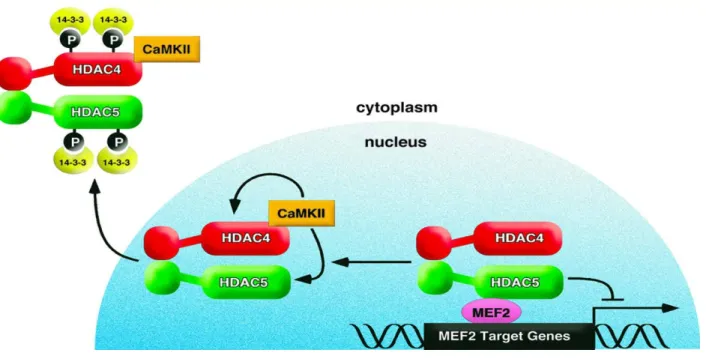
Angiotensin II (AngII), a potent vasoconstrictor, has been shown to activate several signaling pathways linked to cellular hypertrophy, growth, migration and proliferation in various cell types including VSMCs. One of its targets is CaMKII, which transduces downstream signaling responses of AngII upon activation (151). A potential transcriptional target for CaMKII is myocyte enhancing factor 2 (MEF2). MEF2, a DNA binding transcription factor which likely promotes the synthetic phenotype of VSMCs, can either act as an activator or a repressor of transcription depending on its interaction with co-activators or co-repressors, respectively (152). HDAC4 and HDAC5 have been shown to directly interact with MEF2 in the nucleus to promote its repressive activity. Consequently, HDAC4 and HDAC5 phosphorylation and nuclear export increases MEF2 transcriptional activity and leads to VSMC hypertrophy (152). As recently reported, CaMKII is capable of mediating AngII-dependent increases in HDAC4 (S467) (153) and HDAC5 (S498) (154) phosphorylation and subsequent nuclear export. This derepresses MEF2, increasing MEF2 DNA binding activity and transcription. Additionally, it was demonstrated that HDAC5 phosphorylation is mediated by HDAC4 in VSMCs, suggesting a potential regulatory mechanism involving protein-protein interaction (154). Figure 5 illustrates CaMKII-dependent nuclear export of HDAC4 and HDAC5.
Figure 5: This model depicts CaMKII-dependent nuclear export of HDAC4 and HDAC5. Following AngII stimulation, CaMKII specifically phosphorylates HDAC4 at S467 and HDAC5 at S498 (sites not shown in model), resulting in 14-3-3 protein-mediated nuclear export. Following HDAC4 and HDAC5 nuclear export, MEF2 is derepressed and MEF2-dependent gene transcription is activated (154).
Based on evidence from different cell types, class IIa HDACs have been shown to have many transcription factor targets in addition to MEF2, including serum response factor (SRF), which has been linked to CaMKII and HDAC4 in cardiomyocytes (155). Davis et al. showed that HDAC4 interacts with SRF in cardiomyocytes and this interaction is enhanced by AngII stimulation (155). Thus, MEF2 is unlikely to be the only transcription factor targeted by CaMKII and class IIa HDACs in VSMCs.
As suggested above, there are many pathways and factors involved in propagating hypertrophic and proliferative signals in VSMCs. As evidenced by Pang et al., G-protein coupled
receptor (GPCR)-kinase 2 interacting protein-1 (GIT1) has been shown to be involved in mediating HDAC5 phosphorylation by AngII (156). AngII stimulation leads to GIT1 phosphorylation, causing PLCγ activation which is required for elevation of intracellular Ca2+ and activation of CaMKII. Once CaMKII is activated, it phosphorylates HDAC4 and HDAC5 leading to nuclear export (152).
In contrast to CaMKII-dependent HDAC4 and HDAC5 phosphorylation, other studies have shown that AngII is able to induce HDAC5 phosphorylation and nuclear export in VSMCs in a calcium independent manner (157). As evidenced by Xu et al., AngII has been shown to induce HDAC5 phosphorylation and export via the protein kinase C (PKC)-protein kinase D1 (PKD1) pathway. PKD1 has been shown to phosphorylate HDAC5 at Serine 259/498 (157). PKC has also been shown to phosphorylate Serine 259 directly in the tissue of failing hearts (158). Once phosphorylated, these residues serve as docking sites for 14-3-3 chaperone proteins. This results in an increase in MEF2 transcriptional activity, which derepresses VSMC growth genes and consequently leads to VSMC hypertrophy (116).
4.6B Growth factor-induced HDAC phosphorylation and activation
In addition to vasoactive peptides, like AngII, growth factors are also involved in HDAC-related vascular pathophysiology. In VSMCs, HDAC4 and HDAC5 have been shown to be phosphorylated by CaMKII in response to Platelet-Derived Growth Factor-BB (PDGF-BB) (159).
PDGF-BB is a key mediator of VSMC phenotype switching from the contractile state to the synthetic state (160). As previously described, this switch is associated with suppressed expression of VSMC marker genes, such as smooth muscle ⍺-actin and smooth muscle myosin heavy chain, as well as increased proliferation and migration rates of cultured VSMCs. It has been shown by Yoshida et al. that PDGF-BB represses the expression of VSMC marker genes through the recruitment of HDAC4 and HDAC5 to the promoters of these genes (161). HDAC-induced hypoacetylation inhibits the accessibility of transcription factors, notably SRF, to interact with the promoters of these genes, reducing transcription (162). Additionally, HDAC5 has been shown to directly interact and inhibit myocardin, SRF’s coactivator, which also contributes to decreased transcription of marker genes (163). In terms of proliferation and migration, PDGF-BB- induced HDAC4 phosphorylation is involved in mediating proliferation and migration of VSMCs (164). HDAC4 knockdown was shown to inhibit PDGF-induced expression of cyclin D1, a cell cycle regulatory protein required for the progression of the G1 phase, which has an inhibitory effect on proliferative signals (165). Furthermore, Usui et al. also illustrated the involvement of HDAC4 in PDGF-BB-induced VSMC migration and cytoskeletal reorganization (164). Both migration and cytoskeletal reorganization were shown to be significantly inhibited by HDAC4 siRNA as well as MC1568, a Class IIa HDAC inhibitor (164).
In addition to VSMC phenotype switching, it is likely that PDGF-BB also increases ROS production by upregulating NOX activity in an HDAC4-dependent manner, as evidenced by