Université de Montréal
Knockout Mouse Model Generated by CRISPR
Technology to Study the Function of BSP Proteins on Male
Fertility in vivo
Par
Marzieh Eskandari Shahraki
Département de pharmacologie et physiologie Faculté de Médecine
Thèse présentée à la Faculté des études supérieures En vue de l’obtention du grade de Philosophie Doctoral (Ph.D.)
En Physiologie moléculaire, cellulaire et Intégrative
Avril 2019
Université de Montréal Faculté de Médecine
Ce mémoire intitulé :
Modèle de souris knockout généré par la technologie
CRISPR pour étudier le rôle des protéines BSP dans la
fertilité masculine in vivo
Présenté par
Marzieh Eskandari Shahraki
A été évalué par un jury composé des personnes suivantes :
Audry Claing, Ph.D., Président-rapporteur Puttaswamy Manjunath, Ph.D., Directeur de recherche
Serge McGraw, Ph.D., Directeur de recherche Frédérick A. Mallette, Ph.D., Co-directeur de recherche
Lawrence C. Smith, Ph.D., Membre du jury Daniel Cyr, Ph.D., Examinateur externe Sylvie Girard, Ph.D., Représentant de la doyenne
Abstract
Infertility is a worldwide problem that affects 15% of couples, with male and female factors contributing equally to cases of infertility. The cause of male infertility in half of the cases is unknown. A series of coordinated cellular events are responsible for sperm-egg interaction and successful fertilization. Therefore, elucidating the mechanisms involved in sperm production and maturation is critical to facilitate the diagnosis and treatment of male infertility, as well as the regulation of fertility. Over the last three decades, the laboratory of Dr. P. Manjunath has been extensively investigating a particular family of proteins that is expressed specifically in the male reproductive tract, called the Binder of SPerm (BSP) proteins. This protein family was first identified in bovine seminal plasma and subsequently, BSP proteins were identified in other mammalian species such as stallion, boar, ram and goat. Depending on the species, these small ubiquitous proteins are expressed by the seminal vesicles and/or epididymis. Systematic work done by Manjunath’s group has demonstrated that these proteins bind to choline phospholipids on the outer leaflet of the sperm membrane upon ejaculation and/or during passage through the epididymis and promote sperm capacitation. In 2007, BSP-homologous genes were identified in human (BSPH1) and mouse (Bsph1 and Bsph2) epididymis. Further studies indicated that the interaction of BSP proteins with the sperm membrane results in lipid (phospholipid and cholesterol) alterations and facilitates epididymal sperm maturation. Additional research on BSP proteins suggested that these proteins may play an important role in male fertility. However, the exact role of BSP proteins in fertility has remained largely unknown. In the current study, I generated the first single and double BSP knockout mouse model to investigate whether the
knockout Bsph1 and Bsph2 mice models, the Clustered Regularly Interspaced Short Palindromic Repeat (CRISPR/Cas9) approach was utilized. I used RT-PCR, Digital Droplet-PCR (dd-PCR) and Liquid Chromatography tandem Mass Spectrometry (LC-MS/MS) techniques to examine Bsp gene expression at the mRNA and protein levels and confirm whether BSP proteins were completely eliminated in knockout animals. The knockout BSP mouse lines were used to evaluate sperm parameters of fertility. No obvious abnormalities were observed in the fertilizing ability nor in the number of pups for knockout compared to wild type male mice. Sperm count and motility analyses indicated no significant differences between BSP-null mice compared to control. Interestingly, significant differences in terms of the pup’s body weight were observed in double knockout compared to wild type.
Based on these results, we conclude that BSP proteins, individually or together, are not essential for proper sperm function and fertilization in mice, and the absence of these proteins does not affect male fertility. Further studies could provide a better understanding of the mechanisms involved and possible pathways by which BSP proteins could be involved in regulating pup weight.
Keywords: Male infertility, Binder of SPerm (BSP) proteins, Epididymal proteins, Knockout Mice.
Résumé
L'infertilité est un problème mondial qui touche 15% des couples, les facteurs masculins et féminins contribuant de façon égale aux cas d'infertilité. La cause de l'infertilité masculine est inconnue dans la moitié des cas. Une série d'événements cellulaires coordonnés est responsable de l'interaction spermatozoïde-ovocyte et de la fécondation réussie. Par conséquent, l’élucidation des mécanismes impliqués dans la production des spermatozoïdes est essentielle pour faciliter le diagnostic et le traitement de l'infertilité masculine, ainsi que la régulation de la fertilité. Au cours des trois dernières décennies, le laboratoire du Dr P. Manjunath a mené des recherches approfondies sur une famille particulière de protéines exprimées spécifiquement dans l'appareil reproducteur masculin, appelées protéines Binder of SPerm (BSP). Cette famille de protéines a d’abord été identifiée dans le plasma séminal bovin, et par la suite, des protéines BSP ont été identifiées chez d’autres espèces de mammifères telles que l’étalon, le verrat, le bélier et la chèvre. Selon les espèces, ces petites protéines ubiquitaires sont exprimées par les vésicules séminales et/ou l'épididyme. Des travaux systématiques effectués par le groupe de Manjunath ont démontré que ces protéines se lient aux phospholipides portant un groupement choline sur le feuillet externe de la membrane des spermatozoïdes lors de l’éjaculation et/ou pendant le passage à travers l'épididyme, et favorisent la capacitation des spermatozoïdes. En 2007, les gènes BSP-homologues ont été identifiés dans l’épididyme humain (BSPH1) et de souris (Bsph1 et Bsph2). D'autres études ont montré que l'interaction des protéines BSP avec la membrane des spermatozoïdes entraîne une altération des lipides (phospholipides et cholestérol) et facilite la maturation des spermatozoïdes épididymaires. D’autres recherches sur les protéines BSP ont suggéré que ces protéines pourraient jouer un rôle important dans la fertilité masculine.
cadre de la présente étude, j'ai créé le premier modèle de souris BSP knockout simple et double pour déterminer si l'absence de protéines BSP pouvait affecter la fertilité masculine. Pour cibler et générer des modèles de souris knockout simple et double Bsph1 et Bsph2, l'approche de courtes répétitions palindromiques regroupées et régulièrement espacées (CRISPR/Cas9) fut utilisée. De plus, les techniques RT-PCR, Digital Droplet-PCR (dd-PCR) et chromatographie liquide – spectrométrie de masse en tandem (LC-MS/MS) ont permis d'examiner l'expression des gènes Bsp au niveau de l'ARNm et des protéines afin de confirmer l'élimination des protéines BSP chez les souris knockout. Les lignées de souris BSP knockout ont été utilisées pour évaluer les paramètres de fertilité des spermatozoïdes. Aucune anomalie évidente n'a été observée dans la capacité de fertilisation ni dans le nombre de souriceaux engendrés par les mâles knockout par rapport au type sauvage. Le décompte du nombre de spermatozoïdes et l'analyse de leur motilité n'ont révélé aucune différence significative entre les souris BSP knockout et les souris contrôle. Il est intéressant de noter que des différences significatives en termes de poids corporel des souriceaux ont été observées dans le double knockout par rapport au type sauvage.
Sur la base de ces résultats, nous concluons que les protéines BSP, individuellement ou ensemble, ne sont pas essentielles aux fonctions spermatiques et pour la fécondation chez la souris, et que l’absence de ces protéines n’affecte pas la fertilité masculine. D'autres études pourraient permettre de mieux comprendre les mécanismes impliqués et les voies possibles par lesquelles les protéines BSP pourraient être impliquées dans la régulation du poids des souriceaux.
Mots-clés : Infertilité masculine, Protéines Binder of SPerm Homolog, épididyme, souris Knockout.
Table of contents
Abstract ... i
Résumé ...iii
Table of contents ... v
List of figures... vii
List of acronyms ... viii
List of abbreviations ... ix
Acknowledgments ... xii
1. Introduction ... 16
2. Infertility ... 16
2.1 Male infertility ... 19
3. Male reproductive system ... 20
3.1 The epididymis ... 22
3.2 Epididymal maturation... 24
3.3 Epididymal proteins ... 26
4. Sperm structure ... 27
5. The Binder of SPerm protein family ... 29
5.1 Background ... 29
5.2 BSP protein structure ... 30
5.3.1 Role of BSP proteins in the first sperm maturation step (Epididymal
maturation) ... 32
5.3.2 Role of BSP proteins in the second sperm maturation step (Capacitation) ... 34
5.3.3 Role of BSP proteins in the formation of the oviductal sperm reservoir ... 36
6. Application of CRISPR/Cas9 in reproductive research ... 37
7. Thesis Objectives ... 40
Article 1. ... 43
Article 2 ... 75
8. Discussion ... 107
8.1 Efficiency of generating Bsph2-KO and Bsph1/2-DKO mice ... 109
8.2 Normal fertility in Bsph2-KO and Bsph1/2-DKO mice ... 111
8.3 Lack of BSP family proteins leads to increased body weight in mouse pups... 112
9. Conclusion and perspectives ... 113
List of figures
Figure 1: Schematic representation of the fertilization site inside the female reproductive
tract……….. ... 20
Figure 2: Diagrammatic representation of male reproductive organ in human... 25
Figure 3: Sperm transit inside the epididymis ... 27
Figure 4: Diagram of the human spermatozoa ... 31
Figure 5: Illustration of the BSP protein structure. ... 34
Figure 6: Schematic representation of the differences of secretion in the BSP proteins and their binding to the sperm membrane in ungulates, primates and rodents. ... 37
Figure 7: Proposed mechanism of involvement of BSP proteins in capacitation and destabilization of sperm membrane ... 39
List of acronyms
~ ... approximately % ... percent
e.g. ... for example i.e. ... that is µl ... microliter µg ... microgram µM ... micromolar °C ... degrees Celsius kDa ... kilodalton mM ... millimolar cm ... centimeter mg ... milligram
List of abbreviations
ADAM... A disintegrin and metalloprotease AI ... Artificial Insemination
AR ... Acrosome reaction
ART ... Assisted Reproductive Technologies BSP ... Binder of SPerm
BSPH ... Binder of SPerm Homolog Ca2+ ... Calcium
cDNA ... Complementary DNA CLU ... Clusterin
CRISP ... Cysteine-rich secretory protein
CRISPR/Cas9 ... Clustered Regulatory Interspaced Short Palindromic Repeats DKO ... Double knockout
DNA ... Deoxyribonucleic acid ddPCR ... Digital droplet PCR DSB ... Double-strand breaks
ExPASy ... Expert Protein Analysis System Fn2 ... Fibronectin type II domain GAG ... Glycosaminoglycan
GLB1 ... b-galactosidase
GPI ... Glycosylphosphatidylinositol GSN ... Gelsolin
HDL ... High-density lipoprotein
ICSI ... Intra-cytoplasmic sperm injection IVF ... in vitro fertilization
KO ... Knock-out
LC/MS ... Liquid chromatography–mass spectrometry LCN5, E-RAPB ... lipocalin 5
Min ... Minute
NPC2 ... Niemann–Pick disease type C2 PC ... Phosphatidylcholine
PCR ... Polymerase chain reaction
rec-BSPH ... Recombinant Binder of SPerm Homolog RT-PCR ... Reverse transcription PCR
SCA ... Sperm Class Analyser
SDS-PAGE ... Sodium dodecyl sulfate polyacrylamide gel electrophoresis SP ... Seminal plasma
SSNs ... Site-Specific Nucleases s ... Second
TALENs ... Transcription activator-like protein nucleases TF ... Transferrin
WHO ... World Health Organization ZFNs... Zinc-finger nucleases ZP ... Zona pellucida
Acknowledgments
In addition to my own efforts, the successful completion of this project depended largely on the encouragement and guidance of many others. I would like to take this opportunity to express my appreciation to the people who tremendously supported and helped me.
I would like to express my sincere thanks and gratitude to my research director Dr. Puttaswamy Manjunath, for giving me the courage and opportunity to undertake this wonderful project. He supported and helped me to overcome difficulties in many different aspects of my studies and life, and I am very thankful to him.
I would also like to thank Bruno Prud’homme, who has willingly helped me throughout this research project by providing his help and consultation.
In my journey through this degree, I had the opportunity to have Dr. Serge McGraw and Dr. Frédérick-Antoine Mallette as my research directors to complete my degree. I would like to thank them for their kind support and help in completing this project.
I would like to express my special gratitude towards my entire family, especially my beloved parents and siblings, who always have supported and encouraged me, and provided me with moral and emotional support. I would never have reached this stage in my academic career without their support.
I am also grateful to all my friends for their understanding and endless friendship, who allowed me to forget my studies for a few moments, who have always been there to listen to and advise
me. I thank my laboratory colleagues, especially Samin, who have always been there to cheer me up in difficult situations. They made my doctorate studies unforgettable.
I would like to convey thanks to the Département de Pharmacologie et Physiologie, at the Université de Montréal, more specifically to Dr. Réjean Couture, for his help and support. Special thanks to the Hôpital Maisonneuve-Rosemont Research Center for providing laboratory facilities. Thanks to the Faculty of Graduate Studies of the Université de Montréal for the scholarship, specifically Dr. Pierre Belhumeur. Finally, thanks to the Canadian Institutes of Health Research (CIHR) for funding the research project described in this thesis.
1. Introduction
Fertilization is a species-specific physio-chemical process, which is defined as a process of fusion between the male and female gametes to produce a new cell called the zygote. The development of the zygote results in a new individual and guarantees the survival of the species [1]. Reproduction is the basis of life and a necessity for the survival of species. Two forms of reproduction exist: asexual and sexual reproduction. In asexual reproduction, offspring arise from a single parent, to which the progeny are genetically identical [2]. Mammals produce offspring through sexual reproduction, which requires two individual parental gametes from the same species and opposite sexes. Each gamete provides half of the genetic material and the combination of the male gamete (sperm) with female gamete (oocyte) results in the creation of a new organism with equal parental genetic contribution [3]. The results of several studies point to an increased male infertility. The sources of male infertility are varied and 30-40% of men presenting to a fertility clinic will receive a diagnosis of idiopathic infertility [4, 5]. It is therefore important to better understand the molecular mechanisms underlying reproductive process to allow the development of appropriate treatments for these men. This thesis will focus only on the male fertility, the spermatozoon, following its path and the mechanisms which, according to the synthesis of sperm maturation and allow them make them capable of fertilizing an egg.
2. Infertility
Disorders in the reproductive system lead to infertility problems. Infertility is a complex and multifactorial problem that has important effects on health and social issues [6]. Based on the World Health Organization (WHO), infertility is defined as observing no pregnancy following
twelve months of regular unprotected intercourse and is considered as a disease of the reproductive system [7]. In recent decades, an alarming decline in human fertility was observed. Indeed, the latest statistics show that in Canada, 15% of couples are affected by infertility, which is about one in six couples [8]. Reproductive problems can be caused by the man or the woman, or sometimes by both. Infertility can be a frustrating issue and has a massive impact on infertile couples. It can affect the relationship and lead to social isolation, emotional pressure, stress, anxiety, depression and other psychological impacts. Research shows that three-quarters of infertile couples are likely to get divorced [9].
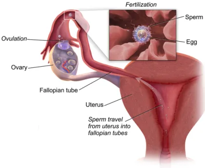
Figure 1: Schematic representation of the fertilization site inside the female reproductive tract. Fertilization usually takes place in the upper third of the fallopian tube. Human sperm are deposited into female reproductive tract, where, sperm swin trought the uterus and uterotubal junctions to reach the fallopian tubes and fertilize the ovulated oocyte
In the industrialized world, enormous changes in social life have led to a remarkable decline in fertility, which has become a worldwide problem [11]. Approximately 15% of couples are suffering from infertility problems, and male and female factors are almost equally responsible for cases of infertility [12]. Primary infertility refers to couples who fail to achieve conception despite at least one year of having sex without using birth control methods. In secondary infertility, the couples have been able to achieve pregnancy at least once, but are subsequently unable to become pregnant [13].
In view of declining fertility rates over the past 20 years, assisted reproductive techniques (ART) have been developed to help couples who are suffering from infertility issues [14]. The first human baby born through in vitro fertilization (IVF) is named Louise Brown and was born in 1978 [15]. Thereafter, the number of annual births by ART has been constantly growing. In order to enhance the success of fertilization, different types of ART have been developed for the different causes of infertility. These include artificial insemination (AI), IVF and intracytoplasmic sperm injection (ICSI) [16]. Couples whose infertility is due to male factors such as low sperm motility or abnormal sperm morphology, may benefit from ICSI, whereas couples diagnosed with female factor infertility may achieve fertilization success with AI or IVF. AI is the least invasive treatment and is used to treat couples who have difficulties with intercourse, or women with normal fallopian tubes but who are unable to conceive due to unknown reasons. In this method, fresh sperm are artificially inserted into the woman’s uterus. The most commonly used method in fertility clinics is IVF, which is employed in a variety of circumstances. In IVF, gametes from parents are collected and fertilized in vitro, then the fertilized egg is developed and the embryo is transferred into the woman’s uterus. ICSI is most often used in cases where the sperm is unable to fertilize the egg for various reasons. In this
treatment, morphologically normal sperm will be selected by a specialist and injected directly into the oocyte. Despite the high cost of ART treatments (~£2,000 per ovulation cycle in the United Kingdom and ~$8,000 in the United States), the use of ART is rising and each year 2 to 3% of births are the result of ART [17]. Several stages of natural selection are bypassed in artificial reproductive treatments, especially in ICSI. Thus, a variety of criteria need to be evaluated to assess the potential risks that could affect the mothers and their offspring in the short and long term [18].
A better understanding of the molecular mechanisms and factors implicated in the male reproductive process could improve existing ART methods and lead to the development of new effective methods of contraception. For all these reasons, this thesis will focus on male fertility in mice and humans.
2.1 Male infertility
Male infertility is a multifunctional disorder and mostly results in low sperm count or no sperm in semen, low sperm motility and high levels of abnormal sperm morphology [19]. Causes for male infertility are classified as being of pre-testicular, testicular or post-testicular origin. Male infertility can be diagnosed by semen analysis, physical examination, medical and reproductive history [20, 21].
Infertility due to pre-testicular factors can be caused by lifestyle as well as genetic history, such as smoking, alcohol intake, nutrition, hormonal abnormalities, toxin exposure, varicocele, medications, and chemotherapy [22]. Physical examination to identify causes of infertility includes an evaluation of the male reproductive system and sex organs. However, semen
analysis is the standard routine diagnostic test in fertility clinics, which assists practitioners in choosing the most appropriate treatment option and is used to predict ART outcome [21]. Despite extensive research on male infertility, the majority of reasons for male-factor infertility are still not clear, and are thus diagnosed as idiopathic male infertility [23]. On a global scale, male factors are responsible for 50% of infertility cases [24]. There exists significantly less accurate information about male compared to female infertility.
Any conditions affecting spermatogenesis such as hypogonadism, genetic defects, radiotherapy, trauma, and testicular cancer, could result in low sperm count or sperm morphological abnormalities. These conditions are classified as testicular problems and can cause male infertility. Most post-testicular problems arise from ejaculatory duct obstruction, infections and retrograde ejaculation [19, 25]. A better understanding of the causes of male infertility would allow the most appropriate treatment to be prescribed. Treatments include [13]:
- Improving sperm quality by making lifestyle changes - Surgery for duct obstruction, varicocele problem - IVF for men with low sperm numbers
- ICSI for men with low or no sperm motility
It is therefore important to better understand the molecular mechanisms underlying the reproductive process to allow the development of novel and more effective treatments for male infertility.
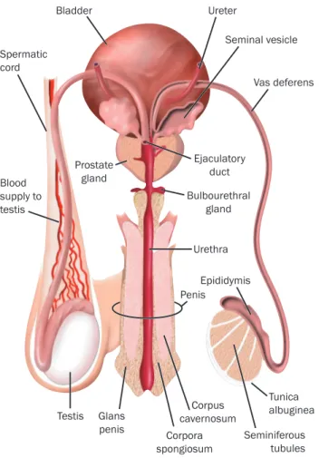
3. Male reproductive system
The following sex organs together constitute the male reproductive system [26] (see Fig 2): - The testicles
- The duct system, which is made up of the epididymis and the vas deferens - The accessory glands, which include the seminal vesicles and prostate gland - The penis.
In the seminiferous tubules of the testes, the synthesis of spermatozoa occurs in two main stages; spermatogenesis and spermiogenesis. Spermatozoa originate from germinal stem cells called spermatogonia, which are located in the seminiferous tubules [27]. A continuous complex process comprising mitotic divisions, differentiation, and meiosis leads to haploid cells called spermatids and eventually millions of tiny microscopic spermatozoa, which are stored in the cauda epididymis [27, 28].
A large number of sperm cells are continually produced in the testis by a process named spermatogenesis [29]. During spermatogenesis, the spermatocyte, which originates from the mitotic division of spermatogonial stem cells, undergoes meiotic division and generates two secondary spermatocytes (Meiosis I). Then, each secondary spermatocyte will continue into Meiosis II and divide into two equal haploid spermatid cells. Spermatids mature into spermatozoa in a process referred to as spermiogenesis [27]. The two testicles or testes in sexually mature males also produce testosterone, which is the major male sex hormone and plays a key role in the development and maintenance of the male reproductive organs [30]. The process of spermatogenesis is highly dependent on the activity of two types of cells in testes: Sertoli cells and Leydig cells. These two major cell types line the seminiferous tubules. Sertoli cells provide nourishment and structural support to the developing sperm cells, whereas Leydig cells are responsible for the synthesis of steroid hormones necessary for germ cell differentiation. The testes produce around 95% of testosterone in males, and a small percentage
Figure 2: Diagrammatic representation of male reproductive organs in human. Spermatozoa are first form in the testes and become mature by trnsit through the epididymis, they are stroed
until ejaculation. (adapted from [13]).
3.1 The epididymis
The epididymis is a narrow and highly segmented organ that serves as a connector tube between the testicles and the vas deferens, as well as providing a milieu for sperm to acquire their motility and fertilizing capacity [32]. It is a tightly-coiled and extremely long organ, with lengths varying from 1 meter in mice to 6-7 meters in human, which is located above the testes [33]. The
ejaculation, ejaculatory duct obstruction, hypogonadism or congenital bilateral absence of the vas deferens. The presence of sperm in a post-ejaculatory urinalysis in someone who is azoospermic or has low volume ejaculate (less than 1 ml) may indicate retrograde ejaculation (Jarrow et al, 2011). Men should also be screened for chlamydia (NICE, 2013).
If the problem is thought to be caused by an obstruction somewhere along the vas, epididymis or ejaculatory duct (Figure 1) then investigations to locate the obstruction and ascertain if this is reversible would also involve a scrotal ultrasound besides the investigations already mentioned. Physical examination of the testicle may indicate that there is epididymal enlargement, nodules in the epididymis or vas deferens, and the vas may be absent or partially missing or malformed. Varioceles, spermatoceles, epididymal induration and testicular masses are also palpable on physical examination. The ultrasound would confirm dilatation of the testis or enlarged cysts in the epididymis or the missing vas. Ultrasound is indicated especially in an individual in whom physical examination of the scrotum is difficult or may not have been adequate or in whom the physical findings were ambiguous (Faraj et al, 2014; Jungwirth et al, 2015).
Scrotal ultrasound is indicated if a testicular mass is suspected (Jarrow et al, 2011). A transrectal ultrasound may also be indicated
is thought to be distal so as to visualise the prostate and the semincal vesicles (BAUS, 2016). These patients are described as having OA, i.e. these patients are able to produce sperm but these are not able to progress to the point where they become part of semen produced and so the ejaculate is semen-free. OA is less common than NOA; it occurs in 15-20% of men who are azoospermic and most of these are acquired forms of obstruction rather than congenital. These patients may also go on to have testicular biopsies and extraction of testicular spermatozoa (Jungwirth et al, 2015).
Another investigation that may be used is for antisperm antibodies (ASA) in the semen. Men at risk of ASA are those who have had genital infections, testicular trauma or ductal obstruction. ASAs may reduce pregnancy rates (Jarrow et al, 2011). However, NICE (2013) does not recommend this as standard as it points out that the significance of antisperm antibodies is as yet unclear.
Treatment
Treatment for azoospermia will depend on why and what is causing the problem, so there is a variety of options.
Improving quality of sperm
Adopting lifestyle changes such as reducing smoking, wearing loose clothing, especially underwear, reducing drug consumption and weight loss may help (Sharma et al, 2013; Faraj et al, 2016). NICE guidelines stated that men with a body mass index (BMI)
greater than 29 kg/m2 are likely to have reduced fertility and
should lose weight (NICE, 2013). Obese patients may benefit from anti-oestrogens and aromatase inhibitors so their FSH and LH levels may rise and potentially improve sperm quality. Naturally, weight loss should also be encouraged. Reducing alcohol intake is also recommended (Sharma et al, 2013) as excessive alcohol may be detrimental to semen quality.
NICE (2013) stated that it is uncertain that wearing loose-fitting underwear improves semen quality but reducing the temperature in the scrotum may. BAUS (2016) also suggest that splashing the scrotum with cold water 2–3 times a day may be beneficial.
Good nutrition is also important, not only to improve general health, but also to help improve the quality of the sperm and sperm production. Infertility Network UK (2016) recommends ensuring that diet includes vital nutrients such as zinc to improve sperm motility; vitamin C, which protects sperm from DNA damage and prevents sperm agglutination; arginine, which is needed to produce sperm; vitamin B12; vitamin E to improve fertility; and coenzyme Q10, which has also been found to improve quality, quantity and mobility of sperm (Sharma et al, 2013; Infertility Network UK, 2016).
It may also be worth having a discussion about the man’s work and workplace environment to identify if any changes can be made to where or how they work, especially if they have a job entailing long periods of sitting down or working in exceptionally hot places. An increase in temperature may result in a decrease in sperm production (Faraj et al, 2016).
The scrotum regulates the temperature of the testis, which should be 2-4o below body temperature so if the temperature
Penis Testis Seminal vesicle Prostate gland Spermatic cord Epididymis Vas deferens Ejaculatory duct Urethra Tunica albuginea Seminiferous tubules Bulbourethral gland Corpora spongiosum Corpus cavernosum Bladder Ureter Blood supply to testis Glans penis
Figure 1. Male sexual and reproductive organs
Peter Lamb
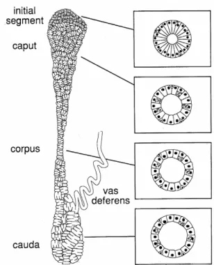
epididymis serves not only as a storage organ for viable sperm but also provides an environment in which sperm undergo many biochemical and morphological changes, allowing them to mature gradually in a process collectively called epididymal maturation [34]. Spermatozoa are stored in the cauda segment of the epididymis for one to two weeks. The most important function of this part of the epididymis is to preserve sperm viability by regulating the motility and metabolism of stored sperm [35, 36]. Sperm motility is repressed by several factors in order to save energy, such as reduction in scrotal temperature, the low oxygen content in epididymal fluid and tissue, the absence of glucose, and variations in the ionic composition of epididymal fluid [37]. The epididymal lumen also provides a specific and continually modified environment in which sperm achieve maturation [34, 38]. Thousands of proteins are actively synthesized in the epididymis in response to androgen stimulation [33]. Studies have estimated that the human epididymal proteome contains 7500 proteins, while 2850 proteins have been identified in the murine epididymis[39]. Regardless of species, sperm transit gradually through the epididymis in a journey that lasts approximately 10 days, and undergo a large number of modifications [34]. Transport, storage, and maturation of sperm are the main functions of the epididymis [40]. Anatomically, the mammalian epididymis is constituted of three main parts: the head (caput), body (corpus) and tail (cauda) (Figure 3).
In rodents, however, the epididymis can be divided into four regions, compared to the three main anatomical regions of epididymides in other mammals. In rodents, the proximal segment of the caput is distinct and is named the initial segment [33]. The luminal fluid of each segment differs in terms of ion concentration, pH and osmolality. Water absorption actively takes place in the initial and proximal segment [33].
Figure 3. Schematic representation of the different regions of mouse epididymis (left panel) and the right are shown cross sectional of the epididymal duct. The epididymis is an organ located above the testicle. It is a long tube and divided into four major parts: the initial segment, the head (caput), the body (corpus) and the tail (cauda). However, these four parts can differs from one species to another. (adapted from [33]).
3.2 Epididymal maturation
During epididymal transit, mammalian sperm are exposed to the epididymal milieu. During their journey through the epididymis, the sperm plasma membrane undergoes a variety of sequential modifications, which result in the acquisition of sperm motility and fertilization capacity. This process is known as epididymal maturation [41]. The composition of the epididymal milieu varies from one segment to another [42]. During epididymal maturation, the major modifications occurring on the outer leaflet of the sperm plasma membrane allow sperm to acquire motility and the ability to fertilize an oocyte [43]. Before sperm are transferred into the
Copyright © American Society of Androloy Handbook of Andrology – What does the epididymis do and how does it do it?
10-1 10-2
What does the epididymis do and how does it do it?
B.T. Hinton
“If anyone asks what the epididymis is, we shall answer that it is a vessel constituting by various twists a body affixed to the back of the testicle” (de Graaf, 1668; see Jocelyn & Setchell, 1972).
Spermatozoa leaving the testis are neither motile nor able to recognize or fertilize an egg; they must traverse a long duct, the epididymis, to acquire these abilities. These post-testicular transformations of spermatozoa are collectively called sperm maturation. The epididymis is a single highly convoluted duct/tube of approximately 1 meter in length in the mouse, 3 meters in the rat, 6 meters in the human and a remarkable 18 meters in the stallion. Hence, it can take anywhere from 1 to 14 days for spermatozoa to traverse the epididymis. Early investigators considered the epididymis as a holding tube for spermatozoa and that it was a place where spermatozoa aged. It was thought that the maturation process was inherent to spermatozoa and had little to do with the epididymis. It is now clear that the epididymis is very much an active participant in the maturation process, not only providing an appropriate luminal fluid microenvironment, but also supplying many of the molecules required by spermatozoa for the acquisition of fertility. The challenge for many investigators has been to identify those molecules. In addition to its sperm maturational role, the epididymis places a premium on protecting spermatozoa as they mature; it also provides an environment for storage following the maturation process. Since spermatozoa are immotile, they require assistance to move along this very long duct. This movement is aided by contractions of smooth muscle that surround the duct as well as pressure from fluid and spermatozoa entering the duct from the testis. From a clinical perspective, an improper functioning epididymis results in male infertility and therefore, the epididymis is considered to be a prime target for the development of a male contraceptive. Interestingly, unlike the testis and prostate, cancer is rarely observed in the epididymis.
Structure of the epididymis
The gross anatomical structure of the epididymis in a variety of species is divided into several regions that include: the initial segment, caput, corpus and cauda regions. Proximally, the efferent ducts connect the testis to the epididymis and distally, the vas deferens extends from the cauda region
(FIG. 1). Within each region there are multiple segments separated by septa, with the numbers of segments within each region being variable. The challenge for investigators is to relate the different regions and segments to epididymal function and sperm maturation.
FIG. 1. Schematic representation of an epididymis showing the different regions: initial segment, caput, corpus and cauda. To the right are shown cross-sectional representations of the epididymal duct at each region. Note how the luminal diameter increases and the cell height decreases from the initial segment to the cauda.
The epithelium of the epididymis is comprised of several cell types including: principal, basal, apical, halo, clear and narrow cells, each of which vary in number and size along the epididymal duct. For example, principal cells in the initial segment are tall resulting in a duct with a small luminal diameter whereas in the cauda region, the principal cells are low columnar and luminal diameter is much larger (Fig. 1, 2). Through extensive analyses a much clearer picture is beginning to emerge regarding the function of each cell type within each epididymal region. Principal cells are known to actively secrete ions, organic solutes and proteins. They are involved in endocytosis and many receptors and transporters are localized to their apical and plasma membranes. Clear and narrow cells play a significant role in the acidification of the luminal fluid and also contain endocytotic machinery. Maintaining an acidic pH luminal fluid microenvironment is important for sperm maturation.
female reproductive tract upon ejaculation, the epididymal mixture is blended with fluid secreted from the accessory glands (seminal vesicles, ampullae, prostate, and bulbourethral glands). This organic fluid, which is known as semen or seminal plasma (SP), is critical for sperm function. It serves as a source of energy for sperm, allows further sperm maturation, and ensures sperm survival and transport [44]. Semen is a mixture fluid secreted from the testes, the excurrent ducts and the accessory glands, and is constituted of two types of components; cellular and non-cellular. The non-cellular portion is enriched with various proteins that are synthesized and secreted in a region-dependent manner from the male reproductive tract, and contains lipids, carbohydrates, proteins and some minerals [44]. Several biochemical and structural modification events occur during epididymal maturation, rendering sperm fertilization-competent cells and preparing them for the second extra-testicular maturation step that takes place in the female reproductive tract, known as capacitation [45]. The two maturation steps (epididymal maturation and capacitation) alter the concentration and distribution of lipids in the sperm membrane, resulting in a decrease in the cholesterol/phospholipid ratio [46, 47].
In addition to the previously mentioned sperm modifications occurring during epididymal transit, the protein composition of the sperm membrane also undergoes several modifications during this journey. Sperm lose several membrane proteins and acquire others, such as secreted epididymal proteins including decapacitation factors. These decapacitation factors bind to the sperm membrane and protect sperm from premature capacitation [41, 48].
Upon ejaculation, sperm are deposited into the female genital tract along with SP, and decapacitation factors are removed from sperm surface in order for sperm to undergo the second maturation step known as capacitation. [45]. Capacitation includes a number of membrane
Studies indicate that the isthmus of the oviduct is the region of the female reproductive tract in which the sperm cell surface undergoes the capacitation process, resulting in the full acquisition of fertilizing ability.
3.3 Epididymal proteins
All of the fundamental sperm modifications required to create a fertile sperm occur inside the epididymal tubule. It is therefore important to better understand all the transformations involved in sperm maturation as well as the proteins implicated in these modifications in order to develop appropriate treatments for male infertility [37]. Since the 1970s and 1980s, many investigations have undertaken to elucidate the protein composition of the epididymis. Spermatozoa travel a long distance in the epididymis and undergo multiple sequential changes in each of the epididymal sections, which have different luminal fluid compositions [38]. The majority of epididymal luminal proteins are secreted by the surrounding epithelium. The secretory activity of each epididymal section is different, leading to distinct protein compositions and concentrations in each region of the epididymis [34, 50]. Epididymal fluid consists of a large pool of soluble (hydrophilic) and insoluble (hydrophobic) proteins, most of which are soluble [51]. The proximal region of the epididymis is responsible for secreting 60 to 83% of epididymal proteins [35]. Several of these proteins are epididymal region-specific and are common among different species. Examples of such proteins are glutathione peroxidase (GPX5), prostaglandin D2 synthase (PTGDS) and RNAse10, which have been found in the proximal epididymal regions [38]. Proteins expressed in the middle and distal parts of the epididymis include lactoferrin (LTF), Niemann–Pick disease type C2 (NPC2), several glucosidases and gelsolin (GSN) [38]. Several of these proteins are common to different species, such as Clusterin (CLU),
transferrin (TF), GSN, NPC2, LTF, lipocalin 5 (LCN5), actin, and b-galactosidase (GLB1) [35, 52, 53]. However, However, epididymal proteome is a quite dynamic entity and the proportion of expressed proteins varies from one species to another [54]. In human for example, 77% of the total luminal proteins are represented by albumin (ALB) (43.8%), CLU (7.6%), NPC2 (6%), LTF (5.9%), extracellular matrix protein (ECM1) (3.2%), a1-antitrypsin (SERPINA1 (A1AT)) (2.7%), PTGDS (2.2%, 1.7%), TF (1.3%), and actin (1.2%) [32].
4. Sperm structure
Spermatozoa are highly specialized cells with minimal cytosol and organelles [55]. The mission of these haploid cells with forward motility is to travel towards the ovum in the female reproductive tract and deliver the paternal genome by combining with the ovum to generate a diploid embryo [56]. These small haploid cells possess remarkable features that make them distinguishable from the other cells. Their motility allows them to travel a long distance in the male and female genital tracts, where they undergo multiple modifications to acquire their fertilizing ability, which enables them to accomplish their mission [57]. Sperm cells are composed of four major parts. The first compartment is the head of the sperm, which contains a very condensed cytosol with no organelles such as ribosomes, endoplasmic reticulum or Golgi apparatus. A very large secretory granule known as the acrosome is located on the anterior part of the sperm head [58]. The acrosome contains the hydrolytic enzymes necessary for sperm to penetrate the egg. The nucleus is located behind the acrosome and contains tightly packed DNA. Special proteins named protamines ensure that sperm DNA is tightly condensed in order to stabilize and maintain DNA integrity during sperm transit through the male and female
The second section is the neck, which connects the head of the sperm to the midpiece, and contains two centrioles for chromatin segmentation [60]. The third section is the midpiece, which contains many spiraled mitochondria that produce ATP and thus provide the energy source for sperm motility [61]. The tail or flagellum is the fourth section, which beats and allows the sperm to move [62] (Figure 4).
Figure 4. Diagram of the human spermatozoon. Taken from:
5. The Binder of SPerm protein family
5.1 Background
Seminal plasma (SP) is a heterogeneous fluid that contains numerous different proteins secreted by the epididymal epithelium, seminal vesicles and other accessory glands. SP induces alterations in the sperm membrane, and mediates the exposure of several sperm membrane proteins and receptors essential for sperm maturation, sperm-egg interaction and fertilization. Despite years of investigations, the function and role of many SP proteins are still unknown. A highly conserved superfamily of proteins, named Binder of SPerm (BSP) proteins, was identified in the SP of more than 15 different mammalian species. The BSP protein family, which is a ubiquitous superfamily among mammals, has been investigated by our laboratory over the past three decades [63]. Depending on the species, BSP proteins are expressed by the seminal vesicles and/or epididymis [64-66]. The members of this family share many similar characteristics such as their expression site, a common structure, and similar binding properties to various ligands and to the sperm membrane [67-70].
Proteins from the BSP superfamily were first identified and characterized in bovine SP. In the bovine species, three BSP proteins (BSP1, BSP3 and BSP5; previously named BSP-A1/A2 or PDC-109, BSP-A3 and BSP -30K, respectively), constitute more than 60% of total SP proteins, and have been shown to be essential for sperm capacitation [63]. Following this discovery in bovine, BSP genes and proteins were identified in other species such as ram, goat, boar, bison and buffalo [69, 71-73]. In 2006, a study reported the existence of two other BSP-related genes in bovine, which are expressed in the epididymis [74]. In farm animals, BSP proteins are mainly
expressed by seminal vesicles in large quantities, whereas BSP proteins originating from the epididymis are present in very low quantities in SP [75].
Shortly thereafter, BSP homologues expressed in mouse and human epididymis were identified by Lefebvre et al. [74]. The investigations of the mouse and human genomes resulted in the discovery of three and one BSP sequences, respectively [64, 76, 77]. The mouse Bsp genes were named mouse Bsp Homologue 1, 2 and 3 (mBsph1, mBsph2 and mBsph3). No mRNA transcripts were identified for mBsph3, which appears to not be an active gene and likely represents a pseudogene [76]. The BSP-homologous gene in human was named hBSPH1 [74]. Important differences exist between mouse/human BSP proteins and bovine BSPs, such as the location of protein expression and the quantity of these proteins in SP. Approximately 60% of bovine SP is constituted by BSP proteins, whereas these proteins form <0.01% of total SP proteins in mice and humans [78].
5.2 BSP protein structure
Most BSP proteins and their homologs are acidic and are relatively small, with molecular masses of 12-30 kDa. The fibronectin type 2 (Fn2) structure is common in this family (function described below) [63]. BSP family proteins possess a conserved secondary structure constituted of a variable N-terminal domain comprising 15 to 71 amino acids, followed by two highly conserved and tandemly arranged Fn2 (Fn2-A and Fn2-B) domains (38-42 amino acids), which are linked by a seven amino acid polypeptide linker as shown in Fig. 5 [77, 79]. A very short variable C-terminal domain (1-5 residues) exists in some members of the BSP family [80-82]. Each Fn2 domain comprises four cysteine residues and two disulfide bonds with connectivities 1-3 and 2-4, to form a hydrophobic pocket [80, 81, 83, 84]. Studies have shown that the two Fn2
domains are responsible for many binding properties of BSP proteins. BSP proteins interact with a wide range of partners including glycosaminoglycans (GAG) such as heparin and chondroitin sulfate B (CBS) [85], choline phospholipids [86], gelatin [87], calmodulin, high-density lipoproteins (HDL) [88], ApoA-I, low density lipoproteins (LDL), insulin-like growth factor II, PLA2, and milk proteins (casein micelles, α-lactalbumin, β-lactoglobulin) [63, 79].
The two active BSP family genes in mice are mBsph1 and mBsph2, which span 24 and 21 kb, respectively. Both genes include 5 exons and 4 introns and are located on chromosome 7. The human Bsp gene is located on chromosome 19 and is composed of six exons and five introns [74].
Figure 5. Illustration of the BSP protein structure (adapted from Plante et al. (2016) [67]).
5.3 Biological roles of BSP proteins
Studies have shown that BSP proteins are involved in many stages of fertilization, including epididymal maturation (development of sperm motility), capacitation, formation of the oviductal sperm reservoir and prolonging sperm viability [67].
5.3.1 Role of BSP proteins in the first sperm maturation step
(Epididymal maturation)
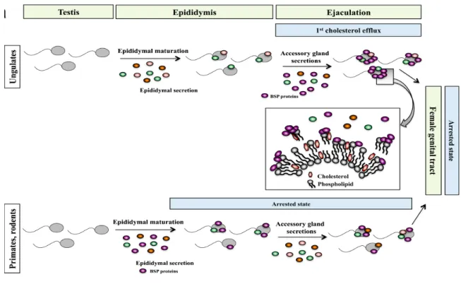
The primary function associated with BSP proteins is their ability to promote sperm capacitation. This capacitation-inducing role of BSP proteins was shown in bovine by Thérien et al. in 1995 [89]. The binding sites for BSP proteins on the sperm plasma membrane are choline phospholipids. The most predominant choline phospholipid derivatives on the sperm membrane are phosphatidylcholine, plasmalogen and sphingomyelin, which account for 70% of the total phospholipids of the sperm membrane [66, 90]. As sperm pass through the epididymis in mouse and human, BSP proteins expressed by the epididymal epithelium bind to the sperm membrane and remove phospholipids and cholesterol from the membrane, prompting a first cholesterol efflux (Figure 6) [91]. However, in ungulates and other farm animals for whom BSP proteins are expressed in the seminal vesicles and not in the epididymis, the first cholesterol efflux occurs during ejaculation when sperm mix with SP and encounter a high concentration of BSP proteins (Figure 6). As sperm are transferred to the female genital tract and the excess free BSP proteins is removed, the first cholesterol efflux ends [91]. However, in the female tract the interaction of BSP proteins with HDL and GAGs on the surface of sperm induces a second cholesterol efflux, resulting in capacitation (Figure 7) [92, 93].
As sperm transit through the epididymis, the protein, lipid and sugar content of the sperm membrane is changed, and it acquires new proteins [42, 94]. BSP proteins are among the proteins that bind and coat the sperm membrane. Gwathmey et al. showed that BSP proteins maintain sperm viability and motility in the cauda epididymis by preventing excessive membrane alterations (Fig. 6) [91, 95].
Cholesterol and phospholipid efflux from the sperm membrane cannot occur unless free BSP proteins in SP bind to the sperm surface and induce the first cholesterol efflux [96, 97]. In ungulates upon ejaculation, free BSP proteins found in SP are mixed with sperm and interact with the membrane and mid-piece of the flagellum. This interaction induces the first cholesterol efflux, resulting in the activation and/or enhancement of calcium (Ca2+)-ATPase (PMCA4) and increase sperm motility following ejaculation (Fig.6) [98-101]. In vitro studies have shown that bovine BSP1 binds to the sperm plasma membrane, and this interaction enhances the activity of PMCA4 [88, 102, 103]. The maintenance and regulation of the swimming behavior of spermatozoa is mainly controlled by Ca2+ signaling. As Ca2+ concentration increases, the sperm flagellar beats increase, thus enabling sperm hyperactivation [56]. As indicated in Fig. 6, in ungulates BSP proteins are expressed by the seminal vesicles and constitute the major proteins of SP (1-50 %). In human and mice, these proteins are expressed in the epididymis and account for a minute amount (0.01%) of epididymal and SP proteins. Remaining unbound BSPs are removed from SP as sperm enter into the female genital tract.
Figure 6. Schematic representation of the differences in the secretion of BSP proteins and their binding to the sperm membrane between ungulates, primates and rodents (adapted from Plante et al. (2016) [67]).
5.3.2 Role of BSP proteins in the second sperm maturation
step (Capacitation)
Six decades ago, two investigators (Austin [104] and Chang [105]) demonstrated that when sperm pass through the uterus and oviduct they undergo multiple biochemical and physiological modifications. These modifications are collectively considered as the second sperm maturation step and referred to as capacitation [27]. The efflux of cholesterol initiates capacitation, which includes several changes such as increased intracellular pH, increased permeability to Ca2+, activation of several signaling pathways, increased phosphorylation of tyrosine residues and the
development of hyperactivity of flagellum [106]. The duration of this synchronized event is very short, taking between 50 minutes and 4 hours in human [107-111]. Several studies have demonstrated that one of the main functions of BSP proteins in bull, stallion, ram, and goat is to trigger sperm capacitation [73, 89, 112-114]. The interaction of the three BSP proteins found in bull SP with decapacitation factors such as HDL and/or GAGs results in the removal of cholesterol and phospholipids from the sperm membrane, causing an alteration in membrane permeability (Figure 7) [65, 91, 95, 115]. Studies on bovine spermatozoa indicated that BSP proteins have a dual effect on the sperm plasma membrane [90]. The high concentration of BSP proteins in SP causes the first cholesterol and phospholipid efflux upon ejaculation. As sperm pass through the cervical mucus, the first phospholipid efflux ceases because of removal of excess BSP proteins as SP is left behind. However, the interaction of sperm-bound BSP proteins with follicular and oviductal fluid mediates a second phospholipid and cholesterol efflux, leading to the destabilization of the sperm membrane and the initiation of capacitation [65]. BSP family members showed different levels of affinity in binding to the sperm membrane. However, despite these differences, studies indicate that the BSP proteins play an important role in the preparation of sperm to fertilize the egg [65, 116].
Figure 7. Proposed mechanism of the involvement of BSP proteins in sperm capacitation and the destabilization of the sperm membrane (adapted from Plante and Manjunath (2016) [67]).
5.3.3 Role of BSP proteins in the formation of the oviductal
sperm reservoir
Among the hundreds of millions of spermatozoa that enter the female reproductive tract at ejaculation, only a few thousands can reach the fertilization site. Once these sperm reach the oviduct (Fallopian tubes in human), they bind to the oviductal epithelium and form a sperm
reservoir. This contact with the oviductal epithelium preserves the viability and motility of sperm, and prevents their premature capacitation [117, 118]. Sperm liberation from the oviductal reservoir is synchronized with the time of ovulation [119]. Studies have shown that one of the factors implicated in the formation of the sperm reservoir is sperm-bound BSP proteins, which bind to molecules on the epithelial surface [120, 121]. Sperm are released from the reservoir as GAGs in follicular fluid interact with sperm-bound BSP proteins at the reservoir site [122].
6. Application of CRISPR/Cas9 in reproductive research
Recent advances in genome editing is unravelling its benefits in the study of reproductive biology and other wide areas of scientific researches. Nowadays, generating mutant organisms has been widely developed by breakthrough genome editing technology named the Clustered Regularly Interspaced Short Palindromic Repeat (CRISPR)/CRISPR-associated (Cas) system. This system allows modulation of gene expression, targeted gene cleavage using site-specific nucleases (SSNs) and gene editing in a variety of eukaryotic cells with high efficiency outcomes [123]. Over the years, several techniques have been applied to manipulate specific DNA sequences in cells and a variety of animal models, including generation of random mutation by chemical or radiation exposure resulting DNA damage, zinc-finger nucleases (ZFNs), transcription activator-like protein nucleases (TALENs) to create double-strand DNA breaks and RNA interference (RNAi) [124].Investigation of the adaptive immune system against plasmids and phages in prokaryotes composed of non-contiguous direct repeats interspaced by variable sequences results in the creation of a novel technique called the CRISPR/Cas9 system [125, 126]. This discovery was
factors involved in a variety of cellular pathways. Adapting this recent approach has emerged as the most popular tool for the precise alteration of the genome with dramatically less effort and expenses. CRISPR/Cas9-mediated genome editing requires a short RNA to target its sequence (protospacer) and make double-strand brakes in the genome [127].
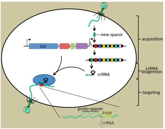
Briefly, CRISPR/Cas9 is an adaptive and defence system in Bacteria and archaea, which exploit a short RNA to generate accurate breaks in the specific inserted DNA sequence of bacteriophage and/or invader plasmid to cleave and repel the foreign nucleic acids [128]. CRISPR loci are composed of considerable repeat and non-repeat (spacer-segments derived from cleaved foreign DNA) sequences, while Cas gene is located upstream of these sequences and encode putative nuclease or helicase proteins (Figure 8).
CRISPR-associated nuclease called Cas9 is guided by short single guide RNA (sgRNA) to recognize and cleave a target of 20 nucleotides in the genomic sequence located at downstream of recognition site of Cas9 named Protospacer Adjacent Motif (PAM) sequence [129]. The sgRNA is transcribed from CRISPR loci of incorporated exogenous DNA, which serves as a genetic record and composed around 30 bp sequences. PAM sequence (5′ NGG 3′) is the target binding site for Cas protein and is located immediately upstream of the targeted sequence [129]. The sgRNA and PAM are the two specific and essential features of the CRISPR/Cas9 complex to track and develop an incision at the desired sequence [130]. CRISPR/Cas 9 introduces double-strand breaks (DSBs) at sites of interest approximately three nucleotides upstream of the PAM sequence. The DSBs can repair by either the imprecise non-homologous end joining (NHEJ) or precise homologous recombination (HR) DNA repair pathways. NHEJ rejoins the broken ends of DNA with random insertions or deletions of DNA nucleotides (indels) [131].
Homology-directed repair, in the presence of a donor DNA sequence, could be harnessed to edit or replace the sequence of any given gene, resulting in gene correction or gene addition [131].
Figure 8. Three stage of defence immune system in Bacteria and archaea. A short DNA sequences of invader is captured and incorporated into the CRISPR loci as new spacers. Upon second infection, the CRISPR locus is transcribed and processed to generate mature CRISPR RNAs, crRNA along with Cas protein guide to target the foreign DNA match with crRNA (adapted from Barrangou and Marraffini (2014) [126]).
Genome editing is used in wide areas of scientific development to modify endogenous genes in a wide variety of cell types and in organisms that have traditionally been challenging to manipulate genetically. The CRISPR/Cas9 system has already been applied to produce genetically modified mice by CRISPR delivery into mouse zygotes to easily and quickly
been widely used in reproductive research to find out the cause of infertility and create therapeutic approach at the molecular level.
In recent years the CRISPR/Cas9 technology is used widely to study several fertility-related genes in males to explore the vital factors of fertility and develop therapeutic approaches. Additionally, the CRISPR/Cas9 systems are used to generate knockout mouse models to study the role of an individual or multiple genes in male reproductive systems in vivo to unlock the mechanisms behind fertility and explore the vital genes in reproduction. Due to the rapid reproductive cycle and the high genetic similarity to the human, mice are the ideal models to investigate the essential genes for male fertility. [132]. A combination of a mouse model and new techniques such as CRISPR/Cas 9, can be used to elucidate the function of genes that are similar to those in humans by genetically modifying mice. Human and mouse BSP proteins are orthologues proteins. Mouse BSPH1 shares 56% identity and 78% sequence similarity with human BSP protein. While, mouse BSPH2 shares 40% identity and 55% sequence similarity with Human BSPH [133]. Therefore, the use of the mouse as an animal model for human BSP can help to elucidate the role of epididymal BSP proteins in sperm maturation, as well as the other possible roles of this protein family in fertilization.
7. Thesis Objectives
The main roles that have been assigned to BSP proteins in ungulate species are their interaction with the sperm membrane, their ability to trigger sperm capacitation and prepare to fertilize the ovum [71, 72, 91, 134, 135]. Homologs of BSP proteins were also found in the SP of mouse and human, though in very minute amounts [74]. Studies indicate that these proteins may be involved in the maturation of mouse and human spermatozoa, allowing them to gain fertilizing
ability. However, since BSP proteins are found in negligible quantities in human and mice SP, the isolation of these proteins from SP is not feasible. Consequently, the study of their biological functions in vitro has been restricted.
Male fertility has been declining over the last three decades; therefore, understanding the mechanisms underlying fertilization and identifying the key factors required for this process is highly important in order to develop new diagnostic tests and treatments for infertility. The current study thus aimed to gain more insight into the role of BSP proteins in sperm maturation and fertilization in mice. Studies on BSP proteins have shown that these proteins are crucial for fertilization in cattle. The discovery of genes encoding proteins of the same family in humans and mice has opened new doors to the identification of new factors that may have an impact on the fertility of these species. Our working hypothesis for this thesis was that BSPs in humans and mice are added to spermatozoa during epididymal maturation and have roles in sperm functions and fertilization. The overall objective of this thesis was to specifically determine the roles of epididymal Bsph1 and Bsph2 in sperm maturation in mice, as well as to determine whether these proteins are essential for fertilization. To answer these questions, the overall objective was divided into six specific objectives:
1. Generation of single (Bsph1 and Bsph2 separately) knockout mice. The novel gene-editing technology CRISPR/Cas9 was selected as a method to target and disrupt the Bsph1 and Bsph2 individually in mice.
2. Screening for homozygous Bsph1 and Bsph2 KOs using different methods. The genotypes of mutant mice were determined by PCR. Homozygous KO mice were confirmed using various methods including Sanger sequencing, quantitative real-time
PCR (qPCR), digital droplet-PCR (ddPCR), reverse transcription PCR (RT-PCR) and Liquid Chromatography Tandem Mass Spectrometry (LC-MS/MS).
3. Fertility assessment in Bsph2 single KO mice. To verify whether the KO males are fertile, we mated gene-targeted male mice and WT male controls with WT females. 4. Assessment of sperm functions in Bsph2 single KO mice. Sperm viability and motility
in KO mice was assessed using Sperm Class Analyser (SCA), a computer assisted microscopic analysis system.
5. Fertility assessment in Bsph1/Bsph2 double knockout (DKO) mice. Two Bsp genes were targeted and disrupted simultaneously by CRISPR/Cas 9 in mice.
6. Assessment fertility and sperm functions in Bsph1/Bsph2 DKO mice. To verify the effects of the absence of BSP proteins on sperm function and fertility in vivo, the Bsph1/Bsph2 DKO were mated with WT females, and as well as sperm viability and motility were assessed.
This work may determine if BSP proteins play a role in sperm maturation and functions in mice. The results of this study will provide insight into the role of mouse BSP proteins in sperm functions and fertilization.
Article 1. Published in Molecular Reproduction and
Development, 85: 709-719, 2018
CRISPR/Cas9-mediated mutation revealed BSPH2 protein is
dispensable for male fertility
Marzieh Eskandari-Shahraki
1,2, Bruno Prud’homme
1and Puttaswamy
Manjunath
1,2,31Maisonneuve-Rosemont Hospital Research Centre, Montreal, Quebec, Canada, H1T 2M4.
2Departments of Pharmacology-Physiology, Faculty of Medicine, University of Montreal, Montreal, Quebec, Canada, H3C 3J7
3Corresponding author: Puttaswamy Manjunath, Centre de Recherche de l’Hôpital
Maisonneuve-Rosemont, 5415 boulevard de l’Assomption, Montreal, Quebec, H1T 2M4, Canada.
E-mail: [email protected]
Funding:This work was funded by the Canadian Institutes of Health Research (CIHR).
ABSTRACT
Members of the Binder of SPerm (BSP) superfamily have been identified in both human and mouse epididymis. These proteins are known to bind sperm membrane and promote sperm capacitation. Studies suggest that BSPH2 might play a different role in sperm functions from its counterparts; however, the role of BSPH2 remains mainly unexplored. To investigate whether the absence one member of the BSP family could affect fertility, mice lacking Bsph2 expression were generated using Clustered Regularly Interspaced Short Palindromic Repeats (CRISPR) associated 9 (Cas9) technology. Knockout (KO) male mice were mated with wild-type (WT) females, and the number and weight of the pups were determined. Sperm motility in WT and KO was assessed using Sperm Class Analyzer (SCA). Liquid chromatography tandem mass spectrometry (LC-MS/MS) was used for protein identification. Fertility analysis of null Bsph2 mice did not reveal any phenotype. No differences were noticed in average litter size or average pup weight. Normal testis weight and morphology were observed in Bsph2+/- and Bsph2 -/-compared to the WT. Quantitative RT-PCR analyses revealed that Bsph1 mRNA expression was increased in mutant mice, whereas LC-MS/MS analysis displayed no increase in protein expression level. Taken together, we show the existence of redundant function for murine BSPH2 and the lack of BSPH2 itself does not lead to sterility.
Key words: epididymal protein / Binder of SPerm (BSP) proteins / sperm / CRISPR-Cas9 / male fertility.
INTRODUCTION
In recent decades, the prevalence of infertility has increased significantly. Aberrant functioning of male fertility factors contribute to this problem accounting for almost half of the infertility cases [9]. In order to overcome issues of male infertility, it is important to understand all mechanisms related to sperm maturation and fertilization, both in vitro and in vivo [136]. In mammals, sperm are produced in the testes, with no ability to swim or fertilize the egg unless two consecutive maturation steps, epididymal maturation and capacitation, occur [137]. In epididymal maturation, sperm leave the testes and travel through a convoluted lumen in the epididymis, where they acquire numerous proteins [138, 139]. Multiple lipid/protein modifications on the sperm membrane arise in the male reproductive tract. Sperm are then transferred into the female reproductive tract wherein the late maturation step, capacitation, occurs [118, 140]. Consecutive transformations, such as plasma membrane modifications, ions and pH alterations, occur in the epididymal-matured sperm to make cells that are physiologically and morphologically competent to fertilize the egg and create a zygote [141, 142]. Several studies have been performed illustrating the function of numerous secreted proteins in the epididymis that appear to be key molecules for fertilization [37]. In the past decade, a novel superfamily of protein, named Binder of SPerm (BSP) proteins, which are expressed specifically in epididymis was investigated in our laboratory [143, 144]. Studies in a variety of mammals, including rodents, primates and humans, indicate that BSP proteins are expressed in the epididymis with no expression in female reproductive tract [72, 74, 75, 145-147]. A common structure in BSP proteins is fibronectin type II (Fn-2), which is responsible for all the binding properties in this family [75, 88, 115, 144]. Bsph2 gene in mouse is located on chromosome 7,

![Figure 5. Illustration of the BSP protein structure (adapted from Plante et al. (2016) [67])](https://thumb-eu.123doks.com/thumbv2/123doknet/2039479.4662/31.918.241.670.500.741/figure-illustration-bsp-protein-structure-adapted-plante-et.webp)
![Figure 7. Proposed mechanism of the involvement of BSP proteins in sperm capacitation and the destabilization of the sperm membrane (adapted from Plante and Manjunath (2016) [67])](https://thumb-eu.123doks.com/thumbv2/123doknet/2039479.4662/36.918.133.802.121.740/proposed-mechanism-involvement-proteins-capacitation-destabilization-membrane-manjunath.webp)