2015/2016
PEOPLE‘S DEMOCRATIC REPUBLIC OF ALGERIA يملعلا ثحبلا و يلاعلا ميلعتلا ةرازو
MINISTERY OF HIGHER EDUCATION AND SCIENTIFIC RESEARCH راتخم يجاب تعماج
-تبانع
BADJI MOKHTAR UNIVERSITY – ANNABA
FACULTY OF SCIENCES DEPARTMENT OF BIOLOGY
THESIS SUBMITTED TO OBTAIN A 3rd CYCLE LMD DOCTORATE
DEGREE IN BIOLOGY
SPECIALTY: Environmental Animal Biology
OPTION:Reproduction & Development
ENTITLED
Presented by: Miss
BOUOUZA Fatiha
Jury Members:
Mr KHELILI kamel Pr Chairman University of Annaba
Mm MALLEM Leila Pr Supervisor University of Annaba
Mr ABDENOUR Cherif Pr Examiner University of Annaba
Mr KHENNOUF Seddik Pr Examiner University of Setif
Mr MESBAH Rachid MC Examiner University of Boumerdes
TOXICITY EVALUATION OF SOME
PHYTOSANITARY PRODUCTS WIDELY USED IN
THE FIELD OF AGRICULTURE IN ALGERIA ON
…Acknowledgements…
First, Praise be to “ALLAH”
Thanks are also given to my supervisor Professor Leila MALLEM for her good advice and support. She was an exemplary form of perseverance, for this enriching advice, keen interest, guidance, encouragement and support throughout my doctoral work.
I would also like to show my gratitude to my committee and their willingness to read my thesis and give critiques and comments in order to make it a better dissertation.
I am grateful to Professor Kamel KHELILI for being the chairman of my thesis. My sincere thanks and deepest respect for him
I would love to thank Professor Cherif ABDENNOUR for agreeing to participate in my thesis editorial board, I am extremely thankful and indebted to him for his help every time I needed him, for his sincere and valuable guidance, advice and encouragement extended to me
I would thank Professor Seddik KHENOUF for agreeing to
participate in my thesis editorial board. My gratitude and profound respect are expressed to him
Thanks are given to Doctor Rachid MESBAH for being an examiner in my thesis. My sincere appreciation
I wish to express my sincere thanks to Professor Mouhamed Salah BOULAKOUD Principal of the laboratory Ecophysiology animal, for providing me all the necessary facilities for the research.
I take this opportunity to express gratitude to Mr kamel BOUSTILE,
Mr Farid CHAHMAT, Miss Rym BENCHIKH, and Miss Meriem
HALOUI for their help, support, encouragement, care, understanding and precious friendship.
Finally, I place on record, my sense of gratitude to one and all, who directly or indirectly, have lent their hand in this thesis
…DEDICATION…
ABSTRACT
AbstractThis study aims to evaluate the toxic effects of the used fungicides Methyl Thiophanate (MT) and Thiram on fertility, hematological, biochemical, hormonal parameters and the histology of some organs in the male rabbits.
The different groups of animals (6 in each group) was treated with MT by doses (50, 100 and 150 mg / kg) for 4 weeks and Thiram with doses (20, 30 and 80 mg / kg) for 2 weeks by orally system, with a control group.
The results indicate a rate of 100% of rabbits mortality in the group treated with a high dose of Thiram after only two weeks.
The most important results have revealed that both fungicides may induce reprotoxic effects, as shown by the decrease in testes and epididymis weight with histological alterations, with a decrease in the indicators of male fertility (count, motility, speed and viability of sperm) associated with a decrease in testosterone level in the treated groups compared with the control one.
Concerning the data of the effects of MT in the hematological parameters, we noted a decrease in the number of red blood cells, hemoglobin, hematocrit, white blood cells, platelet, MCV and MCHC and an increase of MCH in the treated groups compared with the control one.
The treatment with Thiram for 2 weeks decreased the concentration of RBC, HCT, PLT, MCV and MCHC, while the concentration of WBC, HGB and MCH increased in the treated groups compared with the control group.
The results of biochemical parameters revealed a perturbation in the levels of glucose, triglyceride, cholesterol, urea, uric acid and albumin. And a decrease in Thyroids hormones T3 and T4 also observed after administration of both fungicides (MT and Thiram) compared with the control group.
Concerning the histological study the results show that the treatment with the used doses of MT and Thiram caused a damage of hepatocyte tissue revealed by necrosis, cytoplasmic vacuolization and dilatation of blood vessels with an increase in liver weight. In addition the observation of the microphotographs of renal tissue in the treated animals showed
histological changes revealed by impairment of tubular and vascular accompanied by degeneration of renal glomerulus as well as a decrease in kidney weight.
In conclusion, the treatment with the fungicides MT and Thiram in the same experimental conditions may affect many biological markers especially the fertility in male rabbits.
Keywords: Male Fertility, Rabbit, Toxicity, Methyl Thiophanate, Thiram, Physiology Histopathology.
ABSTRACT
RésuméCette étude vise à évaluer les effets toxiques des fongicides utilisés (Thiophanate Methyl et Thirame) sur la fertilité, l’hématologie, les paramètres biochimique, hormonale et l'histologie des organes chez le lapin mâle.
Les différents groupes d'animaux ont été traités par Thiophanate Methyl aux doses (50, 100 et 150 mg / kg) durant 4 semaines et le Thiram aux doses de (20, 30 et 80 mg/kg) pendant 2 semaines.
Les résultats indiquent un taux de mortalité de 100% chez le groupe traité à la dose la plus élevée de Thiram seulement après 2 semaines.
Les résultats les plus importants ont révélé que: les deux fongicides, peuvent induisent des effets reprotoxiques révélés par une diminution de la masse du testicules et l' épididymes avec des altération histologique accompagnées d'une diminution des indicateurs de la fertilité masculine (la concentration, la motilité, la vitesse et la viabilité des spermatozoïdes) avec une diminution de la concentration de testostérone comparés au groupe non traité.
Les résultats montrent également le traitement au TM pendant 4 semaines a provoqué une diminution du nombre des globules rouges, de l’hémoglobine, de l’hematocrite, des globules blancs, des plaquettes, du MCV et de la MCHC et une augmentation de MCH chez les groupes traités par rapport au groupe témoin.
En outre, le traitement avec le Thiram pendant 2 semaines a provoqué la diminution de la concentration de RBC, HCT, PLT, MCV et MCHC, alors que la concentration du WBC, HGB et MCH a augmentée chez les groupes traités par rapport au témoin.
L'étude biochimique a révélé une perturbation dans la concentration des paramètres biochimiques étudiés (glucose, triglycérides, cholestérol, urée, acide urique et albumine) et ainsi que dans les hormones thyroïdiennes T3 et T4) après l'administration des deux fongicides (TM et Thirame).
L'étude histologique a montré que le traitement aux fongicides utilisés a causé hépatotoxicité révélée par la nécrose, vacuolisation cytoplasmique et les vaisseaux sanguins dilatés et congestionnés, accompagnée d'une augmentation de la masse du foie. L'observation des coupes histologiques du rein chez les animaux traités ils ont induit des changements histologiques (une insuffisance vasculaire avec une dégénérescence du glomérule rénal) accompagné d'une diminution de la masse du rein.
Nous Concluons que le traitement aux deux fongicides dans les mêmes conditions expérimentales peut affecter plusieurs marqueurs biologiques et surtout la fertilité chez le lapin male
Mots-clés: Fertilité Masculine, Lapin, Toxicité, Thiophanate Méthyl, Thirame, Physiologie, Histopathologie.
ABSTRACT
صخلملا )واشيرنا ٔ خاَافٕيذ ميريي( خايشطفنا خاذيثًن حيًسنا ساثلأا ىييقذ ٗنإ حساسذنا ِزْ فذٓذ ٗهع خاششؤي طعت .ةَاسلأا سٕكر ٖذن حجسَلأا ٔ خإَيشٓنا , حيئايًيكٕيثنا خاششؤًنا, وذنا خاششؤي ,حتٕصخنا دجنٕع ( حذٔافري خاعشجت ةَاسلأا 50 , 100 ٔ 150 جذًن ميريي خاَافٕيذ ٍي )غك/غي 4 ( خاعشجت ٔ عيتاسأ 20 , 30 ٔ 00 ىفنا قيشغ ٍع ٍيعٕثسأ جذًن واشيرنا ٍي )غك/غي . دحظٔأ جئارُنا خايفٔ حثسَ حعفذشي 100 % .واشيرنا ٍي حعشج ٗهعأت حجناعًنا حعًٕجًنا يف حعذا ختشثنا ٔ يصخنا ٌصٔ ٗهع خشثأ حساسذنا ِزْ يف حيذخرسًنا خايشطفنا خاذيثي ٌأ اًك يف ضافخَا اُهجس حيجيسَ خاشيغذ ,)حيًُٕنا خاَإيحنا حيٕيح ٔ حعشسنا ,حكشحنا ,ضيكشرنا( حيًُٕنا خاَإيحهن حيٕيحنا خاششؤًنا طعت ٔ ختشثنا ٔ ٍيريصخنا يف إ ٌٔشيرسٕرسرنا ٌٕيشْ ضافخَ ت حَساقي ذْاشنا حعًٕجً . خشٓظأ ذ ّن خاَافٕيذ ميريي ٌأ جئارُنا أ شيث ٗهع ,ءاشًحنا حيٕيذنا خايشكنا ضيكشذ يف ضافخَإ اُهجس ثيح وذنا خاششؤي ديشكٕذاًيٓنا ,وذنا باعخ ضيكشذ , ءاعيثنا حيٕيذنا خايشكنا ضيكشذ , ,حيٕيذنا حئافصنا دذع MCV ٔ MCHC ٔ جدايص يف MCH .ذْاشنا حعًٕجًت حَساقي حجناعًنا خاعًٕجًنا ذُع إ جذًن واشيثنات حهياعًنا ،كنر ٗنإ حفاظ أ ٍيعٕثس يف دثثسذ إ ، ديشكٕذاًيٓنا ءاشًحنا حيٕيذنا خايشكنا ضيكشذ ضافخَ حيٕيذنا حئافصنا MCV ٔ MCHC ايشكنا ضيكشذ جدايص اُظحلا متاقًنات ءاعيثنا حيٕيذنا خ ٔ وذنا باعخ ضيكشذ ، MCH ذُع .ذْاشنا حعًٕجًت حَساقي حجناعًنا خاعًٕجًنا ,حيَٕيشٓنأ حيئايًيكٕيثنا خاسايقنا صخي اي يف خاشيغذ دهجس ميريًت حجناعًنا خاعًٕجًنا ٖذن خاسايقنا طعت يف نا, حيثلاثنا ٌْٕذنا ،صٕكٕهجنا ضيكشذ جدايص قيشغ ٍع واشيثنأ خاَافٕيذ كيسٕينا طًح ،ايسٕينا ،لٔشرسينٕك ٔ . إ ضيكشذ ضافخَ لأا حيقسذنا خإَيشٓنأ , ٍييٕثن T3 ٔ T4 . دهجس غذ ٍيذيثًنا ٗهكت حجناعًنا خاعًٕجًهن ٗهكنا ٔ ذثكنا ٖٕرسي ٗهع جشثرعي حيجيسَ خاشي حهثًرًنا نا يف سٕٓظ ،شخُ ٔ حيٕشح خإجف عسٕذ حيٕيذنا حيعٔلأا ٔ جدايص ٌاصٔأ ذثكنا . اًك دأ خ ٗنإ إ ٗهكنا ٌصٔ يف ضافخَ ٔ حيُثنا يف خاشييغذ حيجيسُنا حهثًرًنا يف حيٕيذنا حيعٔلأا سٕصق ٔ .حيٕهكنا حثيثكنا حيُت سْٕذذ جرُرسَ وارخنا يف أ حجناعًنا ٌ ت ذيثًنا خا ذخرسًنا حي حساسذنا ِزْ يف ٓن ا حياس ساثآ خاسايقنا ٗهع حيجٕنٕيثنا حصاخ حتٕصخنا ةَاسلأا سٕكر ذُع . تاملكلا ةيحاتفملا : نا ،حتٕصخ سٕكر ةَاسلأا ، نا ،حيًس ميثيي خاَافٕيذ ، ،واشيث فئاظٔ ءاععلأا .LIST OF FIGURES
Figures Tittle Page
Fig 01 Fungal diseases……… 04
Fig 02 Behavior of pesticides in the environment……… 05
Fig 03 The chemical structure of Methyl Thiophanate ……….. 07
Fig 04 Metabolism of MT in organism………. 09
Fig 05 Chemical structure of Thiram………. 11
Fig 06 Metabolism of Thiram in organism……… 13
Fig 07 Morphological malformation of spermatozoa exposed to hypo- osmotic stress……… 23
Fig 08 Photographs illustrating the gross morphological changes of liver of the treated rabbits with MT and the control rabbit……….. 30
Fig 09 Epididymis sperm count (x106/ml) of treated groups with MT Compared with the control after 4 weeks. ………. 31
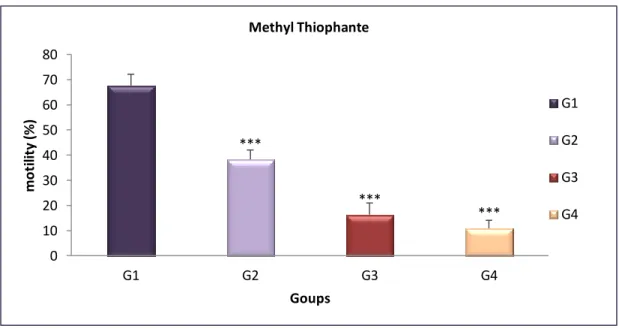
Fig 10 Epididymis sperm motility (%) of treated groups with MT compared with control after 4 weeks……… 31
Fig 11 Epididymis sperm speed (µm/s) of treated groups with MT compared with the control group after 4 weeks. ………. 32
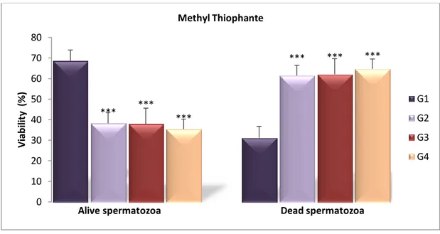
Fig 12 Epididymal sperm viability (%) of treated groups with MT compared with the control one after 4 weeks ………. 32
Fig 13 Epididymis sperm vitality (%) of treated groups compared to control after 4 weeks ( Hypo-osmotic swelling test) ………... 33
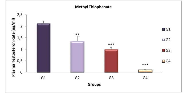
Fig 14 Variation of Testosterone level (ng/ml) of treated groups compared with the control group after 4 weeks. ……… 34
Fig 15 Variation of plasma T3 rate (ng/ml) of the treated groups with MT compared with the control group after 4 weeks ……… 36
Fig 16 Variation of plasma T4 rate (ng / ml) of the treated groups with MT compared with the control group ……….. 36
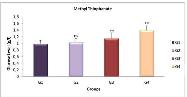
Fig 17 Variation of Glucose level (g/l) of the treated groups with MT compared with the control group after 4 weeks ……… 37
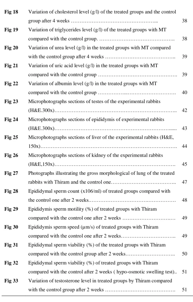
Fig 18 Variation of cholesterol level (g/l) of the treated groups and the control
group after 4 weeks ………... 38
Fig 19 Variation of triglycerides level (g/l) of the treated groups with MT
compared with the control group. ……….. 38 Fig 20 Variation of urea level (g/l) in the treated groups with MT compared
with the control group after 4 weeks ……….. 39 Fig 21 Variation of uric acid level (g/l) in the treated groups with MT
compared with the control group ………. 39 Fig 22 Variation of albumin level (g/l) in the treated groups with MT
compared with the control group ………...………. 40 Fig 23 Microphotographs sections of testes of the experimental rabbits
(H&E.300x)……….. 42
Fig 24 Microphotographs sections of epididymis of experimental rabbits
(H&E.300x)………... 43
Fig 25 Microphotographs sections of liver of the experimental rabbits (H&E,
150x)………. 44
Fig 26 Microphotographs sections of kidney of the experimental rabbits
(H&E,150x)………. 45
Fig 27 Photographs illustrating the gross morphological of lung of the treated rabbits with Thiram and the control one……….. 47 Fig 28 Epididymal sperm count (x106/ml) of treated groups compared with
the control one after 2 weeks.……….………. 48 Fig 29 Epididymis sperm motility (%) of treated groups with Thiram
compared with the control one after 2 weeks ……….… 49 Fig 30 Epididymis sperm speed (µm/s) of treated groups with Thiram
compared with the control one after 2 weeks……….. 49 Fig 31 Epididymal sperm viability (%) of the treated groups with Thiram
compared with the control group after 2 weeks……….. 50 Fig 32 Epididymal sperm viability (%) of treated groups with Thiram
compared with the control after 2 weeks ( hypo-osmotic swelling test).. 51 Fig 33 Variation of testosterone level in treated groups by Thiram compared
Fig 34 Variations of Tri-iodothyronine level (ng/ml) in the Treated groups
with Thiram compared with the control group after 2 weeks………….. 53 Fig 35 Variations of Thyroxin level (ng/ml) in the treated groups with Thiram
and the control group after 2 weeks ……… 53 Fig 36 Variation of glucose level (g/l) in the treated groups with Thiram
compared with the control group after 2 weeks………... 54 Fig 37 Variation of cholesterol level (g/l) in the treated groups with Thiram
compared with the control group after 2 weeks……….. 55 Fig 38 Variation of triglycerides level (g/l) in the treated groups with Thiram
compared with the control group after 2 weeks………... 55 Fig 39 Variation of urea level (g/l) in the treated groups with Thiram
compared with the control group after 2 weeks……….. 56 Fig 40 Variation of uric acid level (g/l) in the treated groups with Thiram
compared with the control group after 2 weeks ……….… 56 Fig 41 Variation of albumin level (g/l) in the treated groups with Thiram
compared with the control group after 2 weeks……….. 57 Fig 42 Photomicrograph sections of testes of the experimental rabbits
(H&E,300x)……….. 59
Fig 43 Microphotographs sections of epididymis of the experimental rabbits
(H&E, 300x)……….……….….. 60
Fig 44 Microphotographs sections of liver of the experimental rabbits (H&E,
150x)………. 61
Fig 45 Microphotographs sections of kidney of the experimental rabbits
(H&E, 150x)……… 62
LIST OF TABLES
Tables Tittle Page
Table 01 Chemical groups of fungicides according to their generic names … 6
Table 02 Some physical and chemical properties of MT……… 8
Table 03 Some physical and chemical properties of Thiram………. 12
Table 04 Organs weights of experimental rabbit treated with MT compared
with the control group………..……….. 29
Table 05 Variation of hematological parameters of treated groups by MT
compared with the control after 4 weeks. ……… 35
Table 06 Percentage of mortality level observed in the treated rabbits with
Thiram……… 46
Table 07 Weight of the liver, kidney, testes and epididymis in experimental rabbit treated with Thiram compared to the control group after 2
weeks………. 48
Table 08 Variation of hematological parameters of treated groups with Thiram compared with the control one after 2 weeks……… 52
ADP : Adenosine-5-Diphosphate Bw : Body Weight
CAS : Chemical Abstracts Service CHE : Cholesterol esters
DAP : Dihydroxyacetone Phosphate
EDTA : Ethylene Diamine-Tetraacetic Acid- EPA : Environmental Protection Agency FSH : Follicle-Stimulating Hormone GOD : Glucose Oxidase
G3P : Glycerol-3-phosphate
GPO : Dihydroxyacetone Phosphate GLDH : Glutamate Dehydrogenase HGB : Haemoglobin
HCT : Hematocrite
H&E : Hematoxylin and Eosin LD50 : Lethal Dose
LH : luteinizing hormone LPL : Liberate Glycerol MT : Methyl Thiophanate MBC : Carbendazime
MCV : Mean Corpuscular Volume MCH : Mean Corpuscular Hemoglobin
MCHC : Mean Corpuscular Hemoglobin Concentration Mg / kg : Milligram / kilogram
Mm Hg :Millimeters of mercury
NADH : Nicotinamide Adenine Dinucleotide Hydrogen NAD : Nicotinamide Adenine Dinucleotide
PLT : Platelet Count Ppm : Parts per million POD : Peroxidase P : Probability value
RBC : Red Blood Cells RT : Reagent SD : Deviation Standard SPZ : Spermatozoa T3 : Tri-Iodothyronine T4 : Thyroxin µl : Micro Liter WBC : White Blood Cell
WHO : World Health Organization
x
: MeanINTRODUCTION ………. 1. REVIEW OF LITERATURE
1.1. Definition of fungicides……… 1.2. Fate processes of fungicides in environment……..………... 1.3. Toxicokinetics of fungicides……….……… 1.4. Fungicides classification …..………... 1.5. Methyl Thiophanate………..………. 1.5.1. Definition………..……….. 1.5.2. The Chemical structure of MT……….…….. 1.5.3. Physical and chemical properties of MT……….…….…….. 1.5.4. Toxicokinetics of MT…………..………... 1.5.5. Toxicodynamics of MT…….………. 1.6. Thiram……….……….. 1.6.1. Definition of Thiram……….……….. 1.6.2. Chemical structure of Thiram…..………... 1.6.3. Physical and chemical properties of Thiram…..……… 1.6.4. Toxicokinetics of Thiram…………..………. 1.6.5. Toxicodynamics of Thiram…..……….. 2. MATERIALS AND METHODS
2.1. Fungicides…………..……… 2.2. Animals……….………. 2.3. Treatment….……….. 2.4. Semen sampling …..……….………. 2.5. Blood collection …..……….. 2.6. Organes remove ………..…….………. 2.7. Experimental protocol……….……….. 2.8. Methods……….……… 2.8.1. Semen snalysis………... 2.8.1.1. Concentration of spermatozoas……….. 2.8.1.2. Motility of spermatozoas.……… 2.8.1.3. Speed of spermatozoas.……….. 2.8.1.4. Viability of spermatozoas…….………... 01 04 04 05 06 07 07 07 08 88 08 00 10 10 12 12 14 16 16 16 17 07 08 09 20 20 20 20 20 22
2.8.2. Hematological analysis (Blood Count)……..……….… 2.8.3. Hormons assay ……….……….…. 2.8.4. Biochimical assay ….………. 2.8.4.1. Quantitative determination of Glucose………….…………. 2.8.4.2. Quantitative determination of Cholesterol…..………….….. 2.8.4.3. Quantitative determination of Triglycerides.………. 2.8.4.4. Quantitative determination of Uric acid……… 2.8.4.5. Quantitative determination of Urea…….……….. 2.8.4.6. Quantitative determination of Albumin……….………….... 2.8.5. Histology examination…….………..…….… 2.8.6. Statistical analysis…………..………. 3. RESULTS
3.1.Impact of Methyl Thiophanate during the first experimental protocol.. 3.1.1. Experimental observation of effect of MT in experimental groups...
3.1.1.1. Variation of organs weight………...… 3.1.1.2. Macroscopic observation of liver………..… 3.1.2. Impact of MT on fertility ……….……….……….… 3.1.2.1.Count of spermatozoa……….……… 3.1.2.2. Motility of spermatozoa….…………..………. 3.1.2.3. Speed of spermatozoa ………..……. 3.1.2.4.Viability of spermatozoa….……….... 3.1.2.5. Plasma Testosterone level….………. 3.1.3. Impact of MT on hematological parameters……….. 3.1.4. Impact of MT on Thyroids hormones….………..……….….
3.1.4.1. Tri-iodothyronine (T3)………... 3.1.4.2. Thyroxin (T4)………. 3.1.5. Impact of MT on biochemical parameters….…….…………... 3.1.5.1. Glucose ……….……….... 3.1.5.2. Cholesterol………... 3.1.5.3. Triglycerides………... 3.1.5.4. Urea ………... 3.1.5.5. Uric acid………. 3.1.5.6. Albumin………... 23 23 23 24 24 25 25 26 27 27 28 29 29 29 29 31 31 31 32 32 33 34 35 36 36 37 37 38 38 39 39 40
3.1.6.2. Histology of epididymis .……….. 3.1.6.3. Histology of liver….……….. 3.1.6.4. Histology of kidney….……….. 3.2. Impact of Thiram during the second experimental protocol..………... 3.2.1. Experimental observation of the effect of Thiram..……….…...
3.2.1.1. Mortality level….……….………... 3.2.1.2. Macroscopic observation of Lung in the experimental Groups………. 3.2.1.3. Variation of organs weight…..………..….… 3.2.2. Impact of Thiram on fertility …….……….…... 3.2.2.1. Sperm concentration …….……….…... 3.2.2.2. Motility of spermatozoa……….… 3.2.2.3. Speed of spermatozoa……… 3.2.2.4. Viability of spermatozoa………..…………..….... 3.2.2.5. Plasma Testosterone level….……….... 3.2.3. Impact of Thiram on hematological parameters…..……...……..…. 3.2.4. Impact of Thiram on thyroids hormones ………..…...…..
3.2.4.1. Tri-iodothyronine (T3)………..….
3.2.4.2. Thyroxin (T4)……….….
3.2.5. Impact of Thiram on biochemical parameters……….…... 3.1.5.1. Glucose ……….….... 3.1.5.2. Cholesterol………... 3.1.5.3. Triglycerides………... 3.1.5.4. Urea ………... 3.1.5.5. Uric acid………. 3.1.5.6. Albumin………... 3.2.6. Impact of Thiram on the histology of organs ………….………..….
3.2.6.1. Histology of testes………..…….……. 3.2.6.2. Histology of epididymis ……….…. 3.2.6.3. Histology of liver ……….….…... 3.2.6.4. Histology of kidney ………... 41 41 41 46 46 46 46 48 48 48 49 49 50 51 52 52 53 53 54 54 55 55 56 56 57 57 57 57 58 58
4. DISCUSSION
4.1. Impact of Methyl Thiophanate………..……….….. 4.2. Impact of Thiram……….…..… 5. CONCLUSION ………. 6. PERSPECTIVES ……….….… 7. REFERENCES ………..…… 63 70 75 76 77
INTRODUCTION
1 INTRODUCTION
Phytosanitary products have been used in modern agriculture for a while of time that causes big concerns about the health, particularly in the reproductive system. These products are wide spread worldwide, the fact that makes a difficulty to remove them from agriculture in the near future. Pesticide risks may be greater in developing countries where there are bad conditions of workplaces and safety for workers who are out the priority of the authorities.
Many experimental studies and some observations have suggested that alterations of the male reproductive functions especially semen characteristics are an important health problem all over the world. This may be due to toxic factors present in our environment which particularly involves molecules originally industrial or anthropogenic grouped under the generic term endocrine disruptor, one of the most famous of which are pesticides (Ferreira, 2010; Hossain et al., 2010).
These pesticides can alter estrogens functions (Xenoestrogenic ), androgens, thyroidien hormones, and even the pituitary gland hormones. Indeed, Endocrine disruptors interact with the endocrine system; it is a complex system composed of many organs scattered around the Organisms ( pancreas, adrenal, testes, ovaries, thyroid and parathyroid). Each body secretes hormones released into the body through the bloodstream. The functioning of the endocrine system and strict internal control involved in homeostasis, or biological balance necessary for life. These organs are under control of regulator substances or releasing Factors (hypothalamus) and stimulines substances (pituitary) (Ferreira, 2010).
There are many possible ways in which humans can be exposed to the toxic effects of these pesticides may have consequences from food consumers, production workers, formulators, farmers and other applicators. They are widely applied as insecticides, herbicides, and fungicides (Bozdogan, 2014).
Fungicides property is to monitor, repel or destroy the fungi that may develop on crops. They are used against fungal diseases such as mildew, powdery mildew and mold. On the other hand, some of the fungicides have shown toxicity to humans, animals, and useful plants, in addition to their persistence (long life) in the environment. Long half-life of chemical in the environment equal a more likely to be exposed to it by the target organisms
2
and it accumulates making a hazardous dose. Moreover, these chemicals were shown to be present in fruit products prepared for human consumption (Cabras et al., 2000).
Recently, it has been confirmed that reprotoxic effects of fungicides decreases the sperm quality and in other disorders of the male reproductive tract (Mallem et al., 2007; Gallo and Tosti, 2015). These fungicides are known to interfere with spermatogenesis by damaging the testes (Kristensen, 1999). The severity of affection that depends on the stage of differentiation can be reversible or irreversible and may even temporarily lead to decrease fertility by modifying sperm count, structure motility, or viability of spermatozoa. These effects are transient after removal of the offending chemical; the spermatogenesis can be restored from stem cell populations (Bretveld et al., 2007). In fact, normal fertility is based on normal spermatogenesis. In the complex process of spermatogenesis, disturbance can easily occur and result in decreased or absent spermatogenesis and sub-infertility, as a consequence. Assessment of the semen parameters is considered a key indicator in male reproductive capacity, by identificating the adverse effects of the related toxicants on fertility (Oliva et al., 2001; Mehrpour et al., 2014).
Two fungicides are the most widely used in the field of agriculture in Algeria namely: Methyl Thiophanate from Benzimidazole class and Thiram from Dithiocarbamate.
Methyl Thiophanate is generally metabolized to Carbandazim (Roberts et al., 1998; Cardone, 2012). Both Methyl Thiophanate and Carbendazim have been reported to be capable of causing endocrine disruption, embryotoxic and teratogenic effects ( Yang et al., 2011 ), Carbendazim is poorly catabolized and remains in tissues such as gonads, liver, adrenals, adipose tissue, skin and other organs (WHO, 1993). It is a well-recognized testicular toxicant (Yu et al., 2009).
Evidences available support the deleterious effects of Methyl Thiophanate and its metabolite Carbendazim on various aspects of male reproduction in hamsters, rats, rabbits and humans. The effects include the decrease of average testes weight, average seminiferous tubular diameter (Carter et al., 1987) total sperm count, motility, increased incidence of sperm abnormalities (Akbarsha et al., 2001) and disruption of microtubule formation (Nakai et al., 2002).
Thiram is moderately toxic by ingestion, but it is highly toxic if inhaled. Symptoms of chronic exposure to Thiram in humans include drowsiness, confusion, loss of sex drive,
INTRODUCTION
3
incoordination, slurred speech and weakness. It has been reported to have adverse effects on the hepatic system, (Dalvi, 1986) the reproductive system and on the developmental processes (Hemavathi et al., 1993; Mishra et al., 1998). Studies have shown that Dithiocarbamate decreases the quality of spermatozoa and affects male fertility and Thiram interfere with the differentiation process of spermatogenic cells (Agrawal et al., 1997)
Objectives:
The objective of this study is to experimentally evaluate the toxicity consequences of the used fungicides Methyl Thiophante and Thiram on the bio-indicator parameters in male rabbits and mainly their effect on male fertility by:
Evaluation of some male reproductive parameters. Determination of the concentration of thyroid hormones. Evaluation of some biochemical and hematological parameters. Determination of histological study of some organs.
REVIEW OF LITERATURE
4 1. REVIEW OF LITERATURE
1.1. Definition of Fungicides
Fungicides are a specific type of pesticides that controls fungal disease specifically inhibit or kill fungi causing important diseases (mildew and mold) (Mc-Grath, 2004). Understanding mechanisms of fungicide actions and toxicity is important because humans and domesticated animals encounter these pesticides through a wide variety of applications. In agriculture, fungicides are used to protect tubers, fruits and vegetables during storage. They are also applied directly to ornamental plants, trees, field crops, cereals and turf grasses (Gupta and Aggarwal, 2007).
Figure 1. Fungal diseases/ a: mildew, b: mold (Anon., 2007).
1.2. Fate Processes of Fungicides in Environment
For nearly fifty years, fungicides (generally all pesticides) have been highlighted in all environmental compartments. As well in river waters, groundwater, air, rainwater in addition to the fruits, vegetables and grains. Failure to comply with good agricultural practices can result in contamination of three biosphere compartments namely water, soil and air. Thus, the geochemical cycle of pesticides is very complex for they can be found at all levels.
The following simplified figure shows the various possible uses and drifts of pesticides (El Mrabet et al., 2008).
5
Figure 2. Behavior of pesticides in the environment (El Mrabet et al., 2008).
1.3. Toxicokinetics of Fungicides
Fungicide exposure pathways are multiple. Whatever is the path of penetration; humans or animals are exposed to fungicides through ingestion or absorb them through the skin or the respiratory system. Fungicides are transported by blood to all organs. In general, the liver is the primary site for biotransformation and may include detoxification as well as activation reactions. They are accumulated in adipose tissue and organs containing a high content of lipid membranes especially in nervous system (brain, spinal cord and nerves), bones, liver and muscles. Then they are excreted and eliminated through expiration, sweat, bile, feces and urine (Gupta and Aggarwal, 2012).
REVIEW OF LITERATURE
6 1.4. Fungicides Classification
Table 01: Chemical groups of fungicides according to their generic names (Burpee, 2006).
CHEMICAL GROUP GENERIC NAMES
TRIAZOLES PROPICONAZOLE TRIADIMEFON MYCLOBUTANIL TRITICONAZOLE TETRACONAZOLE PYRIMIDINES FENARIMOL STROBILURINS FLUOXASTROBIN TRIFLOXYSTROBIN AZOXYSTROBIN PYRACLOSTROBIN POLYOXINS POLYOXIN D BENZIMIDAZOLES THIOPHANATE-METHYL BENOMYL CARBANDAZIM DICARBOXAMIDES IPRODIONE VINCLOZOLIN PHENYLAMIDES MEFENOXAM CARBAMATES PROPAMOCARB
PHOSPHONATES FOSETYL ALUMINUM
PHOSPHONATE
DITHIOCARBAMATES THIRAM
MANCOZEB
AROMATIC HYDROCARBONS QUINTOZENE
CHLORONEB ETHAZOLE
PEROXIDES HYDROGEN DIOXIDE
NITRILES CHLOROTHALONIL PHENYLPYROLLES FLUDIOXONIL CYANOIMIDAZOLE CYANOFAMID CARBOXAMIDES FLUTOLANIL BOSCALID BIOFUNGICIDES ECOGUARD SONATA SOILGUARD
7
Under chemical classification of fungicides, two fungicides are chosen: Methyl-Thiophante and Thiram, the first from Benzimidazol’s and the second from Dithiocarbamat class.
1.5. Methyl Thiophanate 1.5.1. Definition
Methyl Thiophanate 1, 2-Bis (3-(methoxycarbonyl)-2-thioureido) benzene) is a classic Benzimidazole Carbamate. It is a systemic broad-spectrum fungicide that has been used for many years controlling various fungal pathogens of various food crops, ornamental plants, trees and grasses. It is used also as a preseative in paint, textile, papermaking, leather industry and warehousing practices (Giry et al., 2001; Berglof et al., 2002). The LD50 of Methyl Thiophanate in the male rabbit by oral route is 2270 mg/kg body weight (Tomlin, 1994). It is well absorbed (80–85%) after oral exposure and is subsequently metabolized into many compounds within the organisms. The main metabolite is Carbendazim (Methyl 2-Benzimidazolyl Carbamate) (Roberts et al., 1998; Cardone et al., 2012). Carbendazim is poorly catabolized and remains in tissues such as gonads, liver, adrenals, adipose tissue, skin and other organs (WHO, 1993) and it is a well-recognized testicular toxicant (Yu et al., 2009).
1.5.2. The Chemical Structure of MT
The molecular formula of Methyl Thiophanate is C12H14N4O4S2 (fig 03). It has several synonyms: [4, 4 '- (o-Phénylén) bis (3-thioallophanate) of dimethyl], Metoben, Methyl topsin, Mildothane, [1, 2-Bis (Methoxycarbonylthioureido) Benzene] and Cercobin Methyl (Mackay et al., 2006 ).
REVIEW OF LITERATURE
8 1.5.3. Physical and Chemical Properties of MT
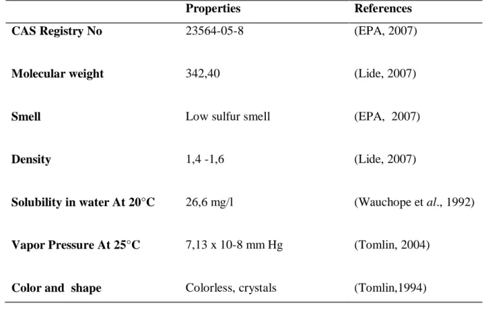
Table 02: presents some physical and chemical properties of MT.
Properties References
CAS Registry No 23564-05-8 (EPA, 2007)
Molecular weight 342,40 (Lide, 2007)
Smell Low sulfur smell (EPA, 2007)
Density 1,4 -1,6 (Lide, 2007)
Solubility in water At 20°C 26,6 mg/l (Wauchope et al., 1992)
Vapor Pressure At 25°C 7,13 x 10⁻⁸ mm Hg (Tomlin, 2004)
Color and shape Colorless, crystals (Tomlin,1994)
1.5.4. Toxicokinetics of MT:
Both Methyl Thiophanate and Carbendazim are classified as Benzimidazole fungicides. Carbendazim is a major metabolite of MT with closely similar structural and toxicological characteristics (Methyl Thiophanate is also structurally related to benomyl).
In studies reported by Noguchi et al. (1971) and Fujino et al. (1973) the administered Methyl-Thiophanate was conjugate to Carbendazim (MBC). MT and Carbendazim are well absorbed after oral exposure (80–85%) but poorly absorbed after dermal exposure (1 or 2%) in rats, mice, dogs and hamsters.
Fujino et al. (1973) suggested the metabolism scheme shown in Fig (04). They reported that the major part of fecal excretion was in the form of unmetabolized Methyl-Thiophanate, while the minor parts consist of 4-hydroxy-thiophanate-methyl (4-OH-TM) and dimethyl-4.4'-O-phenylenebisallophanate. Methyl 2-benzimidazol carbamate (Carbendazim) and 5-hydroxy-MBC (5-OH-MBC) were also observed during the identification of metabolites of fecal extracts. It was, however, questioned whether these
9
two were actual metabolites in faeces or they were compounds produced during the analytical procedures from Methyl-Thiophanate and 4-OH-TM respectively.
Figure 4. Metabolism of MT in organism (funjo et al., 1973)
MT : Methyl-Thiophante
FH-432 : Dimethyl-4.4’-o-phenylenebis (allophanate)
4-OH-FH-432 : 4-Hydroxy-dimethyl-4.4’-o-phenylenebis (allophanate) 4-OH-TM : 4-Hydroxy-thiophanate-methyl
MBC : Methyl-2-benzimidazolecarbamate
REVIEW OF LITERATURE
10 1.5.5. Toxicodynamics of MT
Previous considerations of MT found that it has low acute oral, dermal and inhalational toxicity in animals. The main hazards associated with repeated exposure to their metabolite Carbendazim are systemic effects on liver and thymus and effects on reproduction. Furthermore Carbendazim has also been shown to produce numerical aberrations (aneuploidy) in mammalian cells exposed in vitro and in vivo (APVMA, 2008).
The European Union has classified Carbendazim as a potential genotoxic chemical. Relatively information has highlighted health concerns associated with exposure to Carbendazim. Following an investigation to the effects of Carbendazim on various cell types in the testis, Kadalmani et al. (2002) observed that spermatocytes are a target for Carbendazim. Lu et al. (2004) concluded that Carbendazim causes losses of spermatozoa, decrease in testis weight and decrease in sperm concentration in rats. Carbendazim is associated with adverse reproductive effects, Furthermore both MT and Carbendazim have also been associated with an increased incidence of liver tumors and can cause aneuploidy (abnormal number of chromosomes).
The US Environmental Protection Agency has classified both Carbendazim and Methyl-Thiophanate as probable human carcinogens. Aggregate cancer risk estimates for both Carbendazim and MT from all uses including residential (lawn treatment and post-application exposure) and dietary exposure exceeded EPA’s level of concern. Also it was reported that the liver and thyroid are the primary target organs of MT in several species following sub-chronic and chronic dietary exposure (EPA, 2005).
11 1.6. Thiram
1.6.1. Definition of Thiram
Thiram (fig 05) is a Dimethyl Dithiocarbamate compound used as a fungicide to prevent crop damage in the field and to protect harvested crops from deterioration in storage or transport. Thiram is also used as a seed protectant and to protect fruit, vegetable, ornamental, and turf crops from numerous fungal diseases. Moreover, it is used as a repellent for rodents and certain large animals that cause damage to field crops. Thiram is available as dust, flowable, wettable powder, water dispersible granules, and water suspension formulations, and in mixtures with other fungicides (Dalvi et al., 1988). Thiram has been used in the treatment of human scabies, as a sunscreen, and as a bactericide applied directly to the skin or incorporated into soap. It is also used as a rubber accelerator in tire industry and as a lubricating oil additive (EPA, 2004). The LD50 of Thiram in the male rabbit by oral route is 210 mg/kg body weight (Edwards, 1991).
1.6.2. Chemical Structure of Thiram
The molecular formula of Thiram is C6H12N2S4. It has several synonyms (Thiram, TMTD, tetraalkyl-thiuram disulfide, bis (N,N-diméthylthiocarbamyle) disulfide (Mackay et al., 2006 ).
REVIEW OF LITERATURE
12 1.6.3. Physical and Chemical Properties of Thiram
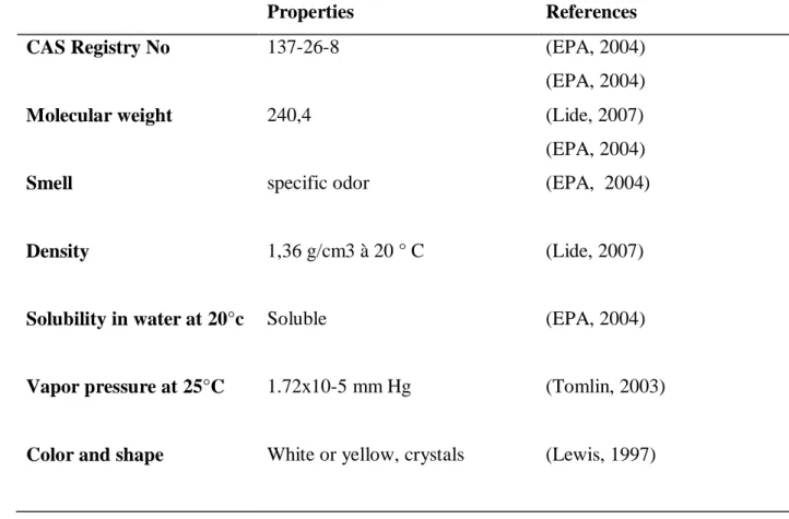
Table 03: presents some physical and chemical properties of Thiram.
Properties References
CAS Registry No 137-26-8 (EPA, 2004)
(EPA, 2004)
Molecular weight 240,4 (Lide, 2007)
(EPA, 2004)
Smell specific odor (EPA, 2004)
Density 1,36 g/cm3 à 20 ° C (Lide, 2007)
Solubility in water at 20°c Soluble (EPA, 2004)
Vapor pressure at 25°C 1.72x10⁻⁵ mm Hg (Tomlin, 2003)
Color and shape White or yellow, crystals (Lewis, 1997)
1.6.4. Toxicokinetics of Thiram:
After absorption (respiratory, dermal, and gastrointestinal), Thiram appears to be readily absorbed through the intestinal tract and the lungs and is quickly and widely distributed throughout the body (ACGIH, 2001).
It is metabolized in the body to toxic metabolites Dimethyldithiocarbamate (This is found either as the free acid or as the S-glucuronide conjugates) and Carbon Disulfide (Aldridge and Magos, 1978), although these compounds have been shown to inhibit hepatic microsomal enzymes (Dalvi and Deoras, 1986).
Gay et al. (1992) summarized the conclusions from a series of metabolism studies on rats with [Thiocarbonyl-14C] Thiram. When rats were dosed orally with [14C] Thiram much of the 14C (40-60%) was eliminated as volatiles in exhaled air, 25-35% was excreted in the urine and 2-5% in the faeces. After an interval of 96 hours 2-3% of the 14C remained in the tissues. Polar metabolites and conjugates (Dimethyldithiocarbamate ) were identified in the
13
urine and other metabolites include Elemental Sulfur, Methionine, Formaldehyde (Aldridge and Magos, 1978; Verschueren, 2001).
REVIEW OF LITERATURE
14 1.6.5. Toxicodynamics of Thiram
Thiram is moderately toxic by ingestion, but it is highly toxic if inhaled. Reported oral LD50 values for Thiram are 620 to over 1900 mg/kg in rats; 1500 to 2000 mg/kg in mice and 210 mg/kg in rabbits (Edwards et al., 1991; Kidd and James, 1991). Acute exposure in humans may cause headaches, dizziness, tiredness, nausea, diarrhea, and other gastrointestinal complaints (U.S. National Library of Medicine, 1995).
The subchronic toxicity profile for Thiram indicates that hematology, clinical chemistry and body weight are affected after subchronic exposure to the compound for all species evaluated (EPA, 2004).
The chronic toxicity profile for Thiram indicates that the liver, blood and urinary system are the target organs for this chemical. Studies have shown evidence of liver damage by Thiram in the form of decreased liver enzyme activity and increased liver weight. Thiram may also cause damage to the nervous system (US National Library of Medicine, 1995).
Symptoms of chronic exposure to Thiram in humans include drowsiness, confusion, and loss of sex drive, incoordination, slurred speech, and weakness, in addition to those due to acute exposure. Repeated or prolonged exposure to Thiram can also cause allergic reactions such as dermatitis, watery eyes, sensitivity to light, and conjunctivitis (Edwards et al., 1991).
In a combined chronic/cancer study in rats, effects included changes in hematology parameters, increased incidence of bile duct hyperplasia (EPA, 2004).
Dithiocarbamate pesticides such as Thiram have a marked spermicidal activity in humans. They also cause viable and non-viable gross morphological alterations of sperm (Rice, 1964). In animals, Dithiocarbamate pesticides given to young and adult domestic chickens produced retarded testicular development and atrophy (Raasul and Howell, 1974).
Many investigations reported that oral doses of Thiram 49 mg/kg/day to rats for 2 years produced weakness, muscle incoordination, and paralysis of the hind legs. Rats fed 52 to 67 mg/kg/day for 80 weeks exhibited hair loss, and paralysis with atrophy of the hind legs. Symptoms of muscle incoordination and paralysis from Thiram poisoning have been shown to be associated with degeneration of nerves in the lower lumbar and pelvic regions (Edwards et al., 1991). Also toxic and some tumorigenic effects have been observed
15
in different animal species exposed to Thiram (Dalvi, 1988; Maita et al., 1991). numerous tests indicated that it is genotoxic (Crebelli et al., 1992; Hemavathi and Rahiman, 1996) and that it reported also effects on cartilaginous tissues in vitro and in vivo, in different animal species exposed to Dithiocarbamtes, including Thiram, (Suzuki et al., 2000, 2001; Rath et al., 2004; Simsa et al., 2007).
16 2. MATERIALS AND METHODS
2.1. Fungicides
Two fungicides have been used: Methyl Thiophanat (70%) as a wettable powder and Thiram (85%) as a wettable powder, which were obtained from a pharmaceutical company.
2.2. Animals
We used mature male rabbits Cuniculus lepus aged between 6 and 8 months with body wight 1500 ± 500 g. The study was carried out in the animalery of the University Badji Mokhtar-Annaba. The Animals were housed in specific cages (50× 60×53 cm3) and were maintained in natural conditions of temperature, photoperiod and relative humidity. Animals had access ad libitum to water and food contains all the necessary elements (salad, carrot, cabbage and beet) the same quantity is given to all the groups of rabbits.
Rabbit Classification
Kingdom: Animalia Order: Lagomorpha
Phylum: Chordata Family: Leporida
Class: Mammalia Species: Cuniculus lepus
2.3. Treatment
The experimental protocol was divided into the first and the second experimentation : First experimentation: animals treated with Methyl Thiophante (Benzimidazol). Second experimentation: animals treated with Thiram (Dithiocarbamate).
MATERIALS & METHODS
17 2.3.1. The First Experimentation
The rabbits were divided into four groups (6 rabbits in each group). G1: Group served as a control one.
And three groups were treated orally with Methyl Thiophanate dissolved in water at doses:
G2: Group treated with 50 mg/1ml / kg body weight. G3: Group treated with 100 mg/1ml / kg body weight. G4: Group treated with 150 mg/1ml / kg body weight. MT was administered by oral system for 4 weeks.
2.3.2. The Second Experimentation
The rabbits were also divided into four groups, each one contained 6 rabbits G1: Group 01 served as a control group.
Three groups were treated orally with Thiram dissolved in water at doses: G2: Group treated with 20 mg/1ml / kg body weight.
G3: Group treated with 30 mg/1ml / kg body weight. G4: Group treated with 80 mg/1ml / kg body weight.
Thiram was administered by oral system over the period of 2 weeks.
After sacrifice by decapitation, blood and semen were collected. Then, the organs of each animal of the different groups were removed and weighed.
2.4. Semen Sampling
To estimate the effects of the used fungicides on fertility of rabbits through the characteristics of spermatozoa and semen quality (count, motility, speed and viability). We proceeded to the sperm test using the method of WHO (1993).
Immediately after dicapitation, semen was collected from a small opening made at the head of the epididymis. 1 µL of sperm was diluted in 49 µL of NaCl 0.9% and placed in an oven at a temperature of 37° C.
2.5. Blood Collection
Blood is immediately collected in three types of tubes, Haparin tubes, dry tubes, and the others contain the EDTA anticoagulant:
18
• The Tubes with EDTA: used for the determination of the formula of blood numeration.
• The Dry Tubes: centrifuged with 5000 tours/minute during 15 minutes; the recovered serums used for the determination of the biochemical parameters (Glucose, Cholesterol, Triglyceride, Uric Acid, Urea and Albumin).
• The Heparin Tubes : containing Heparin anticoagulant, the tubes are centrifuged with 5000 tours/minute during 15 minutes; the recovered plasma used for the determination of hormons (Testosteron, T3 and T4).
2.6. Organs Remove
We made a longitudinal abdominal opening in animals for sampling the different organs (testes, epididym, liver and kidneys). After the separation of their adipose tissues, the organs are weighed, then a piece of liver, kidney, epididymis and testes of each animal are fixed in formol (10%) to make histological sections.
MATERIALS & METHODS
19 2.7. Experimental Protocol
The next figure summarizes the various steps of the experimental protocol of the first experimentation using MT. 30 days of trial G2 :50mg/kg n=6 G3 :100mg/kg n=6 G1 :Control n=6 Sacrifice by decapitation Concentration Motility Speed Viability Testes Epididymis Live Kidney
STATISTIC:Results were evaluated statistically using the Minitab 15. However, the Student's t-test
was used by comparing each of the treated group with the control. The significance level of p ≤ 0.05 was considered.
Semen Collection Blood Collection Organs Remove
EDTA Dry tube Heparin tube
RB WB HB HCT PLT MCV MCH Mchc Glucose Cholesterol Triglyceride Urea……. Uric Acid Albimun Testosterone T3…… T4 Histology Of Organs (Martoja& Martoja1967 Biology Of Spermatozoa (WHO 1993)
24 Male Rabbits
Methyl-Thiophanat
G4 :150mg/kg n=620
The next figure summarizes the various steps of the experimental protocol of the second treatment using Thiram.
15 days of trial G2 :20mg/kg n=6 G3 :30mg/kg n=6 G4 :80mg/kg n=6 G1 :Control n=6 Sacrifice by decapitaion Concentration Motility Speed Viability Testes Epididymis Liver kidney
STATISTIC: Results were evaluated statistically using the Minitab 15. However, the Student's t-test
was used by comparing each treated group with the control. The significance level of p ≤ 0.05 was considered.
Semen Collection Blood Collection Organs Remove
EDTA Dry tube Heparin tube
RB WB HB HCT PLT MCV MCH mchc Glucose Cholesterol Triglyceride Urea……. Acid uric Albimun Testosterone T3…… T4 Histology Of Organs (Martoja& Martoja 1967 Biology Of Spermatozoa(OMS 1993)
24 Male Rabbits
Thiram
MATERIALS & METHODS
21 2.8. Methods
2.8.1. Semen Analysis
Semen analysis is a test that seeks to assess the following characteristics of spermatozoa: (concentration, motility, speed and viability).
2.8.1.1. sperm count
Spermtozoa count was measured using a Malassez cell (WHO, 1993). After mixing the diluted semen sample, we introduce one drop into the Malassez cell then covered with a cover-slip. This study is based on counting the spermatozoa in 05 squares at magnification (× 40) under the microscope. The sperm count calculated via the following method:
D: Dilution ratio (50)
V: Volume of the Malassez cell
n: The number of spermatozoa counted in 05 squares N: The number of small squares.
2.8.1.2. Motility of Spermatozoa
A drop of semen was put between normal slide and cover slip, then examined under optical microscope with magnification (× 40). Sperm motility is determined by the count of mobile spermatozoa in 3 observation fields. Then theaverage percentage of mobile spermatozoa is calculated (WHO, 1993).
2.8.1.3. Speed of Spermatozoa
First,one drop of the diluted semen was placed on the Malassez slide using a micropipette and covered with a cover slip, then the sample under microscope was examined with magnification (× 40) (WHO, 1993).
Finally, we evaluated the speed of sepermatozoas by counting the time (in seconds) required to cross distance of 0.05 mm between two parallel lines of 10 spermatozoas using a timer. Then the speed was calculated by applying the following formula:
Concentration (x106 / ml) =
22
We calculate the speed of 10 spermatozoas, then the average speed.
2.8.1.4.Viability of Spermatozoa
Vital stain: This is a staining technique based on the principle that dead sperm have multiple holes in their membranes, which means they will take up eosin and appear pinkish in colour. Normal live sperm will not be stained as they have intact membranes.
- The used reactive: Eosin (0.1%).
A drop of sperm + drop of eosin were put between normal slide and cover slip and let dry for 2 to 3 min.
After performing the stain test, the preparation is examined microscopically and 100 spermatozoa are recorded. Then, how many of these 100 are stained as well as how many have repeled the staining. This will allow us to count the percentage of dead and normal live sperm (WHO, 1993).
Hypo-osmotic swelling test: This test is carried out to study the viability by studying the morphological changes in spermatozoa tail.
Preparation of the used Solution: Dissolve 0.367g of sodium citrate (Na3C6H3O7; 2H2O) and 0.675g of fructose in 50 ml of distilled water. After thawing, the solution is mixed well before using it.
procedure of work: we put 1 ml of the used solution in Eppendorf tube and placed it in a water bath at a temperature of 37 °C for 3 minutes. Then 0.1 ml of semen is added to the previous solution then incubate at 37 °C for 30 minutes.
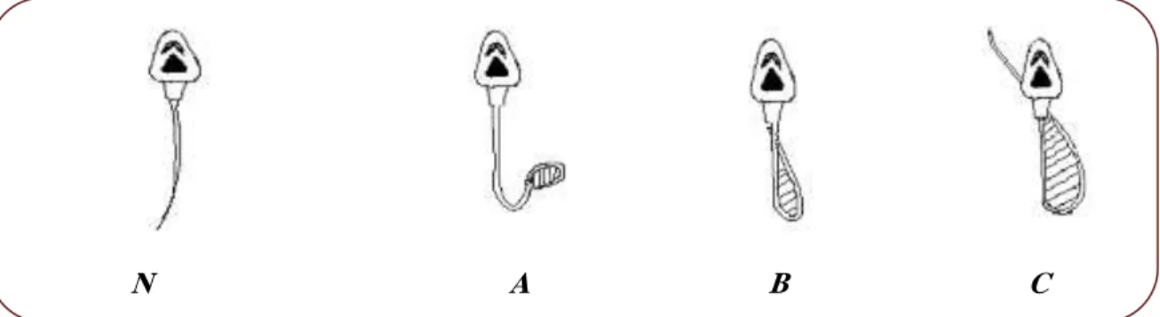
After microscopic observation with magnification [40x], the percentage of spermatozoa with abnormalities was counted (fig 07) on a total counted spermatozoa (WHO, 1993).
MATERIALS & METHODS
23
Figure 7. Morphological malformation of spermatozoa tail exposed to hypo- osmotic stress.
N: No malformation in the tail A: Low malformation of the tail B: Major malformation at the tail
C: Very important malformation in the tail and the midpiece part.
2.8.2. Hematological Analysis (Blood Count)
After decapitation, blood was collected in tubes containing EDTA anticoagulant, and was used for counting and sizing leukocytes subpopulations using automatic Coulter counter machine to calculate each of red blood cells (RBC), haemoglobin (HGB), hematocrite (HCT), mean corpuscular volume (MCV), mean corpuscular hemoglobin (MCH), mean corpuscular hemoglobin concentration (MCHC), platelet count (PLT) and total white blood cell count (WBC).
2.8.3. Hormons Assay
After collecting blood, samples were centrifuged at 5000 tr/min to isolate plasma used for mesearunig hormons. The hormones are proportioned by the conventional method ELISA using commercial kit. Measurement is done using “Tecan ELISA reader” provided with data processing software which calculates the range standard automatically and the level of the testosterone, T3 and T4 to the used unit directly.
2.8.4. Biochimical Assay We used Spinreact kit.
24 2.8.4.1.Quantitative Determination of Glucose
Principle of The Method
Glucose Oxidase (GOD) catalyses the oxidation of Glucose to Gluconic acid. The formed Hydrogen Peroxide (H2O2) is detected by a chromogenic Oxygen acceptor, Phenol-aminophenazone in the presence of Peroxidase (POD):
ß-D- Glucose + O2 +H2O GOD Gluconic acid +H2O H2O2 + Phenol +Aminophenazone POD Quinone + H2O
The intensity of the color formed is proportional to the glucose concentration in the sample
Blank Standard Sample
WR (ml) 1,0 1,0 1,0
Standard (µl) -- 10 --
Sample (µl) -- -- 10
2.8.4.2.Quantitative Determination of Cholesterol Principle of the Method
The cholesterol present in the sample originates a coloured complex, according to the following reaction:
Cholesterol esters CHE Cholesterol + Fatty acids Cholesterol + O2 CHOD 4- cholestenone + H2O2
2H2O2 + Phenol + 4-Aminophenazone POD Quinonimine +4h2O
The intensity of the color formed is proportional to the cholesterol concentration in the sample(Naito, 1984) .
Blank Standard Sample
WR (mL) 1.0 1.0 1.0
Standard (µl) -- 10 --
MATERIALS & METHODS
25 2.8.4.3. Quantitative Determination of Triglycerides
Principle of the Method
Sample Triglycerides incubated with Lipoproteinlipase (LPL), liberate Glycerol and free fatty acids. Glycerol is converted to glycerol-3-phosphate (G3P) and Adenosine-5-Diphosphate (ADP) by Glycerol Kinase and ATP. Glycerol-3-phosphate (G3P) is then converted by Glycerol Phosphate Dehydrogenase (GPO) to Dihydroxyacetone Phosphate (DAP) and Hydrogen Peroxide (H2O2).
In the last reaction, Hydrogen Peroxide (H2O2) reacts with 4-aminophenazone (4-AP) and p-chlorophenol in the presence of Peroxidase (POD) to give a red colored dye
Triglycerides + H2O LPL Glycerol + free fatty acids
Glycerol + ATP Glycerolkinase G3P+ ADP
G3P + O2 GPO DAP + H2O2
H2O2 + 4-AP + p-Chlorophenol POD Quinone + H2O
The intensity of the color formed is proportional to the Triglycerides concentration in the sample (Buccolo et al., 1973).
Blank Standard Sample
WR (mL) 1.0 1.0 1.0
Standard (µl) -- 10 --
Sample (µl) -- -- 10
2.8.4.4. Quantitative Determination of Uric Acid
Principle of the Method
Uric acid is oxidized by Uricase to Allantoine and Hydrogen Peroxide (2H2O2), which under the influence of POD, 4–aminophenazone (4-AP) and 2-4 Dichlorophenol Sulfonate (DCPS) forms a red Quinoneimine compound:
26 Uric acid + 2H2O + O2 Uricase Allantoine + CO2 + 2H2O2 2H2O2 + 4-AP + DCPS POD Quinoneimine+ 4H2O
The intensity of the red color formed is proportional to the Uric acid concentration in the sample (Schultz, 1984).
Blank Standard Sample
WR(ml) 1.0 1.0 1.0
Standard (µl) -- 25 --
Sample (µl) -- -- 25
2.8.4.5. Quantitative Determination of Urea
Principle of the Method
Urea in the sample is hydrolized enzymatically into Ammonia (NH3) and Carbon Dioxide (CO2).
Ammonia ions formed react with α-ketoglutarate in a reaction catalysed by Glutamate Dehydrogenase (GLDH) with simultaneous oxidation of NADH to NAD+:
Urea + H2O + 2 H+ Urease 2 NH3+ CO2
2 NH3+ α- Ketoglutarate + NADH GLDH H2O + NAD+ + L-Glutamate
The decrease in concentration of NADH is proportional to urea concentration in the sample (Kaplan, 1984).
Blank Standard Sample
WR (mL) 1.0 1.0 1.0
Standard(µL) -- 10 --
MATERIALS & METHODS
27 2.8.4.6. Quantitative Determination of Albumin
Principle of the Method
Albumin in the presence of Bromcresol green at a slightly acid pH, produces a colour change of the indicator from yellow-green to green-blue. The intensity of the color formed is proportional to the Albumin concentration in the sample (Gendler, 1984).
Blank Standard Sample
R (mL) 1.0 1.0 1.0
Standard (µL) -- -- --
Sample (µL) -- -- 5
2.8.5. Histology Examination
Histological sections were performed in anatomy laboratory at Ibn Rushd Hospital - Annaba. The technique was conducted according to the method of Martoja and Martoja (1967), which comprises the following steps:
Fixation
The primary function of a fixative is to preserve the cellular structure of the tissue. Fixation is necessary to protect and harden the tissue against the deleterious effects of later procedures, which, otherwise, would disrupt cellular structure beyond recognition. Furthermore, fixation minimizes a process called autolysis. Autolysis is the degradation of the cellular structure which results from the release of degradative enzymes from the excised tissue itself. The fixation process must be started as quickly as possible after removal of the sample. We let the organs section stand in the used fixative (Formol 10%) for 48 hours over the cuts.
Dehydration
As the paraffin is not miscible with water, the samples must then be completely dehydrated prior to paraffin embedding. This latter is not soluble in the alcohol used for dehydration, thus, there is a substitution with xylene. Dehydration is done through a machine making immersing samples in baths of ethanol at increasing concentration (70, 95 and 100%) and in baths of xylene which is a lightening agent giving the tissue more transparency. Then,
28
an oven of xylene evaporates the anatomical pieces; this step is performed by a machine called the circulator.
Inclusion and Achievement of Blocks
The parts are immersed in molten liquid paraffin baths at 60 ° C. The samples are soaked in paraffin and placed in molds, and then it filled with paraffin. This operation uses devices called “inclusion” Chilled to achieve rapid solidification (10 to 15 min) of the paraffin block containing the pieces of the organs.
Making Cuts and Staining
The inclusion blocks are glued on a sample holder.
The bonding surface is softened using a heated metal blade moderately flame then placed on the object carrier block on which the block adheres very well after a slight pressure.
The serial sections are made using a microtome
These series of sections are connected together in strip form, which facilitates reconstitution (three-dimensional) of the observed structures. The slice thickness is 4 to 7 microns.
The obtained tape sections are placed on glass slides
Then covered with distilled water and placed on a hot plate (the temperature of which is lower than the melting temperature of the paraffin).
The heat allows the staggering cuts ribbons.
The Melting paraffin sections were stained with Hematoxylin and Eosin (H&E stain).
Dried preparation in alcohol baths and mounted in Canada Balsam.
Finally, the Observation of sections is performed using a light microscope. 2.8.6. Statistical Analysis
All results were expressed as Mean ± Standard Deviation (M ± SD) and analyzed using
Student's t-test with the Minitab program (version15) comparing each treated group with
the control. The significance level of P ≤ 0.05 was considered. Differences are considered :
Significant (* P ≤ 0.05).
Highly significant compared with the control (** P ≤ 0.01).
0 1 2 3 4 5 6
Category 1 Category 2 Category 3 Category 4
A xi s Ti tl e Chart Title
29 3. RESULTS
3.1. Impact of Methyl Thiophanate During the First Experimental Protocol 3.1.1. Experimental Observation of the Effects of MT
3.1.1.1. Variation of Organs Weight
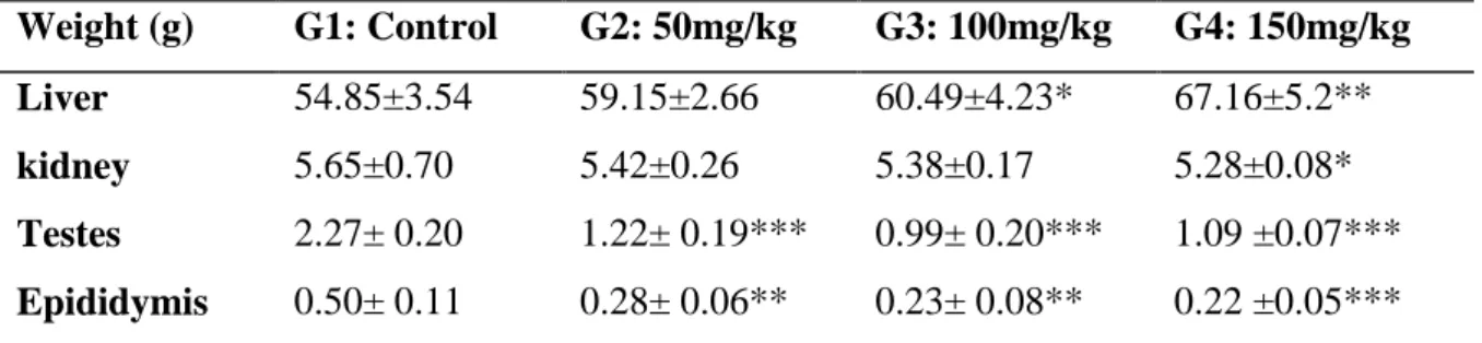
The statistical analysis of these results revealed a significant reduction (p ≤ 0.05) in weight of kidney in the group treated with the high dose of MT. It was also significantly decreased (p≤0.01, p≤0.001) in weight of testis and epididymis in all the treated groups compared with the control group.
However we noted a significant increase (p≤0.05, p≤0.01) of liver weight in the treated groups G3 and G4 compared with the control group.
Table 04: Organs weights of experimental rabbit treated with MT compared with the control group (n=6). Weight (g) G1: Control G2: 50mg/kg G3: 100mg/kg G4: 150mg/kg Liver 54.85±3.54 59.15±2.66 60.49±4.23* 67.16±5.2** kidney 5.65±0.70 5.42±0.26 5.38±0.17 5.28±0.08* Testes 2.27± 0.20 1.22± 0.19*** 0.99± 0.20*** 1.09 ±0.07*** Epididymis 0.50± 0.11 0.28± 0.06** 0.23± 0.08** 0.22 ±0.05*** x SD (n=6) P≤0.05*, P≤0. 01**, P≤0.001***
3.1.1.2. Macroscopic Observation of liver
The Photographs of control rabbit exhibited a normal appearance of liver (fig 8,a ). In the contrast the macroscopic observation (fig 8,b,c,d) of liver of the treated groups with MT illustrating a gross morphological changes revealed by fat accumulation, with presence of irregular nodules emerged on the liver surfaces.
RESULTS
30
Figure 8. Photographs illustrating the gross morphological changes of liver of the treated rabbits with MT and the control rabbit/ a: normal liver of control rabbit,
b: Treated rabbit with 50mg/kg of MT, c: Treated rabbit with 100 mg/kg of MT, d:Treated rabbit with 150 mg/kg of MT. Arrows:irregular nodules (showing yellowish-white coloration and hardening) emerged on the liver surfaces Note: severe fatty change was observed in liver surfaces of rabbit from the treated groups with high doses of MT compared with the control group.