Université de Sherbrooke
Évaluation du potentiel radiosensibilisateur ou
radioprotecteur/antioxydant de quelques composés sélectionnés par
dosimétrie par gel de polyacrylamide et dosimètre de Fricke, et utilisation
de la f:tlamentation par impulsion laser infrarouge femtoseconde comme
un nouveau et puissant faisceau pour la radiothérapie du cancer
Par
Ridthee MEESAT
Département de médecine nucléaire et de radiobiologie Thèse présentée à la Faculté de médecine et des sciences de la santé
en vue de l'obtention du grade de philosophiae doctor (Ph.D.) en Sciences des radiations et imagerie biomédicale
Sherbrooke (Québec) JlH SN4; Canada
Pr Léon Sanche Pr Kim B. McAuley Pr Yue Zhao Pr Daniel Houde Pr Martin Lepage Pr Abdelouahed Khalil Pr Jean-Paul Jay-Gerin © [Ridthee Meesat, 2012] Mars 2012
Membres du jury d'évaluation
Président, Département de médecine nucléaire et
radiobiologie, Faculté de médecine et des sciences de la santé, Université de Sherbrooke
Examinateur, Department of chemical engineering, Faculty
of engineering and applied science, Queen's University
Examinateur, Département de chimie, Faculté des sciences,
Université de Sherbrooke
Examinateur, Département de médecine nucléaire et
radiobiologie, Faculté de médecine et des sciences de la santé, Université de Sherbrooke
Directeur de recherche, Département de médecine
nucléaire et de radiobiologie, Faculté de médecine et des sciences de la santé, Université de Sherbrooke
Directeur de recherche, Département de médecine (Service
de gériatrie), Faculté de médecine et des sciences de la santé, Université de Sherbrooke
Directeur de recherche, Département de médecine
nucléaire et radiobiologie, Faculté de médecine et des sciences de la santé, Université de Sherbrooke
••••••••• •• •••••••••••••• •••• •• •• •••••••
. ..
,_,,-NOTICE:
Library and Archives Canada
Published Heritage Branch
395 Wellington Street Ottawa ON K1A ON4 Canada
The author has granted a
non-exclusive license allowing Library and Archives Canada to reproduce, publish, archive, preserve, conserve, communicate to the public by
telecommunication or on the Internet, loan, distrbute and sell theses
worldwide, for commercial or non-commercial purposes, in microform, paper, electronic and/or any other formats.
The author retains copyright ownership and moral rights in this thesis. Neither the thesis nor substantial extracts from it may be printed or otherwise reproduced without the author's permission.
ln compliance with the Canadian Privacy Act some supporting forms may have been removed from this thesis.
While these forms may be included in the document page count, their removal does not represent any loss of content from the thesis.
C d ...
ana.a
Bibliothèque et Archives Canada Direction du Patrimoine de l'édition 395, rue Wellington Ottawa ON K1A ON4 CanadaAVIS:
Your file Votre référence ISBN: 978-0-494-89650-1 Our file Notre référence ISBN: 978-0-494-89650-1
L'auteur a accordé une licence non exclusive permettant à la Bibliothèque et Archives Canada de reproduire, publier, archiver, sauvegarder, conserver, transmettre au public par télécommunication ou par l'Internet, prêter, distribuer et vendre des thèses partout dans le monde, à des fins commerciales ou autres, sur support microforme, papier, électronique et/ou autres formats.
L'auteur conserve la propriété du droit d'auteur et des droits moraux qui protege cette thèse. Ni la thèse ni des extraits substantiels de celle-ci ne doivent être imprimés ou autrement
reproduits sans son autorisation.
Conformément à la loi canadienne sur la protection de la vie privée, quelques formulaires secondaires ont été enlevés de cette thèse.
Bien que ces formulaires aient inclus dans la pagination, il n'y aura aucun contenu manquant.
Abstract
Evaluation of the radiosensitizing or radioprotective/antioxidant potential of some selected compounds by polyacrylamide gel dosimetry and Fricke dosimeter, and utilization of the femtosecond infrared laser pulse filamentation as a novel, powerful
beam for cancer radiotherapy by
Ridthee MEESAT
Département de médecine nucléaire et radiobiologie
Faculté de médecine et des sciences de la santé, Université de Sherbrooke Sherbrooke (Québec) JIH 5N4, Canada
Thesis submitted to the Faculty of Medicine and Health Sciences for the Degree of Philosophiae Doctor (Ph.D.) in Radiation Sciences and Biomedical Imaging
In radiation treatment, a sufficiently high radiation dose must be delivered to the tissue volumes containing the tumor cells while the lowest possible dose should be deposited in surrounding healthy tissue. W e developed an original approach that is fast and easy to implement for the early assessment of the efficiency of radiation sensitizers and protectors. In addition, we characterized a new femtosecond laser pulse irradiation technique. We are able to deposit a considerable dose with a very high dose rate inside a well-controlled macroscopic volume without deposition of energy in front or behind the target volume.
The radioprotective efficiency was measured by irradiation of the Fricke solution incorporating a compound under study and measuring the corresponding production of ferric ions G(Fe3+). The production of ferric ions is most sensitive to the radical species
produced in the radiolysis of water. We studied experimentally and simulated with a full . Monte-Carlo computer code the radiation-induced chemistry of Fricke/cystamine solutions. Results clearly indicate that the protective effect of cystamine originates from its radical-capturing ability, which allows this compound to compete with the ferrous ions for the various free radicals - especially •oH radicals and H• atoms - formed during irradiation of the surrounding water.
The sensitizing capacity of radiation sensitizers was measured by irradiation of a polyacrylamide gel (P AG) dosimeter incorporating a compound under study and measuring the corresponding increase in the gradient between spin-spin relaxation rate (R2) and absorbed dose. We measured an irradiation energy-dependent increase in Ri-dose sensitivity for halogenated compounds or a decrease for radioprotectors.
Finally, we studied a novel laser irradiation method called "filamentation". We showed that this phenomenon results in an unprecedented deposition of energy and the dose rate thus achieved exceeds by orders of magnitude values previously reported for the most intense clinical radiotherapy systems. Moreover, the length of the dose-free entrance region was adjusted by selecting the duration of femtosecond laser pulses. In addition, we provided evidence that the biological damage caused by this irradiation was similar to other ionizing radiation sources.
Keywords: Radiotherapy, radiosensitization, radioprotection, laser, filamentation, dosimetry
Résumé
Évaluation du potentiel radiosensibilisateur ou radioprotecteur/antioxydant de quelques composés sélectionnés par dosimétrie par gel de polyacrylamide et dosimètre
de Fricke, et utilisation de la filamentation par impulsion laser infrarouge femtoseconde comme un nouveau et puissant faisceau pour la radiothérapie du cancer
par
Ridthee MEESAT
Département de médecine nucléaire et radiobiologie
Faculté de médecine et des sciences de la santé, Université de Sherbrooke Sherbrooke (Québec) JlH 5N4, Canada
Thèse présentée à la Faculté de médecine et des sciences de la santé en vue de l'obtention du grade de Philosophiae Doctor (Ph.D.) en Sciences des radiations et imagerie biomédicale
La radiothérapie repose essentiellement sur le dépôt d'une dose de radiation létale suffisamment élevée à un volume tumoral, tout en minimisant celle délivrée aux tissus sains environnants. Ainsi, une approche originale a été développée, permettant l'évaluation simple et rapide de l'efficacité de composés ayant des propriétés radiosensibilisatrices ou radioprotectrices. En parallèle à cette étude, nous nous sommes intéressés à la caractérisation d'une nouvelle technique d'irradiation utilisant des impulsions laser femtosecondes. Nous avons démontré le dépôt d'une dose considérable avec un débit de dose très élevé au sein d'un volume bien déterminé, sans dépôt d'énergie en amont et en aval du volume cible.
L'efficacité de la radioprotection a été mesurée à l'aide du dosimètre de Fricke via le rendement de production en ions ferrique G(Fe3
l.
Ce dernier est particulièrementsensible aux espèces radicalaires produites au cours de la radiolyse de l'eau. Ainsi, le rendement G(F e3+) dans une solution de Fricke diminue en présence de radioprotecteurs. Sur ce principe, nous étudié expérimentalement et nous avons développé un code Monte-Carlo pour comprendre le mécanisme d'action de la cystamine, un radioprotecteur bien connu.
L'action de radiosensibilisateurs peut être mesurée par la variation du gradient du taux de relaxation spin-spin (R2) en fonction de la dose absorbée. La présente étude repose
sur l'utilisation du dosimètre à gel de polymère afin d'estimer quantitativement les capacités d'ions halogénures et de la thiourée à titre respectivement de radiosensibilisateurs et de radioprotecteur. Nous avons mesuré une dépendence énergétique de la sensibilité Rrdose.
Parallèlement, nous avons étudié le processus de filamentation laser comme une nouvelle source d'irradiation. Il en résulte un dépôt d'énergie considérable et un débit de dose qui surpasse de plusieurs ordres de grandeurs les valeurs précédemment rapportées pour les systèmes de radiothérapie clinique les plus intenses. De plus, il est possible d'ajuster la longueur de la zone libre de tout dépôt de dose à l'entrée du milieu irradié en jouant sur la durée de l'impulsion laser femtoseconde incidente. Finalement, nous nous sommes efforcés d'établir l'équivalence d'un point de vue radiobiologique (dommages induits à un système d'intérêt biologique) entre ce type d'irradiation et d'autres sources de radiations ionisantes plus conventionnelles.
Mots-clés: Radiothérapie, radiosensibilisation, radioprotection, laser, filamentation, dosimétrie
n1nl1:;Ljju,;;'nJ111't'lV11.:in11L~)Jtl1::ivifi111Ylf.:ii n11tlnÜtJ.:if.:iiLLfl::n11iii1utJit1;Jflta1::'1ltJ'lft1nl1mm.Ju1.:i 'liÛl'liJi''lt1î::u1.m111'111f.:iiLLUUL't'lfltl::l'l1fl1L)JrA'L"lfüLfl::)J1f;lrl111f.:iiLLUUmnn LLfl::m:'l.~fimmuut ... l.l'IJtJ'IL'r'J)J
'i6iL'llrlU~U'YhL:lllLflL'litJmift~fl1L)JUi;i"Lf;li\1~1LLft'lft)Jm1ru:::a.:i~1
.
... fuf.:iifm:t1).J:::LÎ'I.. •.Y. ~ 1J1!.ltJ'Vl6 i111c;it1
Département de médecine nucléaire et de radiobiologie, Faculté de médecine et des sciences de la santé, Université de Sherbrooke, Sherbrooke (Québec) JlH 5N4, Canada
~V1tl1ÛYluiffiLilu1bu
...d.:i'lltJ'ln11~m:m::~u1"cvcv1~tt~tl'ruanr;i
(Ph.D.) a1'111 Radiation Sciences andBiomedical lmaging 'Hl'll'ltu:: Medicine and Health Sciences
1.t1m:fm:t1).J:::LÎ'l~'lt1f.:ii 1").J1tuf.:ii~t-nu~1LL'.IU'IL~tlL~tl~LiJU)J:::Lh"lditi.:i~.:iLVlt1.:i'fltll1ltim:
• .- cr ..1 " ., ~.J • 1 .r ~
•t
~.,
.,
~.J" .JVl1fl1tJL'liflfl)JtLN 'l.ti'!Jtu:::vt"l:::f;lfl'll'l'lUf'l)JW)J1tlJNflVllJ11LL'.IU'ILUtlLtJtltlnlJl ... ~'1rulli)J1tlJNflVIU'!ltJVl~I'\ • .J ."I ~-1~" ..: .1 ~ .. " ~ _, .'1 " " ~... • " 1. " .. " " ~
L Vl1Vl"l::L1J'l.l ~1J ~111 a1:L 'fl)J1J :t:::flVlfi111YlN flLLfl:: a111Jn1J'!l.:J'!l'l.lf;l :t1t1 N ri)JPl'l1SJ t111'l cv um:1ntt1).J:::LN1J1'l t11.:i ri
1u~vimilV1ulf~1~w"1t11~fin1muu1.mJ~ft:::111'lmLfl::::'l111Ln 1t1n1:tl1:::LnutJ1:::~vifi111't'l'IJti.:ift1:L~i.1
tl1::aV1fimYlf.:iiLLfl:::ft1nlniJ'!l.:if.:ii
U'fln"l1nd1.um1~n'l:t1füiJ1Vi1m1~m:nin'l:ttu:::Lll't'l1:::'1ltJ'IL'Vlf'lÛl'IL'.1;.l'llti.:i
n1îll'lt1f.:iiiJl'lt1LVll'IÛ11lL'r'J)JLf;lL'l!!Ï'l.l~U'r'J1L:tt'!Lfü'lltJm~t1Ylfl1L)J'l.llllLf;l-i'u iJi''lt1~fiim~Lî1ft1).J'lfCl~"l:::~.:itllmru f.:i;~i°1'81J111i..lh11cuf.:i;~.:ittlti.:i~1LL ... U.:iLiJ1-..)J1tJL\l~'lnfl1.:i
~'îLL -..u.:iiJ\1u ...
-l
1LLfl:::'.I â°'l'IJtl'l~1LL ... U.:JLiJ1'.l)J1tJt11:::~vifim'fl'!JtJ.:ia1nlniltJ.:if.:iia1mml'1J1tiJi'iJl'lt1m:Hi.11l111tl'tJ1f.:iiLLuumnn
i:.itu.i~IJl'llfl.:iL'r'J?JttnLtJflfl'l.l (G(Fe3.)) mt1 ... â'.:i"l1nm:ll1t1f.:iia11fl:::fl1t1mnn ~i'la1:tlm.':ltJ.:if.:iii:.ia)J?JtjLL~'l tll)J1 cu'lltl'IL vltifin
1titi~u;utitjnutiit:.J fl~ft1:::~.:i
...~nt1~Lîi
'1;t1"l1nn ::::u'lum:tftfl1t1t!Ï'l'llti.:itl1Lt'It11 olf .:i~
m:~~t1d1iJIH~.:i
m1VI "1fltJ'ILLfl:::m:H'LLUU~1fltl'I
Monte-Carlof'ltJSJ~'.lLIJlflf
L~tl H~
n'l:t1tl::;~vifim'fl'lltl'I
ft1:1am~u (cystamine) 1.ua11fl::fl1t1mnîi i:.iam:~n'l:t1LiJÎLLfl"1.:i1.'IXL~utJ~1.:ii'i;iL"lu~1 tl:::~Vlfim'flmnlnil'!l.:i
f.:ii'lltJ'lft1~LÙUi:.Jfli.11"11nn1îl'l'l1i.lft1i.117ClLUn1rn1tlDMt11!ÏUtJit~fl~tl5':: ~.:ie111:Jai;i1~'1.lft1i111C1LL'll.:iiu
nrn'r'JtJnt1LtltltiUL~tJ'rÏ1tlDMt11!ÏUfl't~fl~t11:;~.:i'.lfl1t1'i"1mllm:::tJth.:i~.:i
00H LLfl::: H"
~LÎÎ'1"l1nn::::u'.lum1
·aa1t1~'.l'llti.:iJ1~'lt1f.:iimnl1:::Lnu;Ji;i~1!Ï"1'11tl'lft11L~)JU7:::~Vlflll1Ylf.:iia1i.11:ci1'1J1LiJÎ"11nm1Hi.11i;irli;if.:i~LLUUL't'lfltJ:::l'l1
a11i.i t'i"L"lfl 'L111t1~r.i1 :ru1r.i1n m:Ltl~t1ULLtl fl'l'lltJ'I f'l'l1i.JiU'll'Mf'l'.l1i.J t\i.i~ufiL 'i.:iL~'t.u::: ... ~1.:i Phm 1~tiUl'lfl1 t1 LL'l.Juatluatlu (spin-spin relaxation ... -;.'él R2 ) ri'utlli.J1ruf.:ii 111t1 ... i.:ir.i1nm1ll1t1m1J11tl'i;if.:i~'LYl~'ël:::l'l1fl1
11-J'1L<1~ (PAG) ~ii~11L~i.JtJ::::iV1fim'l'l~~"titjLLfi'l 1.um:~n'l:t1eLn~1ii1rn-S'L'lm1L~i.1~u'llti.:il'l'l11-Jiu
(Ri-dose sens itivity) ~1'.lfuft1 ni :r:::ntiU'lltJ'l Ltl LtJ ;lui.:i ... fl1t1 LLfl::mrnr;ia.:i'!l'él'l l'l'l1i.J{u~1-.. fu a1nl nÜ?J.:i f.:ii
qi;il11t11.ul)V1t11ÛYlUfid1iJl~n'l:t1l)fiim:ll1t1f'l~LLUUL'.1;.lLt'lt1H'Lfü'.D'élf~L:t1n~1 "Ylfl1L)JUr;i"Li;ioJu (fil amentation)" 1. um:~m~nd1iJi'LL~"1.:i 1. ,XL ~'l.ILL~'.l~1tl11 n!J m:rruîln'?l 1. 'IX Ln lflYlfl'.:i.:i1u~ rhti L V1!Ïu il'ln a1.:i
iJi''.lt1-è'"171Wl-l1C\J~~'liJ1nn~1~Ll'ltJL-nuf.:iifn'l:t1i.11rltlU i.11nn~1~uiJi''lt1n11ll1tlf'liLLUUdtr.:it111Pl"l1n Ylâ°.:i.:i1u~chmV11.iXnu~'lnfl1.:ii.:iV11.:i;r1u ... ~1LLa:::iJ\1u-..fl'.:i'lltJ.:i~1LL ... u.:iLil1-..mt1~ll1t1f.:ii~niJî'lt1 ~'?l.:i"l1nd
.:i1u~4't1d1ffLLftlfl'l'.linl1Ul'l'l1i.JLitJ'.11tJVl1'1fi'1U=i'l~Vltl1'lltJ'ln1fll1tlf'liiJl'lt1LflL'l!'élfdl'lfl1t1!Ï\lnUn11l'l'l1).J
Table of contents
Abstract
Résumé
List ·of illustrations (Tables and Figures)
Acronyms, abbreviation and symbols
1. Introduction
I. 1. Interaction of the photons 1. 2. Radiolysis of water
1. 3. Radiation protector/antioxidant 1. 4. Radiosensitizers
1. 5. Fricke dosimetry
1. 6. Monte Carlo simulations
1. 7. Polyacrylamide gel (PAG) dosimetry 1. 8. Femtosecond laser pulse filamentation 1.9. Objectives of the research project
II.
Article No.
1
"Evaluation of dose-enhancement of iodinated compounds by polyacrylamide gel dosimetry".
R. Meesat, J.-P. Jay-Gerin, A. Khalil, M. Lepage.
Physics in Medicine and Biology 54,5909-5917 (2009)
III.
Article No. 2
"Utilization of the ferrous sulfate (Fricke) dosimeter for evaluating the
radioprotective potential of cystamine: Experiment and Monte-Carlo simulation", R. Meesat, S. Sanguanmith, J. Meesungnoen, M. Lepage, A. Khalil and J.-P. Jay-Gerin.
This article has been accepted for publication in Radiation Research.
1 11 111 lX 1 1 8 16 18
20
22
24
28
32 34 57IV.
Article
No. 3
"A beam for cancer radiotherapy based on femtosecond IR laser-filamentation yielding ultra-high dose rates and zero entrance dose", R. Meesat, J.-François A., Hakim Belmouaddine, C. Tanguay-Renaud, R. Lemay, T. Brastaviceanu,
L.
Tremblay, B. Paquette, J. R. Wagner, J.-P. Jay-Gerin, M. Lepage, M. A. Huels, and D. Houde.
This article is currently under consideration for publication in The Proceedings of the National Academy of Sciences USA (PNAS), (3 October 2011).
109
V. Discussion
143V. 1. The scope ofthis thesis 143
V. 2. Radiosensitization quantified by polyacrylamide gel dosimetry 143 V. 2.1. Iodine compounds as a radiation sensitizer 144 V. 2.2. Radioprotection quantified by polyacrylamide gel dosimetry 146 V. 3 Radiation protector quantified by Fricke dosimeter 148
V. 3 .1. Experimental study 148
V. 3.2. Monte-Carlo simulation study 150
V. 4. Evaluation absorbed dose of femtosecond laser pulse filamentation by
Fricke and P AG dosimeter 152
V. 4.1. Dosimetry for femtosecond laser pulse filamentation 15"-. V. 4.2. Filamentation ofultrashort light pulses in scattering media 153 V. 4.3. Ultrahigh dose rate from femtosecond laser pulse filamentation 155
V. 4.4. High dose rate for sterilization 15'
V. 4.5. Radiation chemistry of femtosecond laser pulse filamentation lf>1> V. 4.6. "Two bullet theory" of femtosecond laser pulse filamentation for
cancer therapy 161
VI.
Conclusions
163Acknowledgments
167List of illustrations (Tables and Figures)
CHAPTER 1 - Introduction
Table 1
Fig.1 Fig. 2 Fig. 3 Fig. 4 Table2 Fig. S Fig. 6 Fig.7
Fig. 8 Fig. 9Ionization potentials of selected molecules. 2
A diagrammatic representation of the processes from energy transfer 6 to final biological damage.
Diagram showing transmission of photon through an absorber of 7 thickness 6.x.
Mass attenuation coefficients for H20, Br and I. 7
Radiolysis of water by the low-LET radiation. 9
Main reaction scheme and rate constants (k) used in the radiolysis of 15 pure liquid water at ambient temperature (25 °C).
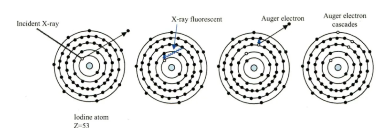
Diagrammatic representation of the absorption of an incident X-ray 20 photon by an iodine atom.
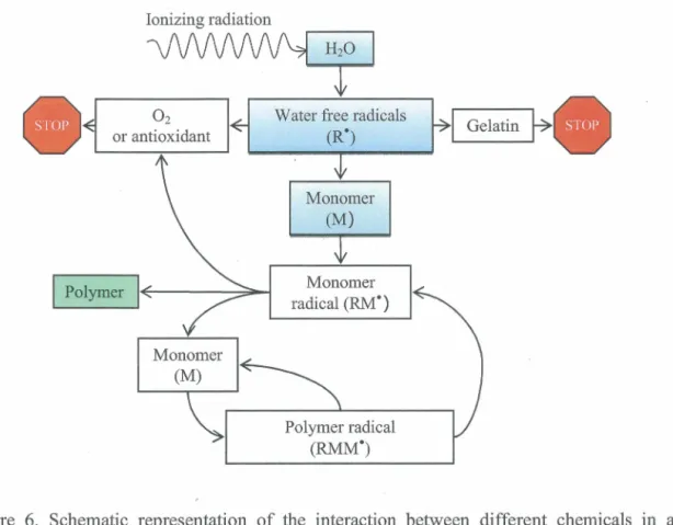
Schematic representation of the interaction between different 27 chemicals in a polyacrylamide gel dosimeter.
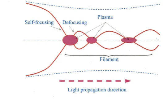
Schematic demonstration of the focusing-defocusing cycles sustaining 29 a long range propagation of light filaments.
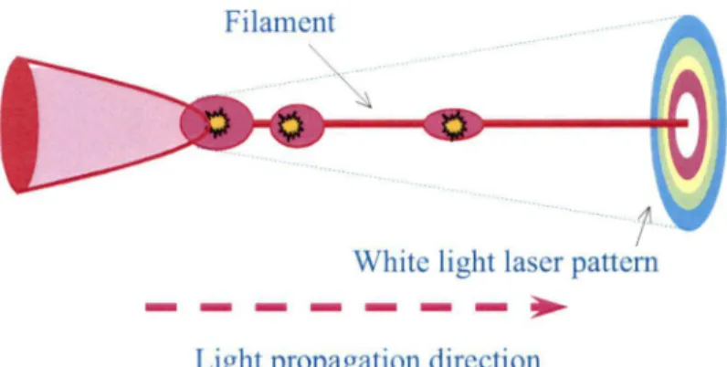
A schematic dia gram of the evolution of a f emtosecond laser pulse 30 propagating in an optical medium. The pulse is demonstrated by the
ellipse at the left. The central slice of the pulse self-focuses to a small area where the resulting high intensity ionizes the air molecules (yellow star). The front part keeps on self-focusing becoming thinner and thinner. At the end of the propagation, the pulse degenerates into a colourful white-light laser pulse, as called conical emission.
Interplay of multiphoton ionization, inverse Bremsstrahlung 31 absorption, and impact ionization in the process of plasma formation.
Free electrons result from inverse Bremsstrahlung absorption events and impact ionization.
Table 1
Fig.1
Fig. 2
Radiosensitization by an iodinated contrast agent (ICA) in a PAG dosimeter. The samples were irradiated by-40 keV X-ray photons. The standard deviations of the dose enhancement ratios (DER) are derived from the uncertainties in the linear fits.
Plot of relative R2 against absorbed dose for P AG dosimeters
containing different concentrations of (a) NaCl, (b) NaBr, (c) Nal, (d) thiourea and (e) ICA (the concentrations of iodine atoms are shown, there are three iodine atoms per ICA molecule) irradiated with 1.25 MeV gamma photons. A lower extent ofpolymerization is noted for every compound added to the P AG dosimeter at this irradiation energy.
Plot of relative
R
2 against absorbed dose for PAG dosimeterscontaining different concentrations of (a) Nal, (b) NaBr and ( c) ICA (the concentrations of iodine atoms are shown) irradiated with 40 kVp X-ray photons. At this energy, a competition between the scavenging of free radicals by the halide ions and a sensitization is observed for Nal (panel a) and NaBr (panel b), which are in an ionic form in the solution. When the iodine atoms are covalently bound to a molecule such as an ICA, only the sensitization is observed (panel c ).
48
49
50
CHAPTER Ill - Article No. 2
Table 1 Reaction added to the pure water reaction scheme to simulate the 92 radiolysis of de-aerated aqueous H2S04 solution, at 25 °C.
Table 2 Chemical reactions and rate constants used in simulations of the 93 radiolysis of cystamine (RSSR) in the Fricke dosimeter in the presence or in the absence of oxygen, at 25 °C.
Fig.1 (Panel a) Time evolution of G(Fe3
l
(in molec./100 eV) for 300-MeV 102incident protons (LET- 0.3 keV/µm) in the radiolysis of
aerated
solution of 1 mMFeS04 in aqueous 0.4 MH2S04 (Fricke solution) at25 °C. The concentration of dissolved oxygen used in the calculations is 2.5 x 10⁴
M.
The solid line shows our simulated kinetics ofFe3+ion formation. The arrow on the right of the figure shows the accepted value (15.5 ± 0.2 molec./100 eV) of the yield of the Fricke dosimeter for 6°Co y-rays and fast electrons. (Panel b) Time dependence of the extents i:\G(Fe3
1
(in molec./100 eV) of the different reactions that contribute to the formation ofFe3+ ions, calculated from ourMonte-Carlo simulations in the interval 10·12-200 s (see text).
Fig. 2 (Panel a) Time evolution of G(Fe3
l
(in molec./100 eV) for 300-MeV 103Fig. 3
Fig. 4
Fig. 5
solution of 1 mMFeS04 in aqueous 0.4 MH2S04 at 25 °C. The solid line shows our simulated kinetics ofFe3+ ion formation. The arrow on
the right of the figure shows the accepted value (8.2 ± 0.3 molec./100 eV) of the yield of the Fricke dosimeter in the absence of oxygen for
6°Co y-rays and fast electrons. (Panel b) Time dependence of the
extents ôG(Fe3+) (in molec./100 eV) of the different reactions that contribute to the formation ofFe3+ ions, calculated from our Monte-Carlo simulations in the interval 10-12-200 s (see text).
Time evolution of G(Fe3} (in molec./100 eV) as obtained from our 104 Monte-Carlo simulations of the radiolysis of Fricke dosimeter
solutions (1 mMFeS04 in aqueous 0.4 MH2S04) containing various
concentrations of cystamine, under both
aerated
(a) anddeaerated
(b) conditions, using 300-MeV incident protons (LET- 0.3 keV/µm) at 25 °C. The different lines correspond to three different concentrations of cystamine added (indicated to the right of the figure): 104 M (dash-dot line), 10-3 M(dotted line), and 0.1 M(dashed line). The solid line
shows our simulated kinetics ofFe3+ ion formation for the Fricke dosimeter without added cystamine (for reference ).
Dependence of ferric ion production from irradiated Fricke solutions 105 (1 mMFeS04 in aqueous 0.4 MH2S04) upon the concentration of
added cystamine in the range 5 x 10⁻⁷ -0.1 M, under both aerated (panel
a) and deaerated (panel b) conditions. The different lines show the Fe3+ ion yields obtained from our Monte-Carlo simulations (at -200 s following ionization) using different values of the fraction of •oH radicals that react with cystamine to form either transient RSSR•+ radical ions [reaction (R3a)] or the non-ionic intermediates RSH and Rso• [reaction (R3b)]. The dotted and dashed lines correspond to a branching ratio of 50% and 70% in favor of the oxidation pathway (R3a), respectively. The solid line is obtained by assuming a negligibly small intervention of reaction (R3b ). As clearly seen, a branching ratio of -100% in favor ofreaction (R3a) is required to achieve the best
simultaneous
agreement between calculated and measured Fe3+ yields as a function of cystamine concentration in the presence and absence of oxygen (see text). Experiment: (•) ref. (25),(à) ref. (26), and (D) this work.
(Panel a) Time evolution of G(Fe3} (in molec./100 eV) for 300-MeV incident protons (LET- 0.3 keV/µm) in the radiolysis of
aerated
Fricke dosimeter solutions containing 1 mMFeS04 and 1 mMcystamine in aqueous 0.4 MH2S04 at 25 °C. The concentration of
dissolved oxygen used in the calculations is 2.5 x 10⁴ M. The solid line shows our simulated kinetics ofFe3+ ion formation. (Panel b) Time dependence of the extents ôG(Fe3) (in molec./100 eV) of the
Fig. 6
Fig. 7
different reactions that contribute to the fotmation of Fe3+ ions, calculated from our Monte-Carlo simulations in the interval 10-12-200 s. The oxidation ofFe2+ ions to Fe3+ involves reactions mainly with
Ho2·, H20 2, and the cystamine-radical species Rs• and RSSR•+ (see text).
(Panel a) Time evolution of G(Fe3) (in molec./100 eV) for 300-MeV 107 incident protons (LET- 0.3 keV/µm) in the radiolysis of deaerated
Fricke solutions containing 1 rn.MFeS04 and 1 mM cystamine in aqueous 0.4 MH2S04 at 25 °C. The solid line shows our simulated kinetics ofFe3+ ion fotmation. (Panel b) Time dependence of the
extents ô.G(Fe3+) (in molec./100 eV) of the different reactions that
contribute to the fotmation of Fe3+ ions, calculated from our Monte-Carlo simulations in the interval 10-12-200 s. The oxidation ofFe2+
ions to Fe3+ involves reactions mainly with H202 and the
cystamine-radical species Rs· and RSSR.+ (see text).
Time dependence of the extents ô.G(Fe3) (in molec./100 eV) of the 108
different reactions that contribute to the fotmation and decay of cystamine (RSSR) (see text and Table 2), calculated from our Monte-Carlo simulations of the radiolysis of Fricke solutions containing 1 rn.MFeS04 and 1 mM cystamine in aqueous 0.4 MH2S04 by 300-MeV incident protoris (LET-0.3 keV/µm) at 25 °C and in the interval -10-12-200 s, when oxygen is present (panel a) or absent (panel b).
CHAPTER IV - Article No. III
Fig. 1 Photographs of containers filled with tissue equivalent polymer 132 dosimeter gel and irradiated with fs laser pulses (-400 µm x 400 µm)
to draw the logo of Université de Sherbrooke (a) top view and (b) side view. The entrance dose was effectively zero up to a certain depth, which in (b) was wave modulated (yellow arrows) by changing the laser pulse duration (input beam from the right of the container), to show depth control of the zero dose region before the target volume in which filamentation occurs. The length of the filamentation tracks, i.e. their end, is controlled in part by the physical geometry of the optics (lenses, etc), as well as the energy (intensity) per laser pulse, at a given pulse width, ceteris paribus, for laser pulse energies above
filamentation threshold, and below dielectric breakdown (22,23). In (c), the polymer gel was irradiated with X-rays (150 kVp) from a clinical X-ray therapy system (Therapax HF150T), at 1, 2 and 3 Gy, and clearly shows that the deposited dose is maximum upon "tissue entry" (blue arrows). (d) is a comparison of depth-dose distributions of various conventional radiation modalities related to radiation therapy.
Fig.2
Fig. 3
Fig. Sl
Fig. 82
Fig. 83
DNA damage induced by 137es gamma radiation, and laser 133
filamentation at 800 nm: (a) a graph of the release of thymine (glycosidic bond cleavage) from the irradiation of thymidine in solution versus absorbed dose. Agarose gel electropholysis separating supercoiled plasmid DNA damage ( circular and linear DNA
conformations correspond to single and double strand breaks) using (b) gamma irradiation and ( c) IR femtosecond laser pulse
filamentation. The absorbed macroscopic doses to the 2 ml target solution are given in Gy, and the two lanes in (b) after the marker are unirradiated controls.
Femtosecond IR (800 nm) laser pulse treatment (left leg) of the mouse 134 mammary carcinoma Me7-Ll tumor compared to control (right leg)
23 days after laser irradiation. Me7-Ll cells were injected into the · right and left thighs of female Balb/c mice. The right side tumor of
mice was used as control. Three to four weeks later, mice bearing tumors with a diameter of 3-5 mm were irradiated by femtosecond IR laser pulses. The best laser treatment result here was obtained by irradiating five separate spots (100 - 300 µm diameter, separated by approximately 1 to 1.5 mm) on the tumors, with 10 min irradiation times for each spot; the laser irradiation and dosimetry conditions were identical to those used for filamentation during chemical dosimetry (10).
Experimental setup for laser filamentation irradiation of samples in 140 solution.
Graph of macroscopic dose (ceric-cerous and Fricke dosimeter) versus 141 exposure time of laser femtosecond filamentation at 800 nm ( • ), and
gamma radiation (y-rays from 137es) (•).
The filamentation of femtosecond laser radiation fixed in polymer gel 142 dosimeters (a) optical side view of the filamentation and (b) optical
image of the cross sections of filamentation ( c) MRI image of the tracks of filamentation ( d) MRI image of the cross sections of the tracks of filamentation. Note that here the laser was set to produce filamentation immediately upon entry in the gel medium, as opposed to Fig la and b in the full text, were the laser pulse width was adjusted to produce a controlled zero entrance dose. Here the filamentation tracks are produced in the single shot mode, one pulse per track.
Fig.1
Plot of relation R
2against the absorbed dose for P AG dosimeter
148
containing different concentrations of cysteine irradiated with Co
60 ,gamma-radiation.
Fig. 2
Plot of relation optical density of conjugated diene formation at 234
149
nm against the absorbed dose for low-density lipoprotein peroxidation
containing different concentrations of cystamine irradiated with Co
60,
gamma-radiation.
Fig. 3
Effect of cystamine upon oxidative modification ofLDL. LDL
150
samples were exposed to free radicals by gamma radiation (dose rate
3.44 Gy/min) in with 50 mM and without cystamine.
Table 1
Rate constants for the reaction of disulfides with hydroxyl radicals.
151
Fig. 4
Photographs of
~ontainersfill with polyacylamide gel dosimeter (a)
156
without milk,
(b)with 0.1
%ofmilk (2
%of fat), (c) with 0.2
%of
milk (2
%of fat). The gels wereirradiated by femtosecondîi-sapphire
laser beams in the same condition with the following properties: pulse
duration was 100 fs; pulse energy was 0.3 mJ/pulse; repetition rate
was 1 kHz; and the central wavelength was 800 nm. The laser beam
was focused by a 30 cm focal length achromatic lens.
In(b ), in order
to compare with laser filamentation, the polymer gel was irradiated by
X-ray from Therapax HF 150T (x-ray therapy system). Fig. al, bl and
cl
shown zoom in to see filamentation track of the sample a, b and c
respectively.
Fig. 5
Graph of dose (ceric-cerous dosimeter) versus yield of total
159
hydroperoxides in de-ionized water;
(O),1 mM
Thymidine(~)and 1
mM Thymidine with catalase
(.à).The sample solutions were
irradiated by Femtosecond Ti-sapphire laser beams with the following
properties generated filamentation: pulse duration was 1 OO fs; pulse
energy was 0.3 mJ/pulse; repetition rate was 1 kHz; and the central
wavelength was 800 nm. The dose rate was 475 Gy/min.
Fig. 6
Graph of wavelenght versus absorbance of goldnanoparticles as
160
function of time of laser filamentation irradiation. Goldnanoparticles
absorb UV at 520 nm. The sample solutions were irradiated by
Femtosecond Ti-sapphire laser beams with the following properties
generated filamentation: pulse duration was 1 OO fs; pulse energy was
0.3 mJ/pulse; repetition rate was 1 kHz; and the central wavelength
was 800nm.
Acronyms, abbreviation and symbols
~Eab ~E1 ~Etr LiX AABIS
BNCT
c
CEDRCF
CNS
DERDNA
DSB
E EDR ET eV f s g(X) G(X)Gx
HPLCIo
ICA
IR IRT Hydrated electron Absorption energy Lost energy Transfer energy Thickness Acrylamide N,N' -methylene-bis-acrylamideBoron-neutron capture therapy Molar concentration
Ceric effective dose rate Conversion faCtor Central nervous system Dose enhancement ratio Deoxyribonucleic acid Double stand break Energy
Effective microscopie dose rate Effective time
Electronvolt F emto-second
calculation 100-eV yield of the final product X Experimental 100-e V yield of the final product X Primary yield of radiolytic species X
High-performance liquid chromatography Incident intensity
Iodinated contrast agent Infrared
lx
kkeV
LEE LET MC7-LlMeV
molec./100 eV
MRI
NaX
OD PAGR2
ROI
RSSR
'SEM
SSB
T2
THPC
X-ray
z
Eex.
'Y
µ
p
µ
pTransmitted intensity
Rate constant reaction
Kilo-electronvolt
low-energy secondary electron
Linear energy transfer
Mouse mammary carcinoma cell
Mega-electronvolt
Molecule/100 eV
Magnetic resonance imaging
Sodium halides (X= Cl, Br, I)
Optical density
Polyacrylarnide gel dosimeter
Spin-spin relaxation rate
Region of interest
Cystarnine
Standard error of mean
Single stand break
Spin-spin relaxation time
Tetrakis (hydroxymethyl) phosphonium chloride
X radiation
Atomic number
Projectile charge number
Molar extinction coefficient
Alpha particle
Gamma radiation
Linear attenuation coefficient
Density
CHAPTER I: Introduction
1. Introduction
I.
1. Interaction of the photons
Radiation therapy is the most widely used modem cancer treatment and continues to play a major rote in the management of an array of potentially curable malignancies (IAEA, 2008). It is vital to deliver the highest radiation dose to the tumor volume while sparing surrounding normal tissues. This is an important challenge to modem radiotherapy since high-energy photons result in a significant entrance dose in front of the tumor and a non-negligible exit dose behind the tumor. Unfortunately, conventional radiation sources (y or X rays, electrons) used over the decades, even with methods using multiple or modulated beams, inevitably deposit the majority of their dose in front or behind the tumor, thereby damaging healthy tissue, and causing secondary cancers years after treatment (Hall and Phill, 2006).
Radiation-sensitizers and radioprotectors are staring to play an important role in clinical radiotherapy (Wardman 2007; Nair et al., 2001). Antioxidants have been shown to prevent diseases and promote health (Garewal, 1997; Valko et al., 2007). It is therefore important to understand the mechanisms of action of such compounds at the chemical level in order to help better control and optimize their biological effects. For example, a chemical compound designed at protecting DNA from radiation damage needs not only to reach its cellular target location but also retain its radioprotectillg activity to produce an effective response. Using clonogenic assays, the radiation survival of cells can be measured, but th~s only provides information on the addition of these two properties. · If a compound proves to be less effective than anticipated, is it because it is in fact nota good radioprotector, or is it that it did not reach its target location? This project aims at characterizing compounds at the molecular or chemical level, such that this information can aid in the design and the optimization of compounds that will be therapeutically useful in vivo.
Radiosensitizers are intended to enhance tumor cell killing while having much less effect on normal tissues. Chemical radioprotectors are, of course, the reverse of radiosensitizers: the aim is to decrease radiosensitivity, especially of normal tissues.
CHAPTER 1: Introduction (Dunne-Daly, 1999). Thus, it is important to understand their interaction with ionizing radiation.
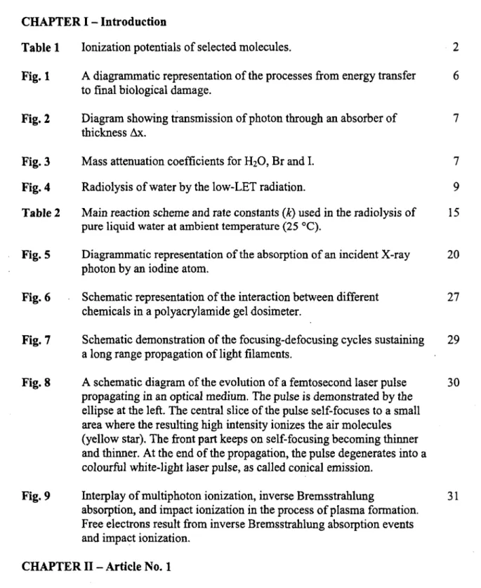
When an ionizing radiation interacts with biological systems or other absorbing matter, a complex series of events takes place as shown in Figure 1. (Alpen, 1998). Typically, absorption of radiation is characterized by an energy transfer from the radiation beam to the medium (Anderson, 1984). The absorption of photon arises from the interaction of the radiation with electrons of the absorber molecule (Bensasson, 1993). In fact, ionizing radiations can be divided into "direct ionizing radiations" and "indirect ionizing radiations", The former are charged particles such as electrons, protons and alpha particles which can ionize by means of particle to particle Coulomb forces. The latter· are uncharged species, such as electromagnetic radiations (e.g., gamma and X-ray) and neutrons which can release energetic charged particles after interaction (Anderson, 1984). Ionization depends on the energy of these radiations. For photons, the energy must exceed the lower limit for ionization (or "ionization potential"). The ionization potential is the minimum energy required to eject an electron from an atom or molecule, values for selected compounds are shown in Table 1.
Table 1. Ionization potentials of selected molecules (Anderson, 1984)
Ionization Ionization
Molecule Molecule
potential (eV) potential (eV)
H2 15.4 C02 13.8
N2 15.6 CH4 13.0
02 12.1 C2~· 10.5
H20 12.6 C6H6 9.3
The probability of ionization depends on the photon energy as well. However, a large ionization potential is not necessarily found whenever the available energy exceeds the ionization potential. In general, the probability of ionization is described by an "interaction coefficient" or a "cross section", which is discussed later in the section.
CHAPTER 1: Introduction
In case of photon interaction, the photon energy (E
=
hv, h is Planck's constant (6.626. x10-34 Js and v is the light frequency) is converted into kinetic energy of high speed charged
particles ( electron or positrons}, whereas some part of this energy is radiated from the medium as scattering radiation (Johns and Cunningham, 1969;· Alpen, 1998).
When a radiation beam passes though biological materials or other absorbing media, energy is lost from the incident beam. The radiation can transfer energy to an absorber; this energy is called energy transferred (~En-). The medium absorbs some part of the radiation energy, which is called energy absorbed (AEab), while· some of it leaves the volume of interaction which is called energy lost (AE1). The total energy transfer is the sum of energy absorbed and energy lost as follows: (Alpen, 1998).
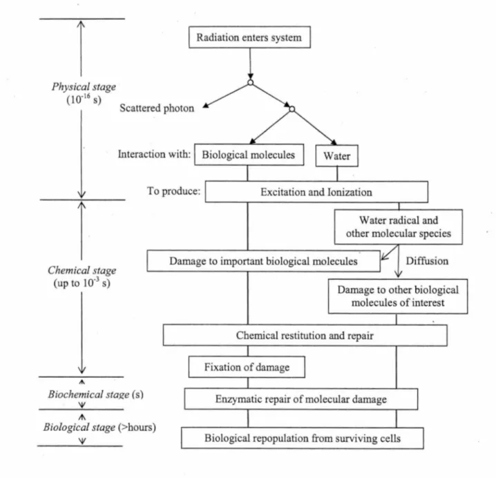
The three main interactions of photons play an important role: photoe/ectric effect, Compton scattering effect and pair production. All these processes result in the partial or complete transfer of the photon energy to electron energy (Knoll, 2000). The fundamentals of the photon interaction are shown in Figure 2. Absorption of the ionizing radiation by matter follows the fundamental Lambert-Heer law (Hughes, 1973).
where /0, lx are the intensities of the incident and transmitted radiation respectively, x is the
thickness of the absorber and
µ
is the linear attenuation coefficient (see Figure 2). In general, the thickness of the absorber is in cm,µ
will be expressed in cm·'. Thus, the linear attenuation coefficient depends on the density of the attenuator. The coefficient is expressed in terms of a mass attenuation coefficient (µ ),
wherep
is the density of the material. Thep
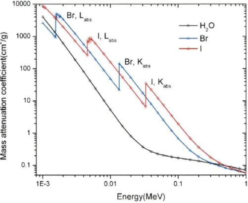
total attenuation coefficient is the sum of the coefficients for the three interactions. Figure 3 shows the total mass attenuation coefficient of H20, Br and 1 as a function of the photon
CHAPTER 1: Introduction
Photoelectric effect
In the photoelectric effect, the entire energy of the ionizing photon (e.g. X-ray or gamma radiation) is transferred to an orbital electron of an atom with the consequent ejection of the electron which is then called photoelectron, typically from the K-shell (Johns and Cunningham, 1969; Hughes, 1973; Alpen, 1998; Knoll, 2000). In this case, the incoming photon disappears and the photoelectron is ejected from its orbital position. Interactions of this kind may occur with electrons in the K, L or M shell as well. The energy of the incoming radiation must be equal to or exceed the binding energy or absorption edges of the electron in the a tom. The binding energies range from -1 OO eV for low atomic weight absorbers to
-100
keV for high atomic weight absorbers. The photo interaction leads to a vacancy of an inner electronic orbital of the atom. This vacancy is quickly filled by an outer electron of the atom. With this process, the outer electron transfers excess energy by emitting a photon called a characteristic ray (or "fluorescent ray"). Similarly, this X-ray can eject other orbital electrons, and so on. The phenomenon is known as the "Auger effect" which is characterized by an electron avalanche or an Auger electron cascade sequence. The photoelectric interaction is the predominant mode of interactfon for photon radiation (gamma rays or X-rays) of relatively low energy. The effect can be enhanced for absorber materials ofhigh atomic number (Z) (Johns and Cunningham, 1969; Knoll, 2000).Thus, at energies above the lowest absorption edge of an atom, photoelectric absorption leads to additional photoelectric effect, this ultimately results in the production of a photoelectron, characteristic X-ray photons, and Auger electrons. In the photoelectric interaction, the process leads to increase radiation absorbed dose in the medium (Johns et al., 1954; Murthy et al., 1976; Fairchild et al., 1982; Regulla et al., 1998) since the cross-section for absorption of electrons is larger than that for photons. In the point of view of medical application, the phenomenon can be applied to enhance radiation dose in a target tissue, a process called "photon activation therapy" (Corde et al., 2004). Applications of this principle led to the evaluation of the sensitizing effect of heavy atoms for example halogen compounds (e. g., bromine and iodine) (Myers et al., 1977; Feinendegen, 1975; Happen et al., 1978; Fairchild et al., 1982; Dawson et al., 1987; Humm and Charlton, 1988; Meesat et al., 2009) and transition elements (e. g., gold) (Regulla et al., 1998; Herold et al., 2000; Hua
CHAPTER 1: Introduction et al., 2001). It should be noted that most authors have been used biological effects as an end point to evaluate the dose enhancing effects of these compounds.
Compton effect
In the Compton effect, the interaction process occurs between the incident ionizing photon and an electron in the absorbing material. The photon radiation provides only part of its energy to an orbital electron. The electron will be free or still bound in an atom. In the former case, the electron will be ejected from atom which is called a
recoi/ electron
and the incident photon will be scattered at an angle with respect to its original direction. The energy transferred to the electron can vary from zero to a large fraction of the radiation energy. The Compton interaction depends only on the number of electrons of the absorber. The number of electrons can be expressed in terms of electron densityi.e.
number of ' electrons per unit volume of material. The Compton absorption is more significant for high energy photons (>l MeV) (Johns and Cunningham, 1969; Hughes, 1973; Anderson, 1984; Alpen, 1998; Knoll, 2000). In this energy range (1-25 MeV), photons mainly interact with bialogical tissue by Compton scattering effect. (Corde et al., 2004).Pair production effect
In the pair production process, the energy of photons must be superior to 1.02 MeV. It is due to the fact that the energy of the radiation will produce a positron-electron pair where the rest mass energy of the particles is 0.511 MeV. In the mechanism of the interaction, the photon, moving close to the nucleus of an atom, is subjected to strong field effects from the nucleus; it may suddenly be converted to become a negative and positive electron pair. The cross section of this interaction continues to increase when the energy of photon increases, while the cross sections for the photoelectric effect and the Compton scattering effect decrease with increase in energy.
In
the high range energy of photons(-25-100
MeV), the pair production interaction in soft tissue is the most important type of absorption (Johns and Cunningham, 1969; Hughes, 1973; Anderson, 1984; Alpen, 1998; Knoll, 2000).CHAPTER I: Introduction
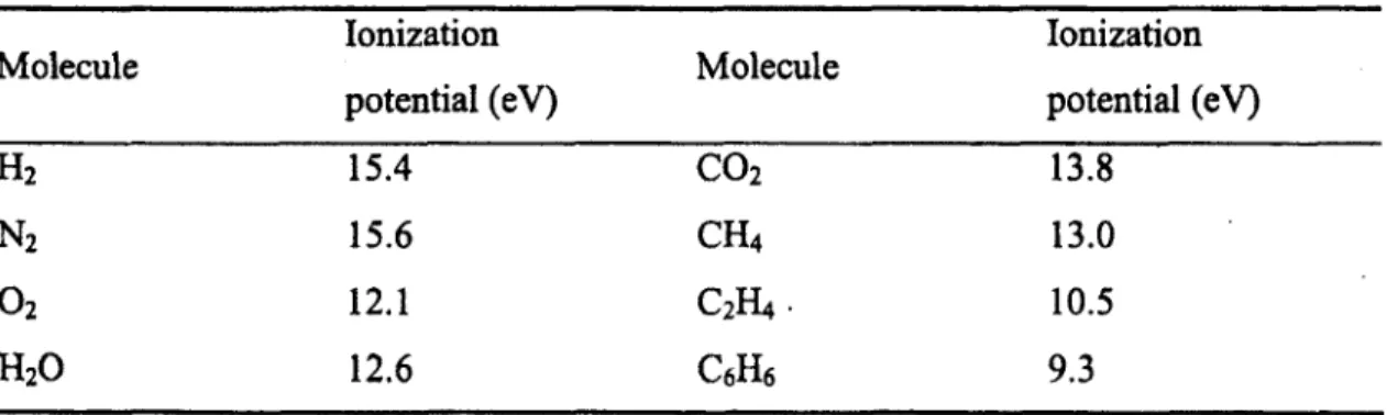
î
Physical stage c10·l6 s)
Radiation enters system
Scattered photon
Interaction with: Biological molecules
To produce: Excitation and Ionization
Water radical and other molecular species
î
Chemical stage (up to 10·3 s)
Damage to important biological molecules Diffusion
,...
Biochemical sta~e ( s)
'V A\
Biological stage (>hours)
'V
Damage to other biological molecules of interest
Chemical restitution and repair
Fixation of damage
Enzymatic repair of molecular damage
Biological repopulation from surviving cells
Figure 1. A diagrammatic representation of the processes from energy transfer to final biological damage. (Adapted from Alp en, 1998. Radiation Biophysics 2nd ed.
CHAPTER I: Introduction
Scattered photon Incident photon (10)
Transmitted photon (/x)
Absorber Scattered photon
Figure 2 Diagram showing transmission of photon through an absorber of thickness t...x (Johns and Cunningham, 1969; Alpen, 1998).
10000 --HO 2 ... 1000 Cl
___.,___ Br
--N E_ ,
u--
c Q) 100·u
!E Q) 0 u c 10 0 :;:: ro :J c Q) :i::: ro !/) !/) ro ::':? 0.1 1 E-3 0.01 0.1 Energy(MeV)Figure 3 Mass attenuation coefficients for H20 , Br and I (Adapted from Hubbel and
CHAPTER I: Introduction
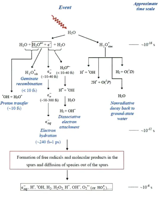
1. 2. Radiolysis of water
The biological effect of ionizing radiations can be illustrated by the schematic shown in Figure 1. Assume that the radiation beam is incident on a living system. The boxes represent stages in a time scale; the arrows indicate particles or molecular species present during the sequence. As an example, suppose radiation photons undergo scattering or absorption processes in the first step, and a high speed electron is ejected. In traveling through the tissue, the fast electron generates a track in which biological or water molecules are ionized and excited. Eventually, all of these processes lead to biological damage (Johns and Cunningham, 1969; Hughes, 1973; Anderson, 1984; Alpen, 1998; Knoll, 2000). Interactions may be separated in direct and indirect effects. The direct mode of action
( often termed the target theory) exerts its effect directly on vital target molecules within the
cell (such as DNA) in form of ionization and excitation. By contrast, the indirect action involves transfer of energy to water molecules with prompt formation of highly reactive species (through the radiolysis of water as shown in Figure 4) (Meesungnoen and Jay-Gerin,
2010) which, in turn, induce damage to the target molecules (Tubiana and Wambersie, 1990). In addition, the biological damage depends not only on the radiation absorbed dose but on the quality of radiation applied as well, a measure of which is given by the "linear energy transfer" or LET1
, which represents, to a first approximation, the nonhomogeneity of
the energy deposition on a sub-microscopic scale. The LET is usually expressed in keV/µm of specific energy traversing the distance (Meesungnoen and Jay-Gerin, 2010).
Figure 4 shows the overall process of radiolysis of water by low LET radiation at ambient temperature and pressure. The process begins with the interaction of water by the ionizing radiation and terminates with the re-establishment of chemical equilibrium. The radiolysis events can be separated into three temporal stages, "physical stage", "physicochemical stage" and "chemical stage" (Meesungnoen and Jay-Gerin, 2010).
1 The LET is defined for charged particle in any medium as the quotient of dEJdl, where dE is the average
energy by a charge particle of specific energy traversing a distance dl (ICRU Report 16 1970). In this way, the (sometimes termed "energy-unrestricted", i.e., when ail permissible energy transfers are included) LET is equivalent to the "stopping power" (which is commonly used in the domain ofradiation physics) of the medium traversed (Anderson, 1984). Usually, LET values are in units ofkeY/µm (the conversion to SI units is: 1keY/µm ~ 1. 602 x 10·19 J/nm) (Meesungnoen and Jay-Gerin, 2010).
CHAPTER I: Introduction
Event Approxima te time scale
~Ho•
' olec - - -10-16 , >\
-1
HO-- 2 H20:;b e,,. (< 10-40 fs) H' + 'OH Hi _,_ 0 (1D ) Geminate (- l01
-40 fs)1
recombination (< 10 fs) If"+ 'OH 'OH + H30"" Proton transfer (-10 fs) eu (---50-300 fs)1
Electron Dissocia rive electron attachment hydration (--40 fs-1 ps)1
H i O Non radiative decay back ro ground-state waterFonuation of free radicals and molecular products in the spurs and diffusion of species out of the spurs
1
--- -10-6 s
Figure 4. Radiolysis of water by the low-LET radiation. (J. Meesungnoen and J.-P. Jay-Gerin 2010)
CHAPTER I: Introduction ( 1) In the physical stage, the passage of ionizing radiation through liquid water leads to the production of free atoms and radicals in an initially nonhomogenous distribution. The duration ofthis stage is of the order of about 10·15 s or less (Draganié and Draganié, 1971). The result of the radiation interaction is the production, along the path of the radiation, of a large number of ionized and electronically excited water molecules (denoted H20-+ and
H20·, respectively) (Meesungnoen and Jay-Gerin, 2010) and secondary electrons. Note that H20• represents here the many excited states (Meesungnoen and Jay-Gerin, 2010). The earliest phenomena in the water radiolysis are:
H20--7 H2Ü:1ec ·
In general, the ejected electrons from ionization process have sufficient energy to ionize or excite one or more other water molecules in the vicinity, and this phenomenon results in, as explained above, to the formation of tracks, or spurs, that are composed of the products of the events. Thus, these secondary electrons of this cascade go on to lead to additional ionization and excitation events resulting in the production of radiation chemical spurs (Mozumder and Magee, 1966; Buxton, 2004; Meesungnoen and Jay-Gerin, 2010).
(2) The physicochemical stage is the period where thermal equilibrium is reached in the system. Its duration is usually taken to be of the order -10-12 s or less. During this period, the ejected electron (secondary electron) moves away from an ionized water molecule. The secondary electron can transfer energy to neighbouring molecules with which it collides and eventually reaches thermal equilibrium with the liquid. Once it bas slowed down to the thermal energy ( e;j), it can be localized or trapped (et;.) in a preformed potential energy well of appropriate depth in the liquid, before it reaches a fully relaxed, hydrated state ( e;q) as the dipoles of the surrounding molecules orient in response to the negative charge of the electron (Meesungnoen and Jay-Gerin, 2010). All of these events (thermalization, trapping, and hydration) can then follow in quick succession (less than
CHAPTER I: Introduction
Moreover, the ejected electron can attach temporarily to a surrounding water molecule to form ff and ·oH according to
Such a process, called "dissociative electron attachment (DEA)", is well known in water vapour and amorphous solid water at low temperature for incident electrons between -5-15 eV (Sanche, 1992; Dugal et al., 1999; Pimblott and Mozumder, 2004). A product of the reaction, hydride anion (Ir), can react with another water molecule through a fast proton transfer as the following reaction: (Meesungnoen and Jay-Gerin, 2010)
Additionally, in the physicocbemical stage, the ejected electron can be recombined to H20-+ radical ions. It is due to the fact that Coulomb attraction between electron and the water positive ion tends to draw them back together to undergo electron-cation "geminate" recombination. As the electron is recaptured, the parent ion is transformed into a (vibrationally) excited neutral molecule. (Meesungnoen and Jay-Gerin, 2010)
Ionized water molecules (H20•+), are unstable. There are electron transfers from neighboring water molecules to the H20•+ molecules. This period can occur in a very short lifetime (-10·14 s) (Mozumder and Magee, 1975). The species produced by the reaction are hydronium ion (H30+) and hydroxyl radical according to
CHAPTER
1:
Introductionwhere H30+ (or equivalently, H;q) represents the hydrated hydrogen ion. Lampe
et al.
(Lampe et al, 1957) have estimated that this reaction takes place in -1.6 x 10⁻¹⁴ s.There are many possible pathways for the decay of the excited molecules (H20*).
The excited molecules may be produced directly as H20:1ec or in directly as
H2o:ib(Meesungnoen and Jay-Gerin, 2010). However .. we have little knowledge about the decomposition of excited water molecules in the liquid phase and the branching ratios associated with each of them (Buxton, 2004). Fortunately, the contribution of the excited compounds to primary yields of radical and molecular species in the radiolysis of water is of relatively minor importance in comparison with that of the ionization processes, such that the lack of information about their decomposition has only limited consequences (Meesungnoen and Jay-Gerin, 2010). Accordingly, they are generally assumed to be essentially the same as those in the water vapour or gas phase (Buxton, 2004) (note that the same decay processes have been reported to occur for the electronically and vibrationally ~xcited water molecules in the gas phase (Meesungnoen and Jay-Gerin, 2010), namely (Cobut et al., 1998; Meesungnoen and Jay-Gerin, 2005):
H20• ~ H2
+
0(1D), H20· ~ 2H•+0(3P),
H20• ~ H20
+
release of thermal energy,where 0(1D) and 0(3 P) represent oxygen atoms in their singlet 1D excited state and triplet
3P ground state respectively (see Figure 4).
(3) In the nonhomogeneous chemical stage, the water reactive species (e.g., e~, ·oH, H•, H2, H30+, Off, •o•) will diffuse and react with each other, resulting in the re-establishment of chemical equilibrium. These products are present at 10-12 sin the spur after the passage of the radiation and then begin to diffuse away from the region where they were originally produced according to macroscopic diffusion principles. They can react with
CHAPTER I: Introduction themselves or with dissolved solutes (if any) present at the time of irradiation~ until all spur or track reactions are complete (Meesungnoèn and Jay-Gerin, 2010). Radical-radical reactions lead to fonn molecular products such as H2, H202 and H20, while the remaining radicals will diffuse out into the bulk of the solution. The lifetime of the spur
in
liquid water for completion of the spur expansion is generally tak.en to be about 10·7 s (Klassen, 1987.). The yield of radicals and molecular products that escape into the bulk solution at this time are known as the "primary" or "escape" yields. The important reactions and reaction rate constants occurring while the spurs expand are shown in Table 2 (Meesungnoen and Jay-Gerin, 2010).In mammalian cells, water mak.es up
-80
% of the cellular mass (Bensasson, et al., 1993) so that,in
general, the indirect action of X or y-rays (low-LET radiation) will predominate as mentioned previously. Various species are released during water radiolysis, the most important being the free radicals eaq-, HO", H" and H02°/02"· (pKa=
4.8), and the molecular products H20 2 and H2 (Spinks and Woods, 1990). The formations of thesereactive species are typically expressed as a radiation-chemical yield (G). The G-value is the number of species produced per 1 OO eV absorbed radiation dose. In general, the product yield is always the yield of "primary reaction", which is called primary yield. The primary yields (at -10·6 s after energy deposition) of these radiolytic species in neutral water
irradiated by 6°Co y-rays or high-energy electrons (-0.3 keV/µm) are (Ferradini and Jay-Gerin, 2000):
G - = 2.65 GH. = 0.60 GH2 = 0.45, t!aq
For acidic solution (0.4 M H2S04), these values are (Ferradini and Jay-Gerin, 2000): G -t!aq +G • H =3.7 GH2 =0.45,
These primary products can then react with solutes to fonn transient species of interest (Wishart, 1998). In a living system, these species will react with cellular macromolecules (such as DNA, proteins, membrane, etc.) and may cause pathological cell _
CHAPTER 1: Introduction
changes and mortality (Nair et al., 2001). This is especially the case for the hydroxyl
radical, which is responsible for about 60-70% of total DNA damage
inmammalian cells
(with low-LET radiation) (Lehnert, 2008).
CHAPTER 1: Introduction
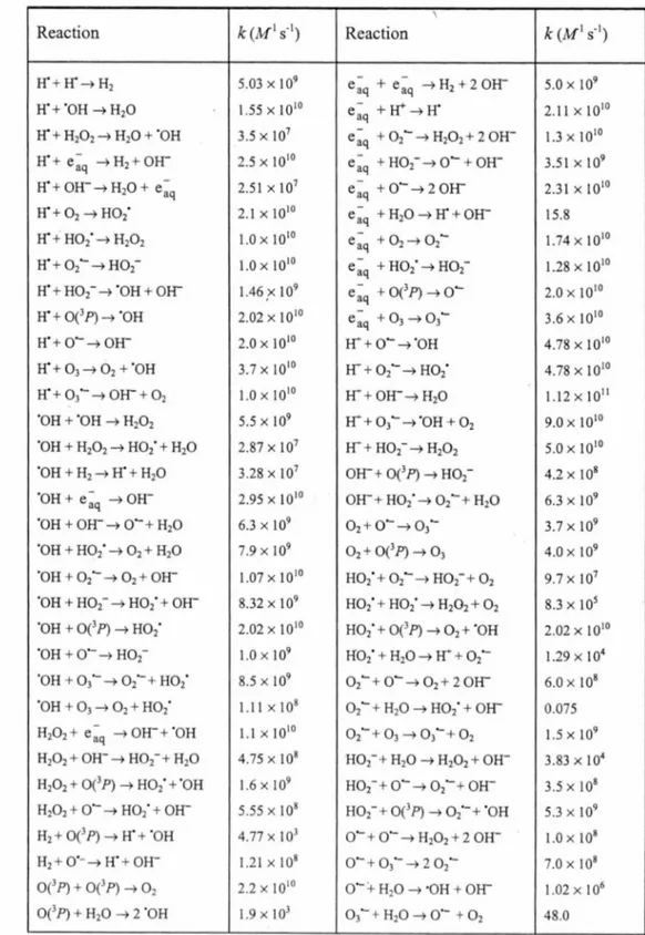
Table 2. Main reaction scheme and rate constants (k) used in the radiolysis of pure liquid
water at ambient temperature (25 °C) (J. Meesungnoen and J.-P. Jay-Gerin 2010).
\
Reaction k(M1 s·1) Reaction k (M1 s·1)
If + If-+ H2 5.03 X 10⁹ e~q + e~q --+ H2 + 2 mr 5.0 X 10⁹ If+ "OH --+ H20 1.55 X J 010 e~q +Ir--+ If 2.J J X 1010 If+ H20 2--+ H20 + "OH 3.5 X 10⁷ e~q + 0 2- --+ H20 2+ 2 OH- J.3 X 1010 If+ e ~q --+ H2 +
mr
2.5 X 10¹⁰ e~q + Ho2---+ o- +mr
3.5Jxl09 If+ OW ....+ H20 + e~q 2.5lx l07 e~q +0- ....+20l1 2.3lxJ 010If+ 02--+ H02" 2.JxlOIO e~q +H20-+lf+OW 15.8
If + H02"--+ H20 2 1.0 X 10¹⁰ e~q + 02--+ 0 2- J.74 X 1010 If + 0 2- ....+H02- 1.0 X 10¹⁰ e~q + H02"--+ H02- J.28 X 1010 If + H02- --7 "OH + mr 1.46 ,x 109 e~q + 0(3P)-+ o- 2.0 X 10¹⁰ If + 0(3 P) --+ "OH 2.02 X 10¹⁰ e~q + 03--+ 03- 3.6 X 10¹⁰ ff+0---7 011 2.0 X 10¹⁰ fr + o-....+ 'OH 4.78 X 10¹⁰ If+ 0 3 --+ 02 + 'OH 3.7 X 10 10 fr + 0 2---+ H02" 4.78 X 10¹⁰ If+ 0 3 - --+
mr
+ 0 2 1.0 X 10¹⁰ fr + OW ....+ H20 J.J2 X 1011 'OH + 'OH --+ H20 2 5.5 X 10⁹ Ir + 0 3 - --+"OH + 0 2 9.0 X 10¹⁰ "OH + H102--+ H02' + H20 2.87 x l07 I-1 + H02 - --+ H102 5.0 X 10¹⁰ 'OH + H2--+ If + H10 3.28 X 10⁷ OW + 0 (3P)-+ H02- 4.2 X 10⁸ 'OH + e ~q --+ OW 2.95 X J 010 OH- + H02'--+ 02-+ H20 6.3 X 10⁹ 'OH +mr
--7 o- + H20 6.3 X 10⁹ 02 + o--+ 03- 3.7 X 10⁹ 'OH + H02"--+ 0 2 + H20 7.9 X 10⁹ 0 2+ 0 (3P)-+ 0 3 4.0 X 10⁹ ·oH + 0 2·---+ 0 2 +mr
J.07 X 1010 H02' + 02- -+ H02- + 0 2 9.7 X 10⁷ "OH+ H02- --7 H02" +mr
8.32 X 10⁹ H02" + H02"--+ H202 + 0 2 8.3 X 10⁵ "OH+ 0 (3P)-+ H02" 2.02 X J 010 H02' + 0(3 P) --+ 0 2 + "OH 2.02 X 10¹⁰ "OH + O"---+ H02- J.Q X 109 Ho2· + H20-+ Ir + 0 2- 1.29 X 10⁴ "OH + 0 3'- --7 02"-+ H02" 8.5 X 10⁹ 02-+ o---+ 02 + 2mr
6.0 X 10⁸ "OH + 03--+ 0 2 + H02" J. [ J X J 08 02 - + H10 --+ H02' +mr
0.075 H20 2+ e~q --7 OW + "OH J.J X 1010 02- + 0 3 --7 0 3"- + 02 J.5 X 109 H20 2+ OW--7 Ho2-+ H20 4.75xJ08 H02-+ H10--+ H10 2+ OH- 3.83 X 10⁴ H20 2 + 0(3 P) --+ Ho2· + "OH J.6 X 109 Ho2-+ o--+ 02-+mr
3.5 X 10⁸ H20 2 + O"---+ H02" + OW 5.55 X 10⁸ H02-+ 0(3P)-+ 0 2·-+ "OH 5.3 X 10⁹H1 + 0(3 P) --+ If + "OH 4.77 X 10³ o- + o---+ H20 2 + 2 OH- J.Q X 108 H1 + o·---+ H" + OH- J.2 J X J 08 o- + 03---+ 2 0 2- 7.0 X 10⁸ 0 (3 P) + 0(3 P) --+ 0 2 2.2 X 10¹⁰ O"-:t H20 --+ ·OH + OI-1 J.02 X [ 06 0 (3 P) + H10 --+ 2 "OH J.9 X [ 03 0 3- + H10--+ o ·-+ 0 2 48.0
CHAPTER 1: Introduction
1.
3. Radiation protector/antioxidant
A radioprotector is a chemical compound that can reduce the toxicity of ionizing radiation (Tubiana et al., l 990). Chemical radioprotectors play a significant role by scavenging water free radicals before they can damage DNA, thus reducing the biological consequences of radiation (Bensasson et al., 1993).
In
addition, some compounds(e.g.,
those containing SH groups) also have radioprotective effects by hydrogen-atom donation to radiation-induced radicals to facilitate direct chemical repair at sites of DNA damage (Bump, 1997; Hall, 2000).Antioxidants are one class of radioprotectors (Weiss, and Landauer, 2003). Thus, their mechanisms of action are similar, although their intended use is different. The former are used in conjunction with a radiation treatment ( administered before radiation exposure ), while the latter are used to protect tissues from the deleterious action of reactive species such as reactive oxygen species (ROS) and reactive nitrogen species (RNS) (Valko et al., 2007; Halliwell, and Gutteridge, 1999) that can be produced during normal cellular metabolism. The major ROS and RNS species (at physiological pH) are HO", 02"-, H202, "NO and ONOO-.
Indirect damage of ionizing radiations originates from reactions of the main radicals of water radiolysis
(e.g.,
H•,"OH,e;q,
etc.) with biological target molecules. It is useful to note that a commonly therapeutic radiation dose (-2 Gy) can produce approximately 2 µM these free radicals (von Sonntag, 1987; Bump, 1997). The hydroxyl radical is a strongly oxidizing species whereas the hydrated electron and the H radical are reducing species (Adams, 1970; von Sonntag, 1987).In
cellular radiobiological damage, the OH radical ande;q
obviously are primary sources because of their high yield (Kiefer, 1990). The cellular radiation effects are potential or mitigated depending on many factors such as for example the presence of oxygen, sulfhydryl compounds (RSH) and other molecule in the cellular milieu (Nair et al., 2001 ).In
presence of oxygen, reaction of oxygen molecules with hydrogen radicals and solvated electrons (e;q)
results in other reactive radical species such as superoxide radical ions (o;-)
and hydroperoxyl radicals(Ho;).
These species play a significant role in oxygen conditions (von Sonntag, 1987; Bump, 1997).CHAPTER I: Introduction
H·+o2 ~Ho;.
However, the radicalsHO; and o;· are in equilibrium. In neutral water the pKa of HO; is
4.8
(Ferradini and Jay-Gerin, 2000). Ho; H H+ +o~·.Most living tissues naturally contain many sulphur-containing compounds such as amino acids, glutathione, methionine and cysteine (Kiefer, 1990; Kamarnisky et al., 2003). Glutathione is the principal nonprotein thiol (Bump, and Brown, 1990) which is the intracellular reducing compound of highest concentration in mammalian cells (Chaudière, 1994). The thiol-containing molecules (RSH) are generally good radical scavengers (Gilbert, and Colton, 1999) especially OH radicals (Kock, 1998). Moreover, the most important feature of their chemistry is probably that they are involved in many cellular redox reactions, illustrated by the general reaction:
2RSH H RSSR + 2e· + 2H+.
This process most lik.ely proceeds via thiyl free radicals, Rs•, as intermediates which are known to readily form disulfides (RSSR) if they lack suitable partners for other reactions (Asmus, 1993). TheRSH can react with other radicals through hydrogen atom abstraction, as an example of the following reaction (Murray, 1998; Wardman, 2007):
DNA •
+
RSH ~ DNA+
Rs·.In radiation biology, the process is commonly known as "repair reaction" (Asmus, 1993). However, the chemical action of these radiation protectors is not well understood. In this project, we study the protecting effect of a disulfide compound, cystamine (RSSR) using Fricke and polyacrylamide gel dosimetry. The Fricke system has the advantage that
it
can be simulated by Monte Carlo techniques such that the mechanism of action of cystamine can be investigated.
CHAPTER I: Introduction
1. 4. Radiosensitizers
Nowadays, radiation therapy is well established and is an important technique for localized cancer treatment (Blattmann et al., 2005). The basic idea of the radiation treatment is to maximize the absorbed dose to the tumeur area white minimizing exposure to normal, healthy cells (Tilikidis et al., 1994). Unfortunately, in this case, the radiation dose is often limited by normal tissue tolerance (Viala et al., 1999; Young et al., 1996). Using a radiation-sensitizing agent is one strategy in order to increase the absorbed dose in the tumor.
Thus, radiosensitizers are intended to enhance tumor cell killing while having much less effect on normal tissues. The enhancement of the radiation damage by radiosensitizers (also called "dose enhancers") originates from the direct action of radiation on the sensitizing compounds. This mechanism of radiosensitization can be generated into three categories: production of free-radical species (W ardman, 1987), nuclear reaction sensitizer (Barth et al., 2005), and heavy element sensitizers (Ertl et al., 1970). The radiation sensitizer that can be used to produce free-radicals is called "electron-affinic radiosensitizer" because the efficiency of sensitization, which is defined by the concentration required to produce a given degree of sensitization, is related to the electron-affinities or the reduction potential of compounds (Adams, 1979; Wardman, 1987), for example, nitroimidazoles (Adams, 1979) and gadolinium (IIn texaphyrin (Viala et al., 1999; Young et al., 1996). The radiation-sensitizing properties of these compounds are similar to that of molecular oxygen (also called "oxygen mimetic radiosensitizer"). Oxygen is itself a free radical, the prototypical radiosensitizer, but it has two unpaired electrons resultuig in rapidly reacting with other species and producing new reactive radicals (W ardman, 1987). The electron-affinic molecules can react with water-radiolytic species especially solvated electrons ( e~q ). This
process forms aggressive radicals as shown in the following reaction.
where, S is radiosensitizer. However, the main drawback of these compounds is their toxicity and the observation that they do not adequately sensitize the entire tumour cell population (Young et al., 1996).