Science Arts & Métiers (SAM)
is an open access repository that collects the work of Arts et Métiers Institute of Technology researchers and makes it freely available over the web where possible.
This is an author-deposited version published in: https://sam.ensam.eu Handle ID: .http://hdl.handle.net/10985/15615
To cite this version :
Peter H. PEDERSEN, Claudio VERGARI, Abdulmajeed ALZAKRI, Raphaël VIALLE, Wafa SKALLI - A reduced micro-dose protocol for 3D reconstruction of the spine in children with scoliosis: results of a phantombased and clinically validated study using stereoradiography -European Radiology - Vol. 29, n°4, p.1874-1881 - 2019
Any correspondence concerning this service should be sent to the repository Administrator : archiveouverte@ensam.eu
DOI : https://doi.org/10.1007/s00330-018-5749-8
1 Orthopedic Department, University Hospital of Aalborg, Hobrovej 18-22, 9000 Aalborg, Denmark
2 Institut de Biomécanique Humaine Georges Charpak, Arts et Métiers Paris Tech, Paris, France, Paris, France 3 Department of Pediatrics, Sorbonne Université, APHP Paris, 26 avenue du Dr Arnold Netter, 75012 Paris, France 4 Orthopedic Department, College of Medicine, King Saud University, Riyadh, Saudi Arabia
in children with scoliosis: results of a phantom based and clinically
validated study using stereo-radiography
Peter H. Pedersen 1,2, 3, Claudio Vergari 2, Abdulmajeed Alzakri 2,4, Raphaël Vialle 3, Wafa Skalli 1
Abstract
PurposeThe aim of this study was to validate the reproducibility of 3D reconstructions of the spine using a new reduced micro-dose protocol.
Methods
First, semi-quantitative image analysis was performed using an anthropomorphic child phantom undergoing low-dose biplanar radiography. This analysis was used to establish a “lowest dose” allowing for acceptable visibility of spinal landmarks. Subsequently a group of 18 scoliotic children, 12 years of age or younger, underwent full-spine biplanar radiography with both micro-dose and the newly-defined reduced micro-dose. An intra- and inter-observer reliability study of 3D reconstructions of the spine was performed according to the International Organization for Standardization (ISO)-5725 standard, with three operators.
Results
The reduced micro-dose setting corresponded to a theoretical reduction of radiation dose exposure of approximately 58%. In vivo results showed acceptable intra- and inter-observer reliability (for instance 3.8° uncertainty on Cobb angle), comparable to previous studies on 3D spine reconstruction reliability and reproducibility based on stereo-radiography.
Conclusion
A new reduced micro-dose protocol offered reliable 3D reconstructions of the spine in patients with mild scoliosis. However, the quality of 3D reconstructions from both reduced micro-dose and micro-dose was inferior to standard-dose protocol on most parameters. Standard–dose protocol remains the option of choice for most accurate assessment and 3D reconstruction of the spine. Still, this new protocol offers a preliminary screening option and a follow-up tool for children with mild scoliosis yielding extremely low radiation and could replace micro-dose protocol for these patients.
DOI : https://doi.org/10.1007/s00330-018-5749-8
Introduction
The evaluation of 3D spine-deformity in scoliosis is challenging and optimally requires comprehension and use of 3D clinical parameters[1, 2]. The correct interpretation of spinal deformities is mandatory to define the optimal treatment strategy for the patients. Different methods for 3D evaluations have been used and evaluated[3], and reconstruction based on stereo-radiography is a commonly used method. Several studies have investigated the possibility of predicting progression of scoliosis based on 3D parameters[1, 2, 4–6], since predicting scoliosis progression at an early stage would be of paramount importance. Apical vertebra rotation (AVR), torsional index of the spine (TI) and intra vertebral rotation (IAR) have been proven significant parameters in determining progression in mild scoliosis (Cobb angle < 25° [2, 4]). In the recent 20 years a lot of effort has gone into defining the “gold standard” for 3D parameters, and to apply these for effective and easy to use tools in daily clinical life[3].
The repeated use of x-ray imaging needed for scoliotic patient follow-up has been of concern in recent years. Ionizing radiation has been associated with a potential risk of developing radiation-induced cancer in scoliotic patients[7–10]. Children have a long life expectancy and are thought to be especially sensitive to long-term stochastic effects from ionizing radiation. Thus, it is of great importance taking steps towards using methods reducing the radiation exposure to our patients. The best approach of course would be to define robust methods of early detection of progressive scoliosis and more efficient methods of treatment in order to limit the number of radiographic exams needed for follow-up. However, although promising results have been reported in the literature, such methods are still not validated or wide spread[4, 6, 11]. The second-best approach is to reduce the ionizing radiation delivered by the radiological exam.
EOS® low dose stereo-radiography(EOS Imaging) is an imaging system that allows for high quality imaging at a radiation dose lower than most conventional systems[8, 12, 13], adhering to the ALARA dose-optimization principle of keeping dose As Low As Reasonably Achievable[14]. 3D reconstruction from EOS imaging stereo-radiography has been described in several previous studies[4, 15–18]. Good reliability on 3D parameters have been reported for both standard-dose and micro-dose protocols[15, 16, 18]. Ilharreborde et al[16, 18] looked at both standard-dose and micro-dose protocols with regards to intra- and inter-observer reproducibility. Results were satisfactory for both modalities and a significant reduction of dose compared with the original standard-dose protocol was described. We hypothesized that the radiation dose delivered to the patient could be reduced even further without compromising reliability of 3D reconstructions. The aim of the present study was to investigate the possibility of reducing the dose of the established micro-dose protocol retaining the possibility of trustworthy 3D reconstructions from the EOS imaging stereo-radiography.
Materials & Methods
Defining the reduced micro-dose protocol
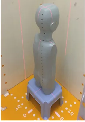
The minimal dose judged to yield sufficient image quality for recognition of anatomical landmarks was defined by imaging a clinically validated ATOM dosimetry child-phantom (ATOM-CIRS, Computerized Imaging Reference System, Inc.)[19]. Figure 1 shows the phantom in posterior-anterior-lateral positioning (PAL) within an EOS scanner. Radiographic expositions were made with sequentially lower dose settings. Radiation dose exposure from the EOS micro-dose protocol was reduced by decreasing the current, milliamps (mA), and the scan speed. Both parameters are directly proportional to radiation dose: a 25% decrease of mA reduces exposure by 25%. A change of scan speed from speed 4 to speed 3, likewise results in a reduction of radiation dose by 25%.
The order of the acquired images was then randomized and an experienced surgeon, blind to the acquisition settings, rated image quality (i.e., contrast, noise and visibility of anatomical structures) in the lumbar and thoracic region on a scale from 1 to 5 (1=optimal, 5=unacceptable). All images were scored twice. A cumulative score for each acquisition setting was determined to find a cut-off value for the images which would yield sufficient recognition of anatomical landmarks to allow for 3D reconstruction. Subsequently this acquisition setting was used for three in vivo pilot radiographies and following 3D reconstructions to test for
Table 1. EOS Scan Protocols Protocols Reduced Micro-Dose Micro-Dose Standard-Dose Morphotype Small Small Small
Scan Speed 2 3 4 Anterior X-Ray Tube kV 60 60 83 mA 50 80 200 DAP1 (mG.cm2) 19 30 222 Lateral X-Ray Tube kV 80 80 102 mA 50 80 200 DAP(mG.cm2) 42 67 371 Total DAP values(mG.cm2)2 72 97 593
Radiographic exposures undertaken with posterior-anterior-lateral stereographic biplanar imaging
a Kilovolts b Milliamps
c DAP = dose area product for a child phantom representing a 5-year-old
at phantom height of 72 cm d Anterior + lateral DAP values
Fig. 1 The anthropomorphic phantom,
representing a 5-year-old child,
placed in posterior-anterior-lateral positioning within the EOS scanner
DOI : https://doi.org/10.1007/s00330-018-5749-8
applicability. Theoretic dose reductions were calculated from proportional differences of dose area product (DAP) values between the standard-dose, micro-dose and reduced micro-dose protocols.
Inclusions
The local ethics review board approved of the study design and methods. A consecutive group of 18 children, 12 years of age or younger, planned for routine clinical and radiological investigation of scoliosis were offered micro-dose and reduced micro-dose images instead of one standard-dose image. An informed consent was obtained for each patient prior to imaging. Images with both protocols were obtained at the same radiological session, one after the other, no more than two minutes apart. This method allowed for direct comparison of 3D parameter reproducibility between the two
modalities. Exclusion criteria were severe obesity, previous spine surgery with implants and mal-positioning of the patients.
3D reconstructions
A validated method of 3D reconstruction of the spine from EOS 2D biplane images was used [15]. Patient data and acquisition settings were blinded and reconstructions took place in random order. Three operators, all trained within 3D reconstructions, did two reconstructions for each obtained image. One operator determined, for each patient, the levels of junctional and apical vertebrae for each scoliotic curve. Table 2 lists the 3D parameters investigated. Figure 3 illustrates 3D reconstruction images using the reduced micro-dose protocol.
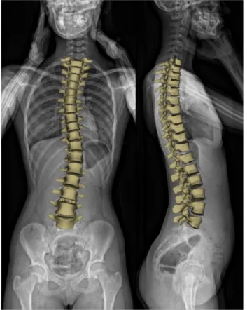
Fig. 2 Examples of 3D reconstruction from reduced micro-dose protocol, coronal and lateral views
Table 2. The different 3D parameters investigated. 3D Parameters: Cobb Angle T1-T12 Kyphosis T4-T12 Kyphosis L1-S1 Lordosis AVR1 TI2 Pelvic Incidence Sacral Slope Pelvic Tilt
1Apical Vertebra Rotation
Statistics
Intra- and inter-operator reproducibility were determined according to the ISO 5725-2:1994 standard, in terms of standard deviation. Bland-Altman plots were used to observe measurement agreement. Results were compared with previously published data on 3D reconstruction based on stereo-radiography and micro-dose[15, 18]. Correlations were analyzed with Spearman’s rank coefficient; significance was set at 0.05.
Results
A cut-off value at 50 mA and 60 kV for frontal imaging and 50 mA and 80kV for lateral imaging, with a scan speed of 2 was found to offer sufficient quality for anatomical landmark recognition. This reduced micro-dose protocol corresponds to a theoretical reduction of radiation exposure of approximately 58% and 93% compared with micro-dose and standard-dose protocols, respectively. Table 1 shows the three scan settings and DAP values for the child phantom.
Preliminary in vivo images with the new reduced micro-dose setting allowed sufficient quality for 3D reconstruction. Figure 2 illustrates an example of dose and reduced micro-dose full-spine imaging.
A group of 18 consecutive children going for routine clinical investigation for scoliosis were then assessed with both micro-dose and reduced micro-dose imaging. Three children were excluded; two were carrying braces during imaging, one had an abnormal number of vertebrae (14 thoracic vertebrae). The remaining 15 children were included in the study. The Mean age was 10.7 years (range 4-12), Gender distribution amongst the included patients: four males and 11 females. Mean reconstruction time was 10 minutes (range 6-21 minutes) for the micro-dose and 9 minutes (range 5-16 minutes) for the reduced micro-dose. Reconstruction time was not correlated with Cobb angle (i.e., with scoliosis severity, p > 0.05).
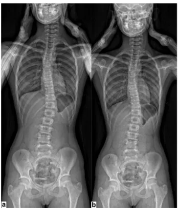
Fig. 3 a Coronal full-spine image in EOS scanner using micro-dose protocol. b Coronal full-spine image in EOS scanner using reduced micro-dose protocol
DOI : https://doi.org/10.1007/s00330-018-5749-8
Reproducibility
A total of 180 3D reconstructions were made (15 patients * 2 modalities * 3 operators * 2 occurrences). 3D reconstructions were possible for all patients, and key anatomical landmarks needed for 3D reconstructions were visible for patients in both protocols. However, for both protocols, mostly the reduced micro-dose group, spinous processes were in some cases difficult to visualize because of increased vertebral rotation. Other anatomical landmarks such as vertebral endplates and pedicles were not affected to the same degree. Tables 3 and 4 show results on 3D repeatability and reproducibility along with results from previously published papers. Both micro-dose and reduced micro-dose showed good reproducibility, however, 3D reconstruction from standard-dose as demonstrated by Humbert et al 2009[15] remained superior. Reproducibility between micro-dose and reduced micro-dose within this study was better for the micro-dose protocol. The highest degree of variability was on AVR and kyphosis parameters. Table 4 shows that reduced micro-dose was better on all parameters except pelvic tilt (PT) and T4-T12 kyphosis, compared with Ilharreborde et al (2016)[18] “fast-spine” micro-dose reconstructions.
Discussion
The purpose of this study was to investigate and validate reproducibility of 3D reconstruction of the spine from stereo-radiography with a reduced micro-dose protocol in scoliotic pediatric patients. For most 3D parameters in mild, reproducibility was comparable to previous studies[15, 16, 18]. As expected, the reduced micro-dose protocol was less reliable than standard-dose and micro-dose for some parameters. 3D transverse rotational parameter uncertainty, AVR and torsion, was higher in this study on both reduced dose and micro-dose protocols than the reported values from Humbert et al (2009)[15] using standard micro-dose, as well as uncertainties on Cobb angle and T4-T12 kyphosis (5.4° and 6.0° in reduced micro-dose, respectively, versus 3.1° and 3.8°in the previous work). However, the reproducibility obtained using reduced micro-dose “full” 3D reconstruction was superior in all clinical parameters except for PT to the results obtained using “fast spine 5min process” (Ilharreborde 2016) using micro-dose in patients with scoliosis severity comparable to this study. Thus, the reduced micro-dose protocol offered acceptable 3D reconstruction reliability of the spine in patients with mild scoliosis. Depending on the objective of the exam, such reliability would be fine for initial screening and follow-up of scoliosis.
Limitations
A reduction of radiation dose exposure to the patients of more than 50% could be beneficial to the patients reducing potential harmful side-effects to ionizing radiation considering the ALARA principle. Still, the risk benefit balance needs to always be evaluated according to the needs of a given radiological assessment. Existing EOS standard-dose protocol already offers high quality images suitable for 3D reconstruction of the spine at a low radiation dose, as shown by Humbert et al[15]. For instance, the reduced micro-dose protocol would not
be accurate enough to calculate the severity index of scoliosis progression[4] simulate or plan surgery. Moreover, the reliability might not be accurate enough for research, where the development of algorithms and decision trees needs higher accuracy.
Images obtained with reduced micro-dose were as expected of lower quality than standard-dose and micro-dose, i.e. more noisy and with less contrast. In standard-dose and micro-dose, the spinous processes are often difficult to visualize, which was generally worse for reduced micro-dose. Spinous process location is, along with pedicles, an important landmarks used to evaluate the axial orientation of the vertebra. However, pedicles were sufficiently recognizable in most patients, except one patient with severe kyphosis. This was independent of the imaging dose as it is inherent to the patient’s spinal geometry; this type of patient would also have been challenging with other 2D modalities, and does in fact put a restriction on usability of 3D reconstruction from stereo-radiography. In some cases regular CT should be advocated for.
For both modalities, T1, which is one of the landmarks needed to initialize the 3D reconstruction was not always visible in lateral projection due to overlapping upper extremities/ shoulders, although correct validated patient positioning was adopted in this work and patient mal-positioning was cause for exclusion. Nevertheless, sagittal inclination of T1 can usually be inferred by the orientations of the adjacent vertebrae. The same applies in the mid-thoracic region where there is low visibility in the lateral view because of the large body span traversed by the x-rays.
As shown above, operator-time is still a limiting factor since the average 3D reconstruction time was 10 minutes, which is often not compatible with everyday clinical routine. A new and faster method is needed to benefit optimally from this 3D analysis method, potentially automated, to reduce user-dependence[15, 16, 18]. We do not recommend this new protocol for children with implants or wearing braces as these cases were not yet investigated.
Table 3, Intra-operator repeatability of clinical parameters, in terms of standard deviation of uncertainty, obtained in the current study and compared with existing literature. All parameters are expressed in degrees
Studies, mean Cobb angle Protocol Main Cobb angle T1-T12 Kyphosi s T4-T12 Kyphosis L1-S Lordosis
AVR Torsion Pelvic
Incidence Sacral slope Pelvic tilt Current study Cobb 16.1 (range 0.2-39) Reduced micro-dose 4.3 5.3 4.7 4.6 4.7 4.7 4.3 3.2 2.6 Micro-dose 2.4 5.3 4.0 3.5 5.3 3.5 3.1 2.6 2.7 Ilharreborde et al 2016 Cobb 24.8 (range 4.6-64.7) Micro-dose 3.6 4.8 4.5 5.8 - - 5.2 5.2 1.3 Ilharreborde Et al 2011 Cobb 62±11* Standard-dose 4.8 5.9 4.4 5.1 5.3 - 4.6 4.3 1.0 *SD
DOI : https://doi.org/10.1007/s00330-018-5749-8
Conclusion
We propose a new reduced-micro-dose protocol for 3D reconstructions based on stereo-radiography which offers reliable 3D reconstructions for preliminary screening and follow-up in children with mild scoliosis. However, standard-dose protocol remains the option of choice for most accurate assessment and 3D reconstruction. The reduced micro-dose protocol is applicable to existing EOS systems and can be taken into use for children being assessed for mild scoliosis right away and could replace micro-dose for these patients.
References
1. Labelle H, Aubin C-E, Jackson R, et al (2011) Seeing the spine in 3D: how will it change what we do? J Pediatr Orthop 31:S37--45 . doi: 10.1097/BPO.0b013e3181fd8801
2. Courvoisier A, Drevelle X, Dubousset J, Skalli W (2013) Transverse plane 3D analysis of mild scoliosis. Eur Spine J 22:2427–2432 . doi: 10.1007/s00586-013-2862-x
3. Donzelli S, Poma S, Balzarini L, et al (2015) State of the art of current 3-D scoliosis classifications: a systematic review from a clinical perspective. J Neuroeng Rehabil 12:91 . doi: 10.1186/s12984-015-0083-8
4. Skalli W, Vergari C, Ebermeyer E, et al (2017) Early Detection of Progressive Adolescent Idiopathic Scoliosis. Spine (Phila Pa 1976) 42:823–830 . doi: 10.1097/BRS.0000000000001961
5. Imager X, Charron G, Beaudoin G, et al (2010) Diagnostic Imaging of Spinal Deformities Reducing Patients Radiation Dose With a New Slot-Scanning. 35:989–994
6. Nault M-L, Mac-Thiong J-M, Roy-Beaudry M, et al (2014) Three-Dimensional Spinal Morphology Can Differentiate Between Progressive and Nonprogressive Patients With Adolescent Idiopathic Scoliosis at the Initial Presentation. Spine (Phila Pa 1976) 39:E601– E606 . doi: 10.1097/BRS.0000000000000284
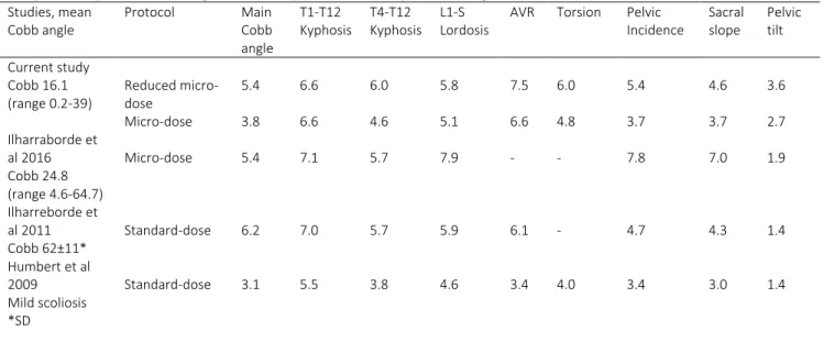
Table 4. Inter-operator reproducibility of clinical parameters, in terms of standard deviation of uncertainty, obtained in the current study and compared with existing literature. All parameters are expressed in degrees
Studies, mean Cobb angle Protocol Main Cobb angle T1-T12 Kyphosis T4-T12 Kyphosis L1-S Lordosis
AVR Torsion Pelvic
Incidence Sacral slope Pelvic tilt Current study Cobb 16.1 (range 0.2-39) Reduced micro-dose 5.4 6.6 6.0 5.8 7.5 6.0 5.4 4.6 3.6 Micro-dose 3.8 6.6 4.6 5.1 6.6 4.8 3.7 3.7 2.7 Ilharraborde et al 2016 Cobb 24.8 (range 4.6-64.7) Micro-dose 5.4 7.1 5.7 7.9 - - 7.8 7.0 1.9 Ilharreborde et al 2011 Cobb 62±11* Standard-dose 6.2 7.0 5.7 5.9 6.1 - 4.7 4.3 1.4 Humbert et al 2009 Mild scoliosis Standard-dose 3.1 5.5 3.8 4.6 3.4 4.0 3.4 3.0 1.4 *SD
7. Levy AR, Goldberg MS, Mayo NE, et al (1996) Reducing the Lifetime Risk of Cancer From Spinal Radiographs Among People With Adolescent Idiopathic Scoliosis. Spine (Phila Pa 1976) 21:1540–1547 . doi: 10.1097/00007632-199607010-00011
8. Law M, Ma WK, Chan E, et al (2017) Evaluation of cumulative effective dose and cancer risk from repetitive full spine imaging using EOS system: Impact to adolescent patients of different populations. Eur J Radiol 96:1–5 . doi: 10.1016/j.ejrad.2017.09.006
9. Doody MM, Ronckers CM, Land CE, et al (2010) Cancer mortality among women frequently exposed to radiographic examinations for spinal disorders. Radiat Res 174:83–90 . doi: 10.1667/RR2022.1
10. A. S, S.B. C, K.E. J, L.Y. C (2015) Incidence of cancer and infertility, in patients treated for adolescent idiopathic scoliosis 25 years prior. Eur Spine J 24:S740 . doi: http://dx.doi.org/10.1016/j.spinee.2015.07.076
11. Kadoury S, Mandel W, Roy-Beaudry M, et al (2017) 3-D Morphology Prediction of Progressive Spinal Deformities from Probabilistic Modeling of Discriminant Manifolds. IEEE Trans Med Imaging 36:1194–1204 . doi: 10.1109/TMI.2017.2657225
12. Damet J, Fournier P, Monnin P, et al (2014) Occupational and patient exposure as well as image quality for full spine examinations with the EOS imaging system Occupational and patient exposure as well as image quality for full spine examinations with the EOS imaging system. Med Phys 063901: . doi: 10.1118/1.4873333
13. Deschênes S, Charron G, Beaudoin G, et al (2010) Diagnostic imaging of spinal deformities: reducing patients radiation dose with a new slot-scanning X-ray imager. Spine (Phila Pa 1976) 35:989–994 . doi: 10.1097/BRS.0b013e3181bdcaa4
14. Strauss KJ, Kaste SC (2006) ALARA in Pediatric Interventional and Fluoroscopic Imaging: Striving to Keep Radiation Doses as Low as Possible During Fluoroscopy of Pediatric Patients-A White Paper Executive Summary. J Am Coll Radiol 3:686–688 . doi: 10.1016/j.jacr.2006.04.008
15. Humbert L, De Guise JA, Aubert B, et al (2009) 3D reconstruction of the spine from biplanar X-rays using parametric models based on transversal and longitudinal inferences. Med Eng Phys 31:681–687 . doi: 10.1016/j.medengphy.2009.01.003
16. Ilharreborde B, Steffen JS, Nectoux E, et al (2011) Angle Measurement Reproducibility Using EOSThree-Dimensional Reconstructions in Adolescent Idiopathic Scoliosis Treated by Posterior Instrumentation. Spine (Phila Pa 1976) 36:E1306–E1313 . doi: 10.1097/BRS.0b013e3182293548
17. Courvoisier A, Ilharreborde B, Constantinou B, et al (2013) Evaluation of a three-dimensional reconstruction method of the rib cage of mild scoliotic patients. Spine Deform 1:321–327 . doi: 10.1016/j.jspd.2013.07.007
18. Ilharreborde B, Ferrero E, Alison M, Mazda K (2016) EOS microdose protocol for the radiological follow-up of adolescent idiopathic scoliosis. Eur Spine J 25:526–531 . doi: 10.1007/s00586-015-3960-8
19. Dose WB, Dose O, Radiation T (2013) ATOM Dosimetry Phantoms. http://www.cirsinc.com/file/Products/701_706/701 706 DS 080715(1).pdf