Université Toulouse 3 Paul Sabatier (UT3,Paul Sabatier)
Laboratoire de Recherche en Sciences Végétales (LRSV,UMR 5546)
Dr. Elodie GAULIN, Maître de Conférences, UT3 Paul Sabatier, HDR
Autres membre du JURY
Ecole Doctorale SEVAB: Interaction Plantes-Microorganismes
Dr. Marie DUFRESNE, Maître de Conférences , Université Paris-Sud Orsay Dr. Franck PANABIERES, DR2 INRA Sophia-Antipolis
Dr. Sylvain JEANDROZ Professeur, AgroSup Dijon
Dr. Christophe ROUX, Professeur Université Paul Sabatier, Toulouse
Diana RAMIREZ-GARCES
Analyses of CRN effectors (Crinkler and Necrosis) of the
oomycete Aphanomyces euteiches
1
Acknowledgments
This work has been fruitful thanks to the support of many people that participated directly or distantly, motivating this four-year journey with their good energy, laughs and dancing-and-chanting moods.
Je remercie tout d’abord ma directrice de thèse, Elodie Gaulin et le chef de l’équipe Bernard Dumas. Merci de m’avoir accueilli dans l’équipe et de m’avoir accordé l’espace, le temps et la confiance pour réaliser mon travail et accomplir cet objectif personnel.
Je remercie Sylvain Jeandroz, Franck Panabières et Marie Dufresne d’avoir accepté d’évaluer mon travail.
Laurent Camborde, le courage avec lequel tu as affronté 50000 femelles d’amphibiens est sans précédents. Et tout ça pour sauver les grenouilles colombiennes des Pyrénées. J’ai beaucoup aimé travailler avec toi. Tu as été une source de bonne humeur et énergie dans les moments de ralenti.
Une partie importante de ce travail a été possible grâce à Yves Martinez, Alain Jauneau, Aurélie Leru et Cécile Pouzet. Je vous remercie de votre disponibilité, grâce à laquelle j’ai pu apprendre et développer les techniques de microscopie. Je garde à l’esprit votre professionnalisme, votre polyvalence scientifique et surtout votre enthousiasme.
Aude Cerutti, merci de m’avoir accompagné durant une partie de ce travail. Une jolie expérience que je n’oublierai pas.
Je pense avec joie et une certaine nostalgie aux anciens membres de l’équipe : Thomas Rey, Mathieu Larroque, Olivier André, Amaury Nars et Yacine avec lesquels j’ai pu partager des moments très agréables. Elo, Sophie, merci pour votre compagnie et votre précieuse aide.
A mis hermanas del alma, Vanessa y Valentina: gracias por llegar a mi vida y acompañarme en este ciclo. Hubo momentos en los que solo deseábamos parar este mundo y bajarnos, pero ahora entendimos que el movimiento es indispensable y que la vida lleva su propio ritmo.
A mi familia, a los que están y también a los que se han ido. Esto es para ustedes.
2
Summary
The oomycete Aphanomyces euteiches is an important pathogen infecting roots of legumes (pea, alfalfa...) and the model legume Medicago truncatula. Oomycetes and other microbial eukaryotic pathogens secrete and deliver effector molecules into host intracellular compartments (intracellular/cytoplasmic effectors) to manipulate plant functions and promote infection. CRN (Crinkling and Necrosis) proteins are a wide class of intracellular, nuclear-localized effectors commonly found in oomycetes and recently described in true fungi whose host targets, virulence roles, secretion and host-delivery mechanisms are poorly understood. We addressed the functional characterization of CRN proteins AeCRN5 and AeCRN13 of A. euteiches and AeCRN13’s homolog of the chytrid fungal pathogen of amphibians Batrachochytrium dendrobatidis, BdCRN13. Gene and protein expression studies showed that AeCRN5 and AeCRN13 are expressed during infection of M.
truncatula’s roots. Preliminary immunolocalization studies on AeCRN13 in infected roots
indicated that the protein is secreted and translocated into root cells, depicting for the first time CRN secretion and translocation into the host during infection. The heterologous ectopic expression of AeCRNs and BdCRN13 in plant and amphibian cells indicated that these proteins target host nuclei and lead to t h e perturbation of host physiology. By developing an in vivo FRET-FLIM-based assay, we revealed that these CRNs target host nucleic acids: AeCRN5 targets plant RNA while AeCRN13 and BdCRN13 target DNA. Both CRN13 exhibit a HNH-like motif commonly found in endonucleases and we further demonstrated that both CRN13 display a nuclease activity in vivo inducing double-stranded DNA cleavage. This work reveals a new mode of action of intracellular eukaryotic effectors and brings new aspects for the comprehension of CRN’s activities not only in oomycetes but, for the first time, also in true fungi.
Keywords: CRN, oomycetes, nucleus, effector, secretion, FRET-FLIM, Aphanomyces
3
Résumé
L’oomycète Aphanomyces euteiches est un pathogène racinaire de légumineuses cultivées (pois, luzerne …) et de la plante modèle Medicago truncatula. Les oomycètes, comme d’autres microorganismes pathogènes eucaryotes, secrètent et transloquent des molécules à l’intérieur des cellules de l’hôte (effecteurs intracellulaires/cytoplasmiques) dans le but de manipuler les fonctions de la plante et de faciliter l’infection. Les protéines CRN (Crinkling and Necrosis) constituent une famille d’effecteurs nucléaires largement répandue chez les oomycètes et récemment décrites chez des espèces fongiques. Leurs cibles et rôle dans la virulence ainsi que leurs mécanismes de sécrétion et de translocation sont encore mal compris. Nous avons entrepris la caractérisation fonctionnelle des protéines AeCRN5 et AeCRN13 d’A.euteiches ainsi que de l’homologue d’AeCRN13 du champignon pathogène d’amphibien Batrachochytrium dendrobatidis, BdCRN13. Les analyses d’expression génique et protéique ont permis de montrer que AeCRN5 et AeCRN13 sont exprimés durant l’infection des racines de M. truncatula. Des résultats préliminaires d’immuno-localisation d’AeCRN13 ont révélé, pour la première fois, la sécrétion et translocation d’un CRN durant l’infection. Leur expression hétérologue, à la fois dans les cellules de plantes et d’amphibiens, a montré que ces protéines se localisent dans les noyaux où leurs activités conduisent à la perturbation de la physiologie de l’hôte. En développant un système in
vivo basé sur la technique de FRET-FLIM, nous avons démontré que ces CRN ciblent les
acides nucléiques: AeCRN5 cible l’ARN des plantes tandis qu’AeCRN13 et BdCRN13 lient l’ADN. Ces deux effecteurs CRN13 exhibent un motif de type HNH, lequel est typiquement retrouvé dans des endonucleases. Nous avons démontré que les CRN13 présentent une activité nuclease in vivo conduisant à la génération de coupures double brin de l’ADN. Ce travail a permis de mettre en évidence un nouvel mécanisme d’action des effecteurs de microorganismes eucaryotes et apporte des nouveaux aspects pour la compréhension de l’activité des protéines CRN d’oomycète mais aussi, pour la première fois, de champignon.
Mots clefs : CRN, oomycète, noyau, effecteur, sécrétion, FRET-FLIM, Aphanomyces
4
List of abbreviations
Ae Aphanomyces euteiches AM Arbuscular Mycorrihiza AT Adenosine Tyrosine Avr Avirulence Bd Batrachochytrium dendrobatidis BFA Brefeldin ABIC Biotrophic Interfacial Complex
CBEL Cellulose Binding ELicitor
CD Cell-Death
CN Cyst Nematode
CRN Crinkling and Necrosis
CSEPs Candidate Secreted Effector Proteins
CWDEs Cell Wall Degrading Enzymes
DBD DNA Binding Domain
EIHM ExtraInvasive Hyphal Membrane ER Endoplasmic Reticulum
ESTs Expressed Sequence Tags ETI Effector-Triggered Immunity
FLIM Fluorescence Life Image Measure
FRET Fluorescence Resonance Energy Transfer
GFP Green Fluorescent Protein
GWAS Genome Wide Association Study HATs Histone AcetylTransferases HDACs Histone DeAcetylases
HIGS Host-Induced Gene Silencing HMTs Histone MethylTranferases
5 JA Jasmoninc Acid
MAMP Microbe-Associated Molecular Pattern MTFs MAD-box Transcription Factors
NE Nuclear Envelop
NES Nuclear Export Signal
NLS Nuclear Localization Signal
NPCs Nuclear Pore Complex
NTR Nuclear Transport Receptor
PAMP Pathogen-Associated Molecular Pattern
PBS Phosphate-Buffered Saline
PCW Plant Cell Wall
PI(3)P PhosphoInositol-3-Phosphate
PR Pathogenesis Related
PTI PAMP-Triggered Immunity
PVX Potato Virus X
QTL Quantitative Trait Loci
R Resistance
RBPs RNA Binding Protein
rDNA ribosomal DNA
RKNs Root Knot Nematodes
RLKs Receptor Like Kinases
RLPs Receptor Like Proteins
ROS Reactive Oxygen Species
RRM RNA Recognition Motif
RZE Repeated Zoospore Emergence
SA Salicylic Acid
6
SIX Secreted In Xylem
SNPs Single Nucleotide Polymorphism
SylA SyringoLin A
T2SS Type 2 Secretion System
T3E Type 3 Effector
T3SS Type 3 Secretion System
T4SS Type 4 Secretion System
TALE Transcription Activator-Like Effector
TBS Tris-Buffered Saline
TE Transposable Element
TFs Transcription Factors
TIR Toll Interleukin Receptor NB-LRR
TMV Tomato Mosaic Virus
Tris Tris (hydroxymethyl)-aminomethane
Ub Ubiquitin
UBCEPs Ubiquitin Cterminal Extension Proteins
WGA Wheat Germ Agglutinin
9
Table of contents
General introduction ... 11
Oomycetes: plant and animal disease agents. ... 13
Oomycetes are distinct from fungi ... 13
Oomycetes: diverse microorganisms and notorious plant pathogens ... 14
Phylogenetic distribution of oomycetes ... 15
Aphanomyces genus ... 16
An ancestral genus harboring diverse species ... 16
A. euteiches and the root rot disease of legumes ... 17
A.euteiches and M. truncatula pathosystem ... 18
Molecular interplay between pathogen effectors and plant immunity ... 19
Plant immunity and effectors ... 19
Effector identification ... 22
Functional characterization of effectors ... 25
Effector delivery and localization ... 27
Function of effectors ... 30
The plant nucleus as a common field for microbial effectors. ... 32
The plant nucleus at the center of plant immunity. ... 32
Nuclear localized effectors and their activities ... 35
Bacterial effectors ... 36
Nematode effectors ... 38
Phytoplasma effectors ... 40
Fungal effectors ... 41
Oomycete effectors ... 42
RxLR effectors ... 42
CRN effectors. ... 44
Chapter 1: Functional characterization of AeCRN13 of A. euteiches and its
ortholog BdCRN13 of B. dendrobatidis. ... 51
10
Materials and methods of complementary experiments. ... 91
Chapter 2: Functional characterization of AeCRN5 of A. euteiches ... 93
Complementary results ... 119
Materials and methods of complementary experiments ... 121
General discussion and perspectives ... 122
11
General introduction
The emergence of agriculture took place 10,000 years ago in eastern China, Mesopotamia, and MesoAmerica (Purugganan and Fuller, 2009) and was a crucial step for human survey and the blowing of civilizations. The cultivation of plants, rather than their gathering from nature, permitted humans to control when and how much food plants were grown and by this way ensure their food supply. This practice resulted in the domestication of wild plant species and conducted progressively to the appearance of plants presenting advantageous traits for humans (as rapid growth, bigger fruits…) and to modern crops. First agro-systems initiated, thus, with the beginning of plant domestication and, ever since their emergence, infectious plant diseases have manifested as a threat for food production and other human activities that depend on it.
Plant-pathogen microorganism associations are as old as 315 Ma, as evidenced in plant fossil records documenting the first pathogenic oomycetes in vascular plants (Strullu-Derrien et al., 2011). Human awareness of plant diseases date back to 3500 BC in Greek civilization and first descriptive reports were brought by Théophraste (370-286 BC). Plant diseases caused by microorganisms posed problems in ancient crop systems: japanese literature of the 600 century AC makes allusion to infectious diseases (blast disease on rice) that resulted in serious famines back in the days (Akai, 1974). But one can only speculate about their causative agents since it was only till principles in biology were cemented that plant pathology and etiology of plant diseases established their biotic origin.
From the beginning of our modern times, serious episodes of plant infectious diseases have taken place on various important crops. An emblematic example of how plant diseases affect humans is the potato blight disease caused by the oomycete P. infestans, responsible for the “Great Irish Famine” in the 1840s that led to important demographic and cultural changes in Ireland. Another oomycete, P. viticola, caused considerable losses for the European grape industry in the beginnings of the 20th century, reaching up to 70% loss in 1915 in France (Gessler et al., 2011).
Today the problem persists. Actually, it has been estimated that only infectious diseases, are responsible for 10% of all crop losses (Strange and Scott, 2005) and recent studies have raised the alarm on novel infectious diseases that are emerging and spreading, putting at risk plant health and thereby food security (Fisher et al., 2012). For instance, the black stem of wheat caused by the strain Ug99 of the fungus P. gramini sf. tritici, which has being threating wheat cultures since 1998, is having a calamitous impact in the Middle East
12
and West Asia and other wheat-growing countries (Flood, 2010; Pennisi, 2010). Another example is the fungus Magnaporthe oryzae (the rice blast disease agent) aggressively infecting rice as well as other grass species including wheat. Only on rice, estimations indicate that harvest losses per year could feed 60 million people (Pennisi, 2010). Hence, it is urgent to provide means to avoid or to better manage these infections. To address this challenge it is crucial to better comprehend plant-pathogen interactions at the molecular level.
Among all microorganisms responsible for the contemporary menaces, the filamentous eukaryotic fungi and oomycetes stand as the most serious (Fisher et al., 2012; Flood, 2010; Pennisi, 2010). Indeed, oomycetes stand as notorious plant pathogens causing dramatic losses on crops, estimated at $ 6.7 billion for P. infestans on tomato and potato (Haas et al., 2009) and from $1 to $2 billion for P. sojae on soybean (Tyler, 2007). In addition to crop systems, oomycetes can severely affect semi-natural and natural ecosystems, like in the case of P. ramorum, responsible for the sudden-oak-death disease (Grünwald et al., 2012). Despite the relevance of these diseases, means to avoid and control oomycete are scarce and often unsuccessful. Oomycete control recommendations include continuous spraying of complex fungicidal mixtures and prophylactic measures (Blum et al., 2010; Hobbelen et al., 2011). Fungicides extensively used for the control of fungal diseases often become ineffective because of the rapid appearance of insensitivity and long standing resistance (Judelson and Senthil, 2006; Pang et al., 2013). In addition, these can be inefficient because the metabolic pathways and key components that they target in fungi can be absent in oomycetes. Studies on the molecular basis governing pathogenicity and plant susceptibility can provide answers of why plants are susceptible to certain pathogens and identify the mechanisms through which pathogens infect successfully their hosts.
The general introduction of the manuscript is divided in different sections. First section is devoted to a general presentation of the current knowledge of the biology of oomycetes with a particular focus on the legume root rot pathogen agent Aphanomyces euteiches. Next section presents main concepts of molecular plant-microbe interactions and the emerging role of the plant nucleus in this context, followed by a description of microbial effectors that target this organelle. Lastly, the objectives of the PhD work are presented.
Figure 1. Schematic representations of the likely phylogeny of eukaryotes and the relationships of main phyla and classes of the Chromalveolata ensemble. A. Scheme of the current consensus of eukaryotic phylogeny
showing the six super-ensembles: Opisthokonta, Plantae, Chromalveolata, Excavata, Rhizaria and Amoebozoa. Stramenopiles (within Heterokonts) are high-lighted in yellow. Adapted from Brinkmann and Phillippe (2007). B. Summary of the phylogenetic relationships of oomycetes (blue squared) within Stramenopiles (yellow squared) and their closest related groups. Adapted from Beakes and Sekimoto (2009).
B A
13
Oomycetes: plant and animal disease agents.
Oomycetes are distinct from fungi
Oomycetes (commonly referred to as water moulds) are eukaryotic microorganisms regrouped in the Stramenipila kingdom (Brinkmann and Philippe, 2007; Beakes et al., 2012). They are heterotrophic organisms acquiring nutrients by absorption (osmotrophs) thanks to the transport into cells of nutrients produced by secreted depolymerizing enzymes on extracellular complex biological material. They present a hyphal tip-growth development that leads to the formation of complex branched mycelia. Because of these characteristics, oomycetes were thought to be related to fungi. It is known, now, that is not the case and that many traits shared with fungi are the result of convergent evolution. In fact, molecular phylogenetic studies based on the mitochondrial cox2 gene (Thines et al., 2008), SSU and LSU rDNA genes (Voglmayr and Riethmüller, 2006) have replaced oomycetes in the super ensemble Chromalveolata (and within it, in Stramenopiles) far distant from true fungi (Opistokonta) (figure 1 A). Oomycetes appear closely related to brown algae and diatoms and quite close to Apicomplexans (figure 1 B). The relatedness to brown algae together with the fact that the most ancestral oomycetes known today (Eurychasma dicksonii and
Haptoglossa spp) are marine habitants, supports a marine origin of oomycetes (Beakes et al.,
2012).
Cytological and biochemical studies corroborate the lack of relatedness to fungi and highlight main differences distinguishing them. Oomycetes present a coenocytic thallus which remains diploid throughout its vegetative stage (only formation of haploid nuclei occurs through meiosis for gamete formation). In contrast, fungal thalli are septate and carry haploid nuclei during vegetative stages. Additionally, their cell-wall polysaccharide composition is different. While chitin, N-acetylglucosamine residus 1,4-linked (1,4-GlcNac), remains largely the primary structural component of fungal cell-walls, oomycetes present a more diverse polymer composition. Cellulose and β-glucans remain the principle structural cell-wall constituents of late-divergent oomycetes (like P. infestans), whereas early-divergent species like Saprolegnia spp and A. euteiches present, in addition to cellulose, different levels of chitin (Badreddine et al., 2008; Guerriero et al., 2010). Recently, the cell-wall analysis of A. euteiches evidenced the presence of original polymers found for the first time in eukaryotes: 1,6-linked GlcNac residus in association to β-1,6 glucans (Mélida et al., 2013; Nars et al., 2013). Another major trait resides in the absence of biosynthetic pathways implicated in the synthesis of sterols in a large majority of oomycetes, compounds that oomycetes acquire from hosts by the secretion of sterol-carrier proteins during infection
14
(Mikes et al., 1998).
Oomycetes: diverse microorganisms and notorious plant pathogens
Between 600 and 1500 species are counted in the oomycetes lineage and, within Stramenopiles, they define a solely branch of highly diverse microorganisms. This is reflected, in a first instance, by the variety of their ecological niches since they occupy fresh- water, marine and terrestrial environments ubiquitously throughout the globe (Thines and Kamoun, 2010). In these environments, they present different life styles: as free-living organisms able to acquire nutrients from complex dead organic matter (saprophytes) and/or in association with other organisms. These symbioses are very often parasitic and ensure the derivation of all or part of their nutrition from the living host. When taking place, they are in a large extent pathogenic as they lead to the establishment of disease and mortality of hosts. It is worth to note that, to date, no mutualistic symbiotic oomycete has been described and that the earliest divergent oomycete, Eurychasma dicksonii, is an obligate pathogen of brown sea weeds (Grenville-Briggs et al., 2011). Thus, the capacity of parasitism seems to have appeared early in the history of oomycetes as well as their pathogenic behaviour which is a hallmark of this lineage.
Pathogenic oomycetes are highly diverse themselves, presenting a wide range of hosts which can be animals (like crustaceans, fishes, mammals including humans, nematodes) as well as algae and plants (Thines & Kamoun, 2010). Their impact can be seriously detrimental for natural ecosystems with important ecological consequences. For example, the plant pathogen oomycete Phytophthora ramorum (the agent of the sudden oak death disease) is destroying entire native North American forests and leading to a decrease of the woody species diversity as well as to changes on carbon and soil nitrogen cycling (Cobb et al., 2013). Another example is the crayfish plague caused by Aphanomyces astaci (listed as a notifiable disease of the World Organisation for Animal Health (http://www.oie.int/en/animal-health-in- causing the disappearance of entire species populations (Filipova et al., 2013; Vrålstad1 et al., 2011). Pathogenic oomycetes also constitute a serious threat to agricultural systems and therefore to food security (Fisher et al., 2012; Phillips et al., 2008). In this regard, phytopathogenic oomycetes merit considerable attention. It is estimated that 60% of oomycete species known today are plant pathogens (Thines & Kamoun, 2010). Among these, several stand as the most deadly pathogens of important cultivated plants. As already mentioned, the “plant destroyer” Phytophthora
infestans, has been and continues to be a threat to potato and tomato representing an
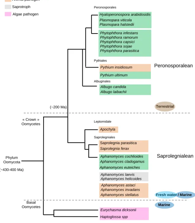
Apochyla Aphanomyces cochlioides Aphanomyces euteiches Aphanomyces cladogamus Aphanomyces laevis Aphanomyces helicoides Aphanomyces invadans Aphanomyces astaci Pythiales Pythium ultimum Pythium insidiosum Peronosporales Saprolegnialean Phylum Oomycota Saprolegnia parasitica Saprolegnia ferax Peronosporalean Basal Oomycetes « Crown » Oomycetes Phytophthora infestans Phytophthora ramorum Phytophthora capsici Phytophthora sojae Phytophthora parasitica Eurychasma dicksonii Haptoglossa spp
Fresh water / Marine Marine
Terrestrial
Hyaloperonospora arabidosidis Plasmopara viticola
Plasmopara halstedii
Figure 2. Summary scheme showing the phylogenetic relationships of main taxa groups of oomycetes, their environmental prevalence and lifestyles. Estimations
place the origin of oomycete between ~430 and 400 million years ago (Ma). Late divergent « Crown » oomycetes are regrouped into two mayor ensembles: the Peronospolean lineage and the Saprolegnialean lineage, having separated ~200 million years ago (Ma). Representative species and their lifestyles are given. The scheme was generated based on information provided by Beakes and Sekimoto (2012), Dieguez-Uribeondo et al (2009) and Matari et al (2014).
Saprolegniales Leptomidale Albugo candida Albugo laibachii Albuginales Plant pathogen Animal pathogen Saprotroph Algae pathogen Aphanomyces stellatus (~200 Ma) (~430-400 Ma)
15
like soybean, grape, broccoli, lettuce and sugar beet endure important diseases hosting species like Phytophthora sojae, Plasmopara viticola, Peronospora parasitica, Bremia
lactucea and Aphanomyces cochlioides, respectively.
Phylogenetic distribution of oomycetes
Oomycetes appeared 430-400 million years ago (Matari and Blair, 2014) and are grouped in two major taxonomic groups/lineages that encompass the most late-diverging groups of oomycetes or “crown oomycetes” (to exclude early-diverged basal lineages) as termed by Beakes and Sekimoto (2009): the Saprolegnialean and the Peronosporalean lineages (figure 2). The latter groups have been estimated to split 200 million year ago in the history of oomycetes and have, thus, different evolutionary histories which can be evidenced by biochemical features as for example the ability to synthesize sterols, a trait found only in Saprolegniales and in their closest relatives, the brown algae (Gaulin et al., 2010). Another distinctive trait concerns their ecological prevalence. Saprolegniales are predominantly found in aquatic environments (fresh water and estuarine) while Peronosporales occupy mainly terrestrial environments. The water-dependence of Saprolegniales is considered an ancestral trait and the tendency to have a lesser dependence on water is thought to be an evolutionary trend in oomycetes (Beakes and Sekimoto 2009).
Phytopathogenic oomycetes species are found in both lineages (figure 2) with a majority in the Peronosporalean lineage which stands as a phytopathogenic line. Species ascribed to genera Phytophthora (over 100 species, Kroon et al., 2012), Albugo and
Hyaloperonospora as well as Pythium are phytopathogenic. Main exceptions are restricted to Pythium species since among the 150 plant pathogenic species some are animal pathogens
like P. insidiosum (infecting mammals, Uzuhashi et al., 2010).
In contrast, the Saprolegnialean lineage is more heterogeneous since in addition to a large number of exclusively saprophytic species the group harbors zoopathogenic and phytopathogenic facultative species. Animal pathogens species of the genus Saprolegnia (i.e: S. parasitica) infect freshwater fishes, insects and amphibians (Sarowar et al., 2013). In
Aphanomyces genus, species A. invadans and A. astaci develop on fishes and crayfishes
species with elevated economic and ecological impacts (Phillips et al., 2008). Few plant pathogens species have been identified in Saprolegniales and are all included in the genus
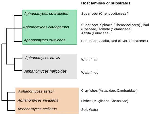
Aphanomyces cochlioides Aphanomyces euteiches Aphanomyces cladogamus Aphanomyces laevis Aphanomyces helicoides Aphanomyces invadans Aphanomyces astaci Aphanomyces stellatus
Figure 3. Close up summary scheme on Aphanomyces phylogeny, lifestyle and principal hosts or substrates.
The phylogenetic relationships were established by analysis of ITS sequences of nuclear rDNA of the principal Aphanomyces spp identified till date. Lifes styles displayed by these species correlates to their phylogenetic regrouping defining three independent lineages: a plant pathogen lineage, a saprophytic/ oportunistic lineage and animal pathogenic lineage. Species A. laevis and A. helicoides are generally assigned as saprotrophs but isolated studies have documented their development on animals (insects). A. stellatus has been found as a free-living species but its ITS sequence analysis assigned it to the zoopathogenic lineage.The scheme was performed based on Dieguez-Uribeondo et al., (2009).
Host families or substrates
Sugar beet (Chenopodiaceae )
Sugar beet, Spinach (Chenopodiacea) , Barley (Poaceae),Tomato (Solanaceae)
Alfalfa (Fabaceae)
Pea, Bean, Alfalfa, Red clover. (Fabaceae.)
Water/mud
Crayfishes (Astacidae, Cambaridae )
Fishes (Mugiladae,Channidae)
Soil, Water
Plant pathogen lineage Saprotroph/opportunistic lineage
Water/mud
16
Aphanomyces genus
An ancestral genus harboring diverse species
Aphanomyces genus regroups about 40 species (Diéguez-Uribeondo et al., 2009).
This is an approximate number because their proper identification is difficult due to the reticence of some species to be isolated and maintained in pure cultures. Hence, its taxonomy is still in progress. Nevertheless, studies converge to the conclusion that Aphanomyces is an ancestral Saprolegniale group (Petersen and Rosendahl, 2000).
In an overall view, most species have prevalence for aquatic niches (fresh water and marine, mostly estuarine) with the exception of plant pathogens which occur in terrestrial wet environments. Phylogenetic studies of Aphanomyces genus regroup species in three independent lineages globally correlated to their life-styles (figure 3). A first line comprises plant pathogenic species, which in the Saprolegnialean line are only restricted to
Aphanomyces genus. Within, species like A. cladogamus have a broad range of hosts of
different families as Fabaceae (Phaseolus vulgaris, common bean), Poaceae (Hordeum
vulgare, barley), Solanaceae (Lycopersicon esculentum, tomato) and Chenopodiaceae
(Spinacia oleacera, spinach). A. euteiches displays a quite marked specialization for Fabaceae species while A. cochlioides is a host-narrow species reported so far only on sugar beet (Beta vulgaris) (Diéguez-Uribeondo et al., 2009). A second lineage harbors species with prevalence for saprophytism as A. laevis and A. helicoides which can exhibit opportunistic parasitism. Lastly, the zoopathogenic lineage regroups A. astaci (infecting freshwater crayfishes, Filipova et al., 2013) or A. invadans (responsible for the epizootic ulcerative syndrome of various species of estuarine fishes, (Boys et al., 2012) as well as A. stellatus, which, consensualy defined as a saprotroph, has been found to develop on crustaceans (Royo et al., 2004).
The diversity of life styles and hosts of Aphanomyces species (animal/ plant pathogens and saprobe species) gives to this genus a special taxonomic position among Saprolegniales and towards Peronosporales.
Aphanomyces life cycle presents sexual and asexual stages. Sexual reproduction
leads to the formation of oospores as the result of the fertilization of oogonia (female reproductive structures carrying the female haploid nuclei) by antheridia (male reproductive structures delivering haploid male nuclei). Oospores are long-resting structures which germinate to produce biflagellate motile zoospores (the primary infection entity). Such zoospores can also be produced as the result of asexual reproduction by live mycelium in roots via specialized hyphae (sporangium). As zoospores reach host surfaces, they encyst and
Figure 4. Infection life cycle of A. euteiches (Saprolegniales). Oospores present in
soil germinate producing a sporangium (1). Primary spores (2N) are formed in the apex of the sporangium and release zoospores (asexual, biflagellate spores) through a pore of their cell wall (2). Zoospores can encyst en germinate to produce a novel sporangium giving rise to a second generation of primary spores and zoospores (3) a phenomenon called Repeated Zoospore Emergence (RZE). Zoospores that reach the host rhizoplane encyst and germinate producing an hyphae that directly penetrates host tissues (4). Infectious hyphae develops intercellularly (5) to completely colonize the root system and subsequently progress to hypocotyls (6). Within roots, sexual reproduction is assured by the differentiation of hyphae into antheridia and oogonia, carrying haploid nuclei (1N) and whose fusion results in the formation of oospores (2N). During the decomposition of dead plant tissue, oospore are then released in soils were they can remain dormant for up to 10 years (7). Adapted from the American Phytopathological Society (APS) https://www.apsnet.org/
17
germinate to penetrate host surfaces (figure 4 ). It is worth to note that some physiological and developmental behaviours are linked to the parasitic life style. Parasitic species present the capacity of Repeated Zoospore Emergence (RZE). This refers to the ability of a motile zoospore to encyst and to produce a second generation of zoospore rather than directly infect the host. It is believed that RZE allows to extend the duration of the infective stage and so the chances to come upon a suitable host. Also, plant parasitic species usually present both sexual and asexual stages whereas in animal pathogens the sexual stage is often absent or rare (Diéguez-Uribeondo et al., 2009).
A. euteiches and the root rot disease of legumes
Aphanomyces euteiches was first described by Dreschler in 1925 from infected pea
(Pisum sativum) in the United States. It is a soil borne pathogen infecting roots of legumes as well as a facultative pathogen which means that it is able to grow as a saprobe outside the host (Papavizas and Davey, 1960). This renders its axenic culture possible. Its developmental cycle includes sexual and asexual stages. The species is homothallic (self-fertile) and presents sexual reproduction typically achieved by the formation of diploid oospores capable to subsist up to 10 years in soils on harsh conditions (Gaulin et al., 2007).
The disease caused by A. euteiches is commonly known as the root rot of legumes. Legumes affected include pea (Pisum sativum), alfalfa (Medicago sativa), snap and red kidney bean (Phaseolus vulgaris), faba bean (Vicia faba,) red clover (Trifolium pratense) and white clover (Trifolium repens). Thus, A. euteiches has a relative large range of hosts within Fabaceae. Nevertheless its occurrence and degree of pathogenicity can differ from one host to another. Its characterization on pea and alfalfa has defined pathotypes: pea-infecting strains and alfalfa infecting strains from the USA and from France (Malvick and Grau 2001; Moussart et al., 2007; Wicker and Rouxel, 2001).
Infection takes place in roots as zoospores, chemo-attracted by root compounds (Sekizaki et al., 1993), encyst in the rhizoplane and germinate to penetrate root cortex tissues. Total colonization of roots is followed by the spread of the pathogen to stem (hypocotyls, epicotyls). Typically, its development provokes the disintegration of cortex tissues denoted by the appearance of softened and water-soaked areas of roots which become orange-brown or blackish-brown at later times of pathogen development. Symptoms can advance to stems and become evident by the necrosis of epicotyls and hypocotyls, and chlorosis of cotyledons followed by total discoloration (death) or foliage milting. Completion of infection is marked by the formation of oospores that ensure the dissemination. When
F8300.5 A17 S a a S b b c d A B C
Figure 5. Infection model in the Medicago truncatula/ Aphanomyces euteiches pathosystem. A. Macroscopic symptoms displayed by M. truncatula F83005.5 (highly
susceptible) and ecotypes A17 (partially resistant) infected with A. euteiches in in vitro conditions (Djébali et al.,2009). B. Scheme of a transversal section of a root infected by
A. euteiches (green). On the left side, the scheme describes infection in F83005.5 were
an asexual spore (S) has landed on the rhizoplane and germinated to produce a germ tube giving rise to an infectious hyphae that directly penetrates root cortex tissues (a). Hyphae develops between root cells of cortex which becomes completely colonized 6 days post inoculation. Cortical cells died as A. euteiches develops leading to root disassembly and water-soaked symptoms typical of root rot disease (b). The pathogen reaches the vascular cylinder before completion of its cycle (not shown) (c). On the right side, the infection is depicted in the tolerant host (A17) were the plant produces supplementary pericyle cell layers with higher levels of lignin in their cell-walls, reinforcing the root stele. In addition to this mechanical barrier, cells produce antimicrobial compounds (d). These cytological responses restrain the advance of the pathogen to the vascular cylinder. C. Cross-sections of infected roots showing full invasion (F83005.5) and partial invasion (A17) by A. euteiches (green) and the production of antimicrobial compounds by A17 (bleu) in the root stele, 15 days post inoculation (Djébali et al.,2009).
Pericycle Cortex
tissue
Vascular tissue
18
conditions are appropriate, oospores germinate in the vicinity of hosts to produce zoosporangia.
Root rot disease occurs wherever host species are grown. Specially, it is a problem for pea and alfalfa-growing regions in North American and European countries. In France, it affects primary forage pea in northern regions (Gaulin et al., 2007). No effective control exists once A. euteiches is installed in soils. In addition, no fully resistant line of pea or alfalfa is available. Only prophylactic measures and crop rotation are preconized to prevent crop losses. Currently, improvement of crop resistance is done via the characterization of broad range resistance and identification of QTLs, which is the only type of resistance towards this pathogen (Djébali et al., 2009; Hamon et al., 2013; Bonhomme et al., 2014).
A.euteiches and M. truncatula pathosystem
The legume Medicago truncatula is a close relative of the cultivated alfalfa (M. sativa) that has become a largely used model plant (Cook, 1999). Protocols suited for laboratory research have been established and a wide collection of mutants and natural genotypes are at disposition for the scientific community. Genomic resources include sequences of over 288 accessions (Stanton-Geddes et al., 2013). More interestingly, it is a host of A. euteiches showing great variability of susceptibility to this pathogen (Moussart et al., 2008).
A. euteiches/M. truncatula pathosystem has been developed and exploited in our
research group to understand the molecular aspects behind the suceptible variability of the plant. The pathosystem makes use of a pea isolate of A. euteiches (ATCC 201684) and different genotypes of M. truncatula that display different degrees of tolerance to this strain. Two accessions exemplifying the opposite resistance degrees are line A17 (partially resistance) and line F83005.5 (highly susceptible). Genetic approaches and the use of an in
vitro infection assay coupled to the characterization of infection phenotypes have led to the
identification of a QTL (Djébali et al., 2009). Recently, the variability of 176 M.
truncatula genotypes towards A. euteiches has been correlated to gene sequence architecture
through a Genome Wide Association Study (GWAS). Single Nucleotide Polymorphisms (SNPs) in the promoter and coding region of an F-box gene have been spotted out and linked to M. truncatulas's variability to A. euteiches (Bonhomme et al., 2014). Although no expression polymorphism between resistance and susceptible lines could be shown, the identified SNPs are preconized to lead to functional and non-functional forms of the F-box protein which are associated to susceptibility and resistance, respectively. In addition to the
F-19
box gene, and consistent with the quantitative type of resistance towards A. euteiches, other genes have also been identified through the GWAS. These latter are associated to hormone regulation, notably the biosynthesis of cytokinine and transcription factors associated to ethylene response as well as giberrilin and abcissic acid (Bonhomme et al., 2014).
Molecular and cytological approaches carried in lines A17 and F83005.5 have evidenced particular plant mechanisms that might be implicated in the contrasting tolerance to A. euteiches. In both lines, upon inoculation of roots with zoospores, A. euteiches penetrates and starts to develop between cells of the outer cortex tissue within 1 day. No specialized infectious hyphae structures as appressoria or haustoria have been evidenced. The pathogen presents an intercellular development and invades the whole cortex area within 3 to 6 days. But, while in F83005.5 total invasion of cortical tissues is followed by the progression of the pathogen to vascular system, in A17 roots the pathogen progression is mostly restrained to cortical tissues. This inability to progress into the vascular system is correlated to the capacity of A17 to deploy a whole set of defense mechanisms like the production of phenolic compounds, the reinforcement of cell walls and, more particularly, the formation of supplementary pericycle cell layers that might act as a physical barrier for the invading hyphae (figure 5). Intriguingly, partial resistance of A17 is accompanied by an increase of lateral roots (Djébali et al., 2009).
Molecular interplay between pathogen effectors and plant immunity
Plant pathogen microorganisms have acquired the ability to take profit of the nutrient- rich niche that may be provided by plants to develop and reproduce. Indeed plants represent a well lasting source of carbon, nitrogen and water as well as a physical protection (Zuluaga et al., 2013). This mode of living implies an intimate association with plants in order to exploit goods. Because the development of associated microorganisms affects host plant health, conducting eventually to plant disease and death, these associations are termed as pathogenic.
Plant immunity and effectors
As any other living organism exposed to a multitude of microorganisms, plants sense these external cues to respond in the best-adapted manner. To defend themselves against pathogens, plants possess constitutive defenses consisting in pre-existing physical or
Table 1. Pathogen Molecular Associated Patterns (PAMPs) of bacteria fungi and oomycetes and their cognate receptors in plants.
Organism PAMP (name) Plant Receptor Reference
Bacteria Flagellin (flg22) FLS2 (A.thaliana) Félix et al. , 1999 Gomez-Gomez et al., 2001
Elongtion factor EF-Tu (elf18/26)
ERF
(A.thaliana) Kunze et al., 2004 Cold shock protein
(RNP) Not identified Félix and Boller, 2003 Lipopolysaccharide Not identified Newman et al., 2005
Peptidoglycan Lym1, Lym2 (A. thaliana)
Erbs et al., 2008a Willmann et al., 2011 Fungi chitin CeBip, (rice) CERK1 (A.thaliana) Kouzai et al., 2014 Petutschnig et al., 2010
Xylanase (EIX) EIX (tomato) Ron and Avni, 2004
Oomycetes
CBEL Not identified Gaulin et al.,2006 Larroque et al., 2013 Pep13 Not identified Nürnberger et al.,1994
Brunner et al., 2002 Β-hepta glucans GEBP (putative, soybean) Umemoto et al., 1997
INF1 NbLRK1
20
biochemical barriers (i.e. hydrophobic cuticles, surface antimicrobial compounds…) that act as a first level of protection to avoid and limit development of potential harmful microorganisms. In addition, they possess an inducible multi-layered immune system that allows them to perceive microorganisms and activate a whole set of modular defenses (figure 6 A) (Dodds and Rathjen, 2010).
The first level of plant immunity is activated by the direct perception of conserved epitopes in commonly occurring molecules of microbes (pathogen or non-pathogen) called “Pathogen/Microbial-Associated Molecular Patterns “P/MAMPs”. These molecular signatures can be of different nature (protein, oligosaccharides...), they identify a whole classe of microbes and are absent from plants (table 1). Their recognition involves Pattern Recognition Receptors (PRRs) associated to the plasma membrane that belong to the family of Receptor-Like-Kinases (RLKs) and Receptor-like Proteins (RLP) (Boller and Felix, 2009) (figure 6). Best characterized examples are the 22-peptide present in the bacteria flagellin protein (flg22) and peptides elf18/26 of the bacterial EF-Tu elongation factor protein perceived by the RLKs FLS2 and EFR, respectively (Boller & Felix, 2009). Fungal chitin is perceived in rice by CEBiP (Kouzai et al., 2014) and by CERK1 in A. thaliana via their LysM extracellular domains (Petutschnig et al., 2010). The activation of the defense signaling after PAMP perception requires the association of these receptors to a central RLK regulator, named BAK1 (Kim et al., 2013) (figure 6). The importance of BAK1 in mediating PAMP-defense signaling and activation is well known for the above mentioned bacterial and fungal PAMPs and has been recently established for defense against nematodes and insects (Peng and Kaloshian, 2014). Oomycete PAMPs that elicite defense in plants have been identified but their cognate receptors and mechanisms behind their recognition remain for the most unknwon. These include the epitote pep13 of transglutaminases (Brunner et al., 2002), the heptaglucans of P. sojae which has been proposed to be perceived by the GEBP (soybean) (Umemoto et al., 1997), the cellulose- binding domain of the cell-wall protein CBEL of Phytophthora parasitica (Gaulin et al., 2006) for which its eliciting activity requires BAK1 (Larroque et al., 2013) and finally, the sterol-binding protein elicitin INF1 from Phytophthora spp (Kamoun et al., 1998) for which a RLK has been identified (Kanzaki et al., 2008) (table 1).
PAMP/MAMPs recognition triggers a level of immunity referred to as PAMP-Triggered Immunity (PTI) also called basal immunity given the broad sprectrum of occurrence of PAMP/MAMPs, not specific to a given pathogen. PTI includes subsets of early responses like the production of ROS species and ion fluxes. More intermediate events include the activation of signaling cascades involving mitogen-activated protein kinases (MAPKs)
A
B
Figure 6. Global concepts of plant susceptibility triggered by microbial effectors (A) and plant immunity (B). A. Pathogens such as bacteria, fungi and
oomycetes establish a close physical encounter with host cells. They secrete effectors into the apoplasm (apoplastic effectors) of inside plant cells (intracellular effectors). Bacteria deliver them into host cells via the Type 3 secretion system (T3SS) and filamentous pathogens via infectious structures (haustoria). Apoplastic effectors interact with apoplastic effector targets (AET) and intracellular effectors with intracellular effector targets (IET). In susceptible plants (A), their interaction with targets perturb different plant processes that benefit the outcome of infection. Among these processes, PTI is an important target whose suppression is required for successful infection. B. Plant immunity activation resides on the perception of pathogens, which can be performed through two modes. A first mode, consists on the perception of PAMPs by PRRs (Patter Recognition Receptors) at the plasma membrane leading to the activation of PTI (PAMP-Triggered Immunity). Initiation of PTI signalling pathway requires the co-receptor BAK1. A second mode, relies on the direct perception of effectors (1) or on the perception of effector activities on plant targets (2) via intracellular nucleotide-binding receptors (NB-LRR), conducting to ETI (Effector-triggered Immunity).
(Modified from Win et al., 2011)
PAMPs F ungi O om y c et e Bacterium Intracellular effectors Haustorium Plant cell PRR BAK1 PTI ETI NB-LRR IET 1 2 T3SS PAMPs F ungi O om y c et e Bacterium Intracellular effectors Haustorium Plant cell PRR BAK1 Perturbation of plant processes Apoplastic effectors Effector Triggered Suceptibility IET AET AET IET PTI T3SS
21
(Pitzschke et al., 2009) that ultimately conduct to the activation of transcription factors (TFs) and expression of pathogenesis-related genes (PR) encoding antimicrobial compounds like chitinases and glucanases which directly degrade cell-wall structural components of pathogens (Dodds & Rathjen, 2010). Phytohormones like salicylic acid (SA) and jasmonic acid (JA) also play key roles in the signal transduction (Tsuda and Katagiri, 2010). All together, these defenses are generally sufficient to arrest pathogen infection and maintain plant health. Nevertheless, adapted pathogens have evolved means to break down or evade such defenses. These pathogens produce secreted molecules termed “effectors” which designates “all proteins and small molecules” produced and secreted by pathogens that “alter host-structure and function” and facilitate infection (Hogenhout et al., 2009). Effectors are secreted into the apoplasm interface (apoplastic effectors) or are addressed inside host cells (intracellular effectors) to interact with plant targets. By doing so, effectors can suppress plant immunity and perturb other plant processes that conduct to host susceptibility or Effector-Triggered Susceptibility, a state benefiting the outcome of the infection (figure 6 A). Manipulation of PTI can be achieved by avoiding PAMP recognition or by directly suppressing PTI. For instance, the fungal LysM apoplastic effectors bind chitin, preventing its recognition by plants (Kombrink and Thomma, 2013). In oomycetes, several apoplastic- secreted protease inhibitors form P. infestans have been shown to directly bind plant apoplastic enzymes, suppressing directly their activities in this host space (Tian et al., 2005, 2007; Song et al., 2009).
Another type of defense relies on the recognition of specific effectors or effectors’s activities and is referred to as Effector-Triggered Immunity (ETI) (figure 6 B). This mechanism for pathogen recognition involves a particular class of receptors called R proteins (Resistance proteins) that are generally cytosolic. Effector recognition by R proteins leads to specific resistance and incompatible interactions, reason why in this context, recognized effectors are called Avirulence (Avr) proteins. This mechanism follows the gene-for-gene model conceptualized by Flor’s work (1971). ETI is similar in nature but is more rapid and strong than PTI (Tao et al., 2003; Tsuda and Katagiri, 2010) and is often accompanied with a cell-death or Hypersensitive Response (HR). Because it relies on the perception of effectors, it is a specific immunity.
R proteins are classified into two classes depending on the type of Nterminal domain: (Toll- like Receptor) TIR-NB-LRR or (coiled-coiled) CC-NB-LRR proteins. Two modes of effector recognition exist: (1) direct physical interaction of R proteins to effectors or (2) indirect interaction of R proteins. In this case, R proteins perceive modifications of plant proteins to which the R protein is associated and monitors (figure 6 B). These modified plant
22
proteins can be genuine virulence target of the effector (the guard model) or a mimic of one (the decoy model). It has been shown that activated R proteins interact with other proteins forming complexes mediating the signaling that conducts to defense response such signaling requires complexes to relocalize to specific subcellular compartments of plant cells (Dodds & Rathjen, 2010; Engelhardt et al., 2012; Qi and Innes, 2013).
In that instance, studies have recently demonstrated the importance of a nucleo-cytoplasmic trafficking of R proteins and other immune components for the activation of defense responses. The accepted model is that such relocalization is induced by pathogen perception and allows immune receptors to accumulate in the plant nucleus where they activate immune responses through transcriptional reprogramming (Qi and Innes, 2013).
Effector identification
Effector proteins were firstly identified thanks to their avirulence activity. In an ETI context, the effector behaves as an avirulent protein and the resultant incompatible interaction was of great aid to identify avirulence proteins in fungi, bacteria and oomycetes. Their cloning and molecular characterization pointed out the presence of secretion leaders either typical of the Type 3 Secretion System (T3SS, for bacteria Avr proteins) or canonical eukaryotic signal peptides (in oomycetal and fungal Avr proteins) (Armstrong et al., 2005; Staskawicz et al., 1984; van Kan et al., 1991). It was then suggested that such key factors were likely to be secreted by pathogens to reach host structures and compounds.
Effector identification has been possible by the use of transcriptomic, proteomic and genomic approaches coupled to functional screening systems based mostly on the capacity of effectors to modulate plant immunity. Transcriptomic studies have allowed to determine genes differentially expressed during infection and even specifically in particular infection stages and pathogen structures as haustoria (Godfrey et al., 2010; Hahn and Mendgen, 1997; Huang et al., 2004). Expressed Sequenced Tags (ESTs) obtained on cDNA libraries coupled with computational searches for secretion peptides provide putative effector secretomes and thus putative effector repertoires. By this means, 31 hautoria-specific in planta-induced genes from the rust fungus Uromyces faba were identified (Hahn and Mendgen, 1997) as well as 100 putative secreted proteins expressed predominantly in haustoria of Blumeria gaminis (Godfrey et al., 2010). Moreover, P. infestans secretome analysis based on ESTs led to the identification of CRN proteins (Torto et al., 2003).
23
proven to be successful on the characterization of key factors of infection. Mass spectrometry analysis of total proteins extracted from xylem sap of susceptible tomato infected with the vascular pathogen Fusarium oxysporum resulted on the identification of SIX (Secreted In
Xylem) proteins of F. oxysporum specifically present during infection (Houterman et al.,
2007). Further characterization of SIX1 and SIX2 proteins demonstrated their requirement for full virulence and as poteins recognized in tomato lines expressing I-3 and I-2 resistance proteins (Rep et al., 2004; Houterman et al., 2009). Proteomic approaches can be also useful when combined with predicted approaches. For instance, mining of P. infestans’s in vivo secretome has not only corroborated repertoires established in silico via gene model prediction on genomic data but has also extended the secretome repertoire (Meijer et al., 2014).
As numerous complete genomes are available, effector repertoires can be established directly on gene models and can be combined to transcriptomic and proteomic data to further support their effector role. In silico criteria to determine candidate secreted effector proteins (CSEPs) on genomic data are principally based on the presence of secretion leaders and the absence of transmembrane domains. For such criteria, sequence surveys make use of computational strategies developed to ensure accuracy of predictions (Bendtsen et al., 2004; Torto et al., 2003). Other criteria can be taken into account for the determination of CSEPs and have become widely used and accepted in the scientific community throughout observations made on some experimental data. As very often, the studied effector proteins display small sizes and a high content of cysteine, such caracters are usually taken into account when defining potential candidates. These criteria have led to determine 929 proteins in the oomycete Albugo candida (Links et al., 2011) and 491 and 365 proteins from fungi Blumeria graminis and Colletotrichum higginsianum, respectively, have been proposed as putative effectors (Pedersen et al., 2012b; O’Connell et al., 2012). Genome survey on Ustilago maydis revealed 426 proteins potentially secreted (Mueller et al., 2008) and tissue-specific expressed, as revealed by transcriptome profiling (Gao et al., 2013).
Further amino acid sequence inspection of CSEPs repertoires has evidenced the presence of commonly occurring amino residues for some of them. These amino acids define motifs that serve as a basis for protein family classification and as a criterion when inspecting novel pathogen genomes (table 2). This was typically the case of oomycetes, for which sequence comparison of Avr proteins like ATR13 of H. parasitica, and Avr3a of P.
infestans (Allen et al., 2004; Armstrong et al., 2005) led to determine a conserved Nterminal
RxLR (Arginine, any amino acid, Leucine, Arginine) motif (Rehmany et al., 2005). Since evidenced as a sequence trait of Avr effectors, this motif has been used on genomic data of
Table 2. Conserved Nterminal motifs identified on Candidate Secreted Effectors Proteins (CSEPs) in fungi anf oomycetes.
Motifs Protein Species Reference
RxLR(dEER) RxLR effectors Phytophthora spp, H. arabidopsidis A. Laibachii Rehmany et al., 2005; Haas et al., 2009; Baxter et al., 2010 Kemen et al., 2011
LSSLR(ILKS)L(KQ)SL Ac-RXL A. candida Links et al. ,2011
LxLFLAK CRN effectors Phytophthora spp H. arabidopsidis, A. euteiches Torto et al.,2003 ; Haas et al.,2009 ; Stam et al.,2013 Baxter et al.,2010 CRN-like R. irregularis Lin et al., 2014
HVVVxxP CRN effectors A. euteiches
Gaulin et al.,2008 ; Gaulin et al., in preparation
LxLYLAR /K CRN effectors P.ultimum Adhikari et al.,2013
CHxC CHxC A. candida, A. laibachii P. infestans Pais et al., 2013 ; Kemen et al.,2011 ; Links et al., 2011 Y/F/WxC Y/F/WxC B. graminis P. graminis M. lini Godfrey et al., 2010b ; Pedersen et al., 2012 Saunders et al., 2012 Duplessis et al. , 2011
RFYR AvrL567 M. lini Rafiqi et al.,2010
RGFLR, KFLK, RDLA AvrM M. lini Rafiqi et al.,2010
RYWT, RTLK AvrLm6 L. maculans Kale et al. , 2010
RMLH and RIYER Avr2 F. oxysporum Kale et al., 2010
FYIQYLxNQPV and/or
24
all oomycetes an d h as led to the identification of a large number of RxLR proteins defining the RxLR class of oomycete effectors (563 coding genes in P.infestans, Haas et al., 2009, 134 in H. parasitica, and 396 in P.sojae, Baxter et al., 2010). The conservation of the Nterminal motif L/Q/FLAK has also allowed the identification and cataloguing of CRN effectors in oomycetes (Baxter et al., 2010; Haas et al., 2009; Stam et al., 2013a). Moreover, a CHXC motif has been recently proposed as a motif characterizing a third large class of effectors in oomycetes (Pais et al., 2013; Kemen et al., 2011; Links et al., 2011). In fungi, RxRL-like motifs (ie, RRLQ RGFLR... Rafiqi et al., 2010; Khang et al., 2010) have been proposed in Nterminal domains of CSEPs. In addition, a Nterminal Y/F/WxC motif, firstly found in the 100 haustoria-expressed CSEPs of Blumeria graminis (Godfrey et al., 2010; Pedersen et al., 2012) have also been reported in CSEPs of Puccinia graminis and Melampsora
larici-populina (Duplessis et al., 2011; Saunders et al., 2012).
In addition to these criteria (presence of secretion signals, absence of transmembrane domains, small size, high content of cysteine residues), other sequence signatures like the signs of diversifying selection can pinpoint their potential effector role as this may infer that the protein in question is submitted to a counter selection by hosts. Indeed, this has been demonstrated for the carboxyl terminal domains of RxLR effector secretomes (Win et al., 2007).
With established effector repertoires in a considerable number of species, it is now possible to perform comparative genomics to provide clues explaining pathogen host-adaptation and particular infection strategies. For example, comparison of putative effectors of the closely related smut fungi U. maydis and Sporisorium reilianum (both infecting maize) have determined specific subsets of effectors in both species. These subsets could be at the basis of their different infection behaviours on maize and may also imply the targeting of distinct maize functions (Schirawski et al., 2010). Another example is the comparison made on seven pathogenic necrotrophic Pythium species that revealed the absence of RxLR effectors. Because RxLR proteins are present in the related hemibiotrophs Phytophthora
spp and the obligate biotroph H. arabidopsidis) and display in planta functions related to the
suppression of host defenses, they have been generaly attributed to the support of biotrophic lifestyles. The lack of RxLR effectors in Pythium spp could further support the latter idea as RxLR functions seem unrelevant for Pythium necrotophic lifestyle (Adhikari et al., 2013). Genomic studies aiming to identify putative effectors can be directed on particular genomic environments. Actually, in Leptosphaeria maculans, putative effector coding genes are mainly found in AT-rich isochores and are often associated to transposable elements (TE) (Rouxel et al.,2011). These effector-TE associations have also been observed for P. infestans
25
RxLR and CRN genes (Haas et al., 2009). Therefore, identifying such genetic environments in species might be used as a strategy to spot out CSEPs.
Functional characterization of effectors
The different predictive approaches for effector identification can result in important number of candidates. Demonstrating their actual contribution to virulence, studying their effect and biochemical activities in plants as well as identifying their targets can be time consuming because of their number, because reverse genetic approaches may not lead to a phenotype and because, for some pathosystems, genetic manipulation of pathogens is not possible. Given this, rapid and efficient functional methods are required in order to screen and functionally characterize effectors.
For this purpose, and based on the concept that effectors are secreted molecules that act in plant cell interfaces, different systems have been developed to heterogously express effectors in planta, in order test directly their cellular localization and any disturbance of plant physiology. Typically, effector proteins tagged with fluorescent proteins are overexpressed with or without their signal peptide (SP) to directly test their cellular localization by plant cell imaging (by means of confocal microscopy) (Caillaud et al., 2012; Stam et al., 2013a/b). Heterologous expression systems are also used to test the “pathogen-independent'” host-delivery of effectors. As the presence of the SP is expected to drive the secretion of effectors into the plant apoplasm, by expressing full length effector proteins (containing their SP), it is inferred that effectors localizing inside cells have re-entered the cytoplasm autonomously via intrinsic translocation signals in the protein. Based on this, it is then inferred that these effectors are likely to localize inside plant cells during infection (Rafiqi et al., 2010; Ribot et al., 2013).
N.benthamiana and N.tabaccum (Solanaceae) have become widely used for molecular plant- microbe interactions studies since they display different degrees of susceptibility to a variety of microorganisms as oomycetes (P.capsici, P.infestans), fungi (Verticillium dahlia,
Colletotrichum orbiculare), bacteria (P. syringae), viruses (TMV, PVX…) and several cDNA
libraries and molecular tools like microarrays are available (Goodin et al., 2008). Their foliar tissues are particularly amenable for genetic transformation via the bacteria Agrobacterium
tumefaciens, compared to other plant model species like A. thaliana and crop species like
barley. Infection with A. tumefaciens strains carrying an effector coding gene allows to, locally and transiently, transform plant cells and constitutes an easy way to express
26
efficiently an important number of effectors in plant cells. As an example, Caillaud and associates (2012) used this approach to screen the localization and cell-death activity of 49 intracellular RxLR effectors of H. arabidopsidis, evidencing the diversity of the subcellular compartments targeted by these proteins. Co-agroinfiltration of N.benthamiana leaves is also possible and has been validated, for example, for the study of Avr/R proteins resulting in the expected HR response (Van der Hoorn et al., 2000; Bos et al., 2006). Besides agroinfiltration, Pseudomonas syringae and its T3SS provide another strategy for delivering effector proteins directly in the cytosol of plant cells by translationnally fusing T3 secretion leaders to the studied effectors. This strategy was used by Whisson and collegues (2007) to demonstrate that the RxLR effector, Avr3a, is recognized by the R protein R3a in the cytoplasm of plant cells and thus that Avr3a is translocated inside plant cells during infection, proving for the first time that RxLR proteins are intracellular effector proteins (Whisson et al., 2007).
Cellular systems have also been developed for rapid and accurate functional studies. Methods based on protoplasts have been described and improved to transiently express effectors and/or reporter genes on various plant crop species like rice, maize, parsley, tomato...(Chen et al., 2006; Sheen, 2001). Such systems have been used to characterize molecular events linked to MAMP/PAMP perception, signal transduction of defense responses and effector activities (Brunner et al., 2014; Kansaki et al., 2014). Recently, a screen on 33 RxLR effectors using protoplasts of tomato and A. thaliana allowed to identify immune suppressive RxLR effectors and to correlate these activities to their cellular localization (Zheng et al., 2014). In addition, screening of 42 in silico-identified putative secreted effectors of M. oryzae permitted to identify 5 effectors capable of triggering cell-death on rice protoplast (Chen et al., 2013). Finally protoplasts can be also used to screen cDNA libraries for protein-protein interactions, analogously to Yeast-two Hybrid approaches (Y2H). This has been performed to identify plant protein partners of effectors of
A.tumefaciens (Lee et al., 2012).
All together, these systems have rendered possible to bridge in silico identification to rapid validation of effector candidates and even their functional molecular dissection. Still, although providing a rapid way to initiate an effector characterization, experimental systems providing a natural host/microorganism context are required to accurately assess effector’s functions.