FACULTÉ DE MÉDECINE
Centre Interfacultaire de Recherche du Médicament Laboratoire de Chimie Pharmaceutique
Professeur Bernard Pirotte
D E S I G N, S Y N T H E S I S A N D P H A R M AC O L O G I C A L E VA LUAT I O N O F O RI G IN A L N I T RO B E N Z E N E S U L F O N Y LU R E A S A N D S U L F O NY L C YA N O G UA NID I N E S A S T H RO M B OX A N E A2 R EC E P T O R A N TAGO N I S T S I n s i g h t s i n t o s e l e c t iv i t y b e t w e e n t h r o m b ox a n e A2 r e c e p t o r i s o f o r m s ( T P α a n d T P β ) Julien Hanson Pharmacien Promoteurs :
Pr. J.-M. Dogné & Pr. B. Pirotte
T h è s e p r é s e n t é e e n v u e d e l ’ o b t e n t i o n d u g r a d e d e D o c t e u r e n S c i e n c e s P h a r m a c e u t i q u e s
ceux-ci n’aurait pas été possible. Ce projet est le fruit de nombreuses collaborations entre différents laboratoires de haut niveau.
J’aimerais tout d’abord exprimer à Monsieur le Professeur Bernard Pirotte ma plus profonde reconnaissance pour m’avoir donné les moyens d’entreprendre cette recherche au sein du Laboratoire de Chimie Pharmaceutique de l’Université de Liège. Son soutien, sa grande expérience et ses conseils judicieux m’ont beaucoup apporté.
Je voudrais également témoigner ici de mon plus profond respect pour Monsieur le Professeur Jaques Delarge qui m’a donné l’envie de poursuivre des études dans le domaine de la chimie pharmaceutique.
J’adresse également mes plus chaleureux remerciements et ma reconnaissance au Professeur Jean-Michel Dogné qui m’a octroyé sa confiance dès le premier jour et m’a permis de travailler dans les meilleures conditions. Ses qualités scientifiques, ses conseils, son soutien, ses encouragements et sa détermination m’ont aidé et apporté énormément.
During this Ph. D. work, I had the chance to join for four months the lab of Pr. Cecil Pace-Asciak, from the Hospital for Sick Children and University of Toronto (Canada). I wish to express my deepest gratitude to him for his support and for the impulsion to the present work. I also wish to address my warmest thanks for the people I worked with in this lab, particularly Denis and Na.
I also wish to address my gratitude to Pr. Therese B. Kinsella, from the University College Dublin (Ireland) for our active collaboration which allowed to generate many data presented in this dissertation.
Je souhaite également exprimer ma profonde reconnaissance à M. le Professeur Jean-Olivier Defraigne qui m’a gentiment permis de réaliser les cultures cellulaires dans son laboratoire (Centre de Chirurgie Expérimentale, CREDEC).
sans lesquelles ce travail n’aurait pas été possible.
Je tiens particulièrement à exprimer ma profonde gratitude envers le Fonds pour la Formation à la Recherche dans l’Industrie et dans l’Agriculture (F.R.I.A) pour son soutien matériel qui m’a permis de mener à bien cette thèse de doctorat. J’exprime ma gratitude envers la Fondation Léon Fredericq, le Fonds National de la Recherche Scientifique (F.N.R.S) ainsi que l’Université de Liège pour m’avoir accordé les moyens financiers indispensables à la réalisation de ces travaux.
Ces années passées au sein du Laboratoire de Chimie Pharmaceutique de l’Université de Liège m’ont permis de rencontrer de nombreuses personnes que je salue ici. Je tiens plus particulièrement à témoigner ma reconnaissance à Philippe Neven pour sa patience, sa grande compétence, son amitié ainsi que pour les nombreuses conversations enrichissantes que nous avons partagées et qui m’ont aidé à mener à bien cette thèse de doctorat. Je n’oublie pas les personnes qui m’ont aidé tout au long de ce travail, avec qui j’ai collaboré et avec qui j’ai partagé de nombreux bons moments et pris beaucoup de plaisir à travailler, notamment : Annie Ooms, Anne-Lise Moray, Alexandre Ghuysen, Catherine Dujardin, Céline Cherdon, Didier Botty, Jean-François Renard, Jean-Paul Cheramy, Jérémie Ghiotto, Philippe Devel, Philippe Kolh, Pierre Drion, Stéphanie Rolin et Vincent Tchana-Sato. J’adresse également un tout grand merci à Yvette Abrassart pour les spectres infrarouge et Stéphane Counerotte pour les analyses élémentaires.
Je voudrais également remercier ma famille et mes proches pour leurs encouragements et leur soutien quotidiens.
Enfin, je tiens à remercier Domi pour sa présence, son écoute et son aide de tous les jours au long de la réalisation de ce travail.
Design, synthesis and pharmacological evaluation of original nitrobenzenesulfonylureas and sulfonylcyanoguanidines as
thromboxane A2 receptor antagonists
Thromboxane A2 (TXA2) is an important mediator metabolized from arachidonic acid through the cyclooxygenase pathway, mainly in platelets and macrophages. It is a potent inducer of platelet aggregation and smooth muscle contraction. Its overproduction has been detected in pathologies such as stroke, asthma, myocardial infarction or atherosclerosis. The action of TXA2 is mediated by a specific G-protein coupled receptor (TP) of which two alternative spliced isoforms, TPα and TPβ, have been described. The exact role of these two isoforms is not clearly understood. However, recent studies have described their implications in vascular physiology and pathology.
The inhibition of the action of TXA2 on platelets and blood vessels would be interesting as original therapies against cardiovascular diseases. Consequently, the design of TP receptor antagonists remains of great interest in cardiovascular medicine. In the laboratory of medicinal chemistry (University of Liège, Belgium), several nitrobenzenesulfonylureas, derived from torasemide (a loop diuretic), have been previously described as TP receptor antagonists. Two compounds, BM573 and BM613 were among the most interesting molecules identified in that previous work.
The present project is divided in two parts. First, we have determined the pharmacological properties of BM573 and BM613 as thromboxane synthase inhibitors and TP receptor antagonists, in vitro and in vivo. In our assays, these two compounds were proved to have high affinity for both TPα and TPβ, to be potent antiplatelet agents, to inhibit thromboxane synthase and TP-mediated smooth muscle contraction. Additionally, they significantly reduced the size of the thrombus in a rat model of ferric chloride-induced arterial thrombosis. Consequently, we demonstrated that the TP receptor antagonists BM573 and BM613, belonging to the chemical family of nitrobenzenesulfonylureas, could be regarded as antiplatelet and
stroke or myocardial infarction.
Secondly, given the interesting pharmacological profile of BM573 and BM613, we have designed and synthesized several series of compounds derived from these two agents. We have evaluated the binding properties (affinity) of the first generation (+/- 35 original derivatives) of compounds on either TPα or TPβ, transiently expressed in COS-7 cell lines. Additionally, we have measured the ability of our drugs to inhibit the intracellular calcium ([Ca2+]
i) mobilization upon TPα or TPβ stimulation. To confirm our results, we also assessed the antiplatelet properties of our drugs by means of determination of inhibition of human platelet aggregation. On the basis of the results obtained with these in vitro assays, we have synthesized and evaluated a second generation of derivatives (+/- 35 original compounds) and improved the selectivity of several original compounds for TP receptor isoforms.
The originality of this work was to evaluate a large library of synthetic compounds on both TP receptor isoforms, using specific pharmacological tests. By means of structure-activity relationship studies, we were able to identify chemical groups implicated in selectivity and to propose lead compounds for development of highly specific TPα or TPβ antagonists. Besides, we have identified an in vivo drug candidates for prevention of thrombosis and pathological platelet aggregation.
Conception, synthèse et évaluation pharmacologique de nitrobenzènesulfonylurées et sulfonylcyanoguanidines en tant
qu'antagonistes des récepteurs au thromboxane A2
Le thromboxane A2 (TXA2) est un métabolite de la cascade de l’acide arachidonique (AA) par la voie des cyclooxygénases et de la thromboxane synthase, principalement formé dans les plaquettes et les macrophages. Le TXA2 est un puissant inducteur de l’agrégation plaquettaire et de la contraction des muscles lisses vasculaires et bronchiques. Par ailleurs, une augmentation des taux en TXA2 a été constatée dans différentes pathologies : l'infarctus du myocarde, l'atherosclérose, les accidents vasculaires cérébraux, ou encore l'asthme. L’action du TXA2 sur les tissus résulte de la stimulation d’un récepteur appartenant à la famille des récepteurs couplés aux protéines G. Ce récepteur au TXA2 (TP) présente deux isoformes générées par épissage alternatif, TPα et TPβ. Le rôle physiologique exact de ces deux isoformes n'est pas encore connu. Cependant, de récents travaux ont mis en évidence leur importance, notamment dans la physiologie vasculaire et dans certaines pathologies.
L’inhibition de l’action du TXA2 au niveau des plaquettes et des vaisseaux sanguins pourrait donc être une stratégie thérapeutique innovante pour traiter et prévenir les maladies cardiovasculaires. En conséquence, le développement d’antagonistes des récepteurs TP reste d’un grand intérêt en médecine cardiovasculaire. Des études de pharmacomodulation avaient permis au Laboratoire de Chimie Pharmaceutique (Université de Liège, Belgique) d'identifier des nitrobenzènesulfonylurées, dérivées du torasémide (un diurétique de l’anse), présentant un puissant antagonisme des récepteurs TP. Parmi ceux-ci, deux composés, le BM573 et le BM613, faisaient parties des molécules les plus intéressantes identifiées au cours de ces précédentes recherches.
thromboxane synthase et antagonistes des récepteurs TP, in vitro et in vivo. Au cours de nos expériences, ces deux composés se sont révélés posséder une grande affinité pour TPα et TPβ, être de puissants agents antiplaquettaires, des inhibiteurs de la thromboxane synthase et de la contraction des muscles lisses induite par le TXA2. En outre, l’utilisation de ces produits dans un modèle de thrombose artérielle induite par le chlorure ferrique chez le rat a provoqué une réduction significative du thrombus formé. En conséquence, nous avons démontré que le BM573 et le BM613, appartenant à la famille chimique des nitrobenzenesulfonylurées, pouvaient être considérés comme des agents antiplaquettaires et antithrombotiques, potentiellement utiles en tant qu’agents thérapeutiques dans des pathologies associées au TXA2 telles que l’infarctus du myocarde ou l’accident vasculaire cérébral.
Ensuite, nous nous sommes concentrés sur l'activité de cette famille de composés (les nitrobenzènesulfonylurées) vis-à-vis des deux isoformes du récepteur au thromboxane. Pour ce faire, nous avons conçu et synthétisé de nombreuses séries de composés dérivés du BM573 et du BM613. Nous avons tout d’abord évalué l’affinité de la première génération de composés (+/- 35 dérivés) sur des lignées cellulaires (COS-7) exprimant sélectivement soit TPα soit TPβ. De plus, nous avons mesuré la capacité de ces composés à inhiber la mobilisation de calcium intracellulaire ([Ca2+]i) induite par la stimulation des deux isoformes TPα et TPβ séparément. Nos résultats ont été confirmés sur agrégation plaquettaire humaine. Sur la base des résultats obtenus avec cette première génération de produits, nous avons synthétisé une seconde génération (+/- 35 dérivés) de composés, et avons réussi à augmenter la sélectivité en faveur de TPβ pour certains produits.
L’originalité de ce travail réside dans le fait que nous avons évalué un nombre de produits importants sur TPα et TPβ, au moyen de tests pharmacologiques spécifiques. Grâce à des études de relation structure-activité, nous avons identifié des groupements chimiques impliqués dans la sélectivité entre les deux isoformes. Nous pouvons donc proposer des structures "chef de file" pouvant être utiles pour le
ailleurs, nous avons identifié in vivo des candidats pour le développement d’agents thérapeutiques pour la prévention des thromboses et des autres pathologies provoquées par une activation plaquettaires excessive.
AC Adenylate cyclase HUVEC
Human Umbilical Vein Endothelial Cells
ADP Adenosine diphosphate IP Prostacyclin
receptor
cAMP cyclic adenosine
monophosphate IP3
inositide triphosphate
ASA acetylsalicylic acid,
Aspirin LDL low-density lipoprotein BAPTA 1,2-bis(2-aminophenoxy) ethane-N,N,N',N'-tetraacetic acid LOX lipoxygenase
Ca2+ Calcium ions NO nitric oxide
COPD chronic obstructive
pulmonary diseases PUFA
polyunsaturated fatty acids
COS-7 cells
African green monkey kidney fibroblast-like cell line
PGD2 Prostaglandin D2
COX cyclooxygenase PGE2 Prostaglandin E2
CTA2 carbocyclic TXA2 PGF2α Prostaglandin F2α
CYP cytochrome P450 PGI2 prostacyclin
DAG diacylglycerol PG prostaglandin
DP PGD2 receptor PGH2 Prostaglandin H2
ED Extracellular domain PIP2
phosphatidylinositol 4, 5-bisphosphate
Tetra Acetate PKC protein kinase C EP PGE2 receptor PLA1 PLA2 PLC Phospholipase A1 Phospholipase A2 Phospholipase C
EPA eicosapentaenoic acid RGS regulation of G protein signaling
FP PGF2α receptors TM Transmembrane domain
GDP guanosine diphosphate TP Thromboxane A2
receptor
GP glycoprotein TXA2 Thromboxane A2
GPCR G protein-coupled receptors TXB2 thromboxane B2 GRK G proteins receptor kinases TXRA Thromboxane receptor antagonists
GTP guanosine triphosphate TXS thromboxane
synthase HEK293
cells
Human embryonic
kidney cell line TXSI
thromboxane synthase inhibitor HETE hydroxyeicosatetraenoic acids vWF von Willebrand Factor
I. INTRODUCTION 1
I.1.FOREWORD 3
I.2.THROMBOXANE A2 3
I.2.1.BIOSYNTHESIS AND METABOLISM 4 I.2.1.1. Essential fatty acids and eicosanoid family 5
I.2.1.2. Membrane phospholipids 6
I.2.1.3. Arachidonic acid cascade 8
I.2.1.4. Thromboxane synthase 12
I.2.1.5. Thromboxane A2 degradation and Catabolism 15
I.3.THROMBOXANE A2 RECEPTOR 16
I.3.1.PHARMACOLOGY OF G PROTEIN-COUPLED RECEPTORS 16 I.3.2.THROMBOXANE A2 RECEPTOR GENE AND EXPRESSION 20 I.3.3.DISTRIBUTION OF TP RECEPTORS 23 I.3.4.PHARMACOLOGY OF TP RECEPTORS 24
I.3.4.1. Pharmacological characterization 24
I.3.4.2. Signal transduction by TP receptors 28
I.3.4.3. TP receptor desensitization 29
I.3.4.4. TP receptor dimerization 30
I.3.4.5. Ligand-binding site 31
I.3.5.PHYSIOLOGICAL SIGNIFICANCE OF TPα AND TPβ 34 I.3.6.PHYSIOLOGICAL AND PATHOLOGICAL IMPLICATIONS OF TXA2 35 I.3.6.1. Physiology of TXA2 36
I.3.6.2. The thromboxane-prostacyclin system 45
I.3.6.3. Correlation between TXA2 and diseases 46
I.4.1.1. Acetylsalicylic acid and other Cyclooxygenase inhibitors 54
I.4.1.2. Thromboxane synthase inhibitors 56
I.4.2.CHEMICAL COMPOUNDS INTERACTING WITH THROMBOXANE
RECEPTORS 60
I.4.2.1. TP receptor agonists 60
I.4.2.2. TP receptor antagonists 61
I.4.3.THROMBOXANE MODULATORS EXPRESSING COMBINED
PHARMACOLOGICAL PROPERTIES 69
I.4.3.1. Combined thromboxane synthase inhibitors and thromboxane
receptor antagonists 69
I.4.3.2. Combined TXA2 modulators and other pharmacological
properties 72
II. AIMS OF THE WORK 75
II.1.PREVIOUS WORK 77
II.2.AIMS OF THE PRESENT WORK 80
III. RESULTS AND DISCUSSION 85
III.1.BM573 AND BM613 87
III.1.1.IN VITRO ASSAYS 87
III.1.1.1. Human ex vivo platelet aggregation 87
III.1.1.2. Thromboxane synthase inhibitory potency 91 III.1.1.3. Inhibition of rat aorta and guinea pig trachea contraction
induced by U46619 92
III.1.1.4. Radioligand binding assay 94
III.1.3.DISCUSSION 102
III.2.SYNTHESIS OF ORIGINAL COMPOUNDS 106
III.2.1.INTRODUCTION 106
III.2.2.BACKGROUND 106
III.2.3.SYNTHESIS STRATEGY 107 III.2.4.COMPOUNDS SYNTHESIS 111 III.2.4.1. From nitroaniline (41) to
2-chloro-5-nitrobenzenesulfonamide (43) 111 III.2.4.2. From chloro-5-nitrobenzenesulfonamide (43) to
2-(cycloalkyl- or arylamino)-5-nitrobenzenesulfonamide (44) 114 III.2.4.3. From 2-chloro-5-nitrobenzenesulfonamide (43) to 2-aryloxy-5-nitrobenzenesulfonamides (45) 116 III.2.4.4. From 2-chloro-5-nitrobenzenesulfonamide (43) to 2-(arylthio)-5-nitrobenzenesulfonamides (46) 117 III.2.4.5. From 2-(cycloalkylamino or arylamino, aryloxy or arylthio)-5-nitrobenzenesulfonamides (44-46) to 2-(cycloalkylamino or arylamino, aryloxy or arylthio)-5-nitrobenzenesulfonylureas (47-49) 118 III.2.4.6. From 2-(arylamino or aryloxy)-5-nitrobenzenesulfonamides (44-45) to 2-(arylamino or
aryloxy)-5-nitrobenzenesulfonycyanoguanidines (54) 123 III.3.PHARMACOLOGICAL EVALUATION OF ORIGINAL COMPOUNDS 126
III.3.1.FIRST PHARMACOMODULATION 126
III.3.1.1. Background and design 126
III.3.1.2. Radioligand binding assay 130
III.3.1.3. Functional assays 137
III.3.2.EXEMPLIFICATION OF "O BRIDGE" SERIES 150
IV. CONCLUSIONS AND PERSPECTIVES 169
IV.1.CONCLUSIONS 171
IV.1.1.PHARMACOLOGICAL EVALUATION OF BM573 AND BM613 171 IV.1.2.DESIGN, SYNTHESIS AND PHARMACOLOGICAL EVALUATION OF
ORIGINAL COMPOUNDS 173
IV.2.PERSPECTIVES 177
IV.2.1.CHEMISTRY 177
IV.2.2.PHARMACOLOGY 179
V. MATERIALS AND METHODS 181
V.1.SYNTHESIS 183
V.1.1.MATERIALS 183
V.1.2.GENERAL PROCEDURE FOR THE REACTION OF 43 WITH AMINES (47) 183 V.1.3.GENERAL PROCEDURE FOR THE REACTION OF 5 WITH PHENOLS (45) 187 V.1.4.GENERAL PROCEDURE FOR THE REACTION OF 43 WITH
THIOPHENOLS (46) 192
V.1.5.GENERAL PROCEDURE FOR THE PREPARATION OF
SULFONYLUREAS WITH ISOCYANATES (39,40,47,48, AND 49) 193
V.2.PHARMACOLOGICAL EVALUATIONS 220
V.2.1.MATERIALS 220
V.2.2.METHODS 221
V.2.2.1. Radioligand Binding Assays 221
V.2.2.4. Statistical analysis 225
V.2.2.5. Rat aorta relaxation 226
V.2.2.6. Guinea-pig trachea relaxation 226
V.2.2.7. Thromboxane synthase activity 227
V.2.2.8. Ferric chloride-induced rat arterial thrombosis 227
VI. BIBLIOGRAPHY 229
I.1. Foreword
This introduction is intended to give a broad description of several aspects of the lipid mediator thromboxane A2 (TXA2). First, we
will describe the complete human metabolism of TXA2. This will be followed by a complete review of the physiology, pharmacology and biochemistry of its specific receptors. We will then emphasize the therapeutic interests of developing and studying agents able to counteract its actions. A short presentation on the previous works achieved in the TXA2 modulator field will also be proposed.
I.2. Thromboxane A
2TXA2 (1, Figure I-1) belongs to the chemical family of eicosanoïd
acids. Its structure is characterized by a 2,6-dioxabicyclo[3.1.1]heptane ring, a 2-heptenoic acid and a 3-hydroxy-1-octenyl chain, named α and ω (respectively), in a trans configuration (Figure I-1). TXA2 is a product
of the arachidonic acid metabolism. The aim of this section is to present metabolic pathway of this important lipid mediator.
O O
CO2H
OH 1
I. 2. 1.
Biosynthesis and metabolism
Regular diet, including• Animal oils * • Plant oils ** • Fish oils ***
Digestion of lipids and incorporation into membranes
Membranes phospholipids
Phospholipase • Dihomo-γ -linoleic acid * • Arachidonic acid **
• Eicosapentaenoic acid ***
Cyt P450 enzymes Non enzymatic degradation
Epoxydes Isoprostanes Cyclooxygenase I Cyclooxygenase II Prostaglandins Thromboxanes
(1 series*, 2 series**, 3 series***)
Lipoxygenases (5,12,15) Leucotrienes (4 series**, 5 series***) HETEs Lipoxins Hepoxylins
Figure I-2. General pathway for the biosynthesis of thromboxanes, prostaglandins, leucotrienes and other lipid mediators issued from polyinsaturated essential fatty acids
I.2.1.1. Essential fatty acids and eicosanoid family
Eicosanoïds are members of a large family of mediators derived from 20 carbon atoms polyunsaturated fatty acids (PUFAs) released from cell membrane phospholipids, especially arachidonic acid or 5,8,11,14-eicosatetraenoic acid (2, Figure I-3). This family includes prostaglandins (PGs), thromboxanes (TXs), leucotrienes (LTs), and hydroxyeicosatetraenoic acids (HETEs).
COOH1 3 5 6 8 9 11 12 14 15 17 19 20 2
Figure I-3. Arachidonic acid structure.
In mammals, arachidonic acid is the major substrate for eicosanoids of the 2-series synthesis. The arachidonic acid is directly taken from diet or is derived from linoleic acid, or 9, 12-octadecadienoic acid, an essential 18 carbon atoms PUFA (3, Vitamine F, Figure I-4).
COOH H3C
3
18 1
Figure I-4. Chemical structure of linoleic acid.
Linoleic acid is also the precursor of another 20 carbon PUFA, dihomo-γ-linolenic acid or 8,11,14-eicosatrienoic acid (4, Figure I-5), which gives rise to the 1-series of eicosanoids. Eicosanoids of the 3-series are derived from a third type of PUFA, namely
5,8,11,14,17-eicosapentaenoic acid or EPA (5, Figure I-3), which is mainly present in cold water fish oils.
COOH COOH 1 8 9 11 12 14 15 20 1 5 6 8 9 11 12 14 15 17 18 20 4 5
Figure I-3. Chemical structures of dihomo-γ-linolenic acid (4) and 5, 8, 11, 14, 17-eicosapentaenoic acid or EPA (5)
I.2.1.2. Membrane phospholipids
After food intake, essential fatty acids are stored in cellular membrane, mainly as phospholipids. These molecules are glycerophospholipids, characterized by the presence of both hydrophilic and hydrophobic domains. The 3-position of glycerol is hydrophilic and substituted by phosphatidylserine, phosphatidylethanolamine, phosphatidylcholine or phosphatidylinositol. The two other carbons (1 and 2) of the glycerol are substituted by fatty acids, linked by an ester bond. Usually, C1 carries a saturated or monosaturated fatty acid whereas
C2 is mainly bound with an unsaturated fatty acid (i.e. arachidonic acid,
dihomo-γ-linolenic acid and 5,8,11,14,17-eicosapentaenoic acid) (Figure I-4).
O O O P O R' R O O 1 2 3 OH O X
Polar chain, hydrophilic domain
Aliphatic chains, hydrophobic domain
R = saturated or monounsaturated fatty acid R’ = polyunsaturated fatty acid
X = serine, ethanolamine, choline or inositol
Figure I-4. General structure of membrane phospholipids
Release of fatty acid from glycerol by hydrolysis of the ester bond is achieved by the action of phospholipases, specific enzymes activated by several stimuli (i.e. chemical, physical or hormonal).
Several types of phospholipases have been identified :
• phospholipase A1 (PLA1) catalyses the release of saturated
or monounsaturated fatty acids attached to C1;
• phospholipase A2 (PLA2) is responsible for the release of
C2 polyunsaturated fatty acids;
• phospholipase C (PLC) hydrolyses a particular phospholipid (phosphatidylinositol or PIP2) into two compounds : a
diacylglycerol (DAG) and a phosphorylated base (inositide triphosphate or IP3) (Ochocka and Pawelczyk 2003);
• phospholipase D (PLD) catalyses the release of C3
substituents (choline, inositol, ethanolamine, serine).
Consequently, PLA2 and C are the main enzymes of this family
that play a central role in eicosanoid biosynthesis (Smith, Marnett et al. 1991). Once released, free arachidonic acid can enter into several metabolic pathways.
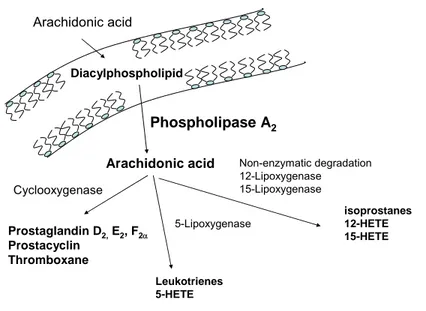
I.2.1.3. Arachidonic acid cascade
Arachidonic acid Phospholipase A2 Diacylphospholipid Arachidonic acid Cyclooxygenase 5-Lipoxygenase Non-enzymatic degradation 12-Lipoxygenase 15-Lipoxygenase Prostaglandin D2,E2, F2α Prostacyclin Thromboxane Leukotrienes 5-HETE isoprostanes 12-HETE 15-HETE
Figure I-5. Arachidonic acid metabolism
Two main enzymatic pathways for arachidonic acid metabolism have been highlighted : the cyclooxygenase (COX) pathway and the lipoxygenase (LOX) pathway. COX catalyses the production of prostaglandins D2 (PGD2), E2 (PGE2), F2α (PGF2α), prostacyclin (PGI2)
and TXA2 (Bergstroem, Danielsson et al. 1964; Hamberg and Samuelsson
1967; Hamberg, Svensson et al. 1975). 5-, 12- and 15-LOX generate respectively leukotrienes (Murphy, Hammarstrom et al. 1979; Samuelsson 1983), hepoxylins (Pace-Asciak and Martin 1984) and lipoxins (Serhan, Hamberg et al. 1984) (Figure I-5). Arachidonic acid can also be oxygenated by cytochrome-dependent monoxygenases. The physiological roles of the metabolites generated by this pathway still has to be clarified.
Although all mediators issued from arachidonic acid metabolism are important, we will focus on COX pathway and TXA2 biosynthesis.
Cyclooxygenase pathway O2 Arachidonic acid (AA) Prostaglandin G2 (PGG2) Prostaglandin H2 (PGH2) Prostacyclin (PGI2) Thromboxane A2 (TXA2) Prostaglandin E2 (PGE2) Prostaglandin F2α (PGF2α) Prostaglandin D2 (PGD2) Cyclooxygenase Prostacyclin synthase Thromboxane synthase Endoperoxyde F reductase Endoperoxyde E isomerase Endoperoxyde D isomerase COOH OOH COOH O O OH COOH O O OH COOH OH COOH OH COOH OH COOH OH O O O HO O HO HO HO O COOH HO
Figure I-6. Prostanoids biosynthesis by the arachidonic acid COX pathway.
Cyclooxygenases (or prostaglandin endoperoxide H synthases) are enzymes that catalyze the formation of PGs, TXA2 and PGI2. All
non-steroidal anti-inflammatory drugs act via COX enzymes inhibition.
Initially, it was thought that there was only one COX, first described by Bergstroem and his collaborators (Bergstroem, Danielsson et
al. 1964). In 1990, another type was discovered, leading to a distinction
to be discovered, is mainly characterized by a constitutive expression. It is expressed in many tissues and cell types, including platelets. Although initial studies concluded that COX-2 expression was inducible, it was recently demonstrated that the expression of COX-2 was also constitutive in some tissues such as kidney (Harris, McKanna et al. 1994), vascular endothelium (Cheng, Austin et al. 2002; McAdam, Catella-Lawson et al. 1999), or cortex (Yamagata, Andreasson et al. 1993)). COX-2 is also expressed in several pathological situations, including inflammation (Seibert, Zhang et al. 1994), and some cancers (colon (Taketo 1998),. liver (Shiota, Okubo et al. 1999), pancreas (Tucker, Dannenberg et al. 1999), lung (Wolff, Saukkonen et al. 1998), and breast (Hwang, Scollard et al. 1998)).
The arachidonic acid oxidation by COXs is a two-steps mechanism. First, COXs catalyse the oxydation and the ring closure of AA to form endoperoxide G2 (Figure I-6). In the second step, the endoperoxidase activity of COX allows the formation of the pivotal endoperoxide prostaglandin H2 (PGH2). Indeed, PGH2 is the common
substrate for the synthesis of all subsequent prostanoids, as described in Figure I-6. Consequently, the specificity of prostanoids formation is achieved by their specific enzymes (Table I-1).
Substrate Enzyme Product
PGH2 Prostacyclin synthase PGI2
PGH2 Endoperoxide D isomerase PGD2 PGH2 Endoperoxide F reductase PGF2α PGH2 Endoperoxide E isomerase PGE2
PGH2 Thromboxane synthase TXA2
Table I-1. Enzymes involved in prostanoid synthesis, using PGH2 as substrate
Non-enzymatic pathways
More recently, it has been highlighted that arachidonic acid could undergo non-enzymatic degradation, giving rise to active compounds, namely isoprostanoïds (Fam and Morrow 2003). The physiological and pathological implications of these products are of particular importance and their formation is increased upon oxidative stress. Several recent reports have proposed that TXA2 and some isoprostanes could indeed
act on the same receptors (Cracowski, Devillier et al. 2001). This effect could be useful for the understanding and treatment of several diseases where TXA2 receptor activation has been demonstrated.
I.2.1.4. Thromboxane synthase
In vivo, TXA2 synthesis involves coordinated action of a series of
enzymes, including phospholipases which release arachidonate from phospholipids, COX-1 or -2 which convert arachidonate into PGH2, and,
TXS catalyzes the isomerization of PGH2 into TXA2. This activity
was first described in platelets (Needleman, Moncada et al. 1976) and the enzyme was later purified and described as an hemoprotein belonging to the cytochrome P450 family (CYP) (Haurand and Ullrich 1985). This assumption was confirmed when the cDNA of the enzyme was cloned (Ohashi, Ruan et al. 1992; Yokoyama, Miyata et al. 1991). Human TXS was assigned as CYP 5A1. Unlike other microsomal CYPs that require the ubiquitous P450 reductase to shuttle electrons for the mono-oxygenation reaction, thromboxane synthase undergoes an isomerization reaction without reductase or molecular oxygen.
The TXS gene is transcribed as a 2.1-kb mRNA in hematopoietic cells, such as platelets, macrophages, monocytes and leukocytes, as well as in various tissues, particularly in lung, kidney, liver, spleen, prostate, placenta and thymus (Miyata, Yokoyama et al. 1994; Zhang, Xiao et al. 1997). The expression in these tissues is consistent with the biological activity of TXA2, and the cell type specificity to produce TXA2 is indeed
Figure I-7. Proposed mechanism for TXA2 formation by TXS, according to Wang et al.
(Wang and Kulmacz 2002)
The proposed mechanism for the isomerization of PGH2 into
TXA2 postulates that in the first step, TXS heme iron interacts with the
oxygen attached to C-9 of PGH2 (Figure I-7, (Hecker and Ullrich 1989)). Then, The TXS heme is proposed to undergo a redox transition from resting Fe(III) to Fe(IV) upon opening of the endoperoxide in Step 2 of the mechanism in Figure I-7. Oxygen- and carbon-centered radicals are predicted to occur during TXS catalysis (Steps 2 and 3 in Figure I-7). Nevertheless, the exact mechanism of subsequent steps remains elusive (Wang and Kulmacz 2002).
I.2.1.5. Thromboxane A
2degradation and Catabolism
TXA2 is a chemical unstable mediator and is rapidly hydrolyzed into inactive thromboxane B2 (TXB2) (Figure I-8). In vivo TXA2 half-life is
30 seconds.
Catabolism of TXA2 leads to the formation of about 20 inactive
compounds in different proportions in humans. The most important plasmatic products are TXB2 and its dehydrogenation product, 11-dehydroTXB2 (Catella, Healy et al. 1986). In urine, metabolites reflecting
TXA2 production are 11-dehydroTXB2 and 2,3-dinorTXB2 (Catella,
Lawson et al. 1987). A third less important metabolite has also been identified in urines, 11-dehydro-2,3-dinorTXB2 by Chiabrando et al.
(Chiabrando, Rivoltella et al. 1993)(Figure I-8).
O COOH OH O O COOH OH HO OH O COOH OH O OH O COOH OH O OH O OH COOH OH HO thromboxane A2 thromboxane B2 2,3-dinorthromboxane B2 H2O 11-dehydrothromboxane B2 11-dehydro-2,3-dinorthromboxane B2 β-oxydation Dehydrogenase β-oxydation
I.3. Thromboxane A
2receptor
The effects of TXA2 are mediated through its specific receptor.
The thromboxane receptor belongs to the most important superfamily of receptors : G protein-coupled receptors (GPCR), characterized by seven transmembrane domains receptors.
I. 3. 1.
Pharmacology of G protein-coupled receptors
Before the complete description of TXA2 receptor properties, we will briefly describe the general properties of GPCR function.
Signal transduction and regulation
These key families of proteins share a common molecular architecture consisting of seven transmembrane helices that are connected by three extracellular and three intracellular loops. GPCRs are characterized by the ability to couple to heterotrimeric G proteins which transduce a specific signal from a ligand-receptor interaction and act as effectors systems inside the cell. These proteins draw their name because they have a high affinity for GTP and are composed of three subunits : α, β and γ. The α subunit possesses a GTPase activity, since it is able to hydrolyze bound GTP.
When a receptor is stimulated by a cognate ligand, the Gα subunit of the G protein exchanges a molecule of bound GDP for GTP. The heterotrimeric protein becomes active until the GTPase activity hydrolyzes GTP for GDP thus turning off the system, which becomes available for a novel stimulation. Different classes of G protein exist and
are classified upon their properties as effectors but also on sequences homologies. To date, up to twenty Gα subunits have been described and are grouped into four major families, classified by their main functional properties (Nguyen Hwangpo and Iyengar 2005):
- Gαs : this G protein subunit family stimulates the activity of
adenylate cyclase, thus increasing cAMP levels ;
- Gαi/o : Gαi family (Gαi 1, 2 and 3) inactivates adenylate
cyclase and induces a decrease of cAMP levels. Gαo is mainly present
in the nervous system and modulate the opening probability of voltage-gated Ca2+ channels ;
- Gαq/11 : this family of Gα subunit (including Gα15 and 16) is coupled to the activation of PLC-β, which leads to the hydrolysis of phosphatidylinositol 4, 5-bisphosphate (PIP2) and to the production of IP3 and DAG (see also point I.2.1.2). The principal property of IP3 is
to release calcium ions (Ca2+) from intracellular stores. DAG acts as an
activator of protein kinase C (PKC) which regulates several processes inside the cell ;
- Gα12/13 : the specific role of these fourth family of G
proteins subunit remains elusive. They are abundant in platelets and have been related to several downstream pathways (notably small GTPase RhoA).
On the other side, Gβγ complex forms one functional unit and can affect downstream effectors as well and as much as the Gα subunits.
Agonist activationof a GPCR not only results in the G protein-dependent activationof effector systems, but also sets in place a series of molecular interactions that allows for feedback regulation of G protein coupling, receptor endocytosis, and signaling through G protein-independent signal transduction pathways. For example, RGS (regulation of G protein signaling) proteins accelerate the hydrolysis of GTP by G proteins α subunits, and hence increase the rate of recovery of the effector from activation by GαGTP or by Gβγ (Richman and
Diversé-Pierluissi 2005). GRK (G proteins receptor kinases) and β-arrestins are proteins able to desensitize the receptor after agonist stimulation by endocytosis or by uncoupling of the receptor from its cognate G protein (Luttrell 2005; Ma, Gao et al. 2005). Additionally, activation of kinases by GPCRs (PKC for example) can also regulate the signaling of distinct GPCRs and thus lead to heterologous desensitization.
Another important feature in GPCR signal transduction regulation is their ability to form dimers, heterodimers or oligomers. In the past ten years, the vision of GPCR acting as monomeric entities has quickly evolved. First evidences for the concept that this family of receptor could be able to form dimers or other oligomeric species were obtained in experiments where inactive mutant receptors recovered their binding and functional activity when cotransfected (Maggio, Vogel et al. 1993a; Maggio, Vogel et al. 1993b). Subsequent key steps in this theory were achieved when it was demonstrated that the metabotropic GABAB
was an obligatory heterodimer (Marshall, Jones et al. 1999) and opioids receptors could form heterodimers with new pharmacology (Jordan and
Devi 1999). The questions regarding this phenomenon remain controversial and are an active field of research.
Constitutive activity, inverse agonism and functional selectivity
Pharmacology of GPCR is complex and has quickly evolved during the past decade. We will briefly describe here the main basic principles of these receptors pharmacology.
The classical "receptor theory", evolved from the early days of experimental pharmacology to now and initially proposed that the interaction between a ligand and its receptor was able to elicit a functional cellular response (Rang 2006). Within this theory, a molecule able to "activate" the receptor protein was termed "agonist". It was soon observed that some chemical entities were able to counteract the agonist action, by displacing it from the active site of the receptor. These compounds, namely competitive antagonists, were though to be inert for the protein and to have a better steric affinity for the ligand binding pocket than the displaced agonist, thus preventing it to bind to and activate receptor.
Although this model permitted the fast development of modern pharmacology and was a powerful tool for elucidation of many questions regarding drugs action, it was rather imprecise. In several key papers from the Lefkowitz group in the early nineties it was evidenced that point mutation of a GCPR of the adrenergic receptor family were able to induce activity of the receptor, in the absence of bound agonist (Lefkowitz, Cotecchia et al. 1993). Beside, De Lean and co-workers proposed the concept of inverse agonism, whereas some antagonists may
be able to promote the dissociation of the receptor from G proteins (Wreggett and De Lean 1984). The later experimental confirmation of this concept, together with the discovery that non-mutated GPCR had basal activity, led to the finding of inverse agonists for a wide range of receptors. Indeed, many of the competitive antagonists in clinical practice were found to act as inverse agonists (Hill 2006). Consequently, recent model suggests that in the absence of any ligands, GPCR exists as a dynamic population of active and inactive form (Kenakin 2001). The receptor spontaneously crosses from inactive to active state and the nature of the ligand is indeed determined by its affinity for either active or inactive state. Thus, a compound with high affinity for active state will act as an agonist and induces this receptor species to redistribute in the system.
More interestingly, recent data suggested that ligands induce unique, ligand specific receptor conformations that can result in activation of distinct signal transduction pathways (Urban, Clarke et al. 2007). This phenomenon is generally reported to as "functional selectivity" and this change of perspective has been spurred by data emerging within the past decade in which ligands were shown to have quite diverse functional consequences mediated via a single receptor.
I. 3. 2.
Thromboxane A
2receptor gene and expression
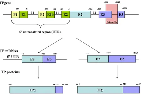
The TXA2 gene has been discovered in 1991 by Shuh Narumiyagroup (Hirata, Hayashi et al. 1991), using an oligonucleotide probe corresponding to its partial amino acid sequence. The cDNA was discovered in placenta and platelet-like MEG-01 cell lines and was found
to encode for a 343 amino acids protein, with seven putative transmembrane (TM) domains. Further work by the group identified that the gene was assigned to chromosome 19p13.3, and had three exons divided by 2 introns and 2 putative promoter regions (Nusing, Hirata et
al. 1993). In 1994, Raychowdhury et al. isolated a TXA2 receptor cDNA
from a human endothelial library. The predicted amino acid sequence revealed a structure of 369 amino acids, in which a novel cytoplasmic tail replaced the carboxyl-terminal portion of the previously characterized TXA2 receptor. The authors suggested that the mechanism for the
generation of the alternative transcript expressed in placenta was a failure to utilize a potential splice site, thus resulting in the encoding of a cytoplasmic domain that, in the endothelial TXA2 receptor, was an intron
in the early discovered TXA2 receptor (Raychowdhury, Yukawa et al.
1994)(Figure I-9).
Consequently, the two isoforms are identical with regard to their N-terminal 328 amino acid residues, but differ exclusively in their C-terminal cytoplasmic domains. The TXA2 receptor was named TP (for
Thromboxane Prostanoid), the two isoforms being subsequently termed TPα, for the initial placental peptide composed of 343 amino acid residues, and TPβ, for the 369 amino acid residues isoform initially found in endothelial cells (Coleman, Smith et al. 1994).
Further genetic analyses of the TP receptor gene confirmed the presence of two promoters, P1 and P2 and the functional regulation of TP expression by numerous transcription factors, P1 having the most important implication (D'Angelo, Davis et al. 1995; D'Angelo, Oliver et al. 1996; Kinsella, O'Mahony et al. 1994b). More recently, Coyle et al. have
demonstrated the existence of a third promoter, P3. This team suggested that expression of TPα and TPβ was subjected to differential regulation through the transcriptional activity of P1/P2 and P3, respectively, since their transcripts differed extensively in their 5’ untranslated region (Jourdan, Aguejouf et al.) sequences (Coyle, Miggin et al. 2002) (Figure I-9). Indeed, there are a number of genetic examples where protein/receptor isoforms arise through differential splicing, being products of the same gene, but may be under the transcriptional control of separate or distinct promoters. Several agents acting on P3 and on TPβ expression have been later identified, including PPARγ, a nuclear receptor playing a prominent role in diabetes, resolution of inflammation or cardioprotective effect (Coyle and Kinsella 2005; Coyle and Kinsella 2006; Coyle, O'Keeffe et al. 2005).
Intron 3b E1 E2 E3 E3 P1 P2 E1b E2 -289 -84 -199 -84 -83 +1 +786 +787 +984 +1642 +1221
5’ untranslated region (UTR)
E2 TPgene TP mRNAs E3 5’ UTR +1 +787 +984 E2 E3 +1 +787 I2 I1 I1 +1029 TP proteins TPα TPβ aa 1 aa 328 aa 343 aa 1 aa 328 aa 407
Figure I-9. Organization of thromboxane receptor gene. See text for details (adapted from (Coyle, Miggin et al. 2002)).
I. 3. 3.
Distribution of TP receptors
Before the discovery of the isoforms, northern blot analyses confirmed the existence of TP mRNA in human MEG-01 cells (platelet-like cell lines), placental and lung tissue (Hirata, Hayashi et al. 1991; Swanson, Lei et al. 1992), in human erythroleukaemic (HEL) cells (Kinsella, O'Mahony et al. 1994a), and in mouse thymus, lung, spleen, ileum, brain and kidney (Namba, Sugimoto et al. 1992; Sugimoto, Narumiya et al. 2000).
Regarding the specific expression of the isoforms, early studies considered that TPα was the platelet/placental receptor (Hirata, Hayashi
et al. 1991) whereas TPβ was the endothelial receptor, since it was initially
discovered in endothelial cells (Raychowdhury, Yukawa et al. 1994).
More recent studies have addressed the specific expression pattern of TPα and TPβ using specific detection of mRNA. For example, Hirata et al. have demonstrated the presence of both mRNA in platelets (Hirata, Ushikubi et al. 1996). Further study conducted in 1998 by Miggin
et al., used PCR technology and specific probes to demonstrate that TP
receptor was abundantly expressed at both the mRNA and protein level in tissues of relevance for TXA2 biology. These tissues and cell types
included erythroleukaemia cells, vascular and uterine smooth muscle, uterus and placental tissue, endothelium, epithelium, trophoblasts, thymus, liver and small intestine (all tissues studied within that study were from human). In that study, TPα was the most expressed isoform in nearly all tissues examined. Moreover, although TPα was expressed at approximately equal levels in all cell/tissue types analyzed, considerable
differences in TPβ mRNA expression were observed. Most strikingly, primary HUVECs (Human Umbilical Vein Endothelial Cells) were found to express low levels of TPβ and approximately 6-fold greater levels of TPα than TPβ (Miggin and Kinsella 1998).
More confusion was added in 1999 when a study conducted by Habib et al demonstrated the absence of TPβ in human platelets using specific antibody for both isoforms. The authors estimated that ≥ 50 fmol/mg of protein of TPβ could be detected with the specific antibodies, and suggested that TPβ was expressed at very low levels in platelets, if any (Habib, FitzGerald et al. 1999).
Collectively, these data point out that the relative expression of both isoforms proteins at the cell surface remains elusive, although one can consider that TPβ has probably a non-significant role in TXA2 action
on platelets.
I. 3. 4.
Pharmacology of TP receptors
Abundant work has been reported on TP receptors pharmacology since the discovery of TXA2. This section reviews the main discoveries and concepts related to TP receptors.
I.3.4.1. Pharmacological characterization
Early pharmacological studies and the two subtypes paradigm
Before the discovery of the human TXA2 receptor gene, several
of binding studies, the TP receptor had been extensively characterized (Hall 1991), using both radiolabelled high affinity antagonists as well as stable TP agonists. Inhibition of specific binding correlated well with affinities predicted from biological activity, suggesting that the binding was meaningful. It is noteworthy that before the prostanoïd receptors nomenclature (TP, IP, DP, etc) the TXA2 receptor was called the
TXA2/PGH2 receptor since early studies had characterized PGH2 as an agonist for TP receptors (Halushka 2000).
Following the first experiments, a controversy over the existence of pharmacologically distinct TP receptors on human platelets and vascular smooth muscle rapidly arose (Halushka 2000). Indeed, respectively in 1980 and 1981, possible existence of distinct platelet and vascular TP receptors was suggested by data produced by Lefer et al. (Lefer, Smith et al. 1980) and LeDuc et al. (LeDuc, Wyche et al. 1981). Additionally, Mais et al. (Mais, DeHoll et al. 1988; Mais, Dunlap et al. 1985), using a series of 13-azapinane TXA2 analogs, provided strong
evidence for the existence of receptors subtypes in vascular tissue and platelets, both issued from human species. Masuda et al. (Masuda, Mais et
al. 1991), using the same compounds, conducted competition radioligand
binding studies in rat and human platelets and cultured rat aortic smooth muscle cells. They found statistically significant differences in the rank order of potencies for these compounds in rat platelets compared to the smooth muscle cells, further supporting the notion that the platelet and vascular receptors represented unique subtypes.
It was also suggested at the time by Morinelli et al. (Morinelli, Niewiarowski et al. 1987) that platelets contained at least two TP receptor
subtypes. This was later supported by Takahara et al., who postulated that platelets contained two TP receptor populations, one which mediated platelet shape change and the other aggregation (Takahara, Murray et al. 1990). Similarly, GR32191, a TXA2 receptor antagonist (vide infra, point
I.4.2.2.A., p. 61) was demonstrated to bind to two specific binding sites on human platelets (Armstrong, Humphrey et al. 1993a; Armstrong, Humphrey et al. 1993b), although further competition binding experiments suggested that these sites were identical (Armstrong and Wilson 1995).
Radioligand binding studies subsequently demonstrated different affinities in platelet and vascular TP receptors for the agonist ligand [125
I]-BOP (Dorn 1989; Morinelli, Okwu et al. 1989). Additionally, Morinelli et
al. (Morinelli, Niewiarowski et al. 1987) demonstrated that the TXA2
agonist, U46619, produced shape change and myosin light chain phosphorylation in platelets (for complete description of platelet aggregation, see point I.3.6.1.A, p. 36) with an EC50 value one-tenth of
that for inducing aggregation, secretion and mobilization of calcium. Dorn subsequently demonstrated that [125I]-BOP also bound to platelet
TP receptors with a high and low affinity state (Dorn 1989). That group provided additional evidence to support the notion that shape change and aggregation were coupled to different receptors (Dorn and DeJesus 1991).
One receptor and two isoforms
Nevertheless, despite the evidence in support of distinct platelet and vascular TP receptors there were several reports refuting that notion. Swayne et al. (Swayne, Maguire et al. 1988) using a series of structurally
dissimilar TP receptor antagonists reported that they could find no evidence to support the notion that the platelet and vascular TP receptors represented unique subtypes. Mihara et al. (Mihara, Hara et al. 1989) failed to find any differences in the binding of receptor agonists and antagonists between platelets and vascular tissue in the pig. Hanasaki et al. (Hanasaki, Nakano et al. 1989) also failed to find any differences in rank order potency for a series of TP receptor agonists and antagonists in rat platelets and aortic smooth muscle cells.
The discovery of the TXA2 receptor gene by Hirata et al. (Hirata,
Hayashi et al. 1991) and the existence of two isoforms generated by alternative splicing (Raychowdhury, Yukawa et al. 1994) shed new light on the controversy. Indeed, genetic and molecular biology showed that there was only one gene coding for the receptor and that the two products only differed in the Carboxyl-terminal tail, leaving the putative ligand-binding site identical.
Moreover, most of the studies that characterized several subtypes for TXA2 receptors used tissue from different species. In 1992, Ogletree et al. showed that there were significant differences between TP receptors
among several species (Ogletree and Allen 1992), although in the same species, the receptors were quite similar. Moreover, the conclusions of those studies using human tissues have to be taken cautiously since they have been conducted on whole organs (Mais, DeHoll et al. 1988).
In 1998, Djellas and co-workers (Djellas, Antonakis et al. 1998) shed some light on the question of the importance of the interactions of TP receptors with G proteins in physiological conditions. They explored a possible molecular mechanism for platelet synergism that is commonly
seen between some aggregating agents in human platelets, for example thrombin and TXA2. They demonstrated that thrombin caused an increase in the affinity for ligands by the TP receptors and that this increase seemed to be due to a raise in the amount of Gαq associated
with the TP receptors. That Gαq can amplify the affinity of TP receptors
for its ligands was further supported by the observations of Allan et al. who found that co-transfection of Gαq or Gα13 with the α isoform of TP
receptors into COS-7 cells resulted in a significant increase in I-BOP affinity (Allan, Higashiura et al. 1996). Similarly, Becker et al. demonstrated that co-transfection of Gα13 with the β isoform increased the affinity for several agonists and also decreased the affinity of both isoforms for several antagonists (Becker, Garnovskaya et al. 1999). Consequently, consistent with recent development of GPCR pharmacological properties (see I. 3. 1., p. 15), Halushka proposed that differential coupling of TP receptor isoforms could change the affinity of the receptor for agonist or antagonist ligands, and could give a possible explanation for apparent TP receptor subtypes (Halushka 2000).
Besides, the presence of two binding sites coupled with two effectors systems has found some possible explanation recently, with the deep exploration of the TP receptor role in platelet aggregation. These mechanisms will be presented extensively at point I.3.6.1.A., p. 36.
I.3.4.2. Signal transduction by TP receptors
Signal transduction by TP receptors can lead to activation of many intracellular mechanisms. At least nine G proteins, Gq, Gi2, Gs, G11,
Gα12, G13, G15, G16, and Gh were demonstrated to some extent to couple
to TP receptors. Coupling with these G proteins will eventually evoke an increase in intracellular Ca2+ concentration ([Ca2+]
i), activation of PLC,
generation of IP3 and DAG, activation of PKC, stimulation of myosin
light chain kinase, exposure of GPIIb/IIIa binding sites, and intracellular alkalinization of platelets (Halushka 2000).
The most relevant signalling processes of TXA2 are extensively
described in section I.3.6.1 which details the physiology of TXA2 with an
emphasis of its roles in platelet aggregation.
I.3.4.3. TP receptor desensitization
Platelet TP receptors are known for a long time to undergo potent agonist-induced desensitization (Liel, Mais et al. 1988) and phosphorylation (Okwu, Ullian et al. 1992). It has subsequently been demonstrated that both TPα and TPβ receptors were subject to homologous, agonist-induced desensitization in transfected HEK293 cell lines (Habib, Vezza et al. 1997). This desensitization involves several mechanisms, including activation of PKC (Spurney 1998) and GRKs (Flannery and Spurney 2002).
Differences in the carboxy-terminal tail between the two isoforms and thus differences in sites for phosporylation imply that TPα and TPβ might undergo differential homologous/heterologous desensitization. For example, some studies indicated that TPβ, but not TPα, can undergo agonist-induced but also tonic internalization in a GRK- and arrestin-dependent manner (Parent, Labrecque et al. 2001; Parent, Labrecque et al.
1999). It is noteworthy that this is in contrast to what was reported for HEL and CHRF-288 (megakaryocytic) cells that express predominantly the α isoform and were found to undergo internalization (Dorn 1992; Parent, Labrecque et al. 1999).
Heterologous desensitization of TXA2 receptors involves many
other receptor/mediator systems, including subtype 1 of PGE2 receptors (EP1) and PGF2α receptors (FP) (Kelley-Hickie and Kinsella 2004).
Additionally, nitric oxide (NO) (Reid and Kinsella 2003) and stimulation of PGD2 receptors (DP) (Foley, Kelley et al. 2001) have been shown to
selectively desensitize TPα. Finally, PGI2/IP mediated heterologous
desensitization through cAMP has been widely documented (Manganello, Djellas et al. 1999; Murray, Shipp et al. 1990) and several data indicate that TPα, but not TPβ was subject to IP-receptor-induced desensitization in a protein kinase A (PKA)-dependent pathway (Walsh, Foley et al. 2000). The PGI2/TXA2 system will be described extensively at point I.3.6.2
(p.45).
I.3.4.4. TP receptor dimerization
Many GPCR have been demonstrated to function as homo-, hetero- or oligomers (see I. 3. 1. p.16). Different works performed on TP receptors have subsequently tried to evidence this phenomenon for TP receptors. In 2005, Laroche et al. demonstrated that TPα and TPβ could form homo- and heterooligomers in an agonist-independent process and suggested that dimers/oligomers constituted the functional unit for the receptor. Interestingly, in that study, TPα which does not undergo
constitutive or agonist-induced endocytosis on its own was subjected to both types of endocytosis when co-expressed with TPβ, indicating that TPα could display intracellular trafficking through heterodimer when complexed with TPβ (Laroche, Lepine et al. 2005). This homo/hetero oligo/dimerization of TP receptors was later confirmed by two studies which showed that TPα/TPβ dimerization enhanced isoprostane (Wilson, McGinley et al. 2007) but lowered U46619 (Sasaki, Miyosawa et
al. 2006) mediated signal transduction. Thus, these data indicate that TP
signaling could be dependent of the receptor isoforms heterodimerization.
Additionally, Wilson et al. postulated and demonstrated that TPα and IP formed functional heterodimers in an agonist-independent manner. Thus, IP/TPα dimerization was coincident with TP-cAMP generation, promoting a "PGI2-like" cellular response to TP activation. Moreover, they demonstrated that IP/TPα interaction permitted reciprocal regulation of receptor endocytosis via the trafficking pathway determined by the activated dimeric partners. These results represented a possible mechanism by which PGI2 and IP receptor may limit the cellular
effects of TP receptors activation (Wilson, Dowling et al. 2007; Wilson, Roche et al. 2004).
I.3.4.5. Ligand-binding site
Although many ligands for TP receptors have been synthesized, the conformation of TP receptor remains elusive. Since the TP receptor belongs to the seven transmembrane-spanning class of receptors, it was
originally proposed that thebinding domain of this receptor might also reside in TM7 (Hirata, Hayashi et al. 1991). Other investigators (Narumiya, Hirata et al. 1993) proposed that while TM7 interacted with the ligand carboxyl group, separate transmembrane regions also participated in ligand binding, i.e. TM3 coordinated with the prostanoid ring, and the TM4 and TM5 regions interacted with the alkyl chains.
More recently, site-directed mutagenesis studies have investigated the ligand coordination site for TP receptors. Specifically, Funk et al.
(Funk, Furci et al. 1993) obtained four mutants with point mutations at TM7of the TP receptor, i.e. between Leu291 and Trp299. Three of these
mutants completely lost binding activity to bothantagonists and agonists. Although the fourth mutant, W299L, didnot recognize the TP receptor antagonist SQ29548, it was ableto bind with two different TP receptor agonists with the same affinitiesthan those observed with the wild type receptor. In addition, Chianget al. (Chiang, Kan et al. 1996) reported that
mutations of S201A and S255A at TM5and TM6, respectively, caused altered affinity to the agonistI-BOP but had no effect on the antagonist SQ29548 binding. Separatestudies by Dorn et al. (Dorn, Davis et al. 1997) used receptor chimeras to evaluate ligand binding activity. They concluded that residues in TM1 constitutedan important portion of the TP receptor binding site. Finally, reports from two different groups suggested that the putativedisulfide bond between Cys105 and Cys183/184 in
extracellular domain (ED) 2 and ED3, respectively, played a critical role in receptor-ligandbinding. In particular, mutants C105A and C183A from the humanplacenta TP receptor (Chiang, Kan et al. 1996) and mutants C105S and C184S from humanK562 TP receptors (D'Angelo, Eubank et
al. 1996) did not show any binding activity for either agonists or antagonists. In addition, both groups reported that Cys102, which is
conserved in most seven transmembrane-spanning receptors including the TP receptor (but absent in other prostanoid receptors),also played an important, yet unspecified role in ligandbinding.
In 1993, Yamamoto et al. (Yamamoto, Kamiya et al. 1993) proposed a receptor model where the ligand binding pocket of the TXA2 receptor included a serine residue from segment V, an arginine residue from segment VII, and a large hydrophobic pocket between these two residues. More recently, Turek et al. (Turek, Halmos et al. 2002), using SQBAzide (a newly synthesized biotinylated photoaffinity probe (Halmos, Turek et al. 1999)) determined that extracellular loop II was critical for ligand binding. Subsequent work by this group led to the conclusion that five key amino acids within this region (Phe184, Thr186,
Ser191, Asp193, and Ser201) participated in TP receptor ligand binding and
function (Khasawneh, Huang et al. 2006).
Using NMR (nuclear magnetic resonance) techniques, some studies determined that the domain within the second extracellular loop and the disulfide bond between the first extracellular loop and the extracellular loop 2 played a major role in forming the ligand recognition pocket (Ruan, Wu et al. 2004; So, Wu et al. 2003).
These collective data of several studies showed that transmembrane domains I, III, IV and VII as well as extracellular loop II and III were involved in ligand binding interaction (Chiang, Kan et al. 1996; D'Angelo, Eubank et al. 1996; Funk, Furci et al. 1993; Hirata, Hayashi et al. 1991; Narumiya, Hirata et al. 1993; Ruan, So et al. 2001;
Yamamoto, Kamiya et al. 1993). One could argue that all these models gave contradictory results. Nevertheless, there is no clue that one model excludes another one, given the important number of ligands (different chemical structures, agonists, antagonists, etc) and the methods used in these studies. Indeed, there is an obvious lack in 3D structure data for TP receptors, given the absence of absolute crystalline conformation.
I. 3. 5.
Physiological significance of TPα and TPβ
The physiological significance for the presence of two isoforms of the TP receptor is still unclear. Nevertheless, several teams tried to decipher the exact role and function of the two isoforms.
Alternative splicing that generates receptor isoforms in the carboxyl-terminal tail is not only found for TP receptors. Among the prostanoid receptors family, mRNA splice variants have been identified for the EP1, EP3 and FP (Pierce and Regan 1998). Interestingly, except
for the EP1 receptors, the mechanisms giving rise to these receptor isoforms involves the use of splice sites located in the cytoplasmic carboxyl-termini of these receptors. Thus, the eight human EP3 isoforms
that have been identified are otherwise identical except for their carboxyl termini. Similarly, the optional use of a potential splice site encoding the carboxyl-terminus gives rise to each of the two FP isoforms. Because the carboxyl-termini of GPCR are generally implicated in interactions with G proteins, it is not surprising that these receptor isoforms differ mainly with respect to their activation of second messenger pathways and not in their pharmacological characteristics. Differences also exist with respect to their levels of constitutive activity and to their desensitization.
Besides its predominant role in platelet aggregation which will be described elsewhere (I.3.6.A. p. 36), TPα has also been proposed to be relevant for vascular homeostasis. Indeed, in 2000, Walsh et al. established that TPα, but not TPβ, was subject to cross-desensitization by IP mediated through direct PKA phosphorylation of the receptor at Serine 329 and the authors concluded that TPα might be the isoform physiologically relevant to TP:IP-mediated vascular homeostasis (Walsh, Foley et al. 2000).
More recently, Ashton and Ware discovered that TPβ was required for VEGF-induced endothelial cells migration and angiogenesis. Consequently, they proposed that selective inhibition of TPβ isoform could enhance myocardial revascularization after infarction (Ashton and Ware 2004).
Collectively, consecutive data resumed and presented in this section indicate that our knowledge of the TP receptor isoforms is still far from complete. The work required for the elucidation of isoform-specific physiological and pharmacological properties has still to be done.
I. 3. 6.
Physiological and pathological implications of
TXA
2TXA2 has an important but diversified role in organism. This part
of the general introduction will describe the TXA2 actions and their
I.3.6.1. Physiology of TXA
2I.3.6.1.A. Hemostasis
In this section, we will briefly describe principal mechanisms of platelet aggregation and will focus on the interest of TXA2 and its major role in hemostasis, platelet activation and aggregation.
Hemostasis literally means (Hemo = blood and stasis = the stop of flowing of blood). At site of vascular injury, thromboresistant endothelium is disrupted and prothrombotic subendothelial vessel wall constituents (eg, collagen) are exposed to blood. Three consecutive phenomena may then occur and their common action eventually conducts to hemostasis :
- vascular constriction ;
- platelet aggregation and plug formation (primary hemostasis) ;
- blood coagulation (secondary hemostasis) ;
Formation of fibrous tissues in the blood clot is sometimes considered as the fourth hemostasis step. It occurs at the cicatrisation and is responsible for definitive obturation of the lesion.
Vascular constriction
Vascular constriction allows sparing blood loss. An important hemorrhage can undeniably have dramatic consequences. Vascular spasm can persist from minutes to several hours and permits the coagulation system to be effective. The origin of this vascular reaction can be
multifactorial, but mainly a consequence of the liberation of several mediators from the wounded endothelium or the activated platelets.
In small vessels, platelets are directly responsible for the vasospams, after secretion of TXA2 which has potent vasoconstrictor
properties besides its proaggregant action. These physiological characteristics will be discussed further in this work.
Platelet aggregation and plug formation (primary hemostasis)
General considerations
Platelets are fundamentally involved in the physiological process of hemostasis. Platelets participation occurs in diversified ways including adhesion to the cut end of vessel or subendothelial components, activation, shape change and secretion, and formation of large platelet aggregates. Although there is no clear separation between these phenomena, we will describe them successively in this chapter.
Platelet adhesion and activation
Under normal conditions, platelets circulate in the blood flow freely as small discoid bodies and do not adhere to normal vascular endothelial cells. Cut end or wound of a vessel provides numerous binding sites in the subendothelial matrix for resting platelets, including collagen and von Willebrand Factor (vWF), a multimeric adhesive protein associated with collagen in the vessel wall. The platelet adhesive receptors include some members of the integrin family, such as the fibrinogen/fibronectin receptor, integrin αIIbβ3, and the collagen receptor, integrin α2β1, but also the glycoprotein Ib-V–IX complex (GPIb-V–IX),