Trace Element - Analytical Chemistry in Medicine and Biology, Vol. 3 Editors: P. Bratter, P. Schramel
© 1984 Walter de Gruyter & Co., Berlin· New York - Printed in Germany
SERUM TRACE ELEMENTS IN HEMOPROLIFERATIVE DISEASES.
Yves Beguin (1), Jeanne-Marie Delbrouck, Iwan Roelandts, Georges I,eber (2)
Department of Medicine and Department of Experimental Nuclear Physics,
University of Liège, Sart-Tilman B 15, B-4000 Liège, Belgium.
Research Assistant (1) and Research Associate (2) of the National Fund for
Scientific Research (Belgium).
Abstract
The PIXE method has been used to compare a reference group (N=100) and
eight groups of patients suffering from hematological malignancies
(Hodgkin's disease, lymphosarcoma, chronic lymphocytic leukemia, acute
lymphocytic leukemia, acute granulocytic leukemia, chronic granulocytic
leukemia, myeloproliferative disorders and myeloma) (N=140).
The serum Cl, K, Ca, Fe, Cu, Zn, Se and Br contents have been measured.
Significative differences were noted in several groups. A statistical
analysis has been performed to correlate these trace element values with
several clinical and biological parameters such as blood cells counting,
clinical stage, histology, inflanrnatory tests, ...
Introduction
Trace elements metabolism has already been extensively studied in various
medical fields (1, 2). In hematology, many authors have reported on serum
copper increase in Hodgkin's disease and lymphosarcoma and return to
normal values when complete remission is achieved (3, 4, 5, 6). Much less
has been described about other trace elements, especially about bromine
who se
role in human metabolism remains obscure.We report here on our experience with serum trace elements dosage and confirm our previous conrnunication (7) on abnormal values in several hemoproliferative diseases.
Trace Element - Analytical Chemistry in Medicine and Biology, Vol. 3 © 1984 Walter de Gruyter & Co., Berlin· New York - Printed in Germany
Patients and methods
The PIXE method has been used to measure the serum content of chlorine
(Cl), potassium (K), calcium (Ca), iron (Fe), copper (Cu), zinc (Zn),
selenium (Se) and bromine (Br), in normal subjects and in patients
suffering from hemoproliferative diseases.
1)
Normal subjects. This group was composed of
100healthy subjects
taking no drug. Although this group was rather younger than our patient
group, no significant difference with age was noted in trace Element
values. We found a statistically significant difference between sexes
in calcium
(103in male versus
100 ug/mlin female, p
=
0.03),copper
(1.05
in male versus
1.17 ug/mlin female, p
= 0.02),zinc
(1.14in
male versus
1.01 ug/mlin female, p
<
0.001).2)
Patients.
319samples were taken from
150patients, many of them being
studied on several occasions, bath in active disease and in remission.
They are divided up as follows : myeloma (MY:
20),Hodgkin's disease
(HO: 12),
lymphosarcoma (lS :
28),chronic lymphocytic leukemia (Cll:
26),
acute lymphocytic leukemia (All :
6),acute myelocytic leukemia
(Ai'll:
26),chronic myelocytic leukemia ((Ml:
11)and
myeloprolifera-tive disorders (MPS :
21).We also studied serum trace elements values according to clinical
status and sorne biological parameters usually regarded as representative
of disease activity. So blood cell counting, erythrocyte, sedimentation
rate, fibrinogen, serum lactate deshydrogenase (LDH) and serum proteins were recorded on the same day as the trace Elements.
Thirteen patients presenting with newly diagnosed or relapsing AMl and
undergoing intensive induction chemotherapy, have been serially
followed for up to three months. The chemotherapy regimen generally
consisted in a seven-day course associating high doses of daunomycine, vincristine and cytosine-arabinoside. Samples were taken before treatment and then about twi ce a Itleek.
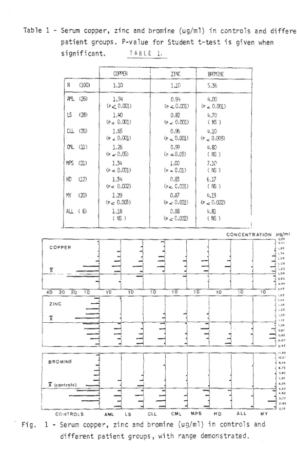
Table 1 - Serum copper, zinc and bromine (ug/ml) in controls and different patieht groups, P-value for Student t-test is given when
significant. ïABLE 1. Ccm:R
,
Z1ff c 1 ,1 ,e N (lOO) 1.10 1 l.K 536 1,
Ii'1L (25) 1,3" 1 0.94 q,OO (p< 0,001) (p..::.O,o]ll Cp ... 0.0)1) LS (28) 1,LiO,
0,82 1 4,70 (p", 0,(01) 1 Cp.: 0,001) ( NS ) Cll (25) 1.55 1 0,95 UD (P ... 0,(01) Cp <. O,OOll (p", 0,005) mL (lil 1,25 o,g::: 1.;,SJ (p .:: 0,(5) 1 Cp .::.0,05) ( NS ) t"IPS (2]) l ,3~ 1 l,CO , 7.10 (P.:: D,COl) (P .... O,OD (NS) HD (2) 1.34 1 (1.83 5,lI (PO:: 0,0...,"2) Cp..:... C,O:::1) ( NS ) MY (10) 1,29 0,87 4,13 (Pc 0,(03) (p..:O,CDll i CP.:: 0.0J21 1 Ali ( 5) 1.18 1 0,88 1 4,8} ( NS ) 1 CP ~ C,\D2) ( NS ) CDNCENTR ATION J'9/ml 11
1~
ll
1 ~ ~,~~ 1 COPPER ~ ~!
~ ;.9' l 1 '."--1
~
-,
-lJ
l"
l ,
..J ~1
---1
1-1
-j
J -,r
-
i
4
~ :i ,.06 1j
1, ".,l
j [).O> 1 10.d 40 30 20"
'0
'0
10 10 "'i 0 10 00 10 """',
! ", ZiNC"
1 .' " ~ 1 - - l~
l
j ~ ,."x
--1 1,
,",0 1 - - - ' ','",
~J
...,
'[ ,...J j'.O'---"1
:j
-j
i-j
1l,
~ ~:::-l
J
:::j
-j
4 1 (.J
1
:::~ "1 1 1 1 i !'Cy BROMINE..1
1 1 11
~..
"J
l
1 ~ •. ro~
ij
j
j
i'·""
"
1x
(CO"''''·)9
.JJ
::~: 1---1
l,
j
~j
11
1 ----=::i "., - !j
--i
::~~ !- l
.Jl
1l
d
~:l
.J1
~."' i"
-{ VO C(!~TROLS AML LS CLL CML MPS HD AL L MYFi g. 1 Serum copper, zi n c and bromine (ug/ml) in controls and
Results
The mean val~es for copper, zinc and bromine in normal subjects and in
patients are listed in Table 1. P-values for student t-test between controls and patients are mentioned when significant. Ranges are shawn in Figure 1, but sorne very high values for bromine are outside this pi cture.
Copper was always increased when compared with controls, and this reached
statistical significance in all groups but ALL which was too small. Zinc
was significantly reduced in all patient groups, though the absolute
difference wasntt important in every groups.
Bromine displayed sorne less systematic pattern. Tt was significantly
decreased in AI-IL, CLL and myeloma, but in A'IL this feature is dependent
upon treatment (see below); it was slightly reduced in lymphosarcoma, (ML and ALL, and slightly enhanced in HD and MPS, but in these groups most values \vere in fa ct very 10w and the mean artificially normalised by very high values.
Selenium showed no difference neither in absolute value nor in range. Chlorine and calcium remained almost constant tram group ta group. Potas-sium was often increased, reflecting extensive cellular turn-over. Iron was sometimes reduced, mostly in CLL and LS groups vlhe}~e copper reached the highest leveis.
Because of the small size of sorne groups, one must be careful in inter-preting apparent correlations betltleen trace Elements and biological para-meters. SA we shall report only on the most important ones. t~ithin the patient group as a whole, copper is correlated with white blood cell counts, inflammatory tests, serum LDH and potassium; zinc disclosed a weak correlation with calcium, iron and selenium, and bramine It,:ith potas-sium; potassium was related ta whale blood cell counts 1 serum proteins
and chiorine; calcium increased with serum proteins, chlorine, potassium and selenium.
In CLL, capper was correlated with l,SC counis (R=O.71, P < 0.001), serum
LDH (R=0.796, P
<
0.001), ESR (R=0.504, P<
0.05) but not with fibrinogen;on the opposite, selenium was inversely correlated with WBC counts (p '" 0,01). In lymphosarcoma, copper shOl'/ed significant correlation with
~n ; Days o • , '0 " o.
=
COPPER (j.l9/m1j C' Nl
BROMINE (jJ9/mll C' N B 6 4,
aFig. 2 - Capper and bromine Evolution in patients undergoing chemo-therapy for ANL? Mean values + Cf are given with comparison with A~L in complete remission (CR) and normal controls (N).
FEB_84 MAR APP
Fig. 3 - A typical pattern of bromine! capper and zinc Evolution during chemotherapy in a 20 year old >!oman with l'ML.
ESR and fibr;nogen! and inverse relationship with hemoglobin.
The A~L patients undergoing intensive chemotherapy bore a characteristic
evolution (Figure 2). The mean values before treatment were 5.79 + 2.07
ug/ml for bromine and 1.57 ~ 0.37 ug/ml for copper. Bromine dropped
dramatically to 2. 74 ~ 1.45 on day 14 and 2.10 ~ 0.48 ug/ml on day 28; the
level remained low for several weeks and then progressively reached normal pre-treatment values. Copper displayed a less constant pattern but usually eut down during chemotherapy and thereafter reaugmented to significantly a
higher level (1.79 ~ 0.43 ug/ml on day 21). A few weeks later, while in
complete remission, dosage reduced to 1.21 ~ 0.28 ug/ml, thus only
slight-ly more than in controls. A typical pattern is presented in Figure 3 with a 20 yeal" old woman undergoing three chemotherapy courses for refractory PML.
Cane lus ions
1) Zinc ~s systematically reduced in hemoproliferative diseases. ïhis has
not 2\·;2.Y5 ~€en confir:71ed (8) and doesn't seem ta be related ta
nutri-tion s-:atus~ treat::lent or other biological abnomalies. It may be due ta t~ss:.;:ar cat2!bolism in cancer patients (1 g 2).
2) Copper is usually enhanced in our patients but considerable overlap
with :1:Jrma: values prevents us fram its excessive ciinical use.None-the'I€SS~ we found a significant correlation between copperj and
infl,a!7i~atory tests in LS (3-6) ~ and WBC chiefly in CLL.
3) Brom~r;e remair,$ misunderstood in human metaboiism. Although we observed a genera"1 tendency tO\.<Jards decreased values in patients ~ very high values were noted~ that viere not reiated ta any l3€culiar disease status nor ta drug intake. The fan noticed in AML chemotherapy \I/as independent of digestive Tolerance ta disease and treatment. This l,<Jas supported by the absence of such a phenomenon in patients undergoing a slimming fast (unpublished data). This drop seemed to be due to chemotherapy itself.
4) Selenium is much the same in our patient groups as in controls. But measured concentration is very low and errors may be relevant.
Bibliography
1. Prasad, A.S., Ed. : Trace Elements in Human Health and Disease,
Academi c Press, New York, 1976.
2. Browner, F., Coburn, J.W., Ed. : Disorders of Mineral Metabolism, Vol. 1., Trace Elements, Academic Press, New York, 1981.
3. Hrgovcic, ~1.J., Schullenberger, C.C., Ed. : Copper and lymphomas, CRC Press, Bora Raton, Florida, 1984.
4. Shah, I., Lewkow, L.M., Khilawani, U. : Cancer 51,851-854 (1983). 5. Wilimas, J., Thompson, E., Smith, K.L. : Cancer 42, 1929-1935 (1978). 6. P.sbjarnsen, G. : Scan. J. Haematol. 22, 193-196 (1978).
7. Weber, G., Robaye, G., Bartsch, P., Collignon, A., Beguin, Y., Roelandts, I., Delbrouck, J.M. : Nucl. Instr. Meth. (In the press) (1984) .
8. Gobbi, P.G., Scarpellini, M., l>1inoia, C., Pozzoli, L., Perugini, S. Haematologica 63, 143-155 (1978).