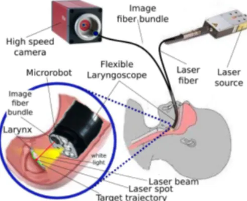
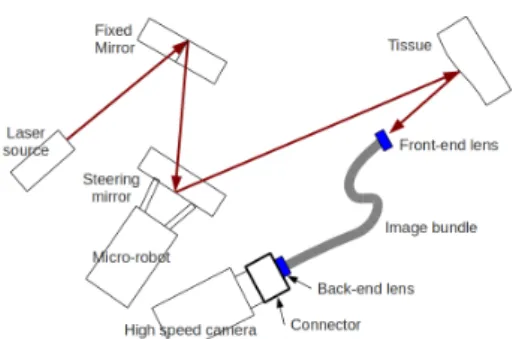
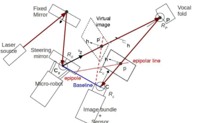
Epipolar geometry for vision-guided laser surgery.
Texte intégral
Figure
Documents relatifs
This paper proposes a review of the existing fast ICP methods and places emphasis on a recently proposed solution that combines the neighbor search algorithm with a
As parameters of the spacecraft at the moment of recording by an optical device are unknown, the problem of the fundamental matrix determination using the
included the theoretical framework, and chapter II of the concept and principles of vocational training and its importance and problems, Chapter III allocated how to build
The obtained pair of projection matrices has made possible, in accordance with specifically proposed steps of the methods 2a and 2b, to obtain a metric
The z-position (in the frame of the current position of the endoscope) of each point on the surface of the organs is individually modelled as a Gaussian distribution (mean and
To set the device in the field: (i) choose an unobstructed point of view and evaluate a typical distance ( D typ ) and distance range ( D min to D max ) at which animals move;
To this end, we propose three calibration subsystems: one automated subsystem to move radioactive sources around the detector, whose main role is to calibrate the energy scale in
Point cloud registration is used to build a new representation, which contains source models from the stereo camera and LIDAR and will be used to correct the output scene.. The
