HAL Id: tel-00771452
https://tel.archives-ouvertes.fr/tel-00771452
Submitted on 8 Jan 2013
HAL is a multi-disciplinary open access
archive for the deposit and dissemination of sci-entific research documents, whether they are pub-lished or not. The documents may come from teaching and research institutions in France or abroad, or from public or private research centers.
L’archive ouverte pluridisciplinaire HAL, est destinée au dépôt et à la diffusion de documents scientifiques de niveau recherche, publiés ou non, émanant des établissements d’enseignement et de recherche français ou étrangers, des laboratoires publics ou privés.
a feeder-cell-free DL-4 system
Christian Reimann
To cite this version:
Christian Reimann. In-vitro Generation of potent T-lymphoid Progenitors in a feeder-cell-free DL-4 system. Human health and pathology. Université René Descartes - Paris V, 2012. English. �NNT : 2012PA05T033�. �tel-00771452�
P
HD
D
ISSERTATIONU
NIVERSITÉP
ARISV
-
R
ENÉD
ESCARTESÉ
COLED
OCTORALE:
GC2
ID
-
G
ÉNÉTIQUE,
C
ELLULES,
I
MMUNOLOGIE,
I
NFECTIOLOGIE,
D
ÉVELOPPEMENTSpecialty: Immunology
Presented by
Christian REIMANN
To obtain the PhD degree from
Paris Descartes University
I
N VITRO
G
ENERATION OF POTENT
T-
LYMPHOID
P
ROGENITORS
IN A FEEDER
-
CELL
-
FREE
DL-4
SYSTEM
This PhD work was performed under the direction of Dr Isabelle André-Schmutz
At INSERM U768 : Développement Normal et Pathologique du Système Immunitaire
Hôpital Necker Enfants Malade, Paris
Thesis defended on November 19th, 2012 Thesis -Jury:
Mme Anne GALY President of the Jury
Mme Isabelle ANDRÉ-SCHMUTZ Thesis director
Mr Christophe BENOIST Reviewer
Mr Jean SOULIER Reviewer
Mr Alain FISCHER Examiner
INDEX
SUMMARY 8
RESUMÉ 10
ZUSAMMENFASSUNG 12
INTRODUCTION 14
1 T cell reconstitution after HSCT 15
2 Potential strategies to enhance immune reconstitution after HSCT 18
2.1 Acceleration of Thymus independent T cell recovery 18
2.1.1Allodepletion 18
2.1.2Injection of pathogen specific T cells 19
2.2 Acceleration of Thymus dependent T cell recovery 20
2.2.1Promotion of intrathymic T cell development 20
2.2.2Improvement of thymic tissue repair 22
2.2.3Adoptive transfer of in vitro-generated T cell precursors 23
3 Early T cell development and Thymopoiesis 25
3.1 Overview on T cell development 25
3.2 Particularities of human T cell development 29
4 Regulation of early steps in T cell development 34
4.1 Signalling pathways in early T cell development 34
4.1.1Notch1/DL-4 signalling 34
4.1.2IL-7 receptor signalling 38
4.1.3Wnt 39
4.1.4CXCR4-SDF1! 40
4.2 Launching the T-developmental programme – a molecular view 43
4.2.1TCF-1 43
4.2.2GATA-3 44
4.2.3Bcl11b 45
5 In vitro systems to recapitulate T cell development 47
5.1 FTOC and RTOC 47
5.2 Feeder cell based approaches 47
5.3 Feeder cell based Notch ligand cultures 48
5.4 Feeder cell free Notch ligand cultures 49
5.6 DL-4 50
6 Potential of in vitro generated T cell progenitors to promote post-transplant T cell reconstitution 51
OBJECTIVE OF PHD PROJECT 54
RESULTS 58
1 Human T-lymphoid progenitors generated in a feeder-cell-free DL-4 Culture system promote T cell reconstitution in NOD/SCID/"c Mice (published Results) 59 1.1 Exposure to immobilized DL-4 induces phenotypical changes consistent with early T-lymphoid
engagement and allows in vitro generation of early T-lymphoid progenitors 59 1.2 DL-4 progenitors display molecular characteristics of early T–lymphoid progenitors 59 1.3 DL-4-primed ETP and proT1 cells have high T-lymphoid potential 60
1.4 DL-4 progenitors seed the thymus, accelerate thymic reconstitution and give rise to mature,
circulating T-lymphocytes in vivo 60
1.5 Co-transplantation of DL-4 progenitors and untreated CD34+ cells promotes thymopoiesis and
accelerates peripheral T cell reconstitution 61
UNPUBLISHED RESULTS 82
2 Comparison of DL-4 progenitors with native thymic progenitors 83 2.1 Phenotypical comparison of DL-4 cells with native thymic progenitors 85 2.2 Native thymic progenitors and DL-4 progenitors have a similar molecular signature 89 2.2.1T-lineage gene expression in native thymic progenitors and DL-4 progenitors follows similar
kinetics 89
2.2.2Stage specific TCR rearrangements occur at the same stages in DL-4 progenitors and in
native thymocytes 92
3 Application of the DL-4 culture for BM derived CD34+ cells 94 3.1 DL-4 induces T cell development in postnatal HSC but with considerably lower efficiency 95 3.2 The CD34-/CD7- DL-4 subset represents a transient myeloid population 97
3.3 Sorting of CD34+/CD38lo cells 98
DISCUSSION 104
1 Characterization of in vitro ETP –potential 105
2 DL-4 progenitors correspond to ETP by means of their in-vivo thymopoeitic potential 106 3 Comparison of the DL-4 and the OP9/DL1 culture for their efficiency to generate early lymphoid
5 Prolonged DL-4 culture favours the emergence of a NK-biased population 111 6 Why is the DL-4 system less efficient for the in vitro generation of T-lymphoid progenitors from
adult HSCs? 112
7 Strategies to improve the generation of T cell progenitors from BM CD34+ cells 115
8 Potential clinical application 116
REFERENCES 120
ANNEXE FEHLER! TEXTMARKE NICHT DEFINIERT.
ACKNOWLEDGEMENTS FEHLER! TEXTMARKE NICHT DEFINIERT.
FIGURE INDEX
Figure 1: The innate and the adaptive immunity recover with different kinetics after HSCT . 17!
Figure 2: Strategies to enhance T cell reconstitution after T cell-depleted haematopoietic stem cell transplantation (from Reimann et al. 2010). ... 24!
Figure 3: The topographie of intrathymic T cell developpment (adapted from Rothenberg, Moore & Yui, 2008) ... 28!
Figure 4: Schema of human haematopoietic differentiation (kindly provided by EM Six) ... 30!
Figure 5: Comparison of known stages of murine and human T cell development ... 33!
Figure 6: Proteolytic cascade induced by activated Notch1 upon interaction with its ligand DL-4 in the thymus (kindly provided by EM Six) ... 35!
Figure 7: Proposed interaction of Notch1, IL-7, Wnt- and CXCR4 signalling during early thymopoeisis ... 42!
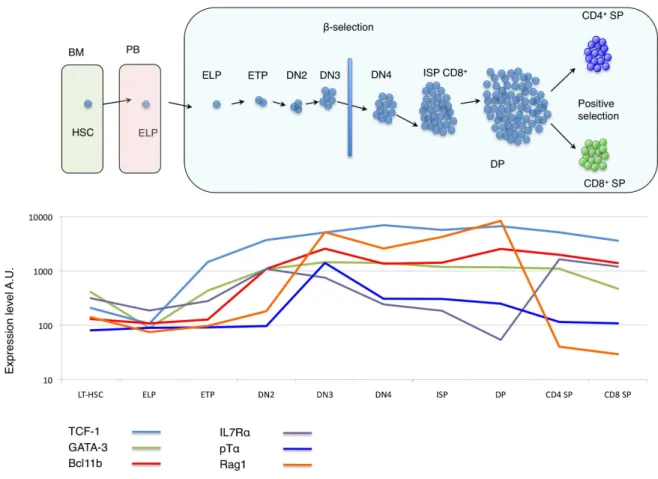
Figure 8: Regulatory gene expression during distinct stages of T-lymphoid differentiation ... 46!
Figure 9: Phenotypical characterization of native thymic progenitor stages ... 86!
Figure 10: Expression of Bcl11b and CXCR4 in DL-4 progenitors and native thymic progenitor subsets ... 88!
Figure 12: Comparison of gene-expression between DL-4 progenitors and native thymic T-lymphoid progenitors ... 91!
Figure 13: DL-4 induced generation of T-lymphoid progenitors from BM CD34+ ... 96! Figure 14: Comparison of T-lymphoid progenitor yields in DL-4 cultures with CD34+ cells from
BM and CB ... 97!
Figure 15: The CD34-/CD7- subset emerging in DL-4 culture displays a myeloid phenotype 98! Figure 16: Impact of sorting the immature CD34+/CD38lo CB subset ... 100! Figure 17: Impact of early renewal of DL-4 ligand ... 101!
ABBREVIATIONS
7-AAD 7-Aminoactinomycin DAhr Aryl hydrocarbon receptor
Bcl11b B-cell lymphoma/leukemia protein bHLH Basic helix-loop-helix protein
BM Bone marrow
BMI1 Polycomb ring finger oncogene BMI1
CB Cord blood
CCR CC chemokine receptor CD Cluster of differentiation
cDNA Complementary Deoxyribonucleic acid CLP Common lymphoid progenitor
CMP Common myeloid progenitor CMV Cytomegalovirus
CTLs Cytotoxic T lymphocytes
CTP T-lineage committed progenitors CXCL12 C-X-C Chemokine ligand
CXCR C-X-C chemokine receptor DC Dendritic cell
DL Delta-like ligand DN Double negative cell DNA Deoxyribonucleic acid DP Double positive cell
E2A E2A helix-loop-helix transcription factors EBF Early B-cell factor
EBV Ebstein Barr Virus EGF Epidermal growth factor ELP Early lymphoid progenitors ETP Early thymic progenitor
FACS Fluoresence activated cell sorting FGF-7 Fibroblast growth factor 7 Flt3 Fms-related tyrosine kinase 3 FTOC Fetal thymic organ culture GATA3 GATA transcription factor 3
GFI1 Growth factor independent 1 transcription repressor
GH Growth hormone
GSK Glycogen synthase kinase GvHD Graft-versus-host disease GvL Graft-versus-leucemia HES1 Hairy and enhancer of split HIV Human immunedeficiency virus HLA Human leukocyte antigen (HLA) HOXB4 Homeobox protein Hox-B4
HSCs Haematopoietic stem cells
HSCT Haematopoietic stem cell transplantation HSZT Hämatopoetische Stammzelltransplantation ICN Intracellular domain of Notch
Id2 Inhibitor of DNA binding
IGFBP1 Insulin-like growth factor-binding protein 1 IGF-I Insulin-like growth factor-I
Ikaros Ikaros family zinc finger protein 1
IL Interleukin
IL2R! Interleukin 2 receptor gamma IL7R" Interleukin 7 recetpor alpha ILC Innate lymphoid cells ISP Immature single positive cell JAG Serrate like ligands Jagged
LCK Lymphocyte-specific protein tyrosine kinase Lck Lymphocyte-specific protein tyrosine kinase LDA Limiting dilution analysis
LEF Lymphoid enhancer-binding factor
LHRH Luteinizing hormone-releasing hormone gene Lin- Lineage negative
LMPP Lymphoid-primed multipotent progenitor
lo Low
LRF Zinc finger and BTB domain containing 7A LSK Lin-Sca1+c-kit+
LT-HSC Longterm repopulating haematopoietic stem cell mAbs Monoclonal antibodies
MAML Mastermind coactivator
MAPK Mitogen activated protein kinase MCSF Messenger ribonucleic acid
MCSF Macrophage colony-stimulating factor MHC Major histocompatibility complex
miRNA MicroRNA
MLP Multipotent early lymphoid progenitors MLR Mixed lymphocyte reactions
MPP Multipotent progenitor MS5 Murine stromal cell MS5
mTECs Medullary thymic epithelial cells Myb Myeloblastosis viral oncogene homolog NK Natural killer
NOD/SCID/!c-/- Non obese diabetic/SCID/common gamma chain null mouse NSG Non-obese-diabetic/SCID/common-gamma-chain-null mouse Pax5 Paired box protein 5
PBMC Peripheral blood mononuclear cell PI3K Phosphoinositide 3-kinase
PSGL-1 P-selectin glycoprotein ligand-1 pTa Pre-T cell receptor alpha PU.1 Transcription factor PU.1 Rag Recombination activation gene
RBP Recombining binding protein suppressor of hairless RORgamma RAR-related orphan receptor gamma
RTE Recent thymic emmigrant
RTOC Rreaggregate thymus organ cultures
Runx1/CBF#
Runt-related transcription factor 1/ Mammalian core binding factor beta complex
S17 Murine stromal cell line S17 SCF Stem cell factor
SCID Severe combined immune deficiency SDF Stromal derived factor
SOX17 SRY-related HMG-box protein 17
SP Spleen focus forming virus proviral integration oncogene SR1 Stemregenin 1
TACE Tumor necrosis factor alpha converting Enzyme TAT HIV transactivating protein
TCF T-cell-specific transcription factor TCR T cell receptor
TEC Thymic epithelial cells TF Transcription factor TPO Thrombopoietin
TREC T-cell receptor excision circles TSLP Thymic stromal lymphopoietin TSP Thymus seeding progenitor TSt-4 Thymic stromal cell line TSt-4
SUMMARY
Human leukocyte antigen (HLA)-mismatched haematopoietic stem cell transplantation (HSCT) represents an important therapeutic option for patients lacking suitable donors. Delayed posttransplant immune recovery constitutes one of its major complications and is most pronounced in the T cellular compartment. A novel strategy to promote de novo thymopoiesis from donor derived HSCs and to accelerate T cellular reconstitution in patients after HSCT consists in the adoptive transfer of in vitro generated T cell progenitor cells. Identification of Notch1 as the key regulator of early T-lineage development has allowed the generation of Notch ligand-based culture systems, which provide a powerful tool to generate T-lymphoid progenitors
in vitro. The efficacy of murine T-lymphoid progenitors to promote T cell reconstitution has been
well demonstrated in conventional mouse models. In consistency, in vitro-generated human T cell progenitors were demonstrated to promote thymic recovery in humanized mice. Yet, positive effects of in vitro generated human T cell precursors on peripheral T cell reconstitution have not been demonstrated. Moreover currently used Notch-based co-culture systems consist of genetically modified murine cell lines. With view to establishing a clinically applicable system, feeder-cell-free Notch-ligand culture systems for the generation of T-lymphopoietic progenitors are warranted.
During my PhD project I developed a new cultu!e system based on the immobilized Notch ligand Delta-like-4 (DL-4).
Exposure of human CD34+ cord blood cells to immobilized DL-4 enabled the in vitro generation of high number of T cell progenitors, which harboured the phenotype of immature early thymic progenitor cells (ETP) and prothymocytes (proT). ETP and proT cell generated during DL-4 culture upregulated essential genes involved in early T-lymphoid development (i.e. IL7R", PT", RAG1 and BCL11b) and had undergone stage-specific recombination of the T cell receptor (TCR) locus in a similar way as in native human thymopoiesis. In limiting dilution analysis after secondary OP9/DL-1 co-culture, DL-4 progenitors displayed a highly increased T-lymphoid potential, which could be entirely attributed to the ETP and proT subset.
When transferred into NOD/SCID/"c-/- mice, DL-4 primed T cell progenitors migrated to the thymus and accelerated intrathymic T cell differentiation and emergence of functional, mature and polyclonal !# T cells in the periphery. In a co-transplantation approach, which more closely
were simultaneously injected in the same recipient. This procedure allowed even more rapid and more robust T cell reconstitution. HLA-tracking of the distinct graft sources further showed, that DL-4 progenitors specifically reconstituted the T-lymphoid compartments.
This work provides further evidence for the ability of in vitro-generated human T cell progenitors to promote de novo thymopoiesis and shows for the first time, that these cells accelerate peripheral T cell reconstitution in humanized mice. The availability of the efficient feeder-cell-free DL-4 culture technique represents an important step towards the future clinical exploitation translation of in vitro generated T-lymphoid progenitor cells to improve posttransplant immune reconstitution.
Key words: T cell development, T-lymphoid progenitor cells, Notch1, DL-4 protein, immunereconstitution after HSCT
RESUMÉ
L’allogreffe des cellules souches hématopoïétiques (CSH) dans les situations d’incompatibilité HLA partielle représente une option thérapeutique irremplaçable pour des patients nécessitant une greffe de cellules souches hématopoïétiques, en absence d’un donneur HLA-identique. Toutefois, le retard de la restauration du système immunitaire en particulier dans du compartiment lymphocytaire après greffe est l'une des complications majeures. Une nouvelle stratégie pour promouvoir la reprise de la thymopoïèse à partir des CSH provenant du donneur et d'accélérer la reconstitution cellulaire T chez des patients après greffe de CSH consiste en le transfert adoptif des progéniteurs T générés in vitro. L’identification de Notch1 comme le régulateur-clé du développement lymphocytaire T a permis l’établissement de systèmes de culture à base de ligands de Notch, qui permettent la génération efficace de progéniteurs lymphoïdes T in
vitro. L'efficacité des progeniteurs T-lymphoïdes murins pour promouvoir la reconstitution des
lymphocytes T a été bien démontrée dans des modèles de greffe chez la souris. De même, des progéniteurs T-lymphopoïétiques humains générés in vitro et greffés aux souris humanisées favorisent la reprise de la thymopoïèse. Pourtant, aucune donnée n’a encore démontré leur capacité à donner naissance à un compartiment lymphocytaire T périphérique. De plus, les systèmes de co-culture à base de ligand de Notch actuellement utilisés consistent en des lignées stromales murines génétiquement modifiées. Afin d'établir un système cliniquement applicable, il est donc indispensable d’établir des systèmes de culture qui soutiennent la génération de progéniteurs T en absence d’un support des cellules nourricières.
Au cours de mon projet de thèse, j'ai développé un nouveau système de culture pour la génération des progéniteurs T-lymphopoïétiques humains T basé sur l’immobilisation du ligand de Notch Delta-like-4 (DL-4) sous sa forme protéique.
La culture des progéniteurs hématopoïétiques CD34+ issue de sang en présence de DL-4 immobilisé permet la génération d’un grand nombre de cellules ayant un phénotype de progéniteurs thymiques précoces (early thymic progenitor: ETP) et de prothymocytes (proT). Les cellules ETP et ProT ainsi générées expriment à des niveaux élevés des gènes impliqués dans le développement lymphocytaire précoce (i.e. pTa, Rag1, IL7Ra et BCL11b). Elles montrent des signes de réarrangement du récepteur des cellules T (TCR) similaires à leurs homologues thymiques. Par des expériences de dilution limite sur une co-culture OP9/DL-1 secondaire, j’ai pu montrer que les progéniteurs générés sur DL-4 possédaient un potentiel lymphoïde T très
Suite à leur transfert dans des souris NOD/SCID/!c-/-, les progéniteurs lymphoïde T générés par exposition a DL-4 sont capable de migrer dans le thymus, d’y poursuivre des étapes ultérieures de leur développement et d’accélérer la différentiation T intra thymique ainsi que l’émergence des lymphocytes T mature, polyclonaux et fonctionnels en périphérie. Dans une approche de co-transplantation, qui se rapproche des conditions cliniques envisagées, j’ai simultanément injecté dans le même récipient des progéniteurs générées sur DL-4 et des cellules CD34+ non traitées (d’un 2èm donneur HLA-incompatible). Cette procédure a permis une reconstitution des lymphocytes T encore plus rapide et plus. Etant donné que les progéniteurs T générées sur DL-4 et les cellules CD34+ non-traitées étaient issue de deux donneurs avec un HLA différent, cette expérience a permis de montrer que les progéniteurs préalablement exposés à DL-4 reconstituaient spécifiquement les compartiments lymphoïdes T alors que les autres lignées hématopoïétiques provenaient des progéniteurs CD34+ non-traités.
Le travail accompli pendant mon projet de thèse renforce l’hypothèse selon laquelle les progéniteurs T générés in vitro promeuvent la thymopoïèse humaine dans des souris humanisées. Alors que les publications antérieures ne faisaient état que d’une thymopoïèse active, nous avons démontré pour la première fois que ces progéniteurs accélèrent également la reconstitution des cellules T périphériques chez des souris humanisées. La mise en place du système de culture cellulaire DL-4, qui soutient la génération des progéniteurs T en absence d’un support de cellules nourricières représente une étape majeure pour une future application clinique des progéniteurs lymphoïde T comme un nouveau traitement pour améliorer la reconstitution immunitaire après greffe de CSH.
Mots clés: développent des lymphocytes T, progéniteurs lymphoïdes T, Notch1, ligand de Notch DL-4 , reconstitution immunitaire après greffe des CSH
ZUSAMMENFASSUNG
Die Wiederherstellung der T-lymphozytären Immunität nach T-Zell depletierter hämatopoetischer Stammzelltransplantation (HSZT) ist ein langwieriger Prozess. Eine potentielle Strategie zur Beschleunigung der Neubildung von T-Zellen aus den transplantierten Stammzellen besteht in der Gabe von T-lymphozytären Vorläuferzellen. Die Entdeckung von Notch1 als wichtigster Regulator der frühen T-Zell-Entwicklung hat zur Etablierung Notchligand-basierter Zellkulturen geführt, mit deren Hilfe T-lymphoide Vorläuferzellen aus hämatopoetischen Stammzellen in vitro gebildet werden können. Das therapeutische Potential dieses Zelltyps wurde eindrucksvoll in konventionellen, syngenen und allogenen Maustransplantationsmodellen belegt, in denen nach Injektion in vitro generierter, muriner T-Vorläuferzellen eine Verbesserung der Neubesiedlung des Thymus sowie eine beschleunigte Wiederherstellung der T-zellulären Immunität erreicht werden konnte. Notchbasierte Co-Kultursysteme wurden ebenfalls für die
in-vitro Herstellung humaner T-lymphoider Vorläuferzellen verwendet. Das in-vivo Potential
humaner T Vorläuferzellen ist bislang jedoch nur lückenhaft charakterisiert: Zwar konnte gezeigt werden, dass humane T-Vorläuferzellen den hypoplastischen Thymus von immundefizienten NOD/SCID/!c-/- Mäusen besiedeln können. Ihre Wirksamkeit, die Wiederherstellung eines funktionellen, peripheren T-Zellkompartiments zu beschleunigen, gelang bislang jedoch nicht. Darüber hinaus werden Notchliganden in derzeit verwendeten Kultursystemen von genetisch modifizierten, murinen Stromazellen präsentiert. Die Entwicklung stromazellfreier, proteinbasierter Notchligand-Kultursysteme ist daher von grosser Bedeutung für eine mögliche therapeutische Nutzung in vitro generierter T-Vorläuferzellen.
Durch Immobilisierung von Notchligand Delta-like 4 (DL-4) habe ich im Rahmen meines PhD Projekts ein stromazellfreies Kultursystem zur Züchtung T-zellulärer Vorläuferzellen aus humanen CD34+ Nabelschnurblutzellen etabliert. In DL-4 Kultur generierte Zellen besitzen phänotypische und molekulare Eigenschaften von frühen thymischen Vorläuferzellen (ETP) und Prothymocyten (proT). ETP und proT Zellen aus DL-4 Kulturen exprimieren wesentliche Gene der frühen T-Zellentstehung (z.B. IL7Ra, PTa, RAG1 und BCL11b). Die entwicklungsstadium-spezifischen TCR-Rekombinationsprozesse in DL-4 Zellen erfolgen nach dem gleichen Muster wie in der nativen Thymusentstehung. Die in DL4 Kultur generierten T-Vorläuferzellen können sich in reife T-Zellen weiterentwickeln und durchlaufen die weitere T-Zelldifferenzierung innerhalb kürzerer Zeit als native CD34+ hämatopoetische Vorläuferzellen.
Darüber hinaus können DL-4 generierte T-Vorläuferzellen nach Xenotransplantation den hypoplastischen Thymus von immundefizienten NOD/SCID/!c-/- Mäusen besiedeln, intrathymische Zellentwicklung begünstigen und die Neubildung reifer und funktionaler T-Zellen in der Peripherie beschleunigen. Zur Simulation einer klinischen Anwendung führte ich weiterhin Co-Transplantationen mit DL-4 Vorläuferzellen und unbehandelten CD34+ Zellen in gleiche Empfänger durch und konnte hiermit eine weitere Verbesserung der Immunrekonstitution erzielen. Durch Verwendung HLA-divergenter Spender in diesen Versuchen konnte ich zeigen, dass DL-4 Zellen sich vornehmlich in T-Zellen weiterentwickelten, während die restlichen Blutzellreihen von unbehandelten CD34-poitiven Zellen gebildet wurden.
Im Rahmen dieses Projekts konnte ich mit einem für die klinische Anwendung geeigneten Kulturmodell wichtige präklinische Belege für das therapeutische Potential in vitro generierter T-Vorläuferzellen erbringen. Diese Arbeit bildet somit eine wichtige Grundlage für eine zukünftige klinische Anwendung von T-Vorläuferzellen zur Verbesserung der T-Zell-Immunität nach HSZT.
Stichwörter: T-Zellentwicklung, T-Vorläuferzellen, Notch1, Notchligand DL-4, Immunrekonstitution nach HSZT
T CELL RECONSTITUTION AFTER HSCT
1 T CELL RECONSTITUTION AFTER HSCT
Allogeneic haematopoietic stem cell transplantation (HSCT) offers an effective treatment for a broad spectrum of malignant and non-malignant disorders. In the early years of the era of bone marrow (BM) transplantation, HSCT was restricted to patients with human leukocyte antigen (HLA)-identical donors. Advances in graft handling and injection of highly purified CD34+ selected grafts have enabled HSCT from partially HLA-mismatched donors to become a widely accepted therapeutic option for patients lacking an HLA-identical donor.
While these procedures allow rapid restoration of haematopoiesis, the transfer of a functional immune system is much harder to achieve. Prolonged posttransplant immune deficiency, which is most evident in the T cell compartment, is a major challenge clinicians have to deal with after HSCT. Opportunistic viral, bacterial and fungal infections are the leading cause of death in recipients of HLA-mismatched transplants most occurring within the first 100 days after HSCT. Moreover, delayed T cell recovery is associated with an increased risk of graft rejection and relapse (Parkman and Weinberg 1997; Ruggeri, Peffault de Latour et al. 2011).
After HSCT, T cell recovery occurs through two mechanisms: one thymus independent and one thymus-dependent.
Homeostatic peripheral expansion (HPE) of mature T cells transferred with the graft occurs within only 10-15 days and can partially correct numerical T cell defects. In the lymphopenic HSCT patients HPE is triggered by (i) high cytokine levels (results of reduced consumption during lymphopenia) and (ii) by interactions with antigen present in the host, which not only include pathogen but also mismatched MHC antigens of the host. The first mechanism explains, why T cells expanded in these condition have activated phenotypes, an inversed CD4/CD8 ratio and contain much less naïve T cells than normal individuals. High expansion via HPE, which continues until a particular threshold of T cell numbers is obtained, further goes along with telomere shortening and leads to higher apoptosis rates of expanded T-lymphocytes. Furthermore, the T cell repertoire generated during HPE is restricted by the limited TCR specificities contained in the graft. The absence of newly produced naïve T cells and the frequent interaction with pathogens in the host (that were not necessarily present in the donor), lead to the dominance of T cells with particular TCRs and to a gradual restriction of the
T CELL RECONSTITUTION AFTER HSCT
TCR repertoire. For all these reasons thymus-independent T cell recovery can indeed provide initial immune competence against (commonly shared) viral infections (e.g. EBV, CMV) but is not sufficient to sustain long term T cell recovery (for review see (Fry and Mackall 2005)).
The functional recovery of T cells with physiological properties and broad TCR-specificities relies on the thymus-dependent generation of naïve T cells. Since the immunological education of de novo generated T cells occurs in the recipients’ thymus, emerging T cells are host-tolerant and provide a broad repertoire of naturally selected naïve T cells. De novo T cell development from donor haematopoietic progenitors requires several successive differentiation steps - first in the BM (to generate a lymphoid progenitor able to migrate to the thymus) and then in the thymus itself (where, after commitment to the T cell lineage, T cell precursors proceed through the complex process of thymopoiesis). Both steps are disturbed after HSCT. The generation of T cell progenitors in the BM and their delivery to the thymus is a limiting step in post-HSCT recovery (Zlotoff, Zhang et al. 2011). Furthermore, intrathymic T cell differentiation is altered for several (often interdependent) reasons: the thymic microenvironment can be damaged by the conditioning regimens, graft versus-host-disease, infectious disease or inflammatory status (Krenger, Blazar et al. 2011). Moreover age-related thymic involution contributes to a further delay of T cell reconstitution in adult patients (Parkman and Weinberg 1997; Small, Papadopoulos et al. 1999; Krenger, Blazar et al. 2011). In view of the above it becomes obvious that thymus-dependent T cell recovery is a long lasting process. The generation of thymus-derived naïve T cells requires 6-12 months in children and may take up to several years in adults, which reflects the age-dependent inefficiency of posttransplant thymic rebound (Weinberg, Annett et al. 1995; Parkman and Weinberg 1997; Ruggeri, Peffault de Latour et al. 2011). Complete restoration of the T cell compartment with a polyclonal T cell repertoire takes even longer and naïve T cells and central T memory cell counts may never reach pre-transplant levels (Komanduri, St John et al. 2007).
T CELL RECONSTITUTION AFTER HSCT
Figure 1: The innate and the adaptive immunity recover with different kinetics after HSCT
After HSCT, reconstitution of innate immunity occurs rapidly, whereas reconstitution of adaptive immunity is delayed. NK cells, monocytes, granulocytes and dendritic cells recover rapidly following HSCT. T cell regeneration is typically delayed and incomplete after HSCT because (i) T-cell progenitors have to be generated in the BM. (ii) the specialized thymic microenvironments is often perturbed due to preparative chemotoxic regimen, GvHD, inflammation. Abbreviations: HSC: Haematopoietic stem cell, MPP: Multipotent progenitor, MMP Myelomonocytic progenitor, MLP: Multipotent lymphoid progenitor, NK: Natural Killer cell. (Figure adapted from (Fry and Mackall 2005)
POTENTIAL STRATEGIES TO ENHANCE IMMUNE RECONSTITUTION AFTER HSCT
2 POTENTIAL STRATEGIES TO ENHANCE IMMUNE RECONSTITUTION AFTER HSCT
In order to shorten T cell reconstitution and to reduce infectious and non-infectious complications, various adoptive immunotherapeutic approaches have been evaluated. The large number of potential strategies can be roughly grouped into therapies supporting thymus-independent T cell recovery versus approaches improving de novo thymopoiesis.
The former includes the adoptive transfer of allodepleted donor T cells and infusion of pathogen-specific T cells and aims to transiently provide overall or antigen-specific specific T cell immunity. The later consists of hormonal or cytokine based therapies to improve the posttransplant thymic microenvironment and adoptive transfer of committed T cell progenitors.
2.1 Acceleration of Thymus independent T cell recovery 2.1.1 Allodepletion
Allodepletion consists of ex vivo depletion of anti-host-activated T cells that shall maintain most of the donor’s T cells’ immunocompetence against infectious agents. Alloreactive donor T cells can be specifically activated against major HLA-incompatible anti-gens by mixed lymphocyte reactions (MLRs). Activated (alloreactive) donor T cells can then be selectively killed using monoclonal antibodies (mAbs) against molecules up regulated during T cell activation (e.g. CD25, CD69, CD71 or CD137 (Fehse, Frerk et al. 2000; Fehse, Goldmann et al. 2000; Andre-Schmutz, Le Deist et al. 2002; Koh, Prentice et al. 2002; Solomon, Mielke et al. 2005; Hartwig, Nonn et al. 2006; Samarasinghe, Mancao et al. 2010)) or by photodynamic cells purging (Chen, Cui et al. 2002). A number of these approaches have been demonstrated to efficiently reduce or prevent GvHD in murine HSCT models. The feasibility and efficacy of ex vivo allodepletion protocols to accelerate posttransplant immune recovery has been demonstrated in a number of preclinical and clinical studies (Cavazzana-Calvo, Fromont et al. 1990; Fehse, Frerk et al. 2000; Fehse, Goldmann et al. 2000; Koh, Prentice et al. 2002; Godfrey, Krampf et al. 2004; Solomon, Mielke et al. 2005; Amrolia, Muccioli-Casadei et al. 2006). Although allodepletion protocols have been shown to markedly reduce GvHD incidence and improve post-transplant immunity in adult patients, they have been rarely used in children to date. One major restriction relates to the fact that today's protocols are not effective enough to provide the drastic degree of allodepletion that is required when used in patients with a low bodyweight. Novel depletion techniques (such as
POTENTIAL STRATEGIES TO ENHANCE IMMUNE RECONSTITUTION AFTER HSCT
results in terms of depletion efficiency. These new approaches seem to hold promise for extending the spectrum of clinical applicability to paediatric patients.
2.1.2 Injection of pathogen specific T cells
Injection of pathogen specific T cells represents another approach for shortening post-transplant immunodeficiency. Current protocols allow expanding pathogen specific cytotoxic T lymphocytes (CTLs) against CMV, EBV and adenovirus in vitro. The potential of these pathogen specific T cells has been in several clinical studies. CMV-specific CTLs can control CMV pneumonia that does not respond to antiviral chemotherapy (Riddell, Watanabe et al. 1992; Walter, Greenberg et al. 1995). EBV-specific CTLs have become an important therapeutic option in the treatment and prevention of post-transplant lymphoproliferative disease (Heslop, Ng et al. 1996; Rooney, Smith et al. 1998). Anti-Aspergillus CTLs have been developed preclinically and their ability to reduce infection-related mortality is currently tested in clinical trials (Beck, Topp et al. 2006; Tramsen, Koehl et al. 2009). More recently, new protocols avoiding long culture periods have been developed. They include sorting with (1) tetramer complexes (i.e. a specific MHC-peptide combinations). This technique is fully adapted to pathogens that induce a strong clonal immune response such as CMV (Feuchtinger, Opherk et al. 2010). (2) Secretion based sorting assays allow to specifically isolate T cells that secrete IFN" after a short term stimulation with given antigens (Parietti, Nelson et al. 2012). To provide combined immunity against all these pathogens, trivirus-specific CTLs (against CMV, EBV and adenovirus) have been generated in a single culture and are currently tested in a clinical trial (Hamel, Blake et al. 2002; Leen, Christin et al. 2009).
HPE generates a T cells with both quantitative and qualitative deficiencies. These T cells only provide a transient T cell pool with restricted TCR specificities. The same holds true for therapies based on in vitro expanded mature T cells: their clinical application requires a seropositive donor for a given pathogen; the monospecificity of the generated T cells affords only limited immune protection to patients, which are often multi-infected; the activated donor T cells retain a residual risk to induce GvHD.
Ideally, approaches to improve immune recovery should not only accelerate the T cell reconstitution within the first months post-transplantation but should provide the transplanted patient with a stable, polyclonal and naïve T cell repertoire.
POTENTIAL STRATEGIES TO ENHANCE IMMUNE RECONSTITUTION AFTER HSCT
2.2 Acceleration of Thymus dependent T cell recovery
A promising approach consists of enhancing the development of T cells from donor-derived HSCs in the recipient’s thymus and to thus accelerate the generation of donor-donor-derived, host-tolerant T cells. As mentioned above, both the altered thymic microenvironment and the delayed delivery of T-lymphoid progenitors to the thymus contribute to prolonged T cell lymphopenia after HSCT. Therefore strategies designed to accelerate thymus dependent T cell recovery follow two different but complementary conceptions: The first consists of enhancing intrathymic T cell development and tissue repair via administration of cytokines, hormones and growth factors. The second is based on the adoptive transfer of committed T-lymphoid precursors (Figure 2).
2.2.1 Promotion of intrathymic T cell development
2.2.1.1 IL-7
IL-7 plays a key role in T cell development, survival and homeostasis in both humans and mice. It signals via a receptor complex containing the IL-7 receptor-alpha (IL-7R!) and the common cytokine receptor-!-chain (!c), the later being shared by other members of the IL2-receptor family (i.e. IL-2R, IL-4R, IL-7R, IL-9R, IL-15R and IL-21R). The IL–7R complex is expressed by early lymphoid progenitors in the BM, early thymic precursors and resting T cells. The IL-7R complex is expressed by early lymphoid progenitors in the bone marrow, early thymic precursors and resting T cells. The importance signalling IL-7 receptor dependent signalling in T cell development is highlighted by the phenotype of two forms of human severe combined immune deficiency (SCID): X-linked SCID, in which patients lack T and NK cells but dispose of B-cells, is characterized by deletions in the IL2RG gene, which encodes for !c (Kondo, Takeshita et al. 1993; Noguchi, Nakamura et al. 1993). In contrast defective IL7R! expression causes T(-)B(+)NK(+) SCID, indicating that T cell deficiency (but not NK- or B-cell-deficiency) results from inactivation of IL-7-receptor alpha signalling in X-linked SCID (Puel, Ziegler et al. 1998). In preclinical studies in murine HSCT models, post-transplant administration of IL-7 enhanced both thymopoiesis and homeostatic proliferation of de novo-generated T cells (Fry, Christensen et al. 2001; Alpdogan, Muriglan et al. 2003). However further studies concluded, that the immune restorative capacity of IL-7 can be mainly attributed to effects on mature T cells and not to stimulation of thymopoiesis (Broers, Posthumus-van
POTENTIAL STRATEGIES TO ENHANCE IMMUNE RECONSTITUTION AFTER HSCT
7 therapy in HSCT patients, the clinical use of IL-7 has to be considered with care, for its potential risk to aggravate GvHD: High serum IL-7 levels in HSCT patients have been linked to a greater risk of acute GVHD suggesting a role in acute GvHD induction (Dean, Fry et al. 2008). While administration of IL-7 was safe and enhanced thymopoiesis in allogeneic murine HSCT models with T cell depleted grafts, it aggravated GvHD when in combination with non-T cell-depleted grafts (Sinha, Fry et al. 2002). A study by our group showed that IL-7 administration did not result in a beneficial effect in the setting of HLA-compatible or partially compatible HSCT but did have a favourable impact on T cell development in a fully HLA-incompatible setting (Andre-Schmutz, Bonhomme et al. 2004). These results indicate that post-HSCT administration of IL-7 should be restricted to recipients of autologous or T cell-depleted grafts.
2.2.1.2 Hormones
Age-dependent changes in two distinct endocrine pathways have been linked to thymic involution and have been shown to exert opposing effects on T cell development. Gonadal steroids (the production of which increases during puberty) inhibit thymopoiesis in an age-dependent manner. By contrast, growth hormone (GH) and insulin-like growth factor-I (IGF-I) stimulate thymopoiesis, but their production declines with age. In view of the hormones’ opposing effects on early T cell development, both endocrinal pathways have been modified their potential benefit in T cell reconstitution.
Sex steroid ablation can delay or reverse thymic involution (Utsuyama and Hirokawa 1989). Castration performed in mice before HSCT enhanced thymic and peripheral T cell recovery, without exacerbating GvHD and maintaining Graft-versus-leukaemia (GvL) activity (Goldberg, Sutherland et al. 2005; Goldberg, Alpdogan et al. 2007; Goldberg, King et al. 2009). In a recent pilot study, LHRH-agonist goserelin was used for temporary sex-steroid blockade in a cohort of aged patients undergoing HSCT. Goserelin accelerated neutrophil and overall lymphocyte recovery and T cell recovery in particular. Improvement of T cell recovery occurred via thymus-dependent regeneration, as indicated CD4 cell counts (both total and naïve), increase of T cell receptor excision circles (TRECs) transcripts. Moreover adjuvant goserelin improved peripheral T cell function, survival and engraftment rates and did not exacerbate GvHD (Sutherland, Spyroglou et al. 2008).
Growth hormone (GH) and its proximal mediator (IGF-I) stimulate lymphopoiesis (Clark 1997). Administration of GH or IGF-I inverts age-related thymic involution and
POTENTIAL STRATEGIES TO ENHANCE IMMUNE RECONSTITUTION AFTER HSCT
enhances T lymphopoiesis (Clark 1997). Besides direct effects on lymphopoiesis (Huang and Terstappen 1994; Hanley, Napolitano et al. 2005), GH and IGF-I further indirectly promote thymic recovery by supporting TEC proliferation (Chu, Schmitz et al. 2008). In murine HSCT models, posttransplant IGF-I administration increased thymic cellularity and enhanced peripheral T cell reconstitution without exacerbating GvHD (Alpdogan, Muriglan et al. 2003). In HIV-infected patients GH administration augmented the volume of the thymus, improved thymic T cell output and increased peripheral CD4cell numbers (total and naïve), emphasizing the importance of GH in T-lymphopoiesis (Napolitano, Schmidt et al. 2008). Very recently, IGF-I was successfully used to restore thymopoiesis in a child with Omenn syndrome with failed T cell reconstitution 6 months after HSCT (Ma, Shah et al. 2010; Wendorff, Koch et al. 2010).
2.2.2 Improvement of thymic tissue repair
Efficient thymopoiesis relies on the crosstalk between developing thymocytes and the non-haematopoietic stromal microenvironment. Stress induced TEC injury (e.g. by irradiation, chemotoxic drugs, GvHD) aggravates the perturbed de-novo thymopoiesis after HSCT. A better preservation of the thymic microenvironment should hence afford improved T cell recovery.
IL22 production by intrathymic innate lymphoid cells (ILC) was recently suggested to promote epithelial tissue repair and thereby the regeneration of the thymic microenvironment after stress induced damage and after HSCT in mice. In preclinical tests, administration of IL-22 increased early proliferation of TECs and enhanced overall thymic recovery after sublethal and lethal irradiation (Dudakov, Hanash et al. 2012).
Fibroblast growth factor 7 (FGF-7) provides cytoprotection against radiation- and chemotherapy-induced damage to TECs and improves thymic tissue repair (Farrell, Bready et al. 1998; Danilenko, Montestruque et al. 1999). FGF-7 also increases intrathymic IL-7 production (by TECs), which contributes to thymocyte survival and maturation in vivo (Min, Panoskaltsis-Mortari et al. 2007). In autologous primate HSCT models, FGF-7 improved the preservation of the thymic architecture and increased thymus-dependent T cell reconstitution (Seggewiss, Lore et al. 2007). In a clinical trial, FGF-7 prevented high-dose chemotherapy induced mucositis in allogeneic HSCT patients (Spielberger, Stiff et al. 2004; Stiff, Emmanouilides et al. 2006). In view of the extensive preclinical data on FGF-7’s
thymopoiesis-POTENTIAL STRATEGIES TO ENHANCE IMMUNE RECONSTITUTION AFTER HSCT
2.2.3 Adoptive transfer of in vitro-generated T cell precursors
In contrast to all other haematopoietic lineages T cells develop at two distinct sites: First in the BM, where T-lymphoid progenitors are generated and then in the thymus itself, where T cell differentiation occurs. The generation of donor derived T-lymphoid progenitors in the recipients BM and their diminished entry to the recipients’ thymus was described as a limiting step for thymus dependent T cell reconstitution after HSCT (Zlotoff, Zhang et al. 2011).
Modification of grafts by inclusion of BM derived committed progenitor cells has therefore been suggested as one strategy to circumvent the limiting step of lymphoid progenitor generation in the BM. Addition of murine BM prone lymphoid progenitors to HSC grafts has been subsequently demonstrated to transiently accelerate thymus-dependent T cell recovery and immunity against murine CMV (Arber, BitMansour et al. 2003). Importantly the inclusion of such progenitor cells in the graft never induced GvHD in mice, because donor progenitors underwent intrathymic education in the recipient. However, T-lymphoid progenitors represent a very rare cell population. The isolation of sufficient numbers is a key limitation even when working in mouse HSCT models. Given that such lymphoid progenitors occur at even lower frequencies in humans and further decline with age, their isolation from donor BM cells is not a valid option for a clinical use (Six, Bonhomme et al. 2007; De Smedt, Leclercq et al. 2011).
Speeding up thymopoiesis by injecting in-vitro-generated T cell precursors represents another strategy to reduce posttransplant T cell lymphopenia. During the past years, a number of seminal discoveries have improved our understanding of early steps in T cell development. The identification of Notch1 as the key regulator in early T cell development provided not only substantial novel biological insights. It also led to the establishment of cell culture systems, which now enable us to generate early T-lymphoid progenitors in vitro.
To further introduce this novel strategy the following chapters will provide an introduction into
(i) Early T cell development and thymopoiesis
(ii) Signalling pathways and molecular regulation of early steps in T cell development
POTENTIAL STRATEGIES TO ENHANCE IMMUNE RECONSTITUTION AFTER HSCT
Figure 2: Strategies to enhance T cell reconstitution after T cell-depleted haematopoietic stem cell transplantation (from Reimann et al. 2010).
Four different approaches to accelerate posttransplant T cell recovery are presented. Strategies A and B are based upon injection of mature T cells. C and D aim to accelerate de novo thymopoiesis. (A) Allodepletion allows selective removal of alloreactive donor cells after activation in mixed lymphocyte reactions with either irradiated PBMC or lymphocytes from the recipient. Specific changes in surface-marker expression (CD25, CD69, CD71 and CD137) or accumulation of photosensitizers allows identification of alloreactive T cells and their subsequent removal with immunotoxins, immunomagnetic depletion or photodynamic purging. (B) Pathogen-specific donor T cells can be selectively expanded by exposure to antigen-presenting cells expressing different viral antigens. Strategies to promote de novo thymopoiesis include pharmaceutical (C) and cellular (D) approaches. (C) Thymopoietic agents include administration of cytokines (IL-7, IL-15) or growth factors (FGF-7) implicated in normal T cell developments and administration or ablation of hormones involved in thymopoiesis. (D) Notch-based cultures promote T-lineage commitment of human HSCs in vitro. Transfer of in vitro-generated T cellular
EARLY T CELL DEVELOPMENT AND THYMOPOIESIS
3 EARLY T CELL DEVELOPMENT AND THYMOPOIESIS 3.1 Overview on T cell development
Thymopoiesis depends on the continuous supply of T cell progenitors from the BM that reach the thymus via circulation. However, thymus settling presumably represents a very rare event and therefore the study of the precise nature of thymus seeding progenitors is very difficult.
Like all haematopoietic lineages, T cell progenitors finally derive from multipotent HSCs residing in BM. T cell differentiation from HSC involves a series of discrete differentiation steps that gradually restrict this multipotency toward more restricted T-lineage precursors. These stages can be identified through specific surface markers.
In mice HSCs are comprised within the most immature Lin-Sca1+c-kit+ (LSK) cell population. Acquisition of Flt3 marks the transition towards non-renewing multipotent progenitors (MPPs), which in turn give rise to Rag1-positive early lymphoid progenitors (ELPs). Expression of IL7R! marks the transition to the common lymphoid progenitor (CLP) that can only differentiate into T- or B-lymphocytes and natural killer (NK) cells. While CLPs were originally proposed as the thymus seeding progenitors, the presence of CLP in the blood remains controversial and subsequent investigation revealed the more advanced CLP-2 and the peripheral blood prone T-lineage committed progenitors (CTPs) could seed the thymus after intravenous injection.
A multitude of progenitors thus have the ability to contribute to T-lymphopoiesis and are candidates for thymus seeding progenitor (TSP). Given the variety of candidates and the fact that thymus settling represents a very rare event, the contributions from each population to thymic input under physiological conditions is difficult to assess.
Blood borne TSP migrate the thymus through post-capillary venules near the thymic corticomedullary junction. TSP are attracted to the thymus by adhesion molecules such as fibronectin laminin, collagen type IV, P-selectin and chemokines. In fetal murine haematopoiesis, progenitor cells expressing C-X-C chemokine receptor type 4 (CXCR4), CC chemokine receptor 7 (CCR7) and CCR9 receptors, whose ligands C-X-C Chemokine ligand 12 (CXCL12), CCL19/21 and CCL25 are expressed by the thymic stroma, enter preferentially
EARLY T CELL DEVELOPMENT AND THYMOPOIESIS
in the thymus (Dando, Tavian et al. 2005). Chemokines also play important role in the migration of thymocytes through the different parts of the thymus.
During further T cell differentiation, thymocytes move in a highly ordered manner through the distinct regions of the thymus. After entrance at the corticomedullary junction, the most immature thymic subsets migrate to the cortex and further on to the subcapsular region before they begin to travel back towards the medulla. At each stage, thymocytes receive distinct, sub-region specific differentiation signals from thymic stromal cells and dendritic cells (DCs). These signals govern the processes of #-selection, positive selection, and negative selection that successively shape immature thymocytes into mature T cells with a diverse array of T cell receptors (TCRs). Apart from that spatial organisation of the differentiation process, developmental steps can be phenotypically defined. The most immature stages are found in the CD4-/CD8- double-negative (DN) compartment. During maturation, DN thymocytes develop – via the transient CD4 (in human T cell development) or CD8 (in murine T cell development) immature single positive (ISP) stage - into CD4+/CD8+ double positive (DP) cells. DP cells then further differentiate into either CD4+ or CD8+ single positive (SP) cells.
In murine T cell development, the DN compartment can be further divided into four differentially advanced substages (DN1-DN4) by differential surface expression of CD44 and CD25. The earliest thymocyte subset, the early thymic progenitor (ETP) is comprised within the most immature CD44+/CD25- DN1 subset. The ETP is phenotypically defined as LSK -/CD44+/CD25- cells and constitutes a very rare subset (approximately 0,01% of the young mouse thymus, even less in adult mice). During the ETP stage, interaction of the immature progenitors with the thymic microenvironment induces the molecular programme for T-lymphoid engagement. While ETPs are considered as canonical T cell precursors for their efficiency in generating downstream T-lineage progeny, they retain NK and DC potential and – depending on specific ETP subsets – very little B cell and myeloid potential. Unlike most genes, which have a stable sequence in each cell, the TCRs are made up of a series of alternative gene fragments. In order to create functional TCRs, DN thymocytes use several DNA-interacting enzymes to cut and rearrange these gene fragments. The outcome of this process assures a different sequence of each TCR. Initiation of the rearrangement process occurs during the CD44+/CD25+ DN2 stage: DN2 thymocytes upregulate recombination-activation gene (Rag) 1 and 2 to induce the VDJ recombinase enzyme complex, which initiates rearrangement of the TCR# locus. The cellular disadvantage in the rearrangement process is
EARLY T CELL DEVELOPMENT AND THYMOPOIESIS
functional TCR rearrangements differentiate into mature T cells, thymocytes have to undergo a first intrathymic selection process, which is called #-selection. This step occurs at the CD44 -/CD25+ DN3 stage: At this stage, rearrangement of the TCR# chain is completed. The TCR# then pairs with the pre-T cell receptor alpha (pT!) to form the pre-T cell receptor (preTCR). If the resulting preTCR is functional, it can then interact with TCR signalling proteins and induces downstream signalling, which allows proliferation and further differentiation of the T cell precursor. Progenitors with non-functional TCR# rearrangements can again undergo TCR# rearrangement for several times. However all progenitors that do not finally succeed to express a productive TCR# chain will eventually be eliminated by programmed cell death. Apart from #-selection, the DN3 stage is characterized by definitive T-lineage commitment (and loss of non-T-lineage potential) of the progenitor. PreTCR signalling further induces rearrangement of the TCR! chain and promotes downregulation of CD25. This marks the progression to the CD44-/CD25- DN4 stage and the end of the DN stage. Thymocytes then upregulate either CD8 (in murine T-development) or CD4 (in human T-development) and thereby reach the intermediate immature single positive (ISP) stage. While developmental steps at DN stages and #-selection occur in the subcapsular regions of the thymus, thymic precursors migrate back towards the central regions afterwards, where they acquire the CD4+CD8+ double positive (DP) stage. DP cells represent more than 85% of all thymocytes and first express a mature TCR!#CD3 complex. However, most DP express TCR!#/CD3 complexes, that cannot interact with MHC-I or II and are thus non-functional. These cells are eliminated during positive selection: DP cells are highly susceptible to apoptosis and their survival depends on on-going TCR signalling. Only DP cells with functional TCR!#/CD3 complexes receive a survival signal and will pass the positive selection checkpoint.
Thymocytes having successfully passed positive selection migrate towards the corticomedullary junction and undergo negative selection, against self-antigens expressed by cortical stromal and DCs. Thymocytes that recognise autologous peptides receive apoptosis signals to avoid immune response against self and thus autoimmunity reactions. Depending on whether a given precursor preferentially recognizes MHC-I, or -II, it then matures into a CD8 or CD4 mature T lymphocyte. CD8 and CD4 cells then exit the thymus via the medulla into the peripheral blood.
EARLY T CELL DEVELOPMENT AND THYMOPOIESIS
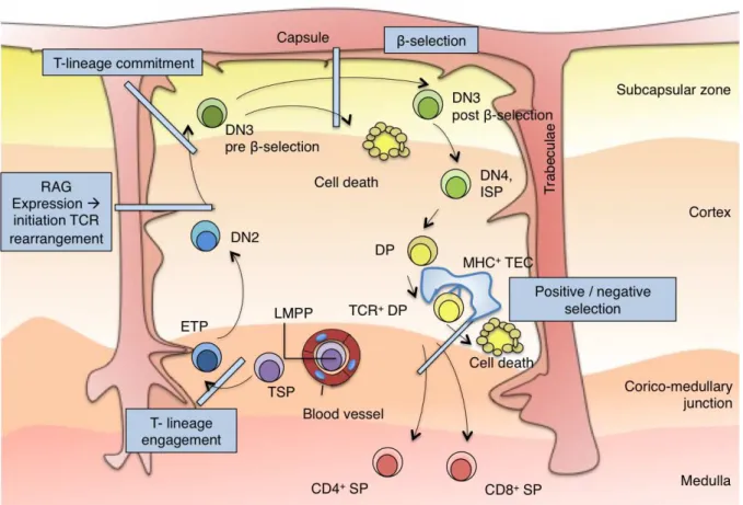
Figure 3: The topography of intrathymic T cell development (adapted from Rothenberg, Moore & Yui, 2008)
Cross-section through a murine adult thymic lobule showing the migration path of T cell precursors during development. Immigrant precursors initially enter the thymus through blood vessels near the corticomedullary junction, the early T cell precursors (ETP) subsequently migrate and differentiate from double negative (DN) to double positive (DP) to single positive (SP) stages, through the distinct microenvironments of the thymus. ETPs appear to have the options to expand in the corticomedullary junction region or to differentiate into DN2 cells that then migrate from the site of entry deep within the cortex to the outer rim of the cortex. $eta-selection occurs during the accumulation of the DN3 T cells in the extreme outer portion - the subcapsular zone - of the thymus. A directional reversal of migration back across the cortex towards the medulla occurs for the later stages of thymocyte development, when thymocytes reach the DP stage. DP cells interact with thymic epithelia cells (TECs) via TCR-MHC interaction and undergo positive and negative selection. ISP: Immature Single Positive; LMPP: lymphoid primed multipotent progenitor, NK: Natural Killer; TCR: T Cell Receptor.
EARLY T CELL DEVELOPMENT AND THYMOPOIESIS
3.2 Particularities of human T cell development
Much of our current knowledge about T cell development has been obtained from highly fine-tuned, genetically modified mouse models that can be diversely combined in an almost mathematical manner. In contrast human T cell development is far less well characterized and much more fragmentary. It is tempting to fill these lacunas by extrapolation from our conception of T cell development in mice, given the high degree of similarity between both species. However this approach is delusive for several reasons.
Progenitor stages traversed during different developmental stages in the BM and in the thymus display substantial differences between the two species by means of their lineage potential, their phenotype and their molecular characteristics. A very recent study shows that lifelong T cell homeostasis depends on on-going thymopoiesis in mice, but much less so in human. This impressively demonstrates that we cannot freely extrapolate our mouse-centred conception of T cell development to physiological conditions in human (den Braber, Mugwagwa et al. 2012).
Human HSC express the CD34 antigen and this marker has been useful in elucidating pathways of distinct haematopoietic lineages. HSCs, MPPs and multipotent early lymphoid progenitors (MLP) are found in the Lin-/CD34+/CD38lo/- fraction of human cord blood or BM. They can be further distinguished based on CD90 and CD45RA expression. The CD90+/CD45RA- subset contains the pluripotent, self-renewing HSCs, MPPs are found in the CD90-/CD45RA- subset and MLP reside in the CD90-/CD45RA+ subset. Human MLP cells have an increased potential to differentiate in lymphoid lineages and have lost their erythroid and megakaryocytic potential. However they still possess potential to generate monocytes, DCs and macrophages. A true CLP, which can only differentiate into T, B and NK cells, has not been identified in human haematopoiesis yet. Likewise the precise nature of the direct thymus seeding progenitors remains elusive. Collective findings of the past years suggest, that different progenitor population can act as MLP and thymus seeding progenitor in human. The ability of human MLP to engage towards either a T-cell or a B-cell fate varies with age. Fetal MLPs have a balanced T and B cell potential. In contrast the T-lymphoid potential of postnatal MLPs is considerably lower and further decreases during childhood. This age-dependent B over T bias of human MLP further complicates the precise phenotypical characterization of the human CLP equivalent.
EARLY T CELL DEVELOPMENT AND THYMOPOIESIS
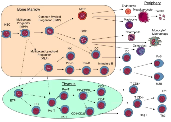
Figure 4: Schema of human haematopoietic differentiation (kindly provided by E.M. Six)
All haematopoietic cells are derived from pluripotent HSC. These cells differentiate into mature haematopoietic cells through various intermediate cell types that are defined by expression of cell surface antigens. Traditionally, it has been assumed that the first step consists in the differentiation of HSC into myeloid precursors on one side and lymphoid precursors on the other side. While myeloid precursors give rise to erythroid, megakaryocyte and mono/granulocytes, lymphoid precursors are supposed to generate T-, B- lymphocytes and NK cells as well as a proportion of DCs. HSC: Haematopoietic Stem cell, MEP: Megakaryocyte–erythroid progenitor cell, DC: dendritic cell, ETP: Early thymic progenitor, Pre-T prethymocyte Th: T-Helper Cell, GMP: granulocyte macrophage progenitor
CD7 and CD10 represent the earliest known markers expressed by the earliest recognizable T- and B-progenitors respectively. Previous attempts to better identify human CLP (and early T-lymphoid progenitors) within the Lin-/CD34+/CD38lo/- compartment have therefore made use of these two markers.
EARLY T CELL DEVELOPMENT AND THYMOPOIESIS
are found at very low frequencies in fetal bone marrow and in CB. CD34+/CD45RA+/CD7+ progenitors can only give rise to lymphoid lineages, but not to myeloid and erythroid cells and thereby fulfil CLP criteria. Yet, CD34+/CD45RA+/CD7+ progenitors are clearly polarized towards the T/NK lineage as compared to their B-lymphoid potential (Allman, Karnell et al. 2001; Hao, Zhu et al. 2001; Haddad, Guardiola et al. 2004; Haddad, Guimiot et al. 2006). Haddad et al. demonstrated that CD34+/CD45RA+/CD7+ progenitors were selectively recruited to the thymus in ex vivo colonization assays and therefore hypothesized that they corresponded to direct prethymic progenitors (Haddad, Guimiot et al. 2006). Interestingly, CD34+/CD45RA+/CD7+ can only be detected in fetal bone marrow and CB, but sharply decline thereafter and cannot be found in adult BM. In postnatal thymopoiesis however, sequential upregulation of CD7 and T-lineage engagement occurs at very immature intrathymic stages only, evoking thymus seeding by a CD7- progenitor (Hao, George et al. 2008). Moreover, the human thymus contains low numbers of progenitors, that can develop into B-lymphoid, myeloid and erythroid lineages in vitro (Weerkamp, Baert et al. 2006). These findings evoke the existence of multipotent progenitor populations distinct from the T/NK biased CD34+/CD45RA+/CD7+ cells that assure lymphopoiesis and especially thymopoiesis after birth.
More than 20 years ago, Lin-/CD10+ progenitors from fetal BM were first shown to be able to undergo primary steps in T cell differentiation in vitro and were thus proposed to correspond to human MLP (Hokland, Hokland et al. 1987). Galy et al. described Lin -/CD34hi/CD45RA+/CD10+ cells in postnatal BM, which contained a broad B, T, and NK potential but lacked non-lymphoid potential. Further studies concluded that bone marrow CD34+CD10+CD19- cells were relatively B-cell committed, since they harboured partial DJh
rearrangements and displayed B-lineage specific gene expression profile (Hokland, Hokland et al. 1987; Galy, Verma et al. 1993; Galy, Travis et al. 1995; Davi, Faili et al. 1997; Dworzak, Fritsch et al. 1998; Rossi, Yokota et al. 2003). In a more recent report by our group the CD34+/CD10+ subset was further separated by depletion of CD24+ cells. The CD34+/CD10+/CD24$ fraction from CB and adult BM was highly enriched for common lymphoid potential (B, T, NK) but still retained residual myeloid potential. In contrast expression of CD24 went along with loss of T cell potential and marked early B cell commitment. Importantly, the multi-lymphoid CD34+CD10+CD24$ cells were detected in peripheral blood and at immature thymic developmental stages, suggesting their potential role as a thymic precursor throughout life. Interestingly this study also demonstrated a sharp
EARLY T CELL DEVELOPMENT AND THYMOPOIESIS
decrease of the CD34+/CD10+/CD24$ population during the first years of age, which went along with a decrease of T-lymphoid potential (Six, Bonhomme et al. 2007).
Collectively these findings suggest that different progenitor population might act as MLP and thymus seeding progenitor in human. The precise contribution of a given progenitor to T- and B-cell development and to thymopoiesis is unknown but appears to depend of age and is most evident between fetal and postnatal stages.
Although the precise nature of the earliest thymic precursor in human T cell development remains elusive, intrathymic CD34+/CD45RA+/CD7+ progenitors are currently accepted to represent the equivalent of the ETP stage. Like in mice, human ETP are comprised within the CD4/CD8 DN compartment at very low frequencies. Early human T cell development is marked by further upregulation of CD7, IL-7R", CXCR4 and CD5. Along with that CD10 is downregulated. The emerging CD34+/CD45RA+/CD7+/CD5+ subset has been named proT stage. Progenitors at the proT stage are T-lineage polarized but still remain at least in vitro B, DC and at a very low degree myeloid potential (Weerkamp, Baert et al. 2006).
Expression of CD1a marks transition to the downstream preT stage and definitive T-lineage commitment. This process goes along with initiation of TCR rearrangements within the TCR-%, TCR-", and TCR-# loci that will determine the developmental outcome (Dik, Pike-Overzet et al. 2005): progenitors with in-frame TCR-% and TCR-" rearrangements will further develop into CD3+/ TCR-"%+ T cells, whereas a TCR-# chain will pair with the pre-T! to form the pre-TCR complex. At the preT stage and the following CD4 ISP stage, precursors undergo #-selection to than become CD4+/CD8+ DP thymocytes (Figure 5).
EARLY T CELL DEVELOPMENT AND THYMOPOIESIS
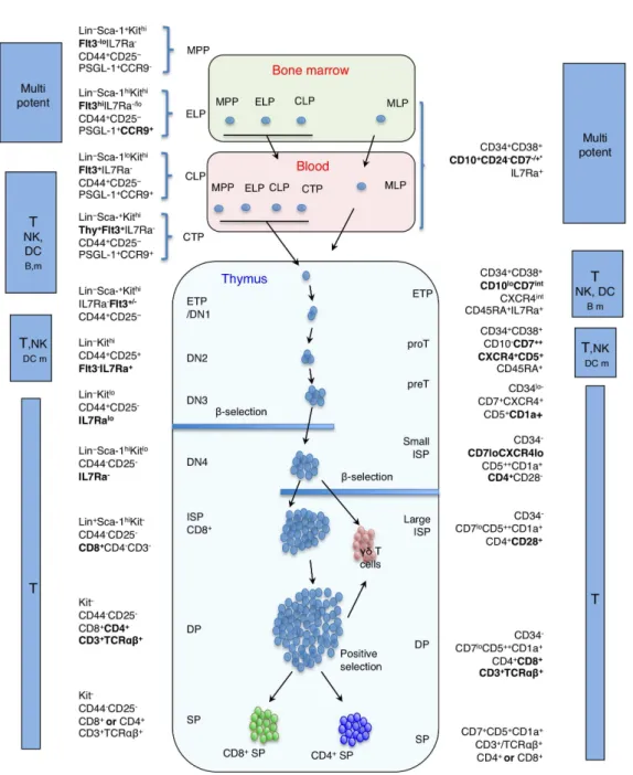
Figure 5: Comparison of known stages of murine and human T cell development
Figure 5: Comparison of known stages of murine (left part) and human (right part) T cell development. Lineage commitment in mice occurs at the transition from the DN2 to the DN3 stage and at the transition from the proT to the preT stage in human. *Phenotype of MLP cells in human BM differs depending on ontogenic stage. In fetal BM and CB, Lin-/CD34+/CD38- MLP express CD45RA and CD7, in adult BM they do not express CD7 and CD45RA but are CD10+/CD24
-Abbreviations: MPP: multipotent progenitor, MLP: multipotent early lymphoid progenitor, ELP: early lymphoid progenitor, CLP: common lymphoid progenitor, CTP: circulating T cell progenitors, DN: CD4/CD8 double negative, ETP: early thymic progenitor, proT: prothymocyte, preT: pre-thymocyte, ISP: immature single positive, DP: double positive, SP: single positive, Lin: Lineage negative.
REGULATION OF EARLY STEPS IN T CELL DEVELOPMENT
4 REGULATION OF EARLY STEPS IN T CELL DEVELOPMENT
Early T cell development depends on the interaction of the thymic microenvironment with the “thymic migrants”. Thymic epithelial cells provide critical signals to the immature lymphoid progenitors to guide them through the processes of T cell engagement, commitment and #-selection. Notch1 is the essential factor for the instruction of T-lineage engagement and interplays with other signalling pathways (IL-7, Wnt and CXCR4) to orchestrate T cell differentiation until #-selection.
4.1 Signalling pathways in early T cell development 4.1.1 Notch1/DL-4 signalling
The most important trigger for a T cell engagement is the activation of Notch1 signalling induced by Delta-like Notch ligands expressed on thymic stroma.
Notch proteins are essential regulators of a broad spectrum of cell fate decisions and differentiation processes during fetal and postnatal development and at several stages of haematopoiesis. Originally identified in Drosophila melanogaster, they are highly conserved among different species. Mammals express 4 Notch receptors (Notch 1-4), which can be activated by 5 different Notch ligands. These ligands can be subdivided into 2 classes: the Serrate like ligands Jagged1 and -2 and the Delta-like ligands -1, -3 and -4 (1, 3, DL-4). Both receptors and ligands are transmembrane proteins with variable numbers of epidermal growth factor (EGF)-like domains. Upon interaction with its ligand the intracellular portion of the Notch receptor undergoes a series of proteolytic cleavages that liberate the intracellular domain of Notch (ICN). The cleaved ICN translocates to the nucleus, where it heterodimerizes with the transcription factor (TF) Recombining Binding Protein suppressor of hairless-k (RBP-Jk). RBP-Jk is thereby converted from a transcriptional repressor into a transcriptional activator. Further recruitment of the Mastermind like co-activator (Maml) then initiates transcription of Notch target genes. The structure of the RBP-Jk/ICN/Maml complex was originally conceived as a monomer bound to DNA, but recent studies show, that it can also bind as a dimer or in cooperation with E2A, a member of E-protein family of basic helix–loop– helix (bHLH) proteins (Figure 6).