HAL Id: tel-00834352
https://tel.archives-ouvertes.fr/tel-00834352
Submitted on 14 Jun 2013HAL is a multi-disciplinary open access
archive for the deposit and dissemination of sci-entific research documents, whether they are pub-lished or not. The documents may come from teaching and research institutions in France or abroad, or from public or private research centers.
L’archive ouverte pluridisciplinaire HAL, est destinée au dépôt et à la diffusion de documents scientifiques de niveau recherche, publiés ou non, émanant des établissements d’enseignement et de recherche français ou étrangers, des laboratoires publics ou privés.
Intervenous immunoglobulins as modulators of immune
response : effect on T cell polarisation, pathogenicity
and trafficking
Shivashankar Othy
To cite this version:
Shivashankar Othy. Intervenous immunoglobulins as modulators of immune response : effect on T cell polarisation, pathogenicity and trafficking. Immunology. Université Pierre et Marie Curie - Paris VI, 2012. English. �NNT : 2012PA066113�. �tel-00834352�
1
THESE DE DOCTORAT DE L’UNIVERSITE PARIS VI PIERRE ET MARIE CURIE
Spécialité: IMMUNOLOGIE
Ecole doctorale: Physiologie et physiopathologie Présentée par Monsieur
Shivashankar OTHY
Pour obtenir le grade de
DOCTEUR DE L’UNIVERSITE PARIS VI Sujet de la thèse
Modulation de la réponse immunitaire par les immunoglobulines intraveineuses : Effets sur la polarisation, la pathogénicité et le trafic des lymphocytes T
Soutenue le 3 May 2012, devant le jury composé de:
Carole ELBIM Président
Fanny MONNEAUX Rapporteur
Christophe JAMIN Rapporteur
Peter SPAETH Examinateur
Jagadeesh BAYRY Srini KAVERI
Directeur de thèse Directeur de thèse
2 Contents Acknowledgements ... 3 Abbreviations ... 4 Résumé ... 6 1. Introduction ... 9 1.1. Immune system ... 9
1.2. Immune homeostasis and activation ... 10
1.3. T cell polarization and Th cells subsets. ... 13
1.3.1. Th1 cells ... 14
1.3.2. Th2 cells ... 15
1.3.3. Th17 cells ... 15
1.3.4. Regulatory T cells ... 18
1.4. mTOR integrates multiple signals during T cell activation ... 21
2. T cells in autoimmunity : shifting paradigms from Th1/Th2 to Th17/Tregs ... 23
2.1. Th1/Th2 hypothesis and the emergence of Th17 cells ... 23
2.2. Dysregulated equilibrium between pathogenic and regulatory T cells leads to immune disease ... 25
3. Intravenous Immunoglobulin ... 26
3.1. Composition, pharmacology and indications of IVIg ... 26
3.2. Immunoregulatory mechanisms of IVIg in autoimmune and inflammatory diseases... 29
3.3. Modulation of the Tregs by IVIg ... 31
4 Multiple sclerosis/ EAE is an organ specific T cell mediated auto-immune disease ... 33
5 Hypothesis and aims ... 35
6 Results ... 38
7 Discussion and Perspectives ... 60
8 References ... 71
3
Acknowledgements
I would like to acknowledge my supervisors Srini Kaveri and Jagadeesh Bayry for
providing me a chance to accomplish the doctoral studies under their guidance. Their kind help,
guidance and patience for the entire period of the thesis will always be remembered. Their
criticisms, suggestions and timely appreciation over the past three years have been a boon for
accomplishment of scientific goals.
I am thankful to Dr. Carole ELBIM for kindly accepting the invitation to be the president
of the jury. I wish to convey my sincere gratitude to Dr. Fanny MONNEAUX and Dr. Christophe
JAMIN for agreeing to be rapporteurs. I am grateful to Dr. Peter SPAETH for accepting the
invitation to be the examiner.
Thanks to all of ‘Team Srini Kaveri’, present and past: Mohan, Pushpa, Sébastien, Marie-Françoise, Sandrine, Sebestien (iSeb), Mr. Sonte, Vani, Ankit, Jordan, Ana, Yann, Justa,
Cyril, Ivan, Devram, Maud, Prathap, Laurent, Julie, Meenu, Nimesh, Fernando, Marcos and
Selma for their kind support as colleagues and friends. I am thankful to all of my friends and
family who kept me sane and made my stay in Paris a fun and memorable time.
4
Abbreviations
Ab Antibody
Ag Antigen
AHR Aryl hydrocarbon receptor APC Antigen presenting cell
BCR B cell receptor
CD Cluster of differentiation
CFA Complete Freund’s adjuvant
CIA Collagen-induced arthritis
CIDP Chronic inflammatory demyelinating polyneuropathy
CNS Central nervous system
CTLA-4 Cytotoxic T lymphocyte antigen 4
CTL Cytotoxic T lymphocyte
CVID Common variable immunodeficiency
DC Dendritic cells
DC-SIGN Dendritic cell-specific intercellular adhesion molecule-3- grabbing non-integrin
DLN Draining lymph node
DNA Deooxribonucleic acid
EAE Experimental autoimmune encephalitis ELISA Enzyme link immunosorbent assay F(ab•')2 Fragment antigen binding
Fc Fragment crystallizable
FcγR Fc gamma receptor
FcRn Neonatal FcR
FCS Foetal Calf Serum
FITC Fluroescein isothiocyanate
Foxp3 Forkhead box p3
GBS Guillain Barrée syndrome
IFN Interferon
Ig Immunoglobulin
IL Interleukin
IBD Inflammatory bowel disease
iTreg Induced regulatory T cell
ITP Immune thrombocytopenic purpura IVIg Intravenous immunoglobulin LAG-3 Lymphocyte activation gene-3
LPS Lipopolysaccharide
MHC Major Histocompatibility complex
5
Mo Monocyte
Mφ Macrophage
MS Multiple sclerosis
NAbs Natural antibodies
NFAT Nuclear factor of activated T cell NK cell Natural Killer cell
nTreg Natural regulatory T cells
PBS Phosphate buffer saline
RA Rheumatoid Arthritis
RORC/ RORγt Retinoic acid-related orphan receptor
SEM Standard error of mean
SLE Systemic lupus erythematosus
STAT Signal transducing and activating transcription factor T-bet T box transcription factor
TCR T cell receptor
Tfh Follicular helper T cells TGF-β Transforming growth factor β Th cell T helper cell
TLR Toll like receptor Tregs Regulatory T cells
6
Résumé
La signalisation au niveau de la synapse immunitaire entre les cellules présentatrices d’antigènes et les lymphocytes T est une phase critique de la réponse immune adaptative. Les lymphocytes T CD4+ naïfs sont polarisés en différentes populations (Th1, Th2, Th17, iTrgs) et les décisions majeures, telles que le choix entre l’homéostasie immunitaire et l’activation du système immunitaire, la tolérance au soi, l’auto-immunité, sont prises à ce moment. Dans certaines conditions, un déclenchement dérégulé de la réponse immunitaire provoque un déséquilibre entre les fonctions effectrice et régulatrice des lymphocytes T, conduisant à des phénomènes d’auto-immunité et d’inflammation systémique. Des doses élevées d’immunoglobulines intraveineuses (IVIg) sont fréquemment utilisées pour traiter ces maladies. Le mécanisme d’action des IVIg sur la polarisation des lymphocytes T reste à ce jour inexploré. Ainsi, j’ai recherché les effets de doses élevées d’IVIg sur la polarisation des lymphocytes T dans le cadre de l’encéphalomyélite immune expérimentale (EAE), une maladie auto-immune mettant en jeu les lymphocytes T et un modèle animal de la sclérose en plaques. L’EAE a été induite chez des souris C57BL/6J en combinant le peptide MOG35-55 avec l’adjuvant complet de Freund (CFA). L’administration d’IVIg a retardé l’apparition de l’EAE et diminué significativement l’intensité des signes cliniques. Les IVIg ont inhibé la différenciation des lymphocytes T CD4+ naïfs en sous-populations effectrices (Th1 et Th17) et ont, de manière concomitante, provoqué la prolifération des lymphocytes T régulatrices CD4+ Foxp3+. En outre, les IVIg ont rendu les lymphocytes T effecteurs moins pathogéniques, en diminuant l’expression de molécules encéphalitogéniques telles que le GM-CSF et la podoplanine. Les IVIg ont diminué l’expression du récepteur à la sphingosine-1 phosphate (S1P1) à la surface des lymphocytes CD4+, séquestrant ces cellules dans les ganglions lymphatiques et diminuant l’infiltrat de l’organe cible (le système nerveux central) en Th1, Th17 et Trég. Le récepteur inhibiteur FcγRIIB n’est pas indispensable pour la modulation des sous-populations de lymphocytes T CD4+ effecteur et régulateur induite par les IVIg in vivo. D’autre part, les portions F(ab)’2 des IVIg ont conservé la fonction modulatrice associée aux IVIg. La cible de la rapamycine chez les mammifères (mTOR) est une kinase qui intègre de multiples signaux de l’environnement et qui est impliqué dans la régulation des réponses effectrice et régulatrice des lymphocytes T. Les IVIg ou les F(ab)’2 ont diminué l’activité de mTOR, rétablissant à nouveau l’équilibre entre les sous-populations de lymphocytes T régulateurs et de lymphocytes T helper pro-inflammatoires. Pris ensemble, ces résultats constituent une base cellulaire et moléculaire,
7 qui sous-tend l’effet bénéfique des IVIg dans certaines maladies auto-immunes et inflammatoires.
Summary
Signaling at the immune synapse between antigen presenting cells and T cells is the critical phase of the adaptive immune response. Naïve CD4 T cells are polarized into various subsets (Th1, Th2, Th17 and iTregs) and crucial decisions such as immune homeostasis versus immune activation; and tolerance to self-versus autoimmunity are made at this point. Dysregulated activation leads to pathogenic immune response to self-antigens and inflammatory pathologies. High dose therapy of intravenous gammaglobulin (IVIg) is widely used in the treatment of several T-cell and autoantibody-mediated autoimmune diseases. However, the comprehension of the mechanisms underlying its therapeutic benefit has remained a major challenge. In particular the mechanisms of action of IVIg in terms of T cell polarization in vivo have remained unexplored. Therefore, I have investigated the effect of high dose IVIg on T cell polarization using actively induced experimental autoimmune encephalomyelitis (EAE), a T cell-mediated autoimmune condition and an animal model of multiple sclerosis. EAE was induced in C57BL/6J mice using MOG35-55 emulsified in complete Freund’s adjuvant (CFA). Concomitant administration of IVIg delays the onset of EAE and significantly decreases severity of the disease. IVIg inhibited the differentiation of naïve CD4 T cells into effector subsets (Th1 and Th17 cells) and concomitantly induced expansion of Foxp3+CD4 cells. Further, IVIg rendered effector T cells less pathogenic by decreasing expression of encephalitogenic molecular players such as GM-CSF and podoplanin. IVIg decreased the expression of sphingosine-1 phosphate receptor (S1P1) on CD4 cells, thus sequestering these cells in the draining lymph nodes and decreasing infiltration of Th1, Th17 and Tregs to the target organ (central nervous system). Inhibitory FcγRIIB appeared dispensable for IVIg-mediated reciprocal modulation of effector and regulatory CD4 subsets in vivo. F(ab’)2 fragments of IVIg also retained the reciprocal CD4 T cell modulatory functions of IVIg. Mammalian target of rapamycin (mTOR) is a kinase which integrates various environmental signals and is involved in regulation of effector and regulatory T cell responses. IVIg or F(ab’)2 decreased activity of mTOR thus restoring the equilibrium between regulatory T cells and pro-inflammatory T helper subsets. Thus, these findings provide a novel cellular and molecular basis underlying the beneficial effect of IVIg in certain T-cell mediated autoimmune and inflammatory conditions.
8
Introduction
9
Intravenous immunoglobulins as modulators of immune response: Effect on T cell polarization, pathogenicity and trafficking
1. Introduction
1.1. Immune system
The immune system comprising innate and adaptive part is a network of interacting molecules and cells evolved to fight against the invading pathogens and regulate aberrant tissue responses such as cancer. Cells of the innate immune compartment include monocytes (Mo), macrophages (MΦ), dendritic cells (DC), mast cells (MC), neutrophils, eosinophils, natural killer (NK) and basophils. The complement system comprises of proteins in the serum that act by opsonizing and inducing cytolysis in pathogens and infected cells. Invasion of host tissues by pathogens leads to cell stress, hypoxia, necrosis, temperature shifts and tissue destruction [1]. Local injury at the site of infection or tissue damage releases various microbial and endogenous products from damaged or dying cells [2]. Molecules originating from pathogens are called as pathogen associated molecular patterns (PAMP), whereas those from insulted cells/tissues are designated as danger associated molecular patterns (DAMP). The cells of the innate immunity residing in various tissue of body are endowed with specialized germline-encoded pattern recognition receptors (PRRs) and multi-protein complexes like inflammasomes. Innate cells recognize PAMPs and DAMPs through these receptors at the site of tissue injury and initiate the early steps of mounting an immune response.
10 Information about local insult to tissues by pathogens or aberrant tissue responses is relayed to the adaptive arm of the immune system to initiate antigen-specific immune response [3]. Adaptive immunity mainly consists of lymphocytes of thymic (T cells) and bone marrow origin (B cells). T cells provide most of the cell-mediated immune response and B cells produce glycoproteins called antibodies that effectors of the humoral immune response. T cells are further classified as CD4+ helper T cells (Th) and CD8+ cytotoxic T cells (CTL). A minor population of γδ T cells and natural killer cells (NKT) also exist which share properties of innate and T cells. In addition to the antigen specific effector responses adoptive immunity also maintains a state of immunological memory to the antigens encountered. This immunological memory helps the host to initiate faster and robust antigen-specific immune response to eliminate the pathogens. Immune responses are constantly checked and regulated at various levels to ensure that body does not attack and destroy the self-organs and tissues. However, under particular conditions a dysregulated immune system might lead establishment of inflammatory conditions and damage to the self-tissues.
1.2. Immune homeostasis and activation
Activation of Th cells marks the key point in initiation of any immune response. Antigen presenting cells (APC) like dendritic cells, B cells and macrophages capture antigen process and present it to CD4 T cells in MHCII- restricted manner. Progress made in intra-vital multi-photon microscopy has revealed many aspects of dynamics of in vivo crosstalk between APCs and T cells involved in the initiation of an immune response [4]. Under homeostatic conditions, a naïve Th cell enters secondary lymphoid
11 organs such as draining lymph nodes (DLN) and arrives at the T cell rich zone. It scans the APCs for cognate antigens during the motion strategy termed as “random walk” at 12 µm/min [5]. Since, the APCs under homeostatic conditions present low levels of self-antigens in a non-stimulatory context; these naïve T cells are leave DLN inactivated to enter into circulation before arriving at another DLN (Figure 1A).
In case of infection or injury, tissue resident immature dendritic cells sense presence of foreign antigens and danger signals through PAMPs and DAMPs; and carry the captured antigen to the nearby DLN. These antigen-primed DCs enter a developmental program called “maturation” during which they decrease their endocytic capacity, and process the captured antigen. The antigen is presented in context of unregulated MHC. Along with antigen presenting molecules mature-DCs also up-regulate surface expression of various co-stimulatory and adhesion molecules like CD28, CTLA-4, PD-1, ICOS, OX40, CD80/CD86, CD40, ICAM-3binding C type lectin and DC-SIGN [6-8]. Additionally mature DCs secrete various cytokines like IL-12, IL-23, IL-2 and interferon-α. Naïve Th cells encountering an antigen loaded, matured APC in DLN changes its motility from “random walk” to “brief contacts” that last for hours. After
brief contacts there is a phase of “sustained contact” between T cell and APC which lasts for 18-20 hour before the first division of T cell [5] (Figure 1B). Naïve T cells recognizing antigen though TCR initiate CD3 signaling and simultaneously integrate various cues from APCs. The period of “sustained contact” is the key phase in T cell activation, as APCs instruct T cells about the nature of antigen encountered in the periphery by presenting the processed antigen together with a combinatorial code of co-stimulatory molecules and cytokines [9, 10].
12 Figure 1: APC and T cell interaction during homeostasis and immune activation
A. Naïve T cells enter the DLN and interact with APCs residing in the T cell rich zone and do a
‘random walk’ and scan for APC for activation cues. Since APCs are in immature phase they T cells leave the LN to enter into circulation to enter another LN.
B. Under conditions of immune activation APC bring antigen from periphery and present it with higher amount of co-stimulatory and adhesion molecules on their surface. Antigen-specific naive T cells make transient serial encounters of 8-10 min with APCs which lasts for 8-10 hrs. Next step is sustained contact between these cells which lasts for 18-30 hours after which T cells enter phase of cell cycle and differentiation. (Adapted from Paul W, Fundamental Immunology 6th Edition 2010 )
13 The activated CD4 T cells are polarized to various subsets, which in turn shape the further course of immune response by providing help to B and CD8 T cell. B cells secrete family of glycoproteins called immunoglobulins which defend the body against invading pathogens. CD8 T cells are involved in clearance of infected cells. Crucial decisions such as immune homeostasis versus activation; memory versus effector response; and tolerance to self-versus autoimmunity are made at this point of T cell activation and polarization.
Different Th lineages generated in immune response include Th1, Th2, Th17, and induced regulatory T cells (iTregs) which are identified by their ability to secrete distinct set of cytokines [11-13]. In addition to these, a different lineage of natural regulatory T cells (nTregs) exist which emerge from thymus [14-16].
1.3. T cell polarization and Th cells subsets.
Th cell polarization occurs in response to activation by APCs and cytokine profile in the milieu of activation. The specific set of cytokines involved in various Th cell polarizations are Th1: IL-12/IFN-γ; Th2: IL-4/ (IL-2); Th17: TGF-β, IL-6, IL-23, IL-21 and IL-1β; iTregs: TGF-β/IL-2. Transforming transcription factors which are the master regulators of differentiation for these lineages are: T-bet/STAT4 for Th1, GATA3/STAT6 for Th2, RORγt (RORC in human)/STAT3 for Th17 and Foxp3/STAT5 for Tregs (Figure 2).
Other putative Th cell lineages include TGF-β-producing Th3cells [17], IL-10-producing Tr1 cells [18], IL-9-producing Th9 cells [19, 20], the follicular helper cells
14 (Tfh) which migrate to follicular regions of lymph nodes and spleen to help B cells [21-23]; and IL-22-producing Th22 cells [24, 25]. Whether these subsets represent lineages distinct from the known Th subsets needs to be further explored, as many of these cytokine are also produced by Th1/Th2/Th17/Tregs.
1.3.1. Th1 cells
TLR activated APCs in presence of type I IFNs, IFNγ or CD40L produce large quantity of IL-12 [26]. IL-12 is the key cytokine in induction of Th1 cells. Signaling through the IL-12 receptor β2 (IL-12Rβ2) results in STAT4-mediated promotion of IFNγ expression which sustains the expression of IL-12Rβ2. Th1 cells initially express IFNγ which acts in autocrine/paracrine manner to activate STAT1. Activated STAT1 strongly promotes expression of T-bet, which increases the transcription of IFN.
T-bet then enhances the transcriptional competence of the IFNγ gene leading to increased production of IFN-γ [27-30]. T-bet, the Th1 master regulator prevents Th2 differentiation by inhibiting GATA3. Thus co-ordinated signaling with IFN-γ and IL-12 leads to full differentiation of Th1 cells. IFN-γ, lymphotoxin-α (LTα), tumor necrosis factor (TNF)-α and IL-2 are the principal cytokines produced by Th1 cells.
IFN-γ plays critical role in innate and adaptive immunity against viral and intracellular bacterial infections. LTα, which is the marker for disease progression in multiple sclerosis (MS) patients, is a potent lymphangiogenesis mediator [31]. TNF-α enhances infiltration of MΦ and neutrophils to a site of infection. CD4+ T cell survival and memory CD4+ and CD8+ T cell generation requires IL-2 production from Th1 cells [32, 33]. Th1 cells are mainly implicated in immune response against intracellular
15 pathogens like mycobacterial infections [34, 35] and also in the pathogenesis of some autoimmune diseases.
1.3.2. Th2 cells
Naïve Th cells activated in presence of IL-4 activate intracellular STAT6 and gata3 genes [36]. The IL-4/STAT6 pathway also induces growth factor independent-1
(Gfi-1), which plays an important role in promoting selective growth of GATA-3
high
cells [37, 38]. Reorganization of chromatin structure in the Th2 locus enhances the transcription of Il4, Il5, and Il13 genes [39]. Feed forward activity of IL-4 further enhances Th2 cell differentiation. Differentiation into Th1 is inhibited by GATA-3 which down regulates expression of IL-12Rβ2 and STAT4. NKT cells, basophils and mast cells are the sources of IL-4 in vivo. Th2 cells are implicated in promoting humoral immunity and promote B cells to up-regulate antibody production to fight extracellular organisms.
1.3.3. Th17 cells
Combination of TGF-β and IL-6 is required to polarize naive Th cells into IL-17 producing Th17 cells [40-42]. Intriguingly TGF-β is an immune regulatory cytokine and IL-6 is pro-inflammatory cytokine. Naïve Th cell activation in presence of TGF-β alone leads to activation of Smad pathway, which generally down regulates immune responses[43]. However in presence of IL-6/IL-21, STAT 3 is also activated. STAT 3 triggers functional expression of retinoic acid-related orphan nuclear receptor (RORγt), the lineage-specific transcription factor for Th17 cells [41, 44]. Th17 cells secrete IL-21 which acts in autocrine manner to enhance differentiation of Th17 cells [45]. IL-1 β and
16 IL-23 are also required for differentiation of Th17 cells [46, 47]. IL-1β is important to enhance the expansion of differentiated Th17 cells and IL-17 production [48]. Th17 cells are stabilized by IL-23, which mediates further expression of IL-22 [49].
T cells are themselves the source of TGF-β for the differentiation of Th17 cells [40, 44, 50]. Cytokines such as IL-1β, TNF, platelet-derived growth factor (PDGF), IL-3, granulocyte macrophage-colony stimulating factor (GM-CSF) and IL-17 act on DCs, monocytes, MΦ, mast cells, and B cells to produce IL-6 [51, 52]. IL-21 is produced by activated and memory T cells and NKT cells, but not by APC [53]. IL-23 is predominantly produced by cells of the innate immune system, including DC and MΦ [54]. IL-23 stabilizes the differentiating Th17 cells to the Th17 lineage but is not involved in the initial differentiation of Th17 cells [49, 55]. Th17 cells participate in immune responses against extracellular bacteria and fungi [56]. Th17 cells are also responsible for many organ-specific autoimmune diseases. Th17 cells exert their effector functions by secreting IL-17A, IL-17F, IL-21, and IL-22 [49]. These effector cytokines target many immune and non-immune cells to induce the production of many pro-inflammatory mediators. Th17 cells use IL-22 as a mediator to communicate with non-immune tissues; such as to enhance the production of protective acute-phase reactants in hepatocytes and β-defensins in keratinocytes thus, enhancing the immune barrier function of epithelium [57].
17 Figure 2: Current schedule of T helper cell differentiation.
The activated naïve Th cells can develop into distinct lineage based on the cytokine milieu in the local environment. A distinct set of cytokines promotes the differentiation processes for each lineage: IL-12/IFN¬γ for Th1; IL-4/(IL-2) for Th2; TGFβ, IL-6, IL-21, IL-1β, IL-23 for Th17 and TGFβ/IL-2 for iTregs. These Th cell lineage express unique set of transcription factor and, cytokines which are critical for exerting effector functions important in host defense as well as in immune-mediated diseases. (Adapted from Maddur et al, 2010 PLos Pathogens)
18
1.3.4. Regulatory T cells
CD4 T cells expressing CD25 and transcription factor Foxp3 are called as Tregs. Population of Tregs includes thymus derived natural (nTregs) and peripherally induced Tregs (iTregs). High affinity interaction between self-peptide-MHC complex and TCR on the developing T cells in thymus leads to selection of nTregs. Cortical and medullary thymic epithelial cells and thymic DC are implicated in the development of Tregs. Engagement of TCR and IL-2R signaling in developing T leads to activation of STAT5, which up regulates Foxp3 gene [58].
iTregs are generated from naïve CD4 T cells if, activated in presence of TGF-β and IL-2 [55]. Activation of Smad3 in presence of TGF-β and NFAT activation by TCR stimulation are the early signaling events in induction of iTregs. Smad3 and NFAT remodel the chromatin structure in Foxp3 enhancer region which facilitates the expression of Foxp3 [59]. IL-2–mediated STAT5 activation is also critical for the
induction of Foxp3 expression [60-62] .
Self-reactive T cells are dominantly controlled by nTregs, contributing to the maintenance of immunologic self-tolerance [63, 64]. Tregs are antigen specific but upon activation can suppress T cells non-specifically. Tregs can inhibit functions of CD4+, CD8+ T cells, DC, B cells, MΦ, monocytes, mast cells, NK cells and NKT cells [65-67]. Tregs use several contact dependent and cytokine mediated mechanism to exert their suppressive functions (Figure 3) [65]. Tregs can secrete suppressor cytokines like TGF-β, IL-10 and IL-35 which suppress responder T cells. Tregs can cause cytolysis through granzyme and perforin. Tregs inhibit the interaction of naive T cells with DC,
19 thereby terminating the T cell activation [68]. Tregs down regulate expression of co-stimulatory molecules CD80 (B7-1), CD86 (B7-2) and CD40, and the MHC-peptide complexes on DC through cytotoxic T lymphocyte antigen 4 (CTLA-4) [69-71]. Treg-modulated DCs produce decreased amount of inflammatory cytokines IL-12, IL-1β, IL-6 and IL-8 and more of anti-inflammatory cytokine IL-10. Treg-express surface CD39, which degrades extracellular ATP to AMP thus, inhibits the ATP-mediated activation of DC [63] [67]. Tregs decrease autoantibody production by B cells by inhibiting T cell-dependent B cell responses. Strategies like increasing Treg numbers and/or enhancing their suppressive function has been proven to be beneficial for treating autoimmune diseases and preventing allograft rejection [72-74].
20 Figure 3. Mechanisms of suppression by Tregs
(a) Tregs produce inhibitory cytokines include IL-10, IL-35 and TGFβ which can suppress the effector T
cell functions.
(b) Target cell can be attached by granzyme (A or B) and perforin dependent cytolysis.
(c) CD25 (high affinity IL-2 receptor) expressed on Tregs might consume the cytokine which induces apoptosis in cells nearby. CD39 is involved in cyclic AMP (cAMP)-mediated inhibition.
(d) Tregs can interfere with DC-T cell interaction leading to premature termination of T cell activation and induction of anergy. (Adapted from Amsen et al., 2009; Curr Opin Immunol. and Rudensky and Campbell, 2006; J Exp Med )
21
1.4. mTOR integrates multiple signals during T cell activation
The differentiation of naïve CD4 cells into specific Th cell type in vivo is a complex process which requires these cells to integrate various environmental cues delivered by APCs in the secondary lymphoid organs [4, 75]. Naïve T cell interacting with APCs often encounter combination of cytokines with both pro and anti-inflammatory activity. There is also a considerable variation in the strength of the signal delivered by APCs in terms of avidity of interaction with MHC-II. How naïve T cells integrate all these signals and enter lineage commitment is a fascinating problem to be understood. The question how biological systems integrate multiple signals is still a “black-box”, However
mammalian target of rapamycin (mTOR) has emerged as a kinase which is involved in integration various environmental signals and regulation of cells energy demands for growth and development [76]. mTOR consists of two protein complexes mTORC1 and mTORC2; mTORC1 is activated by PI3-kinase, Akt and Rheb whereas, mTORC2 is activated by PI3-K and enhances the phosphorylation of Akt (Figure 4) [77]. mTORC1 is rapamycin sensitive and mTORC2 is resistant to rapamycin [76]. Probing into the role of mTOR in immune cells has revealed many exciting observations. With respect to T cell biology, genetic deletion of mTOR resulted in impaired development of Th1, Th2 and Th17 cells. This effect was due to inability of these cells to activate STAT pathway upon stimulation. Interestingly mTOR deficient T cells developed into Foxp3 expressing Tregs independent of TGGβ [78, 79]. Interestingly mTOR keeps a check on expression of Foxp3 in Tregs [80], thus mTOR plays crucial role in differentiation naïve Th cells into effector Th cells (Th1, Th17 and Th2) and negatively regulates expression of Foxp3 in Tregs.
22 Figure 4. mTOR pathway in regulating the effector and regulatory Th Cells
mTORC1 and mTORC2 are the key components of mTOR. mTORC1 is activated by Akt, whereas mTORC2 enhances the phosphorylation of Akt. mTOR facilitates the differentiation of effector Th cells by augmenting the strength of respective STAT signal. Reciprocally, mTOR suppresses the expression of Foxp3 in differentiating Th cells by attenuating TGF-b signaling and also via a TGF-b independent mechanism. The negative effect of mTOR on the induction of Foxp3, although sensitive to rapamycin, can be mediated by mTORC2. But it remains unclear which mTOR complex is responsible for supporting the differentiation of effector Th cells. In contrast to its role in the differentiation of iTreg cells, mTOR probably has little influence on the development of nTreg cells. (Adapted from I-Cheng Ho, Immunity 2009)
23
2. T cells in autoimmunity : shifting paradigms from Th1/Th2 to Th17/Tregs
2.1. Th1/Th2 hypothesis and the emergence of Th17 cells
Initially autoimmunity and many immunological disorders were explained on the basis of imbalance of Th1/Th2 responses. Th1 cells were considered to be pathogenic while Th2 cells were attributed with inhibitory functions [81]. IFNγ, the principle cytokine of Th1 cells was found in the target tissues of at the peak of EAE and CIA [82-84]. Adoptive transfer of Th1 cells was sufficient to induce disease in mouse models of type1 diabetes and EAE [85]. Administration of IFNγ in MS exaggerated the disease [86]. Mice deficient in T-bet and STAT4 were unable to produce IFNγ and were resistant to development of experimentally induced EAE [87, 88]. Administration of anti-IL12 was beneficial in EAE and CIA [86]. All together, these studies supported hypothesis that self-antigen specific IFNγ-producing Th1cells are the pathogenic cells in many autoimmune conditions.
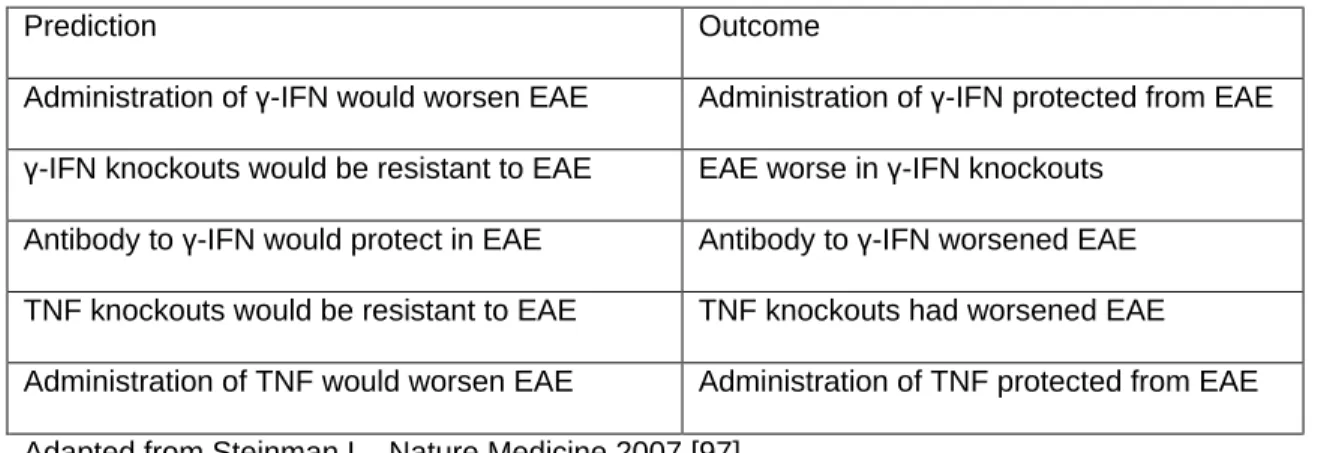
However, some key experiments performed in EAE required the revision of this theory. The flaws of the theory include: IFNγ injections protected against EAE, antibodies to IFNγ worsened EAE, IFNγ knockouts mice were more susceptible to EAE, and TNF knockouts had an exaggerated EAE and administration of TNF protected mice from EAE (Table 1) [89-94].
These contradictory findings and the experiments to understand the role of the cytokine IL-23 in EAE have helped to decipher the paradox of Th1/th2 hypothesis. IL-23 is a heterodimeric cytokine with p40 and p19 subunit. The p40 subunit is common to
24 Th1 inducing IL-12 and IL- 23. Mice deficient in IL-23 were resistant to various animal models of autoimmunity like IBD, Collagen arthritis and EAE [95, 96].
Table 1. Predictions and outcomes of TH1/TH2 hypothesis in EAE
Prediction Outcome
Administration of γ-IFN would worsen EAE Administration of γ-IFN protected from EAE
γ-IFN knockouts would be resistant to EAE EAE worse in γ-IFN knockouts
Antibody to γ-IFN would protect in EAE Antibody to γ-IFN worsened EAE
TNF knockouts would be resistant to EAE TNF knockouts had worsened EAE
Administration of TNF would worsen EAE Administration of TNF protected from EAE
Adapted from Steinman L, Nature Medicine 2007 [97]
Further, IL-23 plays critical role in amplification of myelin-specific Th17 cells which induce more severe form of EAE than IL-12-driven Th1 cells [98]. Treatment of mice with antibodies to IL-23 protected them from EAE [99]. IL-17 produced by Th17 cells has pro-inflammatory effect and is involved in tissue damage and autoimmune diseases [100]. Increased level of IL-17 in clinical samples are associated with RA, MS, inflammatory bowel disease, psoriasis and asthma [49]. Several lines of evidences confirm that Th17 cells are the main pathogenic cells in autoimmunity and systemic inflammatory diseases [73, 101-116]. The fact that Th1 cells can also transfer organ specific autoimmunity [117], the present consensus is that both Th1 and Th17 cells are involved in T cell mediated pathology. Th17 cells which are generated early are known to reach the site of inflammation and initiate migration of other inflammatory cells (example Th1) which propagates the tissue damage [118]. Cytokines produced by Th17 cells such as IL17A, IL17F, IL-21, IL-22 and GM-CSF are involved in recruitment of other inflammatory and effector immune cells at the site of inflammation thus leading to
25 tissue destruction [49, 119, 120]. Fate mapping studies in EAE revealed that Th17 are the main cells infiltrating CNS and subsequently produce IFNγ shutting off the production of IL17 [121]. Th17 express podoplanin which mediates formation of ectopic lymphoid structures which are the hallmarks of many chronic autoimmune and inflammatory conditions [122]. Altogether, Th17 cells are indispensible for induction of autoimmune and inflammatory diseases.
2.2. Dysregulated equilibrium between pathogenic and regulatory T cells leads to immune disease
IPEX (immune dysregulation, polyendocrinopathy, enteropathy, X-linked syndrome) is a condition resulting from mutations in foxp3 gene resulting dysfunctional Tregs [15]. Compromised functions of Tregs are known to be associated with many autoimmune diseases such as MS, autoimmune polyglandular syndrome type II, SLE, type 1 diabetes, psoriasis, myasthenia gravis, RA, and chronic ITP [66, 123-126]. Depletion of Tregs before and after induction of autoimmune disease leads to exaggerated disease with increased cellular and humoral responses [127]. Adoptive transfer of Tregs in the animal models of autoimmunity decreased severity of the disease [14, 18]. Recovery phase of many inflammatory diseases is associated with increase in number of Tregs in the target organs. Thus, Tregs actively regulate autoimmunity throughout the lifespan and are indispensible for maintenance of immune homeostasis [16]. Reprogramming of Tregs by pathogenic T cells and various inflammatory agents renders them less suppressive. Pathogenic Th17 cells are known to develop at the cost of Tregs under severe inflammatory situations of increased IL-6 [41, 128]. Thus, balance between
26 regulatory and effector T cells functions appears crucial for homeostasis and is intricately controlled by various pro and anti-inflammatory mediators [58, 73].
3. Intravenous Immunoglobulin
3.1. Composition, pharmacology and indications of IVIg
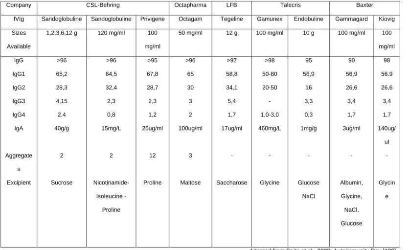
IVIg is a therapeutic concentrate of polyclonal IgG obtained from pools of plasma of a large number of healthy blood donors. Preparations of IVIg contain at least 96% of IgG with traces of IgA and IgM (Table 2). The repertoire of IVIg is relatively wide as it obtained from large number of donors [129]. IVIg has a high content of self-reactive NAbs (Natural antibodies) which can bind to various self-antigens and pathogen specific antibodies [130]. Initially used as replacement therapy for patients with immune deficiencies, IVIg is now widely used for the treatment of a large number of autoimmune and systemic inflammatory diseases including ITP, neuromuscular and neuro-immunological diseases such as acute Guillain–Barré syndrome, myasthenia gravis,
acute or chronic inflammatory demyelinating polyneuropathy, or stiff person syndrome [131] (Table 3). IVIg is also proven valuable for refractory dermatomyositis or multifocal motor neuropathy. The efficacy of IVIg in relapsing–remitting multiple sclerosis (RRMS)
is not as extensively documented as for other disease modifying drugs, but available data suggest its beneficial effects in this condition and IVg is used as second line drug
for patients not responding or not supporting first-line treatment. [132]. Dose regimen of
IVIg is according to the therapeutic goals, As a replacement therapy IVIg is used at 300-500 mg/kg body weight every 3-4 weeks. As an immunomodulatory/anti-inflammatory therapy it is used at 1-2 g/kg, administered at once or divided into 5 daily doses; additional maintenance dose at 4-6 week interval [133, 134]. IgG plasma concentration
27 of 12-14 mg/ml and 20-35 mg/ml is reached after replacement and high dose therapy, respectively [135].
Table 2 : Composition of different IVIg preparations
Company CSL-Behring Octapharma LFB Talecris Baxter
IVIg Sandoglobuline Sandoglobuline Privigene Octagam Tegeline Gamunex Endobuline Gammagard Kiovig
Sizes Available 1,2,3,6,12 g 120 mg/ml 100 mg/ml 50 mg/ml 12 g 100 mg/ml 10 g 100 mg/ml 100 mg/ml IgG >96 >96 >95 >96 >97 >98 95 90 98 IgG1 65,2 64,5 67,8 65 58,8 50-80 56,9 56,9 56.9 IgG2 28,3 32,4 28,7 30 34,1 20-50 16 26,6 26,6 IgG3 4,15 2,3 2,3 3 5,4 - 3,3 3,4 3,4 IgG4 2,4 0,8 1,2 2 1,7 1,0-3,0 0,3 1,7 1,7
IgA 40g/g 15mg/L 25ug/ml 100ug/ml 17ug/ml 460mg/L 1mg/g 3ug/ml 140ug/
ul
Aggregate
s
2 2 12 3 - - - - -
Excipient Sucrose Nicotinamide-
Isoleucine -
Proline
Proline Maltose Saccharose Glycine Glucose
NaCl Albumin, Glycine, NaCl, Glucose Glycin e
28
Table 3: Clinical use of IVIg in autoimmune and inflammatory conditions
Licensed Off-Label
Idiopathic thrombocytopenic purpura Acquired immune thrombocytopenia Guillain Barre syndrome (GBS)a Autoimmune neutropenia
Chronic inflammatory demyelinating polyneuropathy (CIDP)
Autoimmune hemolytic anemia
Kawasaki Disease (KD)a Parvovirus B19-associated red cell aplasia Multifocal motor neuropathy (MMN)a Anti-Factor VIII autoimmune disease Birdshot retinochoroidopathy
(BSRC)
Multiple sclerosis Myasthenia Gravisa
Lambert Eaton myasthenic syndrome Stiff person syndrome
ANCA-positive systemic vasculitis Polymyositis
Dermatomyositisa
Antiphospholipid antibody syndrome Rheumatoid arthritis and Felty’s syndrome Systemic lupus erythematosus (SLE) Juvenile idiopathic arthritis (JIA) Toxic epidermal necrolysis (TEN)
Autoimmune skin blistering diseases (BP, PF, PV)
Steroid-dependent severe atopic dermatitis Graft versus host diseasea
Antibody-mediated rejection (AMR) of the graft Sepsis syndrome
Steroid-dependent severe atopic dermatitis Asthma
a Indicates diseases in which evidence for the effects of IVIg has been obtained in controlled trials.
29
3.2. Immunoregulatory mechanisms of IVIg in autoimmune and inflammatory diseases
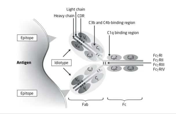
Several mutually non-exclusive immunoregulatory effects of IVIg have been described that apparently contribute in synergy to an effective therapy in various clinical settings [137]. IVIg contains a wide range of anti-idiotypic antibodies that regulate autoreactive B-cell clones and neutralize pathogenic autoantibodies [134, 138, 139]. IVIg saturates the IgG transport receptor [6, 140], which leads to accelerated catabolism of pathogenic auto-antibodies and modulates the affinity of FcγR on phagocytic cells. Studies based on animal models of antibody mediated autoimmunity show that IVIg up-regulates inhibitory FcγRIIB on splenic macrophages [141]. FcγRIIB mediated beneficial effect of IVIg is known to be dependent on α2,6-linked of sialic acid to galactose on the glycan at Asn297 in the CH2 region of Fc fragment small fraction of IgG (sIgG). IVIg
contains a small fraction (1-2%) of sIgG in it. sIgG fraction of IVIg or α2,6 sialylated recombinant human IgG1 Fc protein could reproduce the benefits of IVIg when used at much lower dose [142]. sIgG has been demonstrated to interact with the C-type lectin receptor (SIGN-R1) on myeloid cells that up-regulates the expression of FcγRIIB on ‘effector MΦ’ via TH2 pathway [143].
IVIg attenuates complement-mediated damage by scavenging to the activated C3b fraction of C3 [144, 145]. The interaction of IVIg with complement proteins, therefore, prevents the generation of the C5b–9-membrane attack complex and subsequent
complement-mediated tissue damage in muscle microvasculature and brain [145-147]. IVIg modulates cytokine and chemokine production by various cell types: decreased
30 levels of the pro-inflammatory cytokine IL-1 and increased levels of IL-1R antagonist have been reported in patients following IVIg infusion; TGF-α1 was down-regulated in the muscles of dermatomyositis patients who responded to IVIg therapy; Other studies have shown reduced synthesis of IL-2, IL-3, IL-12, IL-22, and GM-CSF following IVIg therapy [147-150]. IVIg contains antibodies that interact with cytokines and membrane molecules such as the T-cell receptor, cytokine or chemokine receptors, CD4, CD40 and CD95, which have important roles in the balance between auto-reactivity and tolerance [151, 152]. Further, IVIg is shown to exert an impact on the cellular compartment of the immune system; IVIg directly interacts cells of adoptive immunity like B cells, T cells and that of innate immunity and modulate their functions [153]. The maturation state of key APCs like DC is known to regulate immune response and tolerance. Immature and semi-mature DC presenting antigens are known to maintain tolerance by inducing Tregs while mature DC induces strong immune response [154]. IVIg inhibits maturation and function of DC, also modulates the pattern of cytokines secreted by these cells. By down-regulating the interferon-γ-mediated differentiation of DCs, and by inhibiting the uptake of nucleosomes, IVIg might exert an immunoregulatory effect in patients with lupus [155, 156]. In addition, IVIg-treated DC ameliorates ongoing autoimmune disease in vivo upon adoptive transfer [157]. IVIg also modulates in vivo and in vitro T-cell responses by impairing antigen presentation [158].
31
3.3. Modulation of the Tregs by IVIg
The immune system is subject to multiple regulatory mechanisms to control undesired pathogenic immune response to self-antigens and inflammation. One of these regulatory mechanisms involves suppression of auto-reactive T cells by Treg. Treg express GITR, CCR4, CD62L, CTLA-4 and lineage-specific transcription factor Foxp3 [15, 67]. Treg actively regulate autoimmunity throughout an individual’s life and an imbalance in their immune regulation might lead to autoimmunity. Maintenance of immune tolerance by Treg is not attributed to a single mechanism or target cell; rather it involves several pathways targeting multiple cell types. Thus, Treg inhibit the proliferation and cytokine production by conventional T cells and can also regulate the functions of natural killer cells, NKT cells and professional APC. The suppression of immune responses by Treg generally requires direct cell-cell contact implicating CTLA-4, CD39 and LAG-3, but soluble factors, particularly TGF-β and IL-10, have also been implicated. Ability of IVIg to modulate the functions of DC has opened the possibility that the tolerogenic effects of IVIg may implicate the Treg in correcting autoimmunity. Further, the FcγR-mediated effects of IVIg cannot entirely account for its benefit in a number of peripheral and central demyelinating diseases where auto-reactive T cells play critical role. Since the expression of FcγR on T cells has not been established unequivocally, the observed beneficial effects raise certain speculations, that is, if these effects could be attributed to a direct interaction of IgGs with T cells or as mentioned above, an indirect influence via DC. Indeed IVIg manifests its protective effect in T-cell-dependent pathologies via an early modulation of auto-reactive T cells. EAE is a
32 CD4+T-cell-mediated autoimmune disease affecting the CNS [159]. The prophylactic infusion of IVIg prevents the development of EAE [160-165], and this protection conferred by IVIg is associated with a peripheral expansion of CD4+CD25+Foxp3+Treg and amelioration of their functions [165].
The IVIg-expanded Treg were more efficient in suppressing the in vitro response of TCR-stimulated CD4+CD25-T cells as compared to Treg from control group; in adoptive transfer experiments, mice that were reconstituted with Treg from IVIg-treated mice developed milder EAE as compared to non-reconstituted mice; IVIg failed to protect against EAE in mice that were depleted of the Treg [165]. Expansion of Treg by IVIg, described above, suggests that IVIg imposes immune tolerance via modulation of T-cell subsets, in particular, the CD4+CD25+Foxp3+Treg compartment [166]. However, the identification of Th17 cell subset has raised more questions on the mechanisms of action of IVIg in inflammatory and autoimmune conditions. IVIg inhibits in vitro, differentiation and amplification of human Th17 cells [167]. In view of the critical role played by Th17 cells in inflammatory and autoimmune processes, an important question concerns the effect of IVIg on the differentiation and function of Th17 population in vivo.
33
4 Multiple sclerosis / EAE is an organ specific T cell mediated auto-immune disease
Multiple sclerosis (MS) is a demyelinating disease of central nervous system affecting more than 2.5 million people worldwide [168]. MS predominately affects younger women and depending on the region, incidence may rise up to 3 in 1000. MS occurs in various forms like benign, relapsing/remitting, secondary progressive and primary progressive. Perturbation in sensation, motor, autonomic, visual and cognitive systems are the main features of MS in addition to optical neuritis. MS is the primary inflammatory disease of the CNS, characterized by perivascular inflammation and massive leukocyte infiltration leading to axonal loss. Animal model for MS is experimental autoimmune encephalomyelitis (EAE). EAE can be induced in a various species of experimental animals by injection of a myelin peptide emulsified in complete Freund’s adjuvant (CFA) subcutaneously [169].
The pathophysiology of MS is complex and heterogeneous; however some aspects of it are being understood recently. Several lines of evidence suggest that MS has an autoimmune etiology [170, 171]. T lymphocytes and antibodies reactive to myelin were found in the lesion in MS patients [172]. These T cells are mainly CD4+ and specific for the various myelin proteins such as MBP (myelin basic protein), MOG (myelin oligodendrocyte glycoprotein), PLP (myelin proteolipid) and MAG (myelin-associated glycoprotein). Pathogen-(myelin-associated proteins, mainly from hepatitis B virus (HBV) resemble myelin proteins and are antigenic suggesting that, molecular mimicry may be the underlying cause for onset [173]. Further, adoptive transfer of T cells specific for MBP or CNS antigens in the experimental animals leads to MS like
34 syndrome is the proof that MS is T cell mediated pathology [174].
Naïve Th cells reactive to various epitopes of MBP presented in the context of MHC-II by APCs are activated in the periphery. These activated cells migrate to CNS using adhesion molecules like LFA-1 and VLA-4 [175]. Secondary wave of T cell activation and amplification in the CNS is mediated by resident APCs in the CNS [176]. Initially, Th1 cells were thought to be pathogenic in MS. However, recent studies exploring the functions of IL-12 and IL-23 revealed that Th17 cells are the essential lymphocytes in pathogenesis of MS [95]. Th17 cells secrete IL-17 family cytokines like IL-17A, IL-17F and GM-CSF [49, 120]. These cytokines are key players in initiating early events of demyelination. Additionally Th17 cells express podoplanin, which mediates formation of ectopic lymphoid structures in CNS thus resulting in to amplified immune response. The released inflammatory cytokines IFN-γ, IL-23 and TNF-α activate microglia and astrocytes. Chemokines like RANTES, IL-8 recruit other immune cells such as monocytes, CD8+ T cells and B cells from blood.
Mechanisms of demyelination include direct deposition of complement, antibody dependent cellular cytotoxicity, phagocytosis and probably progression to direct attack of axons by cytotoxic T cells, secretion of proteases by neutrophils and apoptosis of oligodendrocytes [177]. The inflammation lasts from few days to two weeks in case of EAE and longer in chronic form of MS. After the attack phase of the CNS, demyelinated axons and apoptotic oligodendrocytes and T lymphocytes were observed [178]. During the resolving phase of inflammation astrocytes proliferate and there is shift to Th2 cytokine profile including IL-10 and TGF-β [58]. Increase in Tregs number in the CNS is another hallmark of resolving inflammation [82].
35
5 Hypothesis and aims
Several lines of evidence show that Th17 cells might be the potent inducers and sustainers of inflammation and play critical role in the pathogenesis of autoimmune and inflammatory diseases. Cytokines produced by Th17 cells such as IL17A, IL17F, IL-21, IL-22 and GM-CSF are involved in recruitment of other inflammatory and effector immune cells at the site of inflammation thus leading to tissue destruction. Tregs on the other hand actively regulate autoimmunity throughout the lifespan of an individual and are indispensible for maintenance of immune homeostasis. Many autoimmune diseases are associated with reduced numbers of Tregs or defects in their functions. Thus, the balance between effector Th cells (Th17) and Treg cells might critically influence the outcomes of many human immune mediated diseases.
IVIg is a therapeutic preparation of normal human polyclonal IgG obtained from pools of plasma from a large number of healthy blood donors. High dose therapy of IVIg is being widely used to treat various autoimmune and inflammatory conditions. Understanding the molecular mechanisms by which IVIg exerts its beneficial effects has been a challenge. IVIg is known to exert its beneficial effects by several mutually non-exclusive mechanisms. Many of these are based on animal models of antibody mediated autoimmunity and in-vitro studies. Nevertheless, IVIg is also widely used in much T cell-mediated autoimmune pathologies. The beneficial effect of IVIg in EAE, an animal model of T mediated pathology, is associated with a peripheral expansion of CD4+CD25+Foxp3+Treg and amelioration of their functions. The emerging knowledge on Th17 cells as potent pathogenic T cells in autoimmunity and Th17 and Treg exercise
36 reciprocal regulatory effects has raised additional interesting questions on the mechanisms of IVIg in vivo. Although, IVIg is shown to interact directly with T cells and inhibit differentiation and amplification of human Th17 cells in vitro, effect of IVIg on differentiation of Th17 cells in vivo remains unexplored. I therefore, hypothesise that IVIg inhibits the Th17 cell development and regulates the balance between Th17 and regulatory T cells in vivo. I have studied this hypothesis with the following objectives:
Extend the knowledge on the protective effect of IVIg in a T cell-mediated
pathology
Understand whether beneficial effect of IVIg in EAE is associated with modulation of Th17 cells along with Th1, Th2 and Tregs
Investigate the molecular mechanisms involved in modulation of T cells by IVIg in vivo in an animal model of T cell mediated autoimmunity
I used active EAE as in vivo model of autoimmune disease to accomplish the objectives. EAE was induced in mice by injecting MOG35-55 peptide emulsified in CFA
with additional PTX. Effect of IVIg on frequency of Th1, Th17, Th2 and Tregs in EAE was measured by using cell specific markers Th1: IFN γ; Th17: IL-17; Th2: IL-4 and Tregs: Foxp3.
37
Results
38
6 Results
Intravenous gammaglobulin reciprocally regulates effector and regulatory CD4 T cell functions in vivo independent of FcγRIIB
(In communication)
Shivashankar Othy,a,b Pushpa Hegde,a,b,d Selma Topçu, a,b Meenu Sharma, a,b.d Mohan S. Maddur,a,b Sebastien Lacroix-Desmazes,a,b,c,e Jagadeesh Bayry, a,b,c,e and Srini V. Kaveria,b,c,e a,Institut National de la Sante et de la Recherche Medicale, Unité 872, 15 rue de l’Ecole de Médicine, Paris-75006, France;
b Université Pierre et Marie Curie – Paris 6, UMR S 872, Centre de Recherche des Cordeliers, Equipe 16-Immunopathology & therapeutic immunointervention, Paris-75006, France ;
c Université Paris Descartes, UMR S 872, Paris; d
Université de Technologie de Compiègne, Compiègne, France; e
International Associated Laboratory IMPACT (Institut National de la Santé et de la Recherche Médicale, France - Indian Council of Medical Research, India), National Institute of Immunohaemotology, Mumbai, India
Correspondance: Srini V. Kaveri and Jagadeesh Bayry Inserm U 872, Centre de Recherche des Cordeliers, Equipe 16 Immunopathology and therapeutic immunointervention, 15, Rue de l’Ecole de Medecine,75006Paris, France;
Phone +33144278201/03 ; FAX : +33144278194
39
Abstract
Despite an increasing use of high dose therapy of intravenous gammaglobulin (IVIg) in the treatment of T-cell and autoantibody-mediated inflammatory and autoimmune diseases, comprehension of the mechanisms underlying its therapeutic benefit has remained a major challenge. Using actively-induced experimental autoimmune encephalomyelitis (EAE) model, a T cell-mediated autoimmune condition, we demonstrate that IVIg inhibits the differentiation of naïve CD4 T cells into effector subsets (Th1 and Th17 cells) and
concomitantly induces an expansion of Foxp3+ regulatory cells. Further, IVIg
renders effector T cells less pathogenic by decreasing the expression of encephalitogenic molecular players like GM-CSF and podoplanin. IVIg decreases the expression of sphingosine-1 phosphate receptor (S1P1) on CD4 cells, thus sequestering these cells in the draining lymph nodes and decreasing infiltration of Th1, Th17 and Treg to the central nervous system. Intriguingly and contrary to the current arguments, the inhibitory FcγRIIB is dispensable for IVIg-mediated reciprocal modulation of effector and regulatory CD4 subsets in vivo.
Additionally, F(ab’)2 part of IVIg also retained this function of IVIg. IVIg or F(ab’)2
fragments decreases the activity of mTOR kinase thus restoring the equilibrium
between regulatory T cells and inflammatory CD4 T cell subsets. Together, our results provide cellular and molecular basis underlying the beneficial effect of IVIg in certain autoimmune and inflammatory conditions.
INTRODUCTION
High dose therapy of IVIg is being widely used to treat various autoimmune and inflammatory conditions1-3. IVIg is a therapeutic preparation of normal human polyclonal IgG obtained from pools of plasma from a large number of healthy blood donors4. Understanding the cellular and molecular mechanisms by which IVIg exerts its anti-inflammatory effects in highly diverse pathological situations incriminating autoantibodies, pathogenic T cells, complement-mediated tissue damage or dysregulated cytokine network has rendered the area particularly challenging. Indeed, IVIg exercises a therapeutic effect in idiopathic thrombocytopenic purpura, Kawasaki disease, myasthenia gravis, dermatomyositis, pemphigus, anti-neutrophil cytoplasmic antibody–associated vasculitis and a number of other diseaseses5
.
IVIg exerts its beneficial effects by several mutually non-exclusive mechanisms
6-10. Many of these effects are deduced based on animal models of antibody-mediated
autoimmunity and ex vivo studies11. IVIg impacts significantly on both innate and adaptive immune compartments7. More recently, it is shown that IVIg up-regulates inhibitory FcγRIIB on macrophages through a Th2 pathway in a K/BxN serum transfer arthritis model12. However, therapeutic benefit of IVIg is also clearly established in several T cell-mediated autoimmune pathologies2,13-15. The beneficial effect of IVIg in EAE, an animal model of multiple sclerosis and a T cell-mediated pathology, is associated with a peripheral expansion of CD4+CD25+Foxp3+ regulatory T cells (Treg)
40 and a significant amelioration of their functions16. Further, IVIg interacts directly with T cells and inhibits differentiation and amplification of human Th17 cells in vitro17. Hence, emerging knowledge that Th17 cells as potent pathogenic T cells in autoimmunity and that Th17 and Treg exercise reciprocal regulatory effects18, have raised additional interesting possibilities on the mechanisms of IVIg in vivo.
We surmised that IVIg restores the dysregulated equilibrium between Th17 cells and Treg in T cell-mediated pathologies. In the present study using actively-induced EAE, we thus set out to investigate whether IVIg modulates T cell polarization in vivo and tilts the balance from pathogenic effector T (Teff) cells towards Treg. We found that
IVIg exerted its beneficial effect in EAE by inhibiting the differentiation of Th17 and Th1 cells and simultaneously increasing Treg. Further, IVIg inhibited encephalitogenic potential of pathogenic T cells and interfered with their trafficking to the target organ. Interestingly, FcγRIIB was dispensable for this effect and F(ab’)2 fragments of IVIg
recapitulated the effect of intact IgG molecules in reciprocally modulating Teff cells and
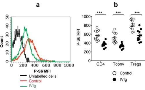
Treg. This reciprocal regulation involved modulation mammalian target of rapamycin (mTOR) kinase in CD4 cells by IVIg.
RESULTS
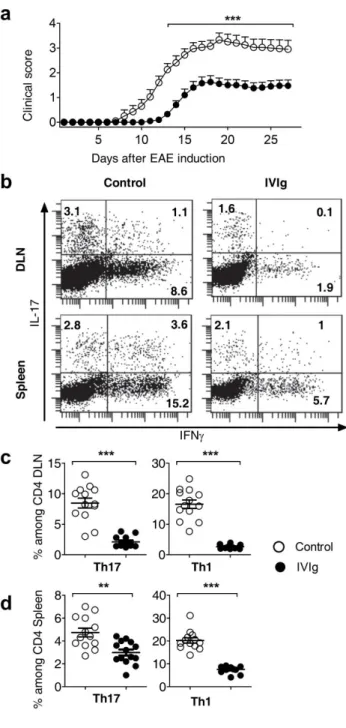
IVIg delays the onset of EAE and decreases severity of the disease by inhibiting Th17 and Th1 cells and increasing Treg
To understand the modulation of effector and regulatory CD4 subsets by IVIg in an autoimmune set up, EAE was induced in WT C57BL/6J mice using MOG35-55
emulsified in CFA. From the day of immunization to the peak of the disease (day16-18), mice in the control and IVIg group were treated with 0.2M Glycine and IVIg (Gamunex ®) respectively. Control mice started to display clinical signs from day 7 and mean score at the peak was 3.5. IVIg significantly delayed the onset of EAE (day 11) and decreased severity of the disease as shown by clinical signs (Fig. 1a). MOG35-55-specific naïve
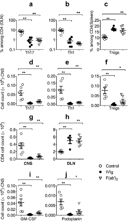
CD4 cells differentiating into Th17 and Th1 cells are known to be the pathogenic in EAE19,20. To investigate whether IVIg affects differentiation of Th17 and Th1 cells in EAE, mice in each group were sacrificed 9 days after EAE induction (onset) and analyzed for their signature cytokines (IL-17 in Th17; IFNγ in Th1) by flow cytometry. We observed decrease in Th17 and Th1 cells in inguinal draining lymph nodes (DLN) of IVIg-treated mice (Fig. 1b, top panel and 1c). Similar trend was also observed in spleens of IVIg-mice (Fig. 1b, lower panel and 1d). Inhibition of Th17 and Th1 cells by IVIg in vivo was further confirmed by the profile of cytokine secretion. Cells from DLN and spleen on day 9 were stimulated ex vivo with MOG35-55 for 24 hours and cell-free
supernatants were analyzed for cytokines. Cells from IVIg-treated mice secreted decreased amounts of IL-17 and IFNγ (Supplementary Fig. 1a, b) as compared to control. IVIg did not affect the CD4+Foxp3+ Treg in DLN (Supplementary Fig. 2a, b). However, we observed concomitant increase in CD4+Foxp3+ Treg in the spleens of IVIg-treated mice (Supplementary Fig. 2c, d), which is in consistent with our previous report16. Thus, IVIg reciprocally modulates Teff and Treg in EAE.
41
IVIg decreases infiltration of lymphocytes to the CNS by inhibiting their egress from the DLN
Protective effect of IVIg treatment in EAE is associated with decreased number of lymphocytes and absence of inflammatory foci in the CNS14,16. It has been previously shown that IVIg interferes with leucocyte recruitment to the CNS in a α 4-integrin-dependent manner21. However, Th17 and Th1 cells use different strategies to invade CNS22,23. To investigate the effect of IVIg on trafficking of Th17 and Th1 in EAE, brain and spinal cords were analyzed on the day of onset (Day 9). IVIg inhibited infiltration of Th1, Th17 and Treg into the CNS (Fig. 2a-c). Accordingly, there was an overall decrease in absolute number of CD4 cells found in the CNS (Fig. 2d). Surprisingly, DLN of IVIg-treated EAE mice were bigger in size and contained two to five-fold more number of CD4 cells than the untreated EAE mice (Fig. 2e). We also observed decrease in total number of CD4 cells circulating in the blood (Fig. 2f). These data suggested that, T cell entry into DLN of IVIg-treated EAE mice was intact; however, their exit from DLN was affected. To further probe into the molecular mechanisms involved in sequestering of CD4 cells into the DLN, we investigated sphingosine 1 phosphate (S1P)–S1P receptor1 (S1P1) axis, which controls the trafficking and migration of lymphocytes24,25. IVIg treatment in EAE mice for 6 days decreased CD4+S1P1+ cells in the DLN (Fig. 2g, h) and MFI of S1P1 on CD4 cells (Fig. 2i). These results suggest that, IVIg down-regulates S1P1 on CD4 cells leading to inhibition of their egress from DLN, thus explaining the increase in the size of the DLN and decrease in infiltration of lymphocytes to CNS.
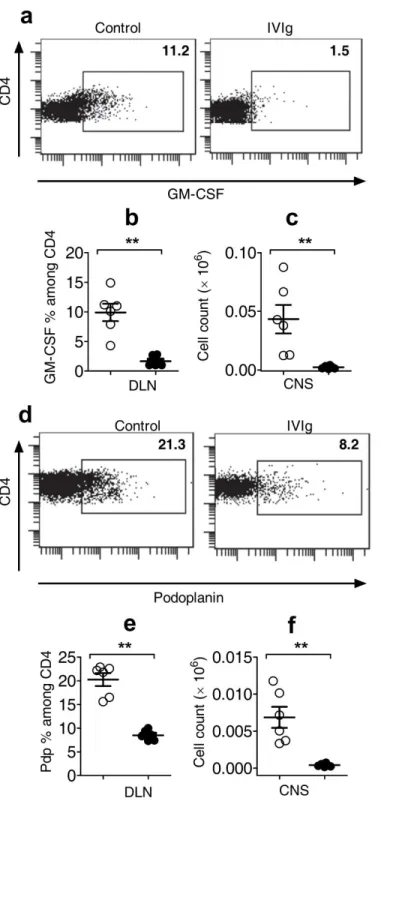
IVIg down-regulates the expression of GM-CSF and podoplanin (PdP) in CD4 T cells
Both Th17 and Th1 cells are involved in EAE, however Th17 cells have emerged as the main pathogenic mediators26. Signature cytokines of Th17 cells like 17A, 17F, IL-21 and IL-22 are dispensable for the induction of EAE27. Encephalitogenic potential of Th17 cells in EAE has been attributed to molecules like GM-CSF28 and podoplanin29. Injected to EAE mice, IVIg significantly decreased CD4+GM-CSF+ cells in DLN (Fig. 3a, b). Additionally, there was a decrease in the absolute number of these cells in CNS
(Fig. 3c). Similarly IVIg also down-regulated the expression of podoplanin on CD4 cells
in EAE (Fig. 3d-f). Thus, in addition to inhibiting the differentiation of Th17 cells, by down-regulating GM-CSF and PdP, IVIg may render these cells less encephalitogenic.
FcγRIIB is dispensable for IVIg-mediated reciprocal modulation of effector and regulatory CD4 subsets in vivo.
Anti-inflammatory effect of IVIg in several animal models of antibody-mediated pathology is attributed to the inhibitory Fc receptor FcγRIIB12,30-32. The role of FcγRIIB in protection of EAE mice by IVIg is however unexplored. To examine whether reciprocal modulation of CD4 subsets by IVIg in EAE is dependent on FcγRIIB, we induced EAE in FcγRIIB-/- mice under C57BL/6J background and analyzed various CD4 subsets 9 days after immunization. IVIg inhibited Th17 and Th1 cells in FcγRIIB-/--EAE mice (Fig. 4a,