HAL Id: tel-01191474
https://tel.archives-ouvertes.fr/tel-01191474
Submitted on 2 Sep 2015HAL is a multi-disciplinary open access archive for the deposit and dissemination of sci-entific research documents, whether they are pub-lished or not. The documents may come from teaching and research institutions in France or abroad, or from public or private research centers.
L’archive ouverte pluridisciplinaire HAL, est destinée au dépôt et à la diffusion de documents scientifiques de niveau recherche, publiés ou non, émanant des établissements d’enseignement et de recherche français ou étrangers, des laboratoires publics ou privés.
Rôle du Granulocyte-Colony Stimulating Factor
(G-CSF) dans le Processus Implantatoire, chez la
Femme et en Modèle Murin ”
Mona Rahmati
To cite this version:
Mona Rahmati. Rôle du Granulocyte-Colony Stimulating Factor (G-CSF) dans le Processus Implan-tatoire, chez la Femme et en Modèle Murin ”. Immunologie. Université Paris Sud - Paris XI, 2014. Français. �NNT : 2014PA11T048�. �tel-01191474�
1
UNIVERSITE PARIS-SUD
ECOLE DOCTORALE BIO-SigNE, ED419Research Team:
“Embryo Implantation and Cytokinic Dialogue between Mother and Conceptus” INSERM UMRS-976, Hopital Saint Louis, Paris, France
SCIENCE THESIS IN REPRODUCTION AND DEVELOPMENT
THÈSE DE DOCTORAT SUR TRAVAUX
Public Presentation and Defence on 26th of September 2014 byMona Rahmati
Granulocyte-Colony Stimulating Factor and Embryo
Implantation Process:
Effects on Human Endometrium and on Murine Abortion
Prone Model CBA/J x DBA/2
Director of Thesis: Mrs Nathalie Ledee, MD PhD MCU-PH, Hopital des Bluets, INSERM U976
Co-Director: Mr Gerard Chaouat, MD PhD DR, INSERM U976
President of the Jury: Mr Michael Schumacher, PhD DR, INSERM U788, Directeur de l’ED419 Reporter: Mrs Elisabeth Menu, PhD DR, Institut Pasteur
Reporter: Mrs Celine Mehats, PhD CR, Institut Cochin
Examiner: Mr Michael Summers, MD PhD PU-PH, London Bridge Fertility Centre
Examiner: Mr Richard Balet, MD PH, Hopital des Bluets, London Women’s Clinic Examiner: Mr Olivier Sandra, PhD CR, INRA, UMR BDR
2
3
Summary
Reproductive Immunology involves general immunology principles and special aspects of reproduction and development. Colony Stimulating Factors (CSFs) are an illustration of the medical application of this domain. In the CSF family, Granulocyte-Colony Stimulating Factor (G-CSF) appears today as a promising therapy in various cases of reproductive failure although its targets and effects are not clearly established. In this work, through a review on CSFs in reproduction, a study dedicated to human endometrial targets of G-CSF, and a study dedicated to systemic G-CSF supplementation effects on murine embryo implantation, we tried to approach some possible mechanisms of action of this cytokine. In the considered non-abortive and abortion-prone murine models, the timed systemic G-CSF supplementation, targeting specifically the pre implantation endometrium, influenced the embryo implantation process. Some pre conceptual human endometrial dysregulations of CSF target genes were also observed in infertile patients. The endometrial influence of G-CSF on these target genes was also illustrated in an ex-vivo model. These molecules under G-CSF influence are described as critically involved in embryo implantation process, by influencing embryo adhesion, cell migration, tissue remodelling and angiogenesis. These data suggest possible pre-conceptual preventive diagnosis of such reproductive failures and future orientated therapies to optimise the endometrial biosensor and the further embryo implantation and ongoing pregnancy.
Key Words
Colony Stimulating Factors, Granulocyte-Colony Stimulating Factor, Reproductive Immunology, Embryo Implantation, Endometrial Biosensor, Human Endometrium, Murine Abortion Prone Model
4
Résumé
L’immunologie de la reproduction englobe les principes de l’immunologie générale et les aspects spécifiques de la reproduction et du développement. Les Colony Stimulating Factors (CSFs) sont une illustration de l'application médicale de ce domaine. Dans la famille des CSFs, le Granulocyte-Colony Stimulating Factor (G-CSF) apparaît aujourd'hui comme une thérapie innovante dans divers cas d'échec de la reproduction, bien que ses cibles et ses effets ne soient pas encore clairement établis. Dans ce travail, à travers une revue sur les CSFs dans la reproduction, une étude consacrée aux gènes cibles du G-CSF dans l'endomètre humain, et une étude consacrée aux effets de la supplémentation systémique en G-CSF sur l’implantation embryonnaire murine, nous avons essayé d'approcher certains mécanismes d'action possibles pour cette cytokine. Dans les modèles murins fertiles et pro-abortifs, la supplémentation systémique en G-CSF, ciblant spécifiquement l’endomètre préimplantatoire, modifie les taux d’implantation embryonnaire. Dans l’endomètre humain, certaines dérégulations préimplantatoires de gènes cibles du G-CSF ont également été observées chez les patients infertiles. L'influence du G-CSF sur ces gènes cibles a été également illustrée dans un modèle ex-vivo de culture endométriale. Ces cibles dont l’expression est influencée par le G-CSF sont décrites comme des molécules clés dans le processus implantatoire, intervenant sur l’adhésion embryonnaire, la migration cellulaire, le remodelage des tissus et l'angiogenèse locale. Ces données suggèrent des possibilités de diagnostic préventif et pré-conceptionnel de certains échecs de reproduction, considérés jusqu’à maintenant comme idiopathiques, et de thérapies innovantes orientées, afin d’optimiser la réceptivité du biosenseur endométrial afin de permettre une implantation embryonnaire harmonieuse et une grossesse évolutive.
Mots Clés
Colony Stimulating Factors, Granulocyte-Colony Stimulating Factor, Immunologie de la Reproduction, Implantation Embryonnaire, Biosenseur Endométrial, Endomètre Humain, Modèle Murin Pro-Abortif
5
Table of Contents
Summary / Résumé ……….……….. pages 3 / 4 Table of Contents ……….…….... page 5 List of Abbreviations ……….……….. page 7 List of Figures and Tables ……… page 10 Preamble ………..………..………. page 11 Acknowledgements ………..………. page 16
1. Introduction ………..……... page 18
1.1. Reminder on Embryo Implantation ………..………. page 19 1.1.1. Overview of embryo implantation steps (page 19), 1.1.2. Endometrial metamorphosis (page 21), 1.1.3. Dialogue between mother and conceptus (page 23), 1.1.4. Placentation process (page 25), 1.1.5. And when something goes wrong …(page 30)
1.2. Reminder on Reproductive Immunology ……….. page 32 1.2.1. Reminder on general immunology (page 32), 1.2.2. History of reproductive immunology (page 34), 1.2.3. Key immune cells in reproductive immunology (page 36), 1.2.4. Key cytokines in reproductive immunology (page 39)
1.3. Hypothesis and Objectives ……… page 43
2. Colony Stimulating Factors in Reproduction ………..…..…. page 44
2.1. Colony Stimulating Factors (CSFs) ……….……….……… page 45 2.2. CSFs Localisation in the Reproductive Tract……….. page 46 2.3. CSFs Functions in Reproduction ……… page 47 2.4. CSFs Medical Applications ……….… page 47 2.5. Dedicated Original Article ………..……….. page 48
3. G-CSF Effects on Human Endometrium ……….. page 68
3.1. G-CSF in Reproduction ……….………… page 69 3.2. Contribution of a Previous Endometrial Microarray Study ……….…….………. page 69
6 3.3. Use of a Previously Described Endometrial Ex Vivo Model ……….…. page 70 3.4. Dedicated Original Article ………..………….. page 71
4. G-CSF Effects on a Pro Abortive Murine Model ……… page 96
4.1. The Pro Abortive Murine Model CBA/J x DBA/2 ……….. page 97 4.2. The Scheme of G-CSF Supplementation ……….... page 98 4.3. Evaluation of Embryo Implantation and Embryo Resorption ………..… page 99 4.4. Dedicated Original Article ……….. page 100
5. Discussion, Conclusion and Perspectives ………..………. page 121
5.1. G-CSF effects on Human Endometrium ………..………. page 122 5.1.1. G-CSF endometrial target genes (page 122), 5.1.2. Target genes expression in fertile women and infertile patients’ endometrium (page 123), 5.1.3. Endometrial target genes variation under ex vivo G-CSF supplementation (page 123)
5.2. G-CSF effects on a Pro Abortive Murine Model ………. page 125 5.3. Conclusion and Perspectives ……… page 127
6. References ……….. page 131
7. Annexes ………. page 140
7.1. Reference Article for Endometrial Microhistoculture ………... page 141 7.2. Reference Article for Endometrial Microarray Study ………..……….. page 153 7.3. Reference Article for Pro Abortive Murine Model ……… page 165 7.4. Summary in French ………..………….. page 174
7
List of Abbreviations
APC: Antigen Presenting Cell
ART: Assisted Reproductive Therapy BCR: B Cell Receptor
CCL: Chemokine Ligand
cDNA: complementary Desoxyribo Nucleic Acid CSF: Colony Stimulating Factor
CTL: Cytotoxic T Lymphocyte DAF: Decay Accelerating Factor DC: Dendritic Cells
DPC: Day Post Coitum
EGF: Epidermal Growth Factor FGF: Fibroblast Growth Factor
Fn14: Fibroblast Growth Factor inducible-14 G-CSF: Granulocyte Colony Stimulating Factor
GM-CSF: Granulocyte Macrophage Colony Stimulating Factor GP130: Glycoprotein 130
HB-EGF: Heparin Binding Epidermal Growth Factor hCG: human Chorionic Gonadotropin
HLA: Human Leukocyte Antigen IDO: Indoleamine 2,3-dioxygenase IF: Implantation Failure
IFN: Interferon
IGF: Insulin Growth Factor
IGF-BP-1: Insulin Growth Factor Binding Protein-1 IL: Interleukin
8
IP: Intra Peritoneal
ITGB3: Integrin alpha-v/beta3
IUGR: Intra Uterine Growth Restriction IVF: In Vitro Fertilization
JAK: Janus Kinase Family
KIR: Killer cell Immunoglobulin-like Receptor KO: Knocked Out
LAKC: Lymphokine Activated Killer Cells LH: Luteinizing Hormone
LIF: Leukaemia Inhibitory Factor MCP: Monocyte Chemostatic Protein
M-CSF: Macrophage Colony Stimulating Factor MHC: Major Histocompatibility Complex MIP: Macrophage Inflammatory Protein mRNA: messenger Ribo Nucleic Acid MUC-1: Mucine-1
NK: Natural Killer
PDGF: Platelet-Derived Growth Factor PGE2: Prostaglandin E2
PIBF: Progesterone Induced Blocking Factor
PLAUR: Plasminogen Activator Urokinase Receptor RM: Repeated Miscarriages
RT: Reverse Transcription
RT-PCR: Real Time Polymerase Chain Reaction
STAT: Signal Transducers and Activators of Transcription TCR: T Cell Receptor
TGF: Transforming Growth Factor
9
TLR: Toll Like Receptor TNF: Tumour Necrosis Factor Treg: T regulatory lymphocyte
TWEAK: TNF WEAK inducer of apoptosis TYMP: Thymidine Phosphorylase
uNK: uterine Natural Killer
VEGF: Vascular endothelial Growth Factor
10
List of Figures and Tables
Figure 1: Overview of Human embryo implantation steps (page 19) Figure 2: Chronology of uterine events in Human (page 20)
Figure 3: Histological slides of proliferative and secretory endometrium (page 21) Figure 4: Early steps of Human placentation (page 26)
Figure 5: Human trophoblast populations in the first trimester of pregnancy (page 27) Figure 6: Decidualization and placentation in mice (page 28)
Figure 7: Comparative anatomy of human and mouse placentas (page 29)
Figure 8: Normal and pathological Human trophoblast invasion and consequences (page 31) Figure 9: Endometrial uNK immunostaining (CD56+) during middle luteal phase (page 37) Figure 10: General model of haematopoiesis, with CSFs localisation (page 45)
Figure 11: Summary of the G-CSF murine supplementation scheme (page 98) Figure 12: CSFs localisations and functions in female reproductive tract (page 127)
Table 1: Overview of Endometrial Immune Cells (page 39)
11
Preamble
My Medical Thesis was about Colony Stimulating Factors as an example of applied Reproductive Immunology in medicine. During the defence, the President of the Jury, yet amongst the most scientifically involved obstetricians in Paris, asked the first question: - But, Mona, why choosing immunology?
So, when you’re a gynaecologist, your interest towards immunology has to be justified. If only he knew.
How to trap a becoming gynaecologist into reproductive immunology
June 2000. I am a medical student since four years. Final annual results are published in Paris V - Rene Descartes University. I passed “Fundamental and Clinical Immunology” with a “B”. I do promise to myself to never ever approach this highly complex, inconstant and unstable field, in any manner and under any circumstances. Surgery seems a more reliable option.
July 2006. I am a specialist registrar since three years and trying from the first day of my nomination in Obstetrics and Gynaecology to get into scientific research, having the prerequisite ready for a master on embryo development. First I was told that I should learn how to perform caesarean sections and hysterectomies - which was done - and then, that the field seemed specifically dedicated to biologists, geneticists or pathologists - which was more frustrating -.
Beginning of July, a very famous foetal medicine congress takes place in our department, and being the youngest registrar, I have the honour to be continuously on call for a couple
12 of days. The last morning, in a particularly good mood, I meet the senior on call for emergencies. I never met her before. Apparently she’s the youngest consultant in reproductive medicine, Nathalie Ledee, who has had the same honour than me. Given my exceptional mood during the first caesarean section, she tries to understand what could be wrong and I explain her that any innovative field seems to be dedicated to older registrars or simply to non-gynaecologists. And she straight tells me that she can accept master students in her lab:
- Well, it’s not just development, it’s about reproduction and development… With very innovative aspects, of course… Working in a European network of excellence… We should organise your research master for next year… Well, the university deadline was yesterday, but we should be able to arrange…
During the second caesarean section, she has a long non-understandable monologue, most probably explaining the subject of her researches. And when noticing my absence of reactivity, she asks:
- Ok, now, do you know endometrial arteries being invaded by trophoblast during placentation?
- Yes.
- Ok, then, you’re going to work on these arteries before their invasion… What’s your e-mail address?
A few weeks later, I am leaving her office with a pile of thick books and congress reports: - By the way, you have to meet Gerard Chaouat. Here is Marie Petitbarat’s e-mail address, she will tell you where and when. Don’t be impressed and don’t be afraid if you don’t understand anything the first time you listen to him. Sylvie Dubanchet will be next to him, she’ll explain you later.
And while coming out, she gives me an extra article from the amount of papers on her desk, by a Japanese guy talking about Th17… I vaguely remember some Th-something somewhere in my studies, Th1 or 2 and certainly not 17. But where was it then? Oh no! That’s it, I’m
13 trapped. And once you’re trapped, your interest towards reproductive immunology can’t be punctual, you get addicted.
September 2008. After the year of research master, Nathalie Ledee calls me about some delayed articles, presentations and posters:
- It was a nice subject. You should follow with a Science Ph.D. We should try for next year. - You know, I’ll need to take at least two years off from hospital. I’m not sure they’ll propose me the same position after a two years’ gap. I don’t have any specific funding for a thesis project. And I already told you, I can’t fiddle mice, I have a phobia.
- I understand. I have a serious plan in mind. The submission deadline was yesterday, but we should be able to arrange.
And her calling me a few weeks later: “Yes, We Can!”
Reproductive immunology today
Immunology today is a widespread field, involved in any medical sub-speciality. Reproductive immunology is a specific domain, involving general immunology principles and special aspects of reproduction and development. A short review of the history of reproductive immunology is suggested in a further chapter.
Some independent international societies are exclusively dedicated to reproductive immunology, such as International Society for Immunology of Reproduction (ISIR), European Society for Reproductive Immunology (ESRI) or American Society for Reproductive Immunology (ASRI). And many other active groups are within national societies for immunology.
Two internationally admitted journals are specifically dedicated to reproductive immunology: the Journal of Reproductive Immunology (JRI) and the American Journal of Immunology (AJRI).
14 And ultimately, reproductive immunology found its new Holy Book, actually a holy e-book. Gerard Chaouat’s Little Red E-Book, entitled “Immunology of Pregnancy” is co-edited by Nathalie Ledee and Olivier Sandra. This book is not only a collection of quotations from our supreme immunological guide, but the most updated collected data, written by the major international specialists of each field. Given the very active evolution of immunological concepts, I do advise you to hurry in getting and reading it.
Why trying to attract gynaecologists towards reproductive immunology
Once the fundamental part fulfilled, the last dam to pass in reproductive immunology is communicating with the gynaecologists. It seems so unfair, when other fundamental immunologists have to deal with rheumatologists, dermatologists or haematologists who are amongst the most receptive people in medical field, reproductive immunologists have to cope with the most inaccessible, delayed, impatient, rushed, sleepless and sometimes aggressive people in hospitals.
Nevertheless, explaining the interest of reproductive immunology to gynaecologists is vital… And it’s vital for reproductive immunologists. Gynaecologists are the last link in this chain, applying the very precious fundamentally demonstrated principles. Knowing the rational of some pathologies may help in improving infertility treatment or a better obstetrical management. But being aware of such fundamental data may also prevent from inadequate, unsubstantial or even deleterious use of so called immunological treatments. Moreover, gynaecologists are generally more reluctant to new approaches than other specialists, given some sensitive aspects of their field, particularly the risks linked to embryonic or foetal toxicity, and high legal pressures. Introducing reproductive immunology during their training might be helpful for considering further innovative approaches during their profession.
15 I was lucky enough, in my personal curriculum, to meet exceptional people who dared to come to my level and adapt their speech to make me discover their interests. I hope I can transmit their knowledge and arouse more interest around me.
16
Some people I would like to thank
I thank Dr Nathalie Ledee for orienting me towards this fascinating field, suggesting this interesting subject and innovative approaches. Thank you for your constant guidance, support, availability and encouragement.
I thank Dr Gerard Chaouat for this initiation to reproductive immunology, for adapting his knowledge to my level and for the enjoyable co-direction of this work.
I thank Mrs Sylvie Dubanchet and Mrs Marie Petitbarat for their invaluable permanent technical help and friendship during these years.
I thank you all for having accepted me as part of your team and sharing your knowledge as well as your material and technical means. I do not consider these acknowledgements as an end, but as a start to further collaborations.
I thank Pr Armand Bensussan and Dr Jean-Yves Picard for hosting this work in their research units.
I thank Dr Elisabeth Menu and Dr Pascale Chavatte-Palmer for tutoring this work, defining its limits and refocusing it on the main subject during its elaboration.
17 I thank the President of the Jury, Pr Michael Schumacher, and all the Jury Members, Dr Elisabeth Menu, Dr Celine Mehats, Pr Michael Summers, Dr Richard Balet and Dr Olivier Sandra, for their availability and interest.
I thank my latest clinical heads for accepting to adapt my clinical time to fulfil this fundamental project: Pr Marc Dommergues, Dr Richad Balet and Dr Sophie Gaudu.
I thank particularly my parents for their constant support in all ways during all these years, helping me to accomplish my multiple projects. And I thank my husband and children for accepting this unusual life rhythm.
I finally and specially thank my brother, to whom I dedicate this work, for his perpetual help and guidance since ever, allowing me to achieve whatever I want and plan in a much easier and prepared manner.
18
1. Introduction
1.1. Reminder on Embryo Implantation
1.1.1. Overview of embryo implantation steps1.1.2. Endometrial metamorphosis
1.1.3. Dialogue between mother and conceptus
1.1.4. Placentation process
1.1.5. And when something goes wrong …
1.2. Reminder on Reproductive Immunology
1.2.1. Reminder on general immunology1.2.2. History of reproductive immunology
1.2.3. Key immune cells in reproductive immunology
1.2.4. Key cytokines in reproductive immunology
19
1. Introduction
1.1. Reminder on Embryo Implantation
1.1.1. Overview of embryo implantation steps
Despite more than 30 years of remarkable progress in Assisted Reproductive Therapies [1], the embryo implantation remains a black box period, and its study in situ remains impossible in Human, given obvious ethical reasons. Still, this is the putative chronology of the early course of the embryo.
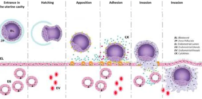
In Human, after a natural fecundation and a ride through the fallopian tube, the embryo reaches the uterine cavity at day 5. The embryo at this time is at the stage of blastocyst, consisting of an inner cell mass enclosed in a trophectoderm shell. After hatching, i.e. emerging from its surrounding zona pellucida, the blastocyst will start its anchoring to a specifically prepared endometrium (Figure 1).
Figure 1: Overview of Human embryo implantation steps From Fitzgerald, Human Reprod Update, 2008
20 Apposition and adhesion are the two first events of the blastocyst attachment to a receptive endometrium. Then the extensive but very tightly controlled process of invasion takes place, to allow the further placentation [2].
The apposition, adhesion and invasion corresponding mechanisms will be detailed in the following paragraphs, together with differentiation of first the ectoplacental cone and then organisation of trophoblast, e.g. placenta, and delimitation of annexes.
The embryo implantation is a highly invasive process, comparable to neoplasia dissemination, which could take place on any organ where the placental vascular bed can spread. Various case reports have already described invasive ectopic pregnancies on peritoneum, spleen, liver, bowel or kidney [2].
Figure 2: Chronology of uterine events in Human
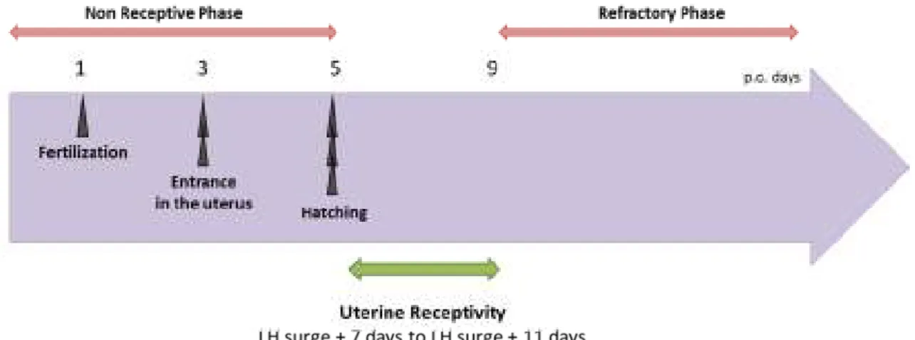
Implantation can occur on any vascularized tissue, but endometrium. During the majority of the endometrial cycle, the embryo will not be able to interact with the endometrium, except during a specific period of five days called the window of implantation (Figure 2).
21
1.1.2. Endometrial metamorphosis
The window of implantation lasts from day 5 to day 9 after ovulation and progesterone secretion from corpus luteum [3]. Specific endometrial changes occurring during this period will define the receptive endometrium.
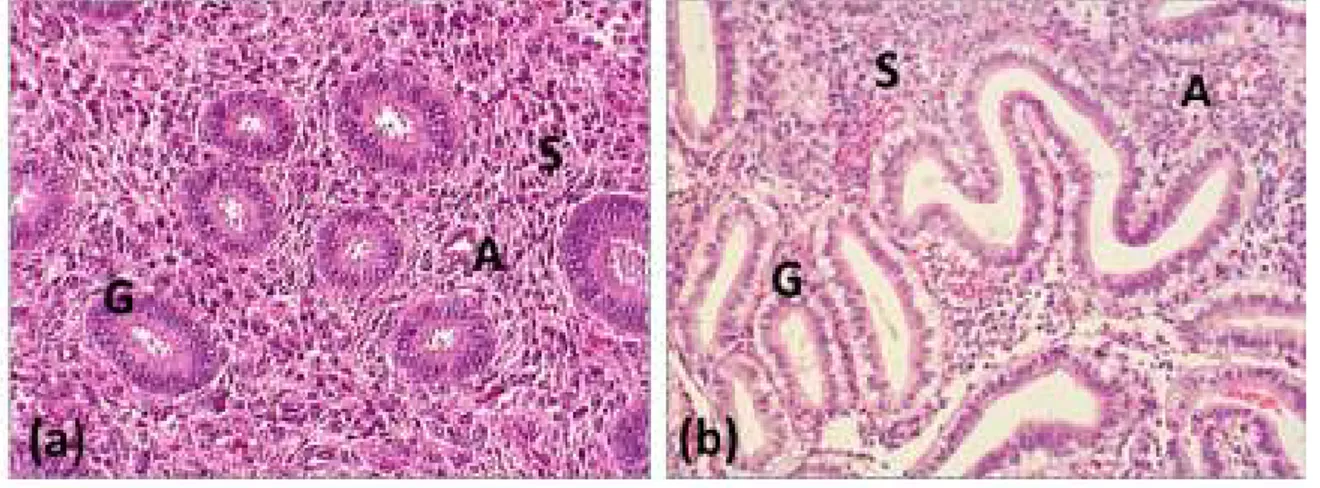
Structural remodelling affects endometrial surface epithelium, endometrial glands, endometrial stroma and endometrial and sub endometrial arteries. This transformation is called endometrial decidualisation and takes place independently from the presence of the embryo (Figure 3). The surface of luminal epithelium increases by emergence of apical pinopodes and microvilli. Endometrial glands turn spiral and secretory. Stromal fibroblast-like cells become large and round. The stromal extra cellular matrix becomes looser and changes its composition [4]. Decidualisation also includes a deep remodelling of endometrial spiral arteries, with endothelial swelling, vacuolisation and vascular smooth muscle disorganisation, essential to a further trophoblast invasion [5]. In Human, this pre implantation decidualisation occurs every cycle, independently from the presence of the embryo itself [4].
Figure 3: Histological slides of proliferative (a) and secretory (b) endometrium Slides from paraffin embedded samples, Haematoxylin and Eosin Staining (x400)
22 At molecular level, the expression and the secretion of various factors underlie the structural endometrial changes. Some specific molecules enable the anchoring process which takes place during the apposition and adhesion steps. The glycocalix which normally recovers the endometrial apex, to turn it anti-adhesive and resistant to microbial attacks, disappears [6]. L-selectin oligosaccharide-based ligands production by endometrial epithelium is up-regulated during the window of implantation, as first receptors to L-selectins expressed on the blastocyst [7] [8]. Tighter anchorage of the blastocyst is allowed by a specific pattern of integrins expression, forming focal adhesion sites on endometrium. Integrins are expressed on both endometrium and blastocyst. Through some extra cellular bridging ligands and the recruitment of a network of cytoskeletal proteins and intracellular signalling complexes, integrins are supposed to mediate cellular adhesion and migration [9]. The swelling extra cellular matrix in receptive endometrium is rich in collagen, fibronectin and laminin, as part of the cellular adhesion and migration system [4].
These expressions are under cyclic regulation of ovarian hormones. Estrogen enhances cell proliferation in uterine epithelium and prepares the specific decidualisation by inducing the local progesterone receptor expression. Progesterone, the essential hormone for embryo implantation and pregnancy maintenance in mammals, has a key role in proliferation and differentiation of stromal, glandular and myometrial cells [10] [11] [12], as well as in the local immune modulation [13] [14] [15]. Further paracrine and embryonic factors help also in creating a transient primary inflammatory reaction followed by a tolerant immune environment necessary to a successful embryo implantation and they will be detailed in the corresponding paragraphs.
Endometrial decidualisation is finally characterised by a local immune switch, from an adaptative immunity protecting the endometrium against infections, to a specific innate immunity, allowing the implantation of the semi-allogenic embryo. The endometrial immune cell population during the implantation window includes uterine Natural Killers cells (uNK), macrophages, regulatory T cells and dendritic cells [16]. The properties of each type of these cells will be detailed further. The majority of this immune population consists of uNK cells [17]. These specific cells do not only take part in the local immune modulation, but above all, promote the spiral arteries remodelling which has already been described [18] through specific secretion of cytokines and angiogenic factors [19] such as, Vascular
23 Endothelial Growth Factor (VEGF), Angiotensin I and II, Interferon gamma (IFNγ) and Nitric Oxide (NO) [20].
1.1.3. Dialogue between mother and conceptus
The decidualised endometrium is described as a biosensor [21] able to establish a dialogue with the coming blastocyst. On one side, the endometrium has to be receptive to start interacting with the embryo, but on the other side, signals from the blastocyst can modulate the maternal answer towards implantation or rejection.
To start the overview of the very delicate, complex and stage specific local equilibrium which is involved in the tissue remodelling that controls uterine receptivity and establishes a mother-conceptus dialogue [22], we can simplify the process by splitting it into two phases: first, a transient pseudo-inflammatory reaction followed by the establishment of local immune modulation.
Schematically, the first inflammatory step in the receptive endometrium, Th1 immunity dominated, is needed for endometrial destabilisation to fulfil the apposition and adhesion process. Endometrial presence of inflammatory cytokines such as Interferon gamma (IFN ϒ),Tumor Necrosis Factor alpha (TNFα), Interleukins 1 , 2 and 6 (IL-1, IL-2, IL-6) or Leukaemia Inhibitory Factor (LIF) enables the anchoring and the cell mobilisation systems for the embryo attachment [23]. During this initial phase the vascular tripod formed by Interleukins 12, 15 and 18 (IL-12, IL-15, IL-18) is essential for the endometrial vascular remodelling through the local control of uNK cells recruitment and activation [24].
Then a local immune modulation, called switch to a Th2 dominated profile, is required to avoid embryo rejection as non self after cell mediated killing and lysis by the endometrial immune cells. Amongst local cytokines, Transforming Growth Factor beta (TGFβ) or
24 Interleukins 4 and 10 (IL-4, IL-10) are described as Th2 cytokines [25]. With this specific environment, endometrial immune cells divert from their cytotoxic capacity whereas uNK cells do not acquire a cytotoxic phenotype, preventing embryo recognition and lysis, while maintaining their trophic and angiogenic factor producer status, helping to maintain embryo invasion and growth [26].
This local immune modulation can be enhanced by the embryo itself. First, the embryo modulates the immune endometrial response by producing several immune suppressive factors such as Indoleamine 2,3-Dioxygenase (IDO) [27], Prostaglandin E2 (PGE2) [28], anti-complement molecules such as Monocyte Chemostatic Protein (MCP) and Decay Accelerating Factor (DAF) [29], and placental microparticles as exosomes [30] [31].
The embryo also enhances this local immune modulation by its specific antigenicity. Trophoblast cells are free of Major Histocompatibility Complex (MHC) Class II (by a phenomenon of hypermethylation) [32], and free of MHC I polymorphic molecules such as Human Leucocyte Antigen (HLA) A or B thus avoiding maternal attacks from cytotoxic T cells. Trophoblastic cells nevertheless express some molecules of the non classical MHC class I monomorphic G and E and the limited polymorphic HLA C, thus limiting maternal attacks from cytotoxic T cells which usually kill non-self cell, while the uterine natural killer KIR system is defusing uNK by expression of HLA-C and on invading extravillous cytotrophoblast HLA-G (see below). Some specific placental components, such as syncytiotrophoblast and extra-villous cytotrophoblast, which are directly in contact with maternal structures, also secrete soluble forms of HLA-G [33] [34] [35]. Both membrane and soluble HLA-G decrease lytic activity of uNK cells and turn their secretions into trophic and angiogenic ones. Soluble HLA-G also stimulates the apoptosis of the local maternal activated Cytotoxic T Lymphocytes (CTL).
When entering the uterine cavity, blastocyst is also shown to be able, via cytokine production of trophectodermic, and later on, for the embryo proper, trophoblastic cells, to modulate endometrial receptivity through regulation of the expression of various adhesion molecules. For example, in vitro models have shown that embryonic secretions, specially Interleukin 1 (IL-1) system, may help the attachment to the endometrial epithelium by
25 enhancing and clustering pro-adhesion membrane molecules such as integrins [36] [37] or by down regulating production of anti-adhesive surface molecules such as mucins [38]. Human Chorionic Gonadotropin (hCG) is another example of embryonic action on the endometrium to facilitate implantation. This glycosylated hormone is mainly produced by syncytiotrophoblast. It stimulates endometrial decidualisation, modulates secretion of Th1 cytokines such as LIF [39] and enhances production of angiogenic factors such as Vascular Endothelial Growth Factor [40] [41]. It also has been demonstrated that hCG influences the recruitment and proliferation of surrounding maternal NK cells [42].
An illustration of this mother-conceptus dialogue, strengthening the hypothesis of the receptive endometrium being a biosensor towards the embryo, was shown in an in vitro confrontation model [43] [44]. Gene expressions varied between decidualised stromal cells in contact with developing blastocysts or non-developing blastocysts. The variation in the decidual expression was orientated towards inhibition of implantation process for non-developing embryos. Such results emphasize the importance of the pre-implantation embryo ability to respond to a receptive endometrium.
These mutual repeated mother-conceptus interactions are essential to enhance embryo implantation but also to control further embryo invasion.
1.1.4. Placentation process
After the adhesion of the blastocyst to maternal endometrium, and the creation of the ectoplacental cone, trophoblast cells differentiate into the outer syncytiotrophoblast and the inner cytotrophoblast. The blastocyst embedding into the endometrial stroma starts with the trophoblast ability to degrade the extra cellular matrix through production of lytic enzymes and triggering apoptosis of endometrial cells. This tissue erosion also affects surrounding capillaries, establishing the first utero-placental circulation system. This process of early embryo implantation ends by the second week of development.
During the third week of gestation, once embryo embedding in the endometrial stroma is completed, primary, secondary and tertiary villi are shaped. Primary villi are formed by
26 penetration of cytotrophoblast into syncytiotrophoblast. Secondary villi are formed by the infiltration of extra-embryonic mesoblast into the primary villi (Figure 4). And the tertiary villi contain blood vessels originated after differentiation from the extra-embryonic mesoblast.
Figure 4: Early steps of Human placentation From Cummings, Pearson Education, 2004
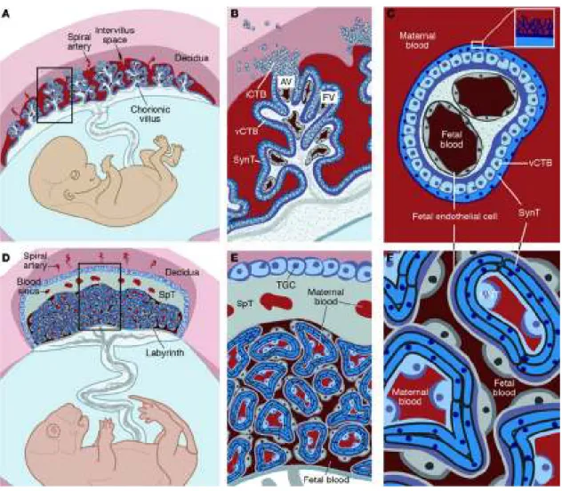
Among the tertiary villi, some will reach the endometrial basal plate, forming the anchoring placental villi by rapid proliferation of cytotrophoblast cells. From these proliferative cells, some highly invasive villous trophoblast cells will start their migration. The extra-villous trophoblast cells can be grouped into two categories [45]: interstitial cytotrophoblast cells invading the endometrium and the proximal third of the myometrium, and the endovascular cytotrophoblast cells, invading and remodelling the decidualised uterine spiral arteries. This process will lead to the establishment of a low resistance vascular system, allowing increased arterial blood flow and facilitating maternal-foetal exchanges in nutrient and gas (Figure 5).
27
Figure 5: Human trophoblast populations in the first trimester of pregnancy From Ashley Moffett-King, Nature Reviews Immunology, 2002
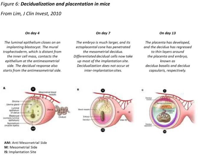
The previously described process in Humans leads to a hemochorial placenta. In mice, placentation is also hemochorial with trophoblast migration and arterial remodelling to a lesser extent (Figure 6).
In this type of placentation, foetal structures, like syncytiotrophoblast in Humans, are directly exposed to maternal blood elements such as leukocytes.
28
Figure 6: Decidualization and placentation in mice From Lim, J Clin Invest, 2010
In mice, where labyrinths form equivalent structures to placental villi in Humans, the outermost labyrinth trophoblast cells are in direct contact with the maternal blood. Human cytotrophoblast cells, equivalent to murine trophoblast giant cells, are also in close association with maternal decidual leukocytes [46] [47]. These similarities in structure and maternal-foetal immune confrontation, suggest that mice might be a fairly close study model in this field (Figure 7).
With this highly invasive placentation, a large contact surface occurs between foetal and maternal structures, permitting an accommodation to the progressive needs of the growing foetus. A defect in this process may then cause pathological consequences.
29
Figure 7: Comparative anatomy of human and mouse placentas From Maltepe, J Clin Invest, 2010
30
1.1.5. And when something goes wrong …
From the endometrial point of view, a defect in decidualisation and receptivity, a local immune hyper activation or a local disruption may induce various pathologies.
When considering early pregnancy, a local immune dysregulation may results in clinical conditions such as early miscarriage (when the embryo implantation starts but the pregnancy spontaneously terminates during the first trimester) or embryo implantation failure (when the embryo implantation process does not even start).
An insufficient initial inflammatory reaction will not help the emergence of endometrial anchoring elements leading to an absence of embryo apposition and adhesion steps.
On the other hand, an uncontrolled initial endometrial inflammatory reaction, particularly by turning local Natural Killer cells cytotoxic, will enhance the recognition of the embryo as non-self, stop the invasion process and provoke embryo lysis [48].
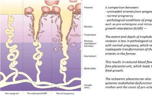
To a lesser extent, the limitation of the invasion process by disequilibrium in the local immune balance, without leading to initial destruction of the embryo, will result in further obstetrical complications through placental pathologies.
An insufficient embryo invasion and specially an inadequate remodelling of the spiral arteries, either by a lack of initial decidualisation or by further trophobast destruction, will lead to a poor trophoblast invasion and limited placental function.
This may result in clinical conditions such as intrauterine growth restriction (IUGR) or pre-eclampsia (Figure 8), a complex and multifactorial pathology which affects 5% of all pregnancies and initially manifests with maternal hypertension and proteinuria [49].
31
Figure 8: Normal and pathological Human trophoblast invasion and consequences From Ashley Moffett-King, Nature Reviews Immunology, 2002
Mechanical endometrial disruption consequences also points out the local influence on placental regulation. On previous uterine scars, after caesarean section or myomectomy, where endometrial barrier has been altered and the control of the invasive potential of the trophoblast could not be limited, we observe a higher incidence of uncontrolled placental invasion, leading to placenta accreta [50]. Placenta accreta occurs by a morbid placental adherence to the uterus. It is divided into three grades based on histopathology: placenta accreta where the chorionic villi are in contact with the myometrium, placenta increta where the chorionic villi invade the myometrium, and placenta percreta where the chorionic villi penetrate the uterine serosa. This severe pregnancy complication is an important cause of maternal morbidity and mortality, specifically through a high risk of [51].
32
1.2. Reminder on Reproductive Immunology
1.2.1. Reminder on general immunology
When simplified to the extreme, immunology deals with t self and non-self recognition, to ensure own defence and integrity. It is classically divided into innate immunity and adaptative immunity, either humoral or cell mediated. Innate immunity concerns mainly continuous, pre-existing recognition of non-self molecules in microorganisms and changes in self molecules or lack of self molecules on cells altered by infection or neoplasia for example. Adaptative immunity is based on self and non-self distinction via generation of large specific receptors repertories recognizing non-self antigens. Cells presenting high avidity receptors toward self molecules have also to be eliminated or inactivated by adaptative immune elements. An imperfection on self and non-self discrimination leads to autoimmune pathologies. Given the enormous variety of antigens and immune receptors, the difference between self and nonself is not absolute. It depends on thresholds of activation, which define the extent of self and nonself discrimination limits and immune responsiveness [52] [53].
Innate immunity includes antigen non-specific elements capable of immediate response to pathogen attacks, within minutes or hours. Apart from physical barriers such as skin or mucous membranes and chemical barrier of pH, innate immunity actors are leukocytes, other than B or T lymphocytes, such as macrophages, dendritic cells, neutrophils and Natural Killer cells [54]. These cells form the first defence against pathogens invasion, for example through Toll-Like Receptors (TLR), recognising pathogen-associated molecular patterns (PAMP) found on microorganisms [55] [56]. Another function of this system is regulating adaptative immunity through cytokines production and assisting antigen presentation [54].
Adaptative immunity manages specific defence and ensures immune memory in case of re-exposure. These functions require steps of antigen presentation, effective response and memorisation. The first step is fulfilled by Antigen Presenting Cells (APC), such as macrophages or dendritic cells, which will process antigens for presentation to T cells, to
33 induce effector lymphocytes activation. Effector functions consist in specific antibody production called humoral immunity, performed by B lymphocytes presenting membrane B Cell Receptors (BCR), and in cytokine production and cell destruction called cell mediated immunity, performed by T lymphocytes presenting membrane T Cell Receptors (TCR). We differentiate two subgroups of T cells given their surface proteins: CD4(+) helper cells involved in inducing production of antibodies as well as activation of the CD8(+) cytotoxic cells, themselves involved in cell destruction, also called Cytotoxic T Lymphocytes (CTL) when activated. Finally, immune memory function is provided by memory B cells created during clonal expansions, ensuring a faster specific response in case of antigen re-exposure [52] [53], as well as memory T cells
The T lymphocytes activation happens via recognition of antigens fragments, as peptides presented by Major Histocompatibility Complexes molecules I and II (MHC I and II). MHC molecules are also called HLA in Human (Human Leucocyte Antigen). These heterodimeric transmembrane glycoproteins are divided into two classes, MHC I and II. MHC I molecules are on all somatic nucleated cells. They include classic isoforms HLA-A, B, C and non-classic isoforms HLA-E, F, G. MHC I molecules present peptides, driven from intracellular digestion, to cytotoxic CD8 T lymphocytes. HLA-A and B isoforms are highly polymorphic, unlike HLA-C which has a restricted polymorphism and non-classic isoforms which are nearly monomorphic. Given these properties, as well of course as their selective expression, HLA-C and HLA-G are the major actors of the transient tolerance towards the foetus. MHC II molecules are on immune cells presenting processed antigens to helper CD4 T cells. In Human, there are three different isotypes (HLA-DR, HLA-DQ, HLA-DP), characterized by a very high level of polymorphism. MHC II molecules functions consist in the presentation of exogenous and endogenous peptides to the TCR of CD4 T cells for adaptative immune response, the establishment of the TCR repertoire of the CD4 T cell population through selective events in thymus, and finally the regulation of peripheral CD4 cells lifespan. [57]. All these immune actions are coordinated by secreted soluble factors such as chemokines, which regulate the immune cells attraction, and cytokines, which are the communication tools between the different immune cells. The cytokinic milieu is crucial for the differentiation of immune cells towards a tolerogenic or inflammatory response. Th1 / Th2 / Th17 type immune responses are named after the corresponding CD4 T helper lymphocytes,
34 classified from a functional point of view in different lineages. Through the dominance of specific cytokine production and cellular orientation, the milieu can turn inflammatory (Th1) or tolerogenic (Th2) [58]. Recently, the reversibility of these phenotypes has been suggested, the shift between tolerogenic to inflammatory dominant immune responses being mediated by Th17 lymphocytes. Th17 lymphocytes possess a functional plasticity allowing them to shift towards the Th1 or the Th2 phenotype in the presence of modulatory cytokines [59].
Some attempts of mathematical modelisation of these highly complex cellular and cytokinic networks are described. With all the redundancies, feedbacks, multi functionality, plasticity and multiplicity of possible combinations in immunity, mathematically modelising the system would be similar to study a chaotic behaviour [60]. Thus, we will not try to draw up an exhaustive report of the current knowledge on the immune system, but, after a brief historical reminder, will try to present some selected major cytokinic or cellular actors intervening in reproductive immunology.
1.2.2. History of reproductive immunology
Reproductive immunology is a domain involving general immunology principles and specific aspects of reproduction and development. From the classical immunology point of view, embryo implantation and pregnancy are exceptional events. The survival and the growth of the semi-allogenic foetus cannot be explained by usually admitted mechanisms of transplantation.
Albeit Reproductive Immunology started as early as the 19th century with the discovery of immunisation against sperm antigens by Landsteiner and Metalnikoff, but as far as pregnancy is concerned, it really started in 1953. Medawar enunciated “the immune paradox of pregnancy” and suggested the first hypotheses to explain this situation, by invoking physical separation or constraint of the immune response in pregnancy [61]. In 1970’s, the first attempt to explain a state of immunosuppression, called “the facilitation theory”, emphasized on systemic immune regulation via maternal antibodies hiding paternal placental antigens [62]. But this theory revealed insufficient, as maternal
pre-35 immunisation or absence of maternal anti-paternal antibodies, in Human or mice, did not impair pregnancy [63]. From the 1980’s, study of local regulatory mechanisms proved more productive.
The initially analysed local mechanism was immunosuppression. On one hand, this immunosuppression was demonstrated to be due to locally secreted factors or particles. On the maternal side this consisted in Transforming Growth Factor β (TGFβ) or Progesterone Induced Blocking Factor (PIBF) [64] [65], and on foetal side, immune modulators like Indoleamine 2,3-Dioxygenase (IDO) [27], Prostaglandin E2 (PGE2) [28], anti-complement molecules as Monocyte Chemostatic Protein (MCP) or Decay Accelerating Factor (DAF) [29], and placental microparticles as exosomes [31] [30]. On the other hand, it was demonstrated that local immune suppression was allowed given a very specific antigenicity on foetal structures in contact with maternal interface, specially due to absence of highly polymorphic surface antigens, Human Leucocyte Antigen (HLA) A or B, and presence of HLA-C [34] and HLA-G [66].
In parallel, Loke and Croy demonstrated the importance of a specific local immune cell population called uterine Natural Killers (uNK) in embryo implantation process [67]. These specific cells were shown to be essential for the local vascular remodelling and thus allowing an extensive placentation, via cytokine expression and further interaction with foetal HLA-C [34].
When considering cytokines at the maternal foetal interface, Wegmann raised the theory of "immunotrophism" [68]. It was demonstrated that, apart from their local immune function, some cytokines secreted by stimulated maternal CD4 T lymphocytes, had a trophic action on placental and embryo growth. The Colony Stimulating Factors action partly belongs to this field.
At the end of 1980's, T helper lymphocytes and their cytokines were categorized in two groups: Th1 for the cytotoxic and pro-inflammatory ones, and Th2 for the immuno-modulatory and anti-inflammatory ones [69]. The same distinction was applied to the cytokines at the maternal foetal interface, and a successful embryo implantation and an ongoing pregnancy were assimilated to a Th2 phenomenon, protecting the conceptus against maternal rejection. Shortly after, the endometrial presence of inflammation actors
36 was shown [70] and it was demonstrated that a lack of some inflammatory molecules would impair embryo implantation [71]. An initial transient Th1 reaction, followed by a Th2 environment, was fundamental to enable an effective embryo attachment and a further sufficient embryo invasion without being rejected [25]. This was designated as the "Th1-Th2 paradigm".
With the evolution of the knowledge in general immunology, soon this paradigm appeared too simplistic to explain pregnancy success [72]. As we can consider the immune interactions and their chronology at the maternal foetal interface with more accuracy, new concepts of local homeostasis implying regulatory T cells [73] [74], dendritic cells [75] or Th17 system [76] [77] are emerging. Environmental influences seem also fundamental on these local immune regulations [78] [79].
Given the revealed differences between classic immunology principles and the mechanisms controlling embryo implantation, we cannot consider anymore this phenomenon as a maternal tolerance to a foetal allograft [80]. Today, we describe a successful implantation and pregnancy as a dynamic and bidirectional dialogue between mother and conceptus, where the receptive endometrium seems to be a selective biosensor [21] towards a competent embryo [43] [44].
1.2.3. Key immune cells in reproductive immunology
Local immune interactions taking place at the maternal foetal interface are enabled by the presence of specific immune cell groups (Table 1) including uterine natural killer cells (uNK), macrophages, dendritic cells (DC) and T regulatory cells (Treg), which invade the endometrium during middle luteal phase via cyclic expression of local chemokines. Chemokines, such as CCL4 (Chemokine Ligand 4) also designated as Macrophage Inflammatory Protein-1β (MIP-1β), enable the recruitment and retention of specific immune cells in endometrium [81] [82].
During implantation window, uterine Natural Killers (uNK) form the majority of immune cell population in a receptive endometrium (Figure 9). Their phenotype and function differ from circulating Natural Killer (NK) cells called after their ability to immediate cellular lysis. The
37 presence of various families of receptors on NK cells helps to understand and predict their functions [83]. When considering their phenotypes, in majority, uterine NK cells are CD56(+) bright and CD16(-) dim whereas peripheral NK cells are CD56(-) dim and CD16(+) bright [84]. CD16 is a membrane protein involved in cellular lysis: CD16(+) NK cells are cytotoxic while CD16(-) NK cells secrete cytokines . If comparing CD56(+) NK cells from peripheral blood and endometrium, their repertoire of activating and inhibiting receptors are significantly different, resulting in distinct regulation of NK related activity. Uterine NK cells main function consists in cytokine production, but they keep their cytotoxic potentiality which could be triggered in an excessive inflammatory environment [85]. Cytokinic secretions of uNK cells are orientated towards angiogenesis and local vascular remodelling [86] allowing a sufficient trophoblastic invasion. When facing trophoblastic cells, particularly via the incoming HLA-C contact, uNK cells will fulfil their maturation and become decidual NK cells (dNK), a specific subset with immune modulatory potential which will direct a further placentation [87] [88].
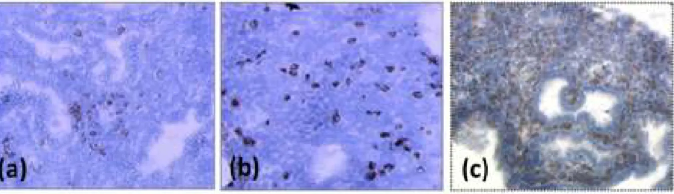
Figure 9: Endometrial uNK immunostaining (CD56+) during middle luteal phase showing a
normal invasion in a fertile control patient (b), an insufficient invasion (a) and an excessive invasion (c) in patients with repeated embryo implantation failure.
Endometrial slides from frozen samples, anti-CD56 Ventana antibodies
Macrophages are the second most abundant immune cell population in the secretory endometrial stroma [89]. Their presence is under cyclic hormonal control [90]: estradiol and
38 progesterone are shown to influence this endometrial influx [91]. Physiologically, macrophages in the endometrium are described as having a tolerogenic phenotype and mostly induce anti-inflammatory cytokine secretions [92]. They are also present at the further maternal foetal interface, contributing to foetal tolerance, trophoblast invasion, vascular remodelling and cellular migration [93]. Their excessive presence in decidua or a Th-1 dominant environment has been linked to preeclampsia and recurrent miscarriages [94].
Recently, a major interest was granted to an endometrial immune cell population interacting with uNK cells: the uterine Dendritic Cells (uDC). Dendritic cells belong to antigen presenting cells family and, besides pregnancy, they are enrolled in promoting immune responses and preventing autoimmunity. They are localised at maternal foetal interface during early pregnancy, where local signals drive them towards tolerogenic or immunogenic potential. In physiological circumstances, in addition to the promotion of an endometrial immune modulation, they are supposed to promote local angiogenesis, via their interaction with uNK cells, down regulating their activation markers and inducing their cytokinic production [95] [75].
A specific subgroup of T lymphocytes has also been identified as intervening at maternal foetal interface [96] [97]. T regulatory lymphocytes (Treg), powerful inhibitors of cell mediated immunity, seem to have a central role in preventing immunity against paternal antigens [98]. Sequential intervention of a series of chemokines and cytokines is suggested to promote Treg endometrial attraction. Apart from cyclic expansion under oestrogen and progesterone influence [82] [99], Treg cells are attracted in the endometrium by chemokines such as CCL3, CCL4 and CCL5 [100]. Their proliferation and maturation are further amplified by seminal plasma exposure through Transforming Growth Factor β (TGFβ) and prostaglandins [101]. Suppressive functions and recruitment of Treg are then supposed to be optimised via antigen presentation [102], reducing the immune attacks towards the conceptus.
39
Table 1: Overview of Endometrial Immune Cells
1.2.4. Key cytokines in reproductive immunology
At the maternal foetal interface, the establishment of functional networks between different immune actors are mediated by small, soluble, labile and inducible glycoproteins called cytokines. Already in late 1980’s, Wegmann described “the embryo bathing in a sea of cytokines” [103] and as underlined earlier in this chapter, given the extreme complexity of cytokines networks [60], only some selected ones which seem essential to embryo implantation are presented.
A major group of cytokines involved in reproduction are members of GP130 (Glycoprotein 130) family, amongst which Leukaemia Inhibitory Factor (LIF), Interleukin 11 (IL-11) and Interleukin 6 (IL-6). LIF is expressed in endometrium trough menstrual cycle with an increase in secretory phase and early pregnancy [104]. It stimulates cell proliferation, differentiation and survival [105] which are necessary to embryo development [106]. Its importance was
40 emphasized with LIF knockout murine models, where embryo implantation failed, whereas LIF knockout embryos did implant in a LIF producing foster mother [71]. Also, patients with primary unexplained infertility showed altered endometrial LIF secretion [107]. Interleukin 11 (IL-11) is also detected at the maternal foetal interface with cyclic variation and a higher expression during late secretory phase [108]. It’s involved in endometrial decidualisation and local Natural Killer cells maturation [109] and further placentation [110]. Fertility is also impaired in IL-11 lacking murine models given an insufficient post implantation endometrial response [111]. Part of the GP130 family, interleukin 6 (IL-6) is a multifunctional cytokine, involved in immediate immune response and haematopoiesis. Its endometrial expression increases during middle luteal phase and its concentration is higher in decidual than in placental tissues [112]. The major epithelial localisation points out the IL-6 role in embryo attachment. Reduced fertility with lower embryo implantation is reported in IL-6 deficient mice [113].
The vascular tripod involving the interleukins 12, 15 and 18 (IL-12, IL-15, IL-18) is a major component of the endometrial cytokine network, particularly through the regulation of uterine Natural Killer (uNK) cells and the local angiogenesis induction [26]. Endometrial production of IL-15 is involved in the recruitment [114] [115] and the maturation of uNKs towards immune-modulatory cytokine producing cells [116] [117]. IL-15 knocked out mice show a lack of endometrial uNK cells and undecidualised spiral arteries [115]. IL-18 is a bivalent cytokine, also produced in the maternal foetal interface. When acting individually, it enhances uNK cells angiogenic cytokine production [118], promoting the local vascular remodelling. But when co-stimulated with IL-12, IL-18 drives uNK cells towards cytotoxicity and enhances local production of pro-inflammatory cytokines [119]. Moreover, some local immune modulators have been recently described for IL-15 and IL-18, which should be considered when evaluating these cytokines effects [120] [121]. During implantation window, variations in the endometrial expression of these vascular cytokines have been described in patients with unexplained reproductive failure when compared to fertile women [122] [123]. They therefore have been suggested as pre conceptual biomarkers to evaluate the immune local profile at the time of endometrial receptivity [24].
The interleukin 1 (IL-1) system is a considerable inflammatory mediator at the maternal foetal interface. This system includes two ligands, two membrane receptors, a non-binding
41 receptor accessory protein, and an antagonist receptor [124]. The components of this multifunctional inflammatory system [125] have been localised in the Human endometrium during the window of implantation, as well as in the placental tissues [36], and suggested to control the trophoblastic invasion [126].
On the other hand, the Transforming Growth Factor Beta (TGF-β) is a key local immune modulator essential to the embryo implantation process [127]. The TGF-β family, including three isoforms (TGF-β 1, 2, 3), has pleiotropic effects on cellular growth, differentiation and immune modulation. TGF-β is expressed in endometrial and decidual tissues [126]. TGF-β is involved in embryo implantation by helping embryo attachment, enabling local immune modulation, and controlling trophoblast invasion through regulation of locally secreted factors such as other growth factors, angiogenic factors, lytic enzymes like metalloproteases or other major pro implantation cytokines like LIF [128] [129] [130].
Other endometrial growth factors seem crucial for the local cell proliferation and differentiation leading to a successful embryo implantation. The Vascular Endothelial Growth Factor (VEGF) is one of the major factors involved in local angiogenesis and vascular remodelling essential to the establishment of a functional hemochorial placentation. Through a series of isoforms and activating or inhibiting receptors, the VEGF family enhances endometrial vascularization and vascular permeability [131]. Their action is under cyclic steroid control [132] [133] as well as locally secreted cytokines [134] and hypoxic conditions [135]. The Epidermal Growth Factor (EGF) is another local growth factor involved in trophoblastic invasion, proliferation and differentiation [136] [137]. EGF is localised in endometrial stroma, decidua and trophoblast [138]. Murine models lacking EGF or its receptors are shown to have impaired infertility, with early embryonic death, abortion or intrauterine growth restriction [139] [140]. Heparin Binding EGF Like Growth Factor (HB-EGF), which also shares receptors with EGF and TGF, is predominantly expressed during the window of implantation. It is localised in endometrial stromal and epithelial cells, regulating the local cell proliferation, secretion and decidualisation [141]. It is also described as a major mediator of embryo implantation by enabling the blastocyst attachment through membrane receptors on trophectoderm [142] [143].
42 Finally, Colony Stimulating Factors are major cytokines involved in reproduction. Their localisation in the reproductive tract and their immunotrophic, anti-apoptotic and immunomodulatory properties will be detailed in this work through dedicated articles.
43
1.3. Hypothesis and Objectives
In the present work, in order to help the understanding of Colony Stimulating Factors (CSFs) and particularly Granulocyte-Colony Stimulating Factor (G-CSF) contribution in reproductive field, we propose a way through three original articles. Our hypothesis was that G-CSF could modulate, by acting on some target genes, the pre-implantation endometrium which is ultimately described as a biosensor towards the embryo.
A first review article is dedicated to CSFs family, each member’s localisation in reproductive tract, biological functions in reproduction and actual medical applications. The two other articles focus on G-CSF, already described as one of the most promising innovative therapies in reproductive medicine. By studying G-CSF and especially by detailing its endometrial actions, in women and murine models, we aim to illustrate how this cytokine could modulate the embryo implantation process.
G-CSF action on Human endometrium is studied in the second article. Based on a former endometrial transcriptomic study, we identified hypothetical targets of G-CSF. Then we described variations of expression for these targets in endometrium of infertile and control women. We finally tested G-CSF action on the specific targets, using a dynamic endometrial microhistoculture model.
The third article is about G-CSF supplementation on murine models. We chose specific crossings where the endometrial biosensor plays a major role between fertile and abortion prone models. The timing of the systemic G-CSF supplementation on females was also chosen in order to target specific endometrial action, avoiding complementary effect on ovulation or embryonic growth. We then studied, in both fertile and abortion prone models, G-CSF systemic supplementation effects on embryo implantation and early embryonic loss and compared it to control mice receiving placebo and to control non-injected mice.
44
2. Colony Stimulating Factors in Reproduction
2.1. Colony Stimulating Factors (CSFs)
2.2. CSFs Localisation in the Reproductive Tract
2.3. CSFs Functions in Reproduction
2.4. CSFs Medical Applications
45
2. Colony Stimulating Factors in Reproduction
2.1. Colony Stimulating Factors (CSFs)
The CSF (Colony Stimulating Factor) family includes: CSF-1 or M-CSF (Macrophage-Colony Stimulating Factor), CSF-2 or GM-CSF (Granulocyte-Macrophage-Colony Stimulating Factor), and CSF-3 or G-CSF (Granulocyte-Colony Stimulating Factor). These cytokines are studied from the mid 1960’s and were named after their action on proliferation and differentiation of leukocytes (Figure 10).
Figure 10: General model of haematopoiesis, with CSFs localisation From Kaushansky, NEJM, 2006
46 CSFs are 18-70 kDa labile glycoproteins, act through specific cell-surface receptors. Each of these transmembrane proteins includes one or two extracellular cytokine-binding domains containing approximately 200 amino acids, a transmembrane domain of 20 to 25 residues, and an intracellular domain of approximately 100 to 500 amino acids with the box 1 and box 2 motifs that recruit kinases of the Janus kinase (JAK) family.
Each of these cytokines supports the survival and proliferation of a number of distinct target cells via JAK-STAT signalling in an endocrine, paracrine or autocrine mode. The binding of the cytokine to its specific receptor induces a major conformational shift, bringing the two tethered cytoplasmic JAKs into close juxtaposition, thereby triggering activation of the kinases by mutual cross-phosphorylation. Once the JAKs are activated, a number of secondary signalling molecules are phosphorylated, each of which activates an overlapping subgroup of tertiary signalling molecules such as transcription factors, cell-cycle activators and inhibitors anti-apoptosis molecules and other growth factors which will be ultimately responsible for the specific effects induced.
Apart from inducing these cascades, the binding of the CSF to its receptor instigates internalization of receptors, activation of phosphatases, and production of suppressors of cytokine signalling, in order to terminate the stimulating signal.
Their involvement in reproduction was raised from early 1970’s when a CSF activity was identified in the Human and murine placenta.
2.2. CSFs Localisation in the Reproductive Tract
All CSFs and their corresponding receptors are localised along the female reproductive tract, but each member is predominantly expressed in some specific locations. At the maternal foetal interface, GM-CSF and its receptor are the most studied. In the ovarian granulosa and follicular fluid, G-CSF seems to raise more interest in fundamental research and medical applications. In the seminal plasma, GM-CSF again is the most studied amongst the CSFs, particularly due to its action on pre implantation endometrium.