Acute and Specific Modulation of Presynaptic
Aromatization in the Vertebrate Brain
Charlotte A. Cornil, Cary H. Leung, Eric R. Pletcher, Kevin C. Naranjo, Sara J. Blauman, and Colin J. Saldanha
GIGA (Groupe Interdisciplinaire de Ge´noprote´omique Applique´e) Neurosciences (C.A.C.), University of Liège, 4000 Liège, Belgium; Biological Sciences (C.H.L., E.R.P., K.C.N., S.J.B., C.J.S.), Lehigh University, Bethlehem, Pennsylvania 18015; Widener University (C.H.L.), Chester, Pennsylvania 19013; and Department of Biology (S.J.B., C.J.S.), American University, Washington, D.C. 20016
Estrogens affect a diversity of peripheral and central physiological endpoints. Traditionally, estrogens were thought to be peripherally derived transcription regulators (i.e. slow acting). More recently, we have learned that estrogens are also synthesized in neuronal cell bodies and synaptic terminals and have potent membrane effects, which modulate brain function. How-ever, the mechanisms that control local steroid concentrations in a temporal and spatial res-olution compatible with their acute actions are poorly understood. Here, using differential centrifugation followed by enzymatic assay, we provide evidence that estrogen synthesis within synaptosomes can be modulated more dramatically by phosphorylating conditions, relative to microsomes. This is the first demonstration of a rapid mechanism that may alter steroid concentrations within the synapse and may represent a potential mechanism for the acute control of neurophysiology and behavior. (Endocrinology 153: 0000 – 0000, 2012)
A
long with their well-known actions as transcription regulators, estrogens also produce membrane-initi-ated, rapid, and transient effects on signaling pathways affecting physiological and behavioral endpoints, includ-ing social behaviors, energy balance, and cognitive pro-cesses (1, 2). Fluctuations in circulating steroids occur rel-atively slowly (3) and do not provide the temporal or anatomical resolution required for the acute activation of specific brain circuits (4 – 6). Estrogens’ ability to rapidly and transiently regulate physiology implies that their syn-thesis may be rapidly fine tuned. Unfortunately, the mech-anisms that control local steroid concentrations in a time and spatial resolution compatible with their acute actions are poorly understood.Estrogens are synthesized from androgens by aroma-tase, a P450enzyme traditionally associated with micro-somes of discrete neuronal populations in the brain of vertebrates (7, 8). Aromatase is also found in presyn-aptic terminals, suggesting that 17-estradiol (E2)
syn-thesis may be achieved with spatial specificity (9 –11).
Further, brain aromatase activity (AA) is rapidly and reversibly modulated by calcium-dependent phospho-rylations resulting from neuronal depolarizations or glutamate release (12–14). Thus, transient and activity-dependent regulation of aromatase may alter its ability to synthesize estrogens. A recent in vivo microdialysis study has indeed suggested that fluctuations in the syn-thesis of estrogens induced by neuronal activity may be a reflection of presynaptic aromatization (13). How-ever, microdialysis is unable to unequivocally discrim-inate between microsomal and synaptosomal E2
syn-thesis. Because estrogens are lipophilic and thus cannot be stored, rapid modulation of their presynaptic syn-thesis is one mechanism that could account for local and rapid changes in bioavailability. However, whether aro-matase can be acutely regulated in specific ultrastruc-tural compartments is unclear, and the mechanism for this potential regulation is unknown.
To clarify this issue, zebra finch telencephalon, tissue that contains the highest concentration of neuronal
aro-ISSN Print 0013-7227 ISSN Online 1945-7170 Printed in U.S.A.
Copyright © 2012 by The Endocrine Society
doi: 10.1210/en.2011-2159 Received December 21, 2011. Accepted March 22, 2012.
Abbreviations: AA, Aromatase activity; BIS, bisindolylmaleimide; E2, 17-estradiol; HPOA,
preoptic-hypothalamic.
B R I E F R E P O R T
Endocrinology, June 2012, 153(6):0000 – 0000 endo.endojournals.org 1 Copyright (C) 2012 by The Endocrine Society
matase in any vertebrate tested, was subjected to differ-ential centrifugation to separate synaptosomal and micro-somal fractions. The modulation of AA across subcellular compartments and sexes was then evaluated using the tri-tiated water assay via exposure to phosphorylating con-ditions with or without protein kinase inhibition.
Materials and Methods
Animals
Fifteen male and 12 female zebra finches (n⫽ 3 per sample, five male and four female samples) were used. All experiments were conducted in accordance with the Institutional Animal Care and Use Committee guidelines at Lehigh University.
Fractionation
Telencephalons were rapidly removed, weighed, placed into ice-cold KTH buffer (150 mM KCL, 10 mM Tris-Base, Hepes, pH7.2) with sucrose (0.32 M) [0.1 mg of fresh tissue per milli-liter] and homogenized, surrounded by ice, with 4⫻ 5-sec bursts of an electric homogenizer. Briefly, as previously described (11), homogenates were centrifuged for 15 min at 1034 ⫻ g. The resulting pellet was discarded and the supernatant (S1) was fur-ther centrifuged for 30 min at 10,081⫻ g. The supernatant (S2) was removed and kept aside, whereas the pellet (P2), containing synaptosomes and mitochondria, was washed twice with 300l of KTH-sucrose buffer followed by another 10-min centrifuga-tion at 10,081⫻ g. The P2 wash solution was added to S2. The resulting mixture was then centrifuged for 1 h at 100,000⫻ g. The microsomal pellet (P3) and P2 were weighed wet, resus-pended in KTH-sucrose (10 and 50 mg of pellet per milliliter, respectively), and stored at⫺80 C.
Electronic microscopy
After the 10,081⫻ g spin, some P2 were washed and fixed in 4% glutaraldehyde for 2 h at 4 C. Pellets were kept in 0.1M
phosphate buffer overnight at 4 C, then washed in 0.9% saline (10 min), and exposed to 2% OsO4in 0.9% saline containing
1.5% KFeCN for 2 h. After serial dehydration, pellets were ex-posed to propylene oxide (30 min), 1:1 propylene oxide and Epon (2 h), 1:2 propylene oxide and Epon (overnight), and then polymerized in 100% Epon at 65 C for 48 h. Ultrathin sections (50 –70 nm) were collected on copper grids, air dried, and ex-amined on a Jeol 1200EX.
Enzymatic assays
AA was quantified by measuring the release of3H-water
pro-duced from each molecule of [1-3H]androstenedione
aroma-tized (15). All samples and reagents were kept on ice at all times unless stated otherwise. Figure 1 illustrates the sequential incu-bation steps and concentrations of drugs added. Aliquots (100 l) were mixed with one volume of KTH buffer (50 l) contain-ing either the calcium chelator EGTA (8 mM) (15), the specific
protein kinase C inhibitor bisindolylmaleimide (BIS) (40 M)
(15, 16) or neither, and another volume of KTH buffer (50l) with or without ATP, Mg2⫹, and Ca2⫹(PO4, 4 – 8 mM;
equimo-lar concentrations of ATP, Mg2⫹, and Ca2⫹). This resulted in a
repeated design with four treatments reaching final concentra-tions (indicated in parentheses) in a preincubation volume of 200 l: control, phosphorylating conditions alone (PO4, 1–2 mM),
phosphorylating conditions with EGTA (2 mM), and
phosphor-ylating conditions with BIS (10M). Samples were then
prein-cubated for 10 min in a water bath at 37 C to allow for the phosphorylation process. Previous experiments conducted in quail brain homogenates or cultured cells expressing human aro-matase demonstrated that incubation in identical conditions (high but physiological concentrations of ATP, Mg2⫹, and Ca2⫹) promotes protein phosphorylation (17) and, indeed, results in aromatase phosphorylation (see Refs. 18 –20; for further details, see Discussion).
The reaction was stopped by placing the samples on ice and adding EGTA to reach a final concentration of 2 mMin all
samples (the concentration added was adjusted to account for the presence of EGTA as a pretreatment in some samples). [1-3H]androstenedione (specific activity ⫽ 26.3 Ci/mmol;
PerkinElmer, Waltham, MA), KTH, and reduced nicotin-amide adenine dinucleotide phosphate were added to the ho-mogenate to reach final concentrations of 25 nM, 1.2 mM, and
40M, respectively. The aromatization reaction was initiated
by incubation at 37 C. After 10 min, the reaction was stopped by adding cold 10% trichloroacetic acid containing 2% char-coal. 3
H-water was purified by centrifugation followed by Dowex cation exchange column separation and quantified with a scintillation counter.
All samples were assayed in duplicate with an additional tube assayed in the presence of an excess of the aromatase inhibitor Fadrozole (gift from Novartis, Basel, Switzerland), whose activity measured was subtracted from the pooled du-plicate value to determine the final specific activity in each sample. Samples were randomly run within three assays for each experiment, each containing a whole homogenate serv-ing as an internal control. AA was expressed in nmol/h䡠 mg fresh weight of pellet after correction of the counts for quench-ing, recovery, blank values, and percentage of tritium in -position in the substrate.
Data analysis
Due to a difference in variance between fractions that cannot be verified statistically (tests for sphericity are invalid when there are less than three repeated modalities), results from each frac-tion were analyzed by separate two-way ANOVA with sex and treatment as independent and repeated factors, respectively. Sig-nificant treatment effects were queried with Tukey post hoc tests.
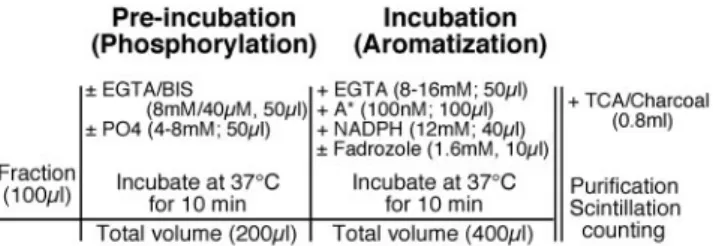
FIG. 1. Schematic presentation of the experimental protocol. All
reagents were used ice-cold and delivered in tubes placed on ice (symbolized by vertical lines). A*, [1-3H]-androstenedione. NADPH, Reduced nicotinamide adenine dinucleotide phosphate; PO4, equimolar concentrations of ATP, Mg2⫹and CA2⫹; TCA, trichloroacetic acid.
All analyses were performed with Statistica 9.1 (Statsoft, Inc., Tulsa, OK).
Results
Verification of the authenticity of subfractions by electronic microscopy revealed numerous synaptosomes containing varying amounts of clear neurotransmitter vesicles and mi-tochondria (Fig. 2). Similar subfractions prepared from quail preoptic-hypothalamic (HPOA) homogenates have been validated previously based on the expression of subcellular-specific enzymatic activities (21). Together, these observa-tions indicate that the AA measured in the P2 pellets is a reflection of synaptosomal aromatization.
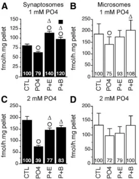
A first experiment tested the effect of a preincubation with 1 mMPO4with or without EGTA (2 mM; calcium chelator), and BIS (10M; protein kinase inhibitor), on AA measured in the two fractions. Separate two-way ANOVA identified a significant effect of treatment in both fractions (P2: F3,21⫽ 33.802, P ⬍ 0.001; P3: F3,21⫽ 5.462, P ⫽ 0.006) but no sex difference (P2: F1,7⫽ 0.439, P ⫽ 0.529;
P3: F1,7⫽ 1.151, P ⫽ 0.319) or interaction (P2: F3,21⫽ 0.758, P⫽ 0.530; P3: F3,21⫽ 1.267, P ⫽ 0.311). Post hoc
analyses revealed that preincubation with 1 mM PO4 caused a weak but significant reduction of AA in both fractions (⬃21–25%) (Fig. 3, A and B). Ca2⫹withdrawal partially (but not significantly) blocked this effect in mi-crosomes but significantly raised AA about 40% above control levels in synaptosomes suggesting the existence of
an endogenous AA inhibition by Ca2⫹ -dependent processes differentially af-fecting microsomal and synaptosomal aromatase. Finally, protein kinase block-ade completely suppressed PO4-induced enzymatic inhibitions in both fractions. Similar to findings obtained in quail HPOA homogenates (18, 19), telence-phalic aromatase (regardless of its subcellular localization) appears to be rapidly altered by Ca2⫹-dependent phosphorylations. However, the pres-ent inhibition was much lower than previously reported with the same con-centration of ATP, Mg2⫹, and Ca2⫹in quail HPOA (15).
Therefore, a higher concentration of PO4(2 mM) was tested on the same
sam-ples. As previously, no sex difference (P2: F1,7⫽ 0.350, P ⫽ 0.529; P3: F1,7⫽
0.430, P ⫽ 0.533) or interaction be-tween sex and treatment was found (P2: F3,21⫽ 0.955, P ⫽ 0.432; P3: F3,21⫽
0.426, P⫽ 0.736). Higher PO4concentrations yielded a
highly significant treatment effect in the synaptosomal fraction (F3,21⫽ 29.115, P ⬍ 0.001). Post hoc analysis indicated that this effect is mainly explained by a robust AA inhibition (⫺60%) after preincubation with 2 mMPO4
(Fig. 3C). In addition, calcium depletion and kinase inhi-bition significantly maintained AA to control levels, thus preventing PO4-induced inhibition. By contrast, the weak drop of AA in the microsomal fraction did not reach sig-nificance (F3,21⫽ 2.91, P ⫽ 0.058). Together, these results
suggest that synaptosomal and microsomal aromatase re-spond differently to phosphorylating conditions.
Discussion
These findings provide additional support for the idea that the rapid regulation of neural synthesis of estrogens is a conserved vertebrate trait (20), with presynaptic aromatization as a primary locus of influence. This in-ference is in excellent agreement with earlier hypotheses about the preferential regulation of presynaptic aroma-tization (13) and offers a temporal and spatial resolu-tion compatible with the rapid effects of estrogens on synaptic physiology (22). Importantly, this property is sexually monomorphic and supports consideration of estrogens as neuromodulators (12) in keeping with the “synaptocrine hypothesis” (23).
FIG. 2. Electrophotomicrographs of P2 pellets. Electron photomicrographs demonstrating the
presence of synaptosomal profiles in the P2 pellets used in the current study. Synaptosomes (1– 6) filled with varying numbers of clear vesicles are visible, sometimes, adjacent to a mitochondrion with visible cristae (m). Also visible is a synapto-dendrosome with an associated postsynaptic density (*) but no visible postsynaptic element. Scale bars, 500 nm.
In synaptosomes, AA is profoundly (⫺60%) reduced by high but physiological concentrations of ATP, Mg2⫹, and Ca2⫹, an effect prevented by calcium chelation and protein kinase inhibition. Identical regimens (including an array of kinase inhibitors, varying concentrations and du-rations of preincubation with ATP, Mg2⫹, or Ca2⫹) have been used to investigate the kinases involved as well as the sites, extent, and consequences of phosphorylation of the aromatase protein in birds (15, 18, 19, 21). Taken to-gether, these data strongly suggest that the treatments used in the present set of studies may indeed induce changes in aromatization via calcium-dependent phosphorylations (12). This influence may be direct, because aromatase con-tains consensus phosphorylation sites (19). Direct evi-dence using antiphospho-residues and radiolabeled phos-phate show that phosphorylations target the enzyme itself, rather than other regulatory and/or associated proteins that would secondarily alter AA (20).
In conditions in which the neuronal integrity and connectivity are preserved, decreases in AA of similar magnitude and temporal pattern are elicited by K⫹ -in-duced depolarization and exposure to glutamate (12– 14). These changes are reversible and independent of the enzyme stability, and kinase inhibition completely pre-vents these activity-dependent enzymatic changes (12, 20). Thus, depolarization and aromatase
phosphoryla-tion appear strongly associated, a pattern corroborat-ing the hypothesis that the synaptosomal changes in AA seen here depend on calcium-dependent phosphoryla-tions and are likely involved in the fine tuning of local synaptic E2synthesis. It is critical, however, to acknowl-edge that the current preparation does not permit us to unequivocally identify the source of calcium flux, nor does it permit identification of region-specific contri-butions to variations in synaptosomal aromatization within the songbird telencephalon.
Previous studies suggested that the calcium involved in the AA regulation was likely released from internal stores. Indeed, in HPOA explants, blockers of calcium channels or complete removal of Ca2⫹from the extracellular milieu failed to abolish the K⫹-induced enzymatic inhibition, whereas a similar inhibition was obtained with thapsi-gargin, a sesquiterpene lactone that increases the intracel-lular pool of free Ca2⫹by blocking its uptake by the en-doplasmic reticulum (12). In contrast, it was recently suggested that the acute control of brain estrogen synthesis depends on calcium entry through presynaptic voltage-dependent calcium channels, an inference supported by the observation that the in vivo retrodialysis of -cono-toxin (a blocker of the presynaptic N-type channels) into the zebra finch caudomedial nidopallium blocks the K⫹ -induced drop in estrogen synthesis (13). Although the present study does reveal an interesting overshoot in EGTA-treated synaptosomes, but not microsomes, with lower concentrations of ATP, Mg2⫹, and Ca2⫹(see Fig. 3), this observation only suggests the existence of a higher calcium concentration in synaptosomes compared with microsomes and, unfortunately, cannot provide any fur-ther information concerning the source of calcium in in-tact neurons.
Although presynaptic aromatase is described in many species (9, 10), its regulation is less well studied. The tran-scriptional activity of testosterone is thought to regulate the concentration of the enzyme and, thus, its activity, in microsomes and synaptosomes in a region-specific man-ner (21, 24). However, this is the first study to report a compartment-specific, phosphorylation-dependent regu-lation of aromatase. The present data strongly suggest that phosphorylations preferentially alter AA in synaptosomes more rapidly than the genomic control of aromatase ex-pression by testosterone (12, 20). Although the molecular underpinnings of this compartment-specific regulation re-main unknown, it may reflect the compartmentalization of specific kinases and/or the dynamics of Ca2⫹ availabil-ity. These hypotheses await testing.
The traditional view about the mechanisms of estro-genic action may require refinement, because estrogens are produced by a variety of tissues, including the brain, and
FIG. 3. Phosphorylating conditions (PO4) differentially inhibit
synaptosomal and microsomal AA (mean⫾SEM). Effect of
preincubation with 1 mM(A and B) and 2 mM(C and D) PO4with or
without 2 mMEGTA (P⫹E) or 10MBIS (P⫹B) on synaptosomal (black,
P2 fraction) and microsomal (white, P3 fraction) AA. Numbers in the bars represent the average percentages of the control mean. E,⌬, and f, P⬍ 0.05 for within-fraction comparison with control treatment (CTL), PO4, and P⫹E, respectively.
act through multiple pathways to affect many physiolog-ical and behavioral processes. Mounting evidence indi-cates that brain E2 synthesis can be regulated in a time
frame compatible with the rapid effects described at the cellular and organismal level (1, 2). However, the subcel-lular dynamics of their synthesis are almost never dis-cussed. Consequently, it is sometimes assumed that estro-gens invariantly diffuse away from their site of synthesis, flooding a large extent of their surroundings in a relatively nonspecific manner. The present finding suggests that the acute control of estrogen synthesis (and their release, be-cause their lipophilic nature prevents their storage) is con-fined to a limited spatial domain offering a spatially re-stricted delivery mechanism of estrogens, perhaps to fine tune synaptic processes (22). Indeed, manipulations of lo-cal estrogen action or synthesis alter neuronal excitability and behavioral output (4 – 6, 25–28). Moreover, fluctua-tions in E2synthesis occur in response to sudden changes
in the social or environmental context (14, 29, 30). Thus, it is likely that activity-dependent fluctuations of local, probably synaptic, E2 bioavailability critically impact behavior. Finally, the present demonstration of acute control of AA preferentially in synaptosomes brings further support to the neuromodulatory role played by locally produced estrogens in the songbird telencephalon possibly for regulation of singing and au-ditory processing (6, 28) and likely extending to other systems and vertebrate species (2).
Acknowledgments
We thank Dr. Jacques Balthazart, Dr. Thierry D. Charlier, and Catherine de Bournonville for their help with data analysis and comments on earlier versions of this manuscript.
Address all correspondence and requests for reprints to: Charlotte A. Cornil, GIGA Neurosciences, University of Liège, 1 Avenue de l’Hopital (Bat 36), 4000 Liège, Belgium. E-mail: [email protected].
This work was supported by National Institutes of Health Grant NS 042767. C.A.C. is a Fonds pour Recherche Scienti-fique – Fonds National pour la Recherche ScientiScienti-fique Research Associate.
Disclosure Summary: The authors have nothing to disclose.
References
1. Roepke TA, Ronnekleiv OK, Kelly MJ 2011 Physiological conse-quences of membrane-initiated estrogen signaling in the brain. Front Biosci 16:1560 –1573
2. Cornil CA, Charlier TD 2010 Rapid behavioural effects of
oestro-gens and fast regulation of their local synthesis by brain aromatase. J Neuroendocrinol 22:664 – 673
3. Cornil CA, Ball GF, Balthazart J 2006 Functional significance of the rapid regulation of brain estrogen action: where do the estrogens come from? Brain Res 1126:2–26
4. Kow LM, Pfaff DW 2004 The membrane actions of estrogens can potentiate their lordosis behavior-facilitating genomic actions. Proc Natl Acad Sci USA 101:12354 –12357
5. Fernandez SM, Lewis MC, Pechenino AS, Harburger LL, Orr PT,
Gresack JE, Schafe GE, Frick KM 2008 Estradiol-induced
enhance-ment of object memory consolidation involves hippocampal extra-cellular signal-regulated kinase activation and membrane-bound es-trogen receptors. J Neurosci 28:8660 – 8667
6. Tremere LA, Jeong JK, Pinaud R 2009 Estradiol shapes auditory processing in the adult brain by regulating inhibitory transmission and plasticity-associated gene expression. J Neurosci 29:5949 – 5963
7. Roselli CE, Resko JA 1997 Sex differences in androgen-regulated expression of cytochrome P450 aromatase in the rat brain. J Steroid Biochem Mol Biol 61:365–374
8. Saldanha CJ, Tuerk MJ, Kim YH, Fernandes AO, Arnold AP,
Schlinger BA 2000 Distribution and regulation of telencephalic
aro-matase expression in the zebra finch revealed with a specific anti-body. J Comp Neurol 423:619 – 630
9. Naftolin F, Horvath TL, Jakab RL, Leranth C, Harada N,
Baltha-zart J 1996 Aromatase immunoreactivity in axon terminals of the
vertebrate brain. Neuroendocrinology 63:149 –155
10. Peterson RS, Yarram L, Schlinger BA, Saldanha CJ 2005 Aromatase is pre-synaptic and sexually dimorphic in the adult zebra finch brain. Proc Biol Sci 272:2089 –2096
11. Rohmann KN, Schlinger BA, Saldanha CJ 2007 Subcellular com-partmentalization of aromatase is sexually dimorphic in the adult zebra finch brain. Dev Neurobiol 67:1–9
12. Balthazart J, Ball GF 2006 Is brain estradiol a hormone or a neu-rotransmitter? Trends Neurosci 241–249
13. Remage-Healey L, Dong S, Maidment NT, Schlinger BA 2011 Pre-synaptic control of rapid estrogen fluctuations in the songbird au-ditory forebrain. J Neurosci 31:10034 –10038
14. Remage-Healey L, Maidment NT, Schlinger BA 2008 Forebrain steroid levels fluctuate rapidly during social interactions. Nat Neu-rosci 11:1327–1334
15. Konkle AT, Balthazart J 2011 Sex differences in the rapid control of aromatase activity in the quail preoptic area. J Neuroendocrinol 23:424 – 434
16. Toullec D, Pianetti P, Coste H, Bellevergue P, Grand-Perret T,
Aja-kane M, Baudet V, Boissin P, Boursier E, Loriolle F, Duhamel L, Charon D, Kirilovsky J 1991 The bisindolylmalmeide GF 109203X
is a potent and selective inhibitor of protein kinase C. J Biol Chem 266:15771–15781
17. Albert KA, Helmer-Matyjek E, Nairn AC, Müller TH, Haycock JW,
Greene LA, Goldstein M, Greengard P 1984
Calcium/phospholipid-dependent protein kinase (protein kinase C) phosphorylates and activates tyrosine hydroxylase. Proc Natl Acad Sci USA 81:7713– 7717
18. Balthazart J, Baillien M, Ball GF 2001 Rapid and reversible inhi-bition of brain aromatase activity. J Neuroendocrinol 13:63–73 19. Balthazart J, Baillien M, Charlier TD, Ball GF 2003
Calcium-de-pendent phosphorylation processes control brain aromatase in quail. Eur J Neurosci 17:1591–1606
20. Charlier TD, Harada N, Balthazart J, Cornil CA 2011 Human and quail aromatase activity is rapidly and reversibly inhibited by phos-phorylating conditions. Endocrinology 152:4199 – 4210
21. Schlinger BA, Callard GV 1989 Localization of aromatase in syn-aptosomal and microsomal subfractions of quail (Coturnix coturnix japonica) brain. Neuroendocrinology 49:434 – 441
Liu F 2011 Rapid estrogen signaling in the brain: implications for the
fine-tuning of neuronal circuitry. J Neurosci 34:16056 –16063 23. Saldanha CJ, Remage-Healey L, Schlinger BA 2011 Synaptocrine
signaling: steroid synthesis and action at the synapse. Endocr Rev 32:532–549
24. Roselli CE 1995 Subcellular localization and kinetic properties of aromatase activity in rat brain. J Steroid Biochem Mol Biol 52:469 – 477
25. Taziaux M, Keller M, Bakker J, Balthazart J 2007 Sexual behavior activity tracks rapid changes in brain estrogen concentrations. J Neurosci 27:6563– 6572
26. Cornil CA, Taziaux M, Baillien M, Ball GF, Balthazart J 2006 Rapid effects of aromatase inhibition on male reproductive behaviors in Japanese quail. Horm Behav 49:45– 67
27. Cornil CA, Dalla C, Papadopoulou-Daifoti Z, Baillien M,
Baltha-zart J 2006 Estradiol rapidly activates male sexual behavior and
affects brain monoamine levels in the quail brain. Behav Brain Res 66:110 –123
28. Remage-Healey L, Coleman MJ, Oyama RK, Schlinger BA 2010 Brain estrogens rapidly strengthen auditory encoding and guide song preference in a songbird. Proc Natl Acad Sci USA 107:3852– 3857
29. Cornil CA, Dalla C, Papadopoulou-Daifoti Z, Baillien M, Dejace C,
Ball GF, Balthazart J 2005 Sexual behavior affects preoptic
aroma-tase activity and brain monoamines’ levels. Endocrinology 146: 3809 –3820
30. Dickens MJ, Cornil CA, Balthazart J 2011 Acute stress differentially affects aromatase activity in specific brain nuclei of adult male and female quail. Endocrinology 152:4242– 4251