Université de Sherbrooke
snoDB: An Interconnected Online Database of Human snoRNA
Par
Philia Bouchard-Bourelle Biochimie et Génomique Fonctionnelle
Mémoire présenté à la Faculté de médecine et des sciences de la santé en vue de l’obtention du grade de M.Sc.
en Biochimie
Sherbrooke, Québec, Canada Janvier 2020
Membres du jury d’évaluation:
Michelle S. Scott, Biochimie et Génomique Fonctionnelle Sherif Abou Elela, Microbiologie et Infectiologie
Alan Cohen, Médecine de Famille Manuel Lafond, Informatique
A
CKNOWLEDGMENTSThank you to Michelle Scott, Jean-Michel Garant, Gabrielle Deschamps-Francoeur and Vincent Boivin for their continued advice and support over the years. I would also like to sincerely thank Joël Simoneau, Fanny Thuriot, Gaspard Reulet, Hoang Dong Nguyen and Étienne Fafard-Couture for their camaraderie and open mindedness. And a special thank you to everyone involved in snoDBs including Clément Desjardin-Henri for developing its sister tool snoTHAW. The project wouldn’t be nearly what it is without them.
I am also grateful to my research directors Michelle Scott and Sherif Abou Elela for their guidance and to all of them including my mentor Alan Cohen for their professional career advice.
R
ÉSUMÉsnoDB: An Interconnected Online Database of Human snoRNA Par
Philia Bouchard-Bourelle Biochimie
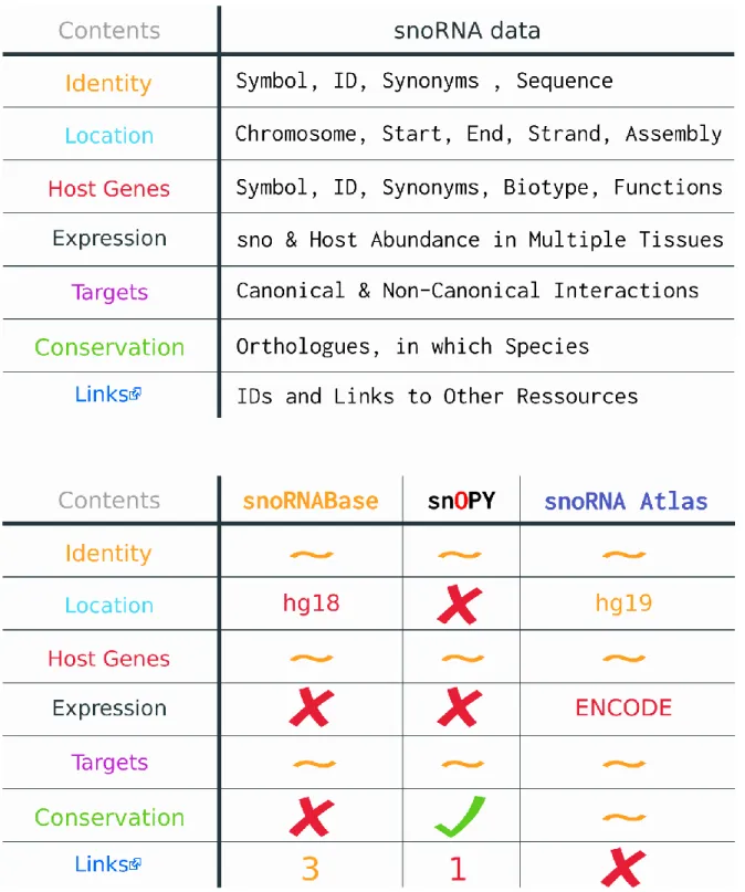
Mémoire présenté à la Faculté de médecine et des sciences de la santé en vue de l’obtention du diplôme de maîtrise ès sciences (M.Sc.) en Biochimie, Faculté de médecine et des sciences de la santé, Université de Sherbrooke, Sherbrooke, Québec, Canada, J1H 5N4 L’ARN est bien plus qu’une molécule transitoire entre l’ADN et les protéines. Au-delà des ARN encodant des protéines, on trouve un vaste éventail d’ARN non-codants qui demeurent encore sous-étudiés. Ces ARN ont été découverts dans les années 1960, mais ce n’est qu’au tournant du siècle que leur incroyable prévalence en cellule a pu être confirmée avec la venue de méthodes de séquençage d’ARN à haut débit. Les expériences à haut débit ont également augmenté de façon exponentielle la quantité de données sur l’ARN créant un besoin pour des outils bio-informatiques permettant leur analyse et leur stockage. Un des premiers, et des plus abondant, type d’ARN non-codant à être découvert sont les petit ARN nucléolaires (snoRNA). Canoniquement caractérisés comme guides de modifications spécifiques dans l’ARN ribosomal, ces petits ARN hautement conservés ont maintenant une liste variée de fonctions non-canoniques, notamment au niveau de l’expression génique, ainsi qu’un nombre croissant d’associations à une panoplie de maladies et de cancer. Considérant la littérature grandissante sur les snoRNA chez l’humain, ainsi que leur connexion maintenant apparente à plusieurs domaines de recherche variés, un regroupement accessible de ce large spectre d’information est maintenant indispensable. Malheureusement, les bases de données en ligne de snoRNA humain, snoRNABase, snOPY, et snoRNA Atlas, ne sont plus à jour ou sont trop pointues au niveau de leurs données. De plus, elles figurent peu ou pas de données d’interactions non-canonique et/ou d’expression. Nous avons donc créé snoDB : une base de données interactive de snoRNA humain qui contient des données sur leurs fonctions non-canoniques, trouvées à travers la littérature, des données d’expression dans une panoplie de tissus, et bien plus. Contrairement à ces prédécesseurs, snoDB offre une visualisions sélectives de son plus large éventail de données, au sein d’une table interactive aux options de recherche abondantes. Les données d’expression peuvent également être visualisées dans la même page, sous forme de carte de chaleur, grâce à l’application sœur de snoDB : snoTHAW. snoDB se démarque aussi par sa connectivité à plus d’une douzaine de ressources incluant le consortium RNAcentral, la plus grande base de données d’ARN non-codant, dont snoDB fais maintenant parti. Les données de ces ressources ont été acquises puis jointe ensemble dans une base de données relationnel postgreSQL. De plus, elles sont toutes en lien dans la table de snoDB afin de facilement pouvoir corroborer l’information visible, ainsi qu’accéder aux fonctionnalités des autres sites. Enfin, snoDB a été construit pour être facile à mettre à jour afin d’assurer ces contributions à la recherche pour de nombreuses années. Mots clés : Petits ARN nucléolaire, Bio-informatique, Base de données (PostgreSQL),
S
UMMARYsnoDB: An Interconnected Online Database of Human snoRNA By
Philia Bouchard-Bourelle Biochemistry
Thesis presented to the Faculty of medicine and health sciences for the obtention of Master degree diploma maîtrise ès sciences (M.Sc.) in Biochemistry, Faculty of medicine and
health sciences, Université de Sherbrooke, Sherbrooke, Québec, Canada, J1H 5N4 RNA is more than just a transitory molecule between DNA and proteins. Beyond the scope of protein-coding RNAs lies a vast underexplored landscape of non-coding RNAs (ncRNA). These RNAs have been slowly uncovered since the 1960s but it took until the turn of the century, and the advent of high-throughput RNA-Sequencing methodologies, for us to finally see how dominated by ncRNAs the transcriptome really is. High-throughput experiments also exponentially expanded the amount of data on RNA and created a need for bioinformatics tools for their analysis and storage. One of the first, and most abundant, ncRNA types to be discovered was small nucleolar RNAs (snoRNAs). Canonically pegged as guides for the modification of pre-ribosomal RNAs, these highly conserved RNAs now boast a diverse list of crucial non-canonical roles, notably in gene expression, as well as being associated to a myriad of diseases and cancers. Considering the growing body of literature surrounding snoRNAs in humans, and their increasing connections to a broad range of fields of study, having an accessible and comprehensive assessment of these data has become essential. Unfortunately, existing online human snoRNA databases, snoRNABase, snOPY, and snoRNA Atlas, are either outdated or too narrow in scope, focusing almost exclusively on canonical snoRNA interactions and lacking expression data. As such, we have created snoDB: a modern, interactive database of human snoRNAs with curated data on non-canonical snoRNA interactions, expression data in a growing range of tissues and cell lines, and more. Unlike the old snoRNA databases, snoDB features extensive visualisation and filtering capabilities, allowing for its larger array of data to be selectively viewed in an interactive and customizable table. Expression data can be further visualised in interactive heatmaps thanks to snoDB’s sister tool: snoTHAW. snoDB also innovates by being much more interconnected with other resources. Data was gathered, and joined together in a relational postgreSQL database, from over a dozen resources, including the RNAcentral database consortium, the largest database of ncRNA sequences, of which snoDB is now a part of. In addition, all resources are linked to in-table, where data they provided appears, to help corroborate the data shown for transparency, as well as to grant access to interesting features housed on remote sites. Finally, snoDB is built to be easily maintainable, updatable and extensible to keep up with ongoing developments and insure that the information it contains will contribute to snoRNA research for years to come. Keywords: snoRNA, Bioinformatics, RNA-Seq, Database, postgreSQL (PSQL), RNA-RNA Interactions, Datatables
T
ABLE OF CONTENT 1 Acknowledgments ... iii 2 Résumé ... iv 3 Summary ... v 4 Table of Content ... vi 5 List of Figures ... ix 6 Abbreviations ... x 1 Introduction... 11.1 History of RNA Biology ... 1
1.1.1 Discovery & Distinction: The two nucleic acids ... 1
1.1.2 Polynucleic Chains & the Tetranucleotide Hypothesis ... 2
1.1.3 The Path to Protein-Coding RNA & the Central Dogma of Biology ... 3
1.1.3.1 The (Actual) Central Dogma of Biology ... 4
1.1.4 Non-Coding RNA, small Nucleolar RNA & the RNA World Theory ... 6
1.1.4.1 Small Nucleolar RNA ... 7
1.1.4.2 Canonical snoRNA Functions ... 9
1.1.4.3 Small Cajal Body-Specific RNA... 11
1.1.4.4 RNA World Theory ... 11
1.1.5 Transcriptome... 12
1.2 Emerging Non-Canonical snoRNA Functions ... 14
1.2.1 SNORD115: Alternative Splicing & Editing ... 14
1.2.2 From Housekeeping Genes to Non-Uniform Expression & Function ... 15
1.2.3 Beyond Alternative Splicing; snoRNAs` Roles in Gene Expression ... 16
1.2.4 snoRNAs & Diseases ... 17
1.2.4.1 Metabolic Stress & Homeostasis ... 17
1.2.4.2 Cancers ... 18
1.2.4.3 snoRNAs as Biomarkers ... 19
1.2.4.4 Mutations in snoRNAs & their Host Genes ... 20
1.3 Simultaneous RNA-Sequencing of snoRNAs & their Host Genes ... 21
1.3.1 RNA-Seq Part 1: Library Preparation & TGIRT ... 22
1.3.1.1 TGIRT-Seq: Holistic RNA-Seq Adressing Structural Bias ... 23
1.3.2.1 CoCo: Count Corrector for Nested & Multimapped Genes ... 24
1.4 High Troughput RNA-RNA Interaction Studies ... 25
1.5 Databases ... 26
1.5.1 SQL & Relational Databases ... 27
1.5.2 The State of snoRNA Data Online ... 29
1.5.2.1 snoRNABase ... 29 1.5.2.2 snOPy ... 31 1.5.2.3 snoRNA Atlas ... 31 1.5.2.4 General Databases ... 32 1.6 Hypothèse/problématique ... 34 1.6.1 Objectifs ... 34 2 Article ... 35 3 Results ... 52
3.1.1 Which Data from Which Sources ... 52
3.1.1.1 Names, Synonyms & snoRNABase IDs: HUGO Gene Nomenclature Committee (HGNC).……….. 52
3.1.1.2 Genomic Annotations: RefSeq/NCBI & Ensembl ... 52
3.1.1.3 Annotations & Cross-Reference Identifiers: RNAcentral ... 53
3.1.1.4 RNA Interactions: snoRNABASE, RISE, the Literature & the Human Protein Atlas ... 55
3.1.1.5 In-House Data: TGIRT-Seq Datasets & Host Genes ... 59
3.1.1.6 Conservation: snoRNA Atlas, snOPY & Ensembl ... 60
3.2.1 Joining Data together ... 60
4 Discussion... 62
4.1 What Other snoRNA Databases Lacked Beyond Their Data ... 62
4.1.1 Interconnectedness ... 62
4.1.2 Fully Downloadable Data ... 64
4.1.3 Maintainability & Extensibility ... 64
4.1.3.1 PostgreSQL Code & Tables ... 65
4.1.3.2 Front-End Plugins ... 65
4.1.4 User Experience ... 66
4.2 What should be Added to snoDB in Future ... 68
4.3 Conclusion ... 70
FIGURES
Figure 1: Rough Timeline of Nucleic Acid Research ... 5
Figure 2: Canonical snoRNAs ... 8
Figure 3: TGIRT-Seq + CoCo Pipeline for Holistic RNA-Sequencing ... 22
Figure 4: Data Models Found in Relational Databases ... 28
Figure 5: Pertinent Information in a snoRNA Databases ... 30
ABREVIATIONS A cDNA C DNA FTP G HGNC LCC lncRNA miRNA mRNA ncRNA NGS piRNA PSQL PWS RDBMS RNA RNA-Seq RNP rRNA scaRNA snoRNA snRNA SQL Adenine, Adénine
Complmentary DNA, ADN complémentaire Cytosine, Cytosine
Deoxyribonucleic acid, Acide déoxyribonucléique File Transfer Protocol, protocole de transfert de fichier Guanine, Guanine
HUGO Gene Nomenclature Committee
Leukoencephalopathy with Calcification and Cysts Long ncRNA, Long ARN non-codant
Micro RNA, Micro ARN
Messenger RNA, ARN messager Non-Coding RNA, ARN non-codant
Next Generation Sequencing, Séquençage de nouvelle génération PIWI-interacting RNA, ARN intéragissant avec les protéines PIWI PostgreSQL
Prader-Willi Syndrome, Syndrome de Prader-Willi
Relational Database Management System, Système de gestion de
base de données relationelle
Ribonucleic acid, Acide ribonucléique RNA-Sequencing, Séquençage d’ARN
Ribonucleoprotein Complex, Complexe ribonucléoprotéique Ribosomal RNA, ARN ribosomal
Small Cajal Body-Specific RNA, Petit ARN spécifique aux corps
de Cajal
Small Nucleolar RNA, Petit ARN nucléolaire Small Nuclear RNA, Petit ARN nucléaire Structured Query Language
T TERC TGIRT tRNA U yadcf Thymine, Thymine
Telomerase RNA component, Composante ARN de la télomérase Thermotable Group 2 Intro Reverse Transcriptase, Transcriptase
inverse thermostable de group 2
Transfert RNA, ARN de transfert Uracil, Uracile
I
NTRODUCTIONRNA biology has come a long way since its inception during the last century. We now have so much data about this diverse class of molecules that innumerable tools and databases have been created to aid in their analysis. But what is RNA and how do specialized bioinformatics resources, such as an online snoRNA database, help drive RNA research forward?
History of RNA Biology
Ribonucleic acids (RNAs) are a diverse class of molecules often transcribed from deoxyribonucleic acids (DNA) (Brosius and Raabe, 2016; Zhang et al., 2018). Both are long chains of nucleotides made up of a sugar phosphate spine (ribose sugar for RNA and deoxyribose for DNA) with one of four bases attached to each sugar molecule. These bases are adenine (A), thymine (T), guanine (G) and cytosine (C) with thymine being substituted for Uracil (U) in RNA. In addition, the familiar double helix structure of DNA is not seen in RNA; rather it is a single helix that folds in on itself in a wide variety of structures (Lodish et al., 2000). Cells are filled with an incredible diversity of RNA types that enact and regulate vital biological functions (Kufel and Grzechnik, 2019). Although the chemical and structural differences between the two types of nucleic acid chains have been identified for a hundred years, their individual importance, their interconnected relationship, and even their names, took several decades to be established.
Discovery & Distinction: The two nucleic acids:
The history of nucleic acids research begins in 1869 with the work of Johann Friedrich Miescher on leukocytes. These white blood cells were easily obtained with relatively good levels of purity from pus soiled bandages of a nearby clinic. Through purification of the infectious fluid, Miescher’s experiment uncovered a cellular substance with unprecedented resistance to chemical methods of protein degradation. This seemingly non-proteic substance was hypothesized to originate from the nucleus which inspired the name it was given at the time: nuclein (His et al.,
1897; Miescher, 1871; Miescher, F., 1869). As it turns out, nuclein was in fact the first recorded precipitate of DNA. (His et al., 1897; Miescher, 1874; Miescher and Schmiedeberg, 1896).
These findings happened around the same time as the nucleus itself was being suggested by prominent biologist Ernst Haeckel to contain the materials responsible for heredity (Haeckel, 1866; Olby, 1969). Of note, though cell theory was technically around at the time, stating that life is built of tiny units called cells, only its bigger constituents such as the nucleus and the nucleolus it contains, could be observed with microscopes (Beale, 1858). The notion that the nucleus and the cytoplasm that surrounds it differed functionally wasn’t yet popular, with the entire contents of the cell, called protoplasm, being deemed the substance of life (Welch, 1995). Miescher did theorize that nuclein could be the material responsible for fertilization, but the carrying of hereditary information was deemed an impossible role for a single molecule to possess, given the wide range of diversity observed in nature (Dahm, 2005; Miescher, 1874; Ralf Dahm, 2008). And so, the link between nuclein (DNA) and heredity was not to be confirmed until over half a century later.
In the meantime, nuclein’s acidic nature would see it re-named to nucleic acid (Altmann, R., 1889). Improved methods for its purification enabled Nobel Prize laureate to be Albrecht Kossel to uncover the components of nucleic acid: adenine, guanine, cytosine, thymine and uracil along with a sugar thought to be a pentose (Hammarsten O., 1894; Jones, 1953). As these experiments were repeated, two kinds of nucleic acids emerged based on the model organisms from which they were extracted: yeast nucleic acid, obtained from plants, and thymus nucleic acid, which was abundant in the thymus gland of animals. These two types of nucleic acids, in fact RNA and DNA respectively, were found to have differing composition of bases: uracil in yeast versus thymine in thymus nucleic acid, as well as different sugars: pentose for yeast and a hypothesized hexose in thymus nucleic acid (P. A. Levene, 1910).
Polynucleic Chains & the Tetranucleotide Hypothesis
At the turn of the 20th century, nucleic acids are still seen as a soup of their constituents. The concept of polynucleic chains came around 1909 with the work of Pheobus Levene (Jacobs W.
A., and Levene P. A., 1909). The Russian physician showed how the bases contained in both types of nucleic acids bind to sugars to form nucleosides and how these subunits link up with a phosphoric acid molecule to form nucleotides (Levene and Jacobs, 1909). Despite World War I halting progress, Levene eventually established that the sugars found in yeast nucleic acid and thymus nucleic acids are ribose and deoxyribose respectively (Levene et al., 1930; Levene and Jacobs, 1909). Based on the different chemical and physical properties of these carbohydrates, they proposed that the nucleic acids be called ribonucleic acids (RNA) and deoxyribonucleic acids (DNA) (Frixione and Ruiz-Zamarripa, 2019; Levene and Tipson, 1935). Around the same time, the long held hypothesis that the two nucleic acids are exclusive to either plants or animals was finally overturned (Allen, 1941).
Despite plentiful contributions, Pheobus Levene is a name synonymous with the tetranucleotide hypothesis, the erroneous theory that nucleotides form into simple closed rings of four nucleotides known as tetradic repeats (Hunter, 1999). This infamous theory is often attributed as holding back research on nucleic acids since a tetradic repeats conformation made them predictably dull. However, historical context suggests otherwise; Levene wasn’t a biologist, focusing instead on the chemical characterization of nucleic acids and leaving it up to biologist to assess their function. At the time, biologist were simply more interested in the study of proteins which they believed were the more likely carriers of genetic information in cells rather than the ‘idiotic’ nucleic acids (Hargittai, 2009; Olby, 1994). This mindset was likely bolstered by the successful crystallization of insulin and other key enzymes around the 1930s coupled with the unavailability of homogenous samples of nucleic acids for study (Lederberg Joshua, 1994; McPherson and Gavira, 2013).
Such pure samples would finally be available in 1944 through DNA viruses with the work of Avery, McCarty and MacLeod, on pneumococci, which went on to finally and rightfully attribute heredity as a characteristic of DNA (Lederberg Joshua, 1994).
The Path to Protein-Coding RNA & the Central Dogma of Biology
variant was proved to be lethal by Fredirick Griffith in 1928. The assumption was that the transformation of the inoffensive type 2 virus into its deadly type 3 counterpart was a protein mediated phenomenon (Griffith, 1928). However, Avery, McCarty and MacLeod, through extensive purification of the deoxyribonucleic acid from the heat-killed type 3 pneumococci, showed that the transformation was fundamentally attributable to DNA. This breakthrough paved the way for the DNA theory of inheritance, and renewed interest in nucleic acid research (Avery et al., 1944).
Many protein-enthusiasts were not so easily convinced, continuing to uphold proteins as the progenitors of genes up until the early 1960s (Eck, 1961). If DNA truly held all genetic information within cells, how was it turned into proteins? One theory at the time assumed the existence of a yet unknown RNA intermediate (Brachet, 1942; Capspersson, 1947). However, back then the only type of RNA to have formerly been identified was ribosomal RNA (rRNA). Known then as microsomal particles, they had been shown in an experiment to turn radiolabeled amino acids that passed through them into proteins (Hoagland et al., 1957; Roberts, 1958). Convinced of the existence of a hypothetical “adaptor” between DNA and the ribosomes, Francis Crick published the now famous central dogma of biology (Crick, 1958).
The (Actual) Central Dogma of Biology
Contrary to what many are taught in school, the central dogma of biology was never meant to be a mere roadmap of protein synthesis. Rather it outlined the possible ways for information to transit between DNA, RNA and proteins based on scientific knowledge at the time, as well as Crick’s own intuition. Conversions from DNA to RNA and from RNA to protein were indeed postulated, but this was alongside established phenomena like DNA’s ability to copy itself during cell division (DNA to DNA), and the ability of RNA viruses to self-replicate (RNA to RNA) (Figure 1). The flow of RNA to DNA was tentatively hypothesized as well, but it would only see confirmation in 1970 with the discovery of the “reverse transcriptase” enzyme (Coffin and Fan, 2016). Most interesting were the paths of information flow that were deemed impossible (protein to protein, protein to RNA, and protein to DNA). They lacked any known mechanisms but at the time so did the transcription of DNA to RNA, as well as the subsequent
translation of RNA into proteins. Therefore, by asserting the unfeasibility of information transits stemming from proteins, the central dogma discredited theories pinning proteins as gene progenitors. Overall, the intended takeaway was that the transfer of genetic information between DNA, RNA and proteins follows defined paths in cells and that once information reaches proteins, reversing it back into DNA or RNA is impossible (Cobb, 2017; Morange, 2008).
The central dogma of biology, and the RNA adaptor hypothesis between the ribosomes and proteins, would be confirmed shortly after the dogma’s publication with the formal discovery and characterization of protein coding RNA, called messenger RNA (mRNA). Several groups published papers on mRNA describing it as a transcribed template of DNA that travels to the cytoplasm to bind with ribosomes, as well as with a specific iteration of amino acid carrying transfer RNA (tRNA), to be translated into protein (Cobb, 2015).
Figure 1 : Rough Timeline of Nucleic Acid Research
Nucleic acids were first discovered in the 1860s under the name nuclein. Later studies around the turn of the century categorized two types of nucleic acids based on the presence of the nucleo-bases thymine or uracil and based on their source of extraction (animal glands such as the thymus or plants such as certain yeasts). During the 1930s, the two types of sugars composing the nucleotides, found in both types of nucleic acids, had been established along with their ability to link up together to form polymeric chains. All of this paved the way for the central dogma of
biology, posited towards the end of the 1950s, which correctly mapped the flow of genetic information between what we now knew as DNA and RNA as well as proteins.
Non-coding RNA, small Nucleolar RNA & the RNA World Theory
The monumental discovery of mRNA, as vectors of genetic information for protein synthesis, was instrumental to the development of RNA biology. It also colored our view of RNA as mostly being passive elements while proteins remained the predominant functional components of the cell. Opposing this view came a string of discoveries implicating non-protein-coding RNA, simply called non-coding RNA, in various novel and essential regulatory pathways.
Analysis of HeLa cell nuclei, in the late 1960s, revealed the existence of new, uridine(U)-rich, non-coding RNA(ncRNA) species beyond rRNA and tRNA, which were named U1, U2, U3, etc. (Pene et al., 1968; Weinberg and Penman, 1968). Further studies on these new RNAs in yeast allowed for their classification into two distinct categories, based on their localization and functionalities (Riedel et al., 1986; Wise et al., 1983) . U1, U2, U4, U5 and U6 were dubbed small nuclear RNA (snRNA) for localizing to the nucleus, while U3 and U8 were dubbed small nucleolar RNA (snoRNA) for specifically localizing to a membrane-less compartment of the nucleus called the nucleolus (Busch et al., 1982). These two types of RNA both form into hairpin-like structures that average over a hundred nucleotides in length. Both RNA species were also found to individually bind with specific proteins to form ribonucleoprotein complexes (RNPs) (example: U1 snRNP and U3 snoRNP) (Busch et al., 1982). However, snRNPs are formed outside the nucleus before returning and assembling together into a big complex called the spliceosome (Patel and Bellini, 2008). The spliceosome then binds pre-mRNA and regulates a vital processing step of theirs called splicing, i.e., the excision of the non-coding regions named introns that separate the multiple coding segments of a nascent gene called exons (Lerner et al., 1980; Mount and Wolin, 2015). Meanwhile, snoRNPs form within the nucleus immediately after snoRNAs are transcribed, before making their way to the nucleolus where they individually act in rRNA processing (Schimmang et al., 1989).
Small nucleolar RNA
We now know that Small nucleolar RNAs (snoRNAs) are a group of highly structured and expressed non-coding RNAs that are conserved across all eukaryotes (Henras et al., 2004). Most snoRNAs discovered thus far have been found in humans and in yeast, but snoRNAs have also been identified in rodents, amphibians, plants and even Achaea (Bertrand and Fournier, 2013). In humans, they are mostly encoded within the introns of mRNAs and long non-coding RNAs (lncRNAs) which can be referred to as snoRNA host genes (Boivin et al., 2018b). Two thousand unique snoRNA sequences have been reported in human RNA databases to date (Bouchard-Bourelle et al., 2019).
We distinguish two main snoRNA families based on their structure and specifically conserved sequence motifs dubbed boxes: the single stem-loop (stem-bulge-stem), RUGAUGA & CUGA motif bearing box C/D family and the dual stem-loop (hairpin-hinge-hairpin-tail), ANANNA & ACA motif bearing Box H/ACA family (Figure 2). In addition, some box C/Ds sometimes possess less well conserved copies of C and D boxes dubbed C’ and D’. All these conserved box motifs, along with a cascade of chaperones and other conserved proteins, enable snoRNAs to bind specific sets of proteins conserved across eukaryotes when they are transcribed in the nucleoplasm. This association prevents the degradation of the bound regions by exonucleases, and results in fully processed small nucleolar ribonucleoprotein complexes (snoRNPs) (Kishore et al., 2013).
The boxes are also responsible for the trafficking of snoRNPs from the nucleus to the nucleolus, which is where they perform their most widely recognized function as guides for specific post-transcriptional RNA modifications (Massenet et al., 2016).
Figure 2: Canonical snoRNAs
snoRNAs are canonically divided into 2 families that bind distinct sets of proteins which catalyze specific modifications in pre-rRNA which snoRNAs bind through complementarity.
Canonical snoRNA Functions
The most well characterized function of snoRNAs is guiding multiple site-specific modifications in pre-ribosomal RNA (pre-rRNA) during ribosome biogenesis in the nucleolus. They do this through regions on the snoRNA called antisense elements which are complementary to specific regions of pre-rRNA (Henras et al., 2004; Kiss, 2001). This mechanism, and snoRNAs’ role as guides for these modification, were first discovered in yeast (Philippe Ganot et al., 1997; Kiss-László et al., 1996) and confirmed in humans shortly thereafter (Balakin et al., 1996; P. Ganot et al., 1997). Orthologous snoRNAs have also been identified in organism as evolutionarily distant as Achaea, pointing to the primeval nature of canonical snoRNA functionalities (Gaspin et al., 2000; Omer et al., 2000).
Box C/D snoRNA antisense elements are composed of 10 to 21 nucleotides and are located upstream of the D’ and/or D box with the modification site on complementary rRNA usually being located 5 nucleotides upstream of the D/D’ box. Box H/ACA snoRNA possess antisense elements within their 3’ and/or 5’-terminal hairpin domains also called pseudouridylation pockets (Philippe Ganot et al., 1997; Kishore et al., 2013).
Once the snoRNAs’ antisense element binds to a complementary strand of rRNA, core proteins forming the snoRNP catalyse the modification of a specific nucleotide. Box C/D snoRNPs are canonically composed of four conserved proteins including the methyltransferase Fibrillarin (FBL) which catalyzes the 2’-O-methylation of pre-rRNA. Box H/ACA snoRNPs are canonically composed of 2 identical protein quartets, on each of their stem-loops, with Dyskerin (DKC1) being responsible for the pseudouridylation of pre-rRNA (Figure 2). In both snoRNA families, the proteins without any catalytic functions, NOP56, NOP58 and SNU13 for box C/Ds, and Nhp2, NOP10 and Gar1 for box H/ACAs, serve as scaffolds to recruit each other and stabilize the resulting complex (Filipowicz and Pogačić, 2002; Jády and Kiss, 2001).
Although the modification of specific, and often conserved, bases in rRNA by snoRNAs haven’t been found to be essential, hints to their involvements in proper folding of pre-rRNA and
the interactions between rRNA and ribosomal proteins have been found pointing to a need for further research on the matter (Bachellerie et al., 2002).
However, there are snoRNAs that have for a long time been known to function as something other than guides for post-transcriptional modifications. The U3 (SNORD3A) and U8 (SNORD118) to name only two, are highly conserved box C/D snoRNAs which are not known to guide any chemical modifications in pre-rRNA while still being key players of pre-rRNA processing and accumulation (Langhendries et al., 2016).
U3 is associated with the proper folding of the 18S small ribosomal subunit. It can execute this function due to the presence within its sequence of an additional motif called a B box. This motif allows U3 to bind a protein called Rrp9 in humans which is essential for the formation of its snoRNP (Zhang et al., 2013). More specifically, it has been shown that nucleotide substitutions in the 5’ETS (external transcribed spacer), located upstream of the 18S ribosomal RNA gene, leads to cell death in yeast. This region is involved in the initial binding of the 18S pre-mRNA with U3, and when plasmids containing a mutated U3 sequence complementary to the altered 5’ETS sequence is expressed, 18S accumulation and cell growth is regained (Dutca et al., 2011). When U3 is properly bound to its core proteins, as well as to the 5’ETS region upstream of the 18S rRNA gene, it mediates early cleavage steps in pre-rRNA processing that are essential for ribosome biogenesis (Cléry et al., 2007).
U8 on the other hand is vitally involved in the accumulation of 5.8S and 28S rRNA which are components of the large ribosomal subunit. Similarly to U3, U8 binds to the 28S pre-mRNA with mutational studies showing this to be essential for maturation of the large ribosomal subunit. In this case however, although the interaction is necessary it must be undone before pre-rRNA processing is concluded to allow for the association of the 28S and 5.8S subunits (Peculis, 1997).
Small Cajal Body-Specific RNA
snoRNAs have also canonically been shown to modify specific conserved positions in snRNAs. These RNA-dependent modifications were at first thought to exclusively occur in nuclear sub-organelles known as Cajal (coiled) bodies, to which a subset of snoRNA localize, and were hence named small Cajal body-specific RNA (scaRNA). Their characterization as snoRNAs might seem odd, as they are non-nucleolar RNAs, but Cajal bodies are actually closely related to nucleoli both physically and functionally, even being originally called nucleolar accessory bodies (Trinkle-Mulcahy and Sleeman, 2016). In addition scaRNAs possess familiar C/D and/or H/ACA box motifs through which they guide Fibrillarin dependant 2’-O-methylation and Dyskerin dependant pseudouridylation, although in snRNA instead of in rRNA (Darzacq et al., 2002). However, emerging evidence points to snoRNA nomenclature potentially needing revision as both scaRNAs and snoRNAs alike can exist and be functional outside of the localization that informed their naming scheme. For example, scaRNA-dependant snRNAs modifications are still carried out in cell-types/species which lack Cajal bodies, while snoRNAs have been found to accumulate in Cajal bodies, with some also being found in the cytoplasm (Deryusheva and Gall, 2009; Michel et al., 2011). Furthermore, scaRNAs have been found to act as guides for the modification of rRNA, just like canonical snoRNAs, further blurring the divide between them (Deryusheva and Gall, 2019).
RNA World Theory
Around that same time as snRNAs and snoRNAs were begin discovered, a fundamental characteristic of non-coding RNAs’ activity was being uncovered: the specific structures they fold into. It was shown to allow the t-shaped tRNAs to successfully bind amino acids, to grant ribosomes the ability to mediate protein synthesis and would be shown to be functionally important in most non-coding RNA. This is reminiscent of the need for proteins to fold into specific structures themselves to become functional, with mutated or denatured protein and RNA alike losing some or all of their activity (Bhartiya and Scaria, 2016; Rich and RajBhandary, 1976). The parallels did not stop there as the discovery of ribozymes came soon after, and showed for the first time that RNAs, just like proteins, could catalyze chemical reactions in cells
(Cech et al., 1981; Guerrier-Takada et al., 1983). Even before having discovered RNAs’ catalytic functions, many researchers were pondering the hypothesis of self-replicating RNA molecules being primordial precursors to both DNA and proteins. This theory, which posits a time where RNA alone permitted life, was formally named the “RNA world hypothesis” in 1986 (Cech, 2012; Neveu et al., 2013).
1992 brought the discovery of the long non-coding RNA (lncRNA) XIST, which is involved in X-chromosome inactivation (Brockdorff et al., 1992; Brown et al., 1992). In 1993, the first micro RNA (miRNA) was discovered in yeast (Lee et al., 1993). It took until the early 2000s for these small non-coding RNAs to be formally characterized as a group and notably linked to the regulation of gene expression through gene silencing (Ambros, 2004).
In summary, ncRNAs were clearly shown to be as functionally diverse and important as proteins, if not more so, potentially enabling life independently from proteins and DNA for billions of years (Cech, 2012). Nonetheless, research into ncRNAs didn’t pick-up steam until the turn of the century. In 2001, a working draft of the human genome had finally been sequenced and published, using first-generation sequencing (one nucleo-base at a time). It confirmed the genome as being overwhelmingly composed of non-coding regions (Venter et al., 2001). Around the same time, the more efficient next-generation sequencing (NGS) technologies were inaugurated, and shortly thereafter adapted, to allow for high-throughput quantification of all RNA transcripts in a population of cells (Weber, 2015).
Transcriptome
Our ability to quantify RNA transcripts using high-throughput RNA-Sequencing represented another revolution in the field of RNA biology. It was the first technique to actually give us a look at the transcriptome, that is which parts of the genome are actively transcribed into RNA, at the moment an experiment was conducted, as well as giving us insights into their cellular functions (Wang et al., 2009). But the term transcriptome had been coined years prior while only low-throughput sequencing technologies were available. The main focus of these first gene profiling experiments was mRNA, with the term transcriptome being originally defined as
representing the sum of mRNA expression in a population of cells (McGettigan, 2013; Piétu et al., 1999). This led to much of transcriptomic research being mRNA-centric, even in the era of next-generation sequencing. This despite the fact that evolutionary studies showed that non-coding RNAs can represent up to 98% of transcriptional output across higher eukaryotes (Mattick and Gagen, 2001). Such insight coupled with the growing body of literature surrounding non-coding RNAs, and their wide range of essential regulatory functions, should have been the final nail in the coffin for the notion that RNAs are merely passive intermediates between DNA and proteins. But half a century of characterizing non-coding DNA as junk (Ehret and De Haller, 1963) seems hard to shake off and an old guard persists as exemplified by Google’s definition of ‘’transcriptome’’ still reading:‘’ the sum total of all the messenger RNA
molecules expressed from the genes of an organism.’’ Relinquishing such a notion and overall
adopting a more fluid view of scientific concepts new and old is more than ever imperative in this new age of fast discoveries.
For example, we have known for a long time that, in terms of the molecules present, the transcriptome is mostly composed of rRNA and tRNA. Like mRNA, these two types of non-coding RNA have also canonically been pegged as static actors in protein synthesis but have since been hinted to have other unsuspected non-canonical functions. Transfer RNA have been shown to be cleaved into tRNA-derived small RNAs (tsRNAs) which are implicated in stress-response signalization and in regulating gene expressions (Shen et al., 2018). Moreover, growing evidence points to ribosomal RNAs being highly regulated and heterogeneous in nature, leading to the preferential translation of certain mRNA, with possible implication in differential development and response to stress (Guo, 2018; Xue and Barna, 2012). mRNAs themselves have recently been attributed new roles beyond mere messengers including participating in the assembly of nuclear bodies like Cajal bodies, which are membrane-less regions of nuclei where splicing complexes are assembled (Shevtsov and Dundr, 2011). When even the most extensively studied RNA-type has secrets left for us to uncover, one can easily posit that we have in fact only begun to scratch the surface of RNA biology. This notion sees itself exemplified in small nucleolar RNAs, which were found in the early 60s and quickly pegged as actors in rRNA processing but have since gradually been ascribed unsuspected and essential multifarious functions.
Emerging Non-Canonical snoRNA Functions
snoRNAs’ vital and non-vital roles in ribosome and spliceosome biogenesis have been the focus of many studies for multiple decades (Jády and Kiss, 2001; Maxwell and Fournier, 1995). This makes sense considering that the most evolutionarily stable snoRNAs across mammals are highly expressed, and located in introns of host genes related to ribosome biogenesis and translation (Hoeppner et al., 2009). Meanwhile, the less conserved snoRNAs, seen only in humans and primates, show strong evidence of being derived from transposable element, and having thus been recently retrotransposed into new genomic location/host genes (Scott and Ono, 2011). In vertebrates, most snoRNAs are co-transcribed with their host gene, while some are independently transcribed by RNA polymerase II or III (Dieci et al., 2009; Tycowski et al., 2004). Interestingly, the less conserved snoRNAs, seen only in humans and primates, do not show enrichments for canonical snoRNA functions, suggesting a broader spectrum of snoRNA functionalities (Hoeppner et al., 2009). Taken together, these studies point to a flexibility in snoRNA expression, which isn’t always correlated with their host gene, because some snoRNAs likely regulate other genes involved in non-canonical functions. Indeed, in recent years a growing number of papers have begun ascribing a wide range of varied non-canonical functions to snoRNAs.
SNORD115: Alternative Splicing & Editing
These discoveries slowly began around the turn of the century with one of two clusters of cerebrally enriched snoRNAs, known then as HBII-52 (now SNORD115). This cluster is located within the locus of chromosome 15 associated with Prader-Willi syndrome (PWS), which is the leading genetic cause of obesity worldwide. The disease was shown to be caused by the deletion of the paternal locus which features genetic imprinting. Imprinting is an epigenetic methylation of a parent’s gene that makes only its expression possible regardless of the presence of a copy of this gene on the complementary allele. The mechanism behind PWS was linked to the SNORD115 cluster present in the affected locus. SNORD115 lacks any known rRNA complement, but it has been found to associate with the alternatively spliced exon 5 of the serotonin receptor 5-HT2CR (Cavaillé et al., 2000). This complementarity mediated binding was
later confirmed to affect the alternative splicing of said exon, with PWS-afflicted individuals exhibiting distinct serotonin receptor mRNA isoforms when compared to healthy individuals (Kishore and Stamm, 2006). However, it remains unclear if a change in alternative splicing alone is responsible for PWS. Other studies have pointed to an additional post-transcriptional modification, mediated by an interaction between SNORD115 and the 5-HT2CR pre-mRNA, known as Adenosine-to-Inosine (A-to-I) editing, which was shown to have behavioral impacts in mice (Doe et al., 2009). Although this editing mechanism is confirmed, its link to PWS remains a matter of debate (Glatt-Deeley et al., 2010).
From Housekeeping Genes to Non-Uniform Expression & Functions
These discoveries meant that snoRNAs could no longer be thought of as constitutively expressed genes solely implicated in site-specific RNA modifications. They can have tissue-dependent expression and enact non-housekeeping functions. But with only about a quarter of snoRNAs being orphans, i.e. having no known function (Falaleeva et al., 2016), could they really have that many undiscovered roles left to uncover?
Bioinformatics-based predictive approaches have found undiscovered complementary rRNA sites for a few orphan snoRNAs in regions that were not known to be modified. This sparked the tentative connection of snoRNAs with ribosome heterogeneity, in a perhaps tissue- or condition-specific manner further (Dieci et al., 2009; Piekna-Przybylska et al., 2007; Xue and Barna, 2012). If true, this would redefine the importance of canonical snoRNA functions in stress-response, and perhaps even development, through the preferential translation of certain mRNAs by so-called specialized ribosomes. Although, it would also reduce the pool of orphan snoRNA with potential non-canonical functions even. This assumption of fewer snoRNA potentially having novel roles is however proven false on two fronts. First, new snoRNA are steadily being discovered (Jorjani et al., 2016; Kishore et al., 2013). Second, snoRNAs have been found which have both canonical and non-canonical functions, broadening the member spectrum with new functionalities to perhaps every known and yet unknown snoRNA. For example, SNORD27 has been shown to both guide rRNA methylation and mediate the alternative splicing of several protein-coding genes, including ABCA8, E2F7, FER and MAP4K3. A potential mechanistic hint
for how this functional diversity is possible came with soluble nuclear extract studies which detected two distinct SNORD27 ribonucleoprotein complexes in biochemically divisible fractions; a canonical fibrillarin-dependant snoRNP and a non-canonical fibrillarin-independent snoRNP (Falaleeva et al., 2016).
Beyond Alternative Splicing; snoRNAs` Roles in Gene Expression
Alternative splicing remains one of the best characterized non-canonical snoRNA functions, with other snoRNAs not previously listed also being implicated in it. However, snoRNAs have now been found to regulate gene expression through a much broader array of mechanisms. For example, dozens of snoRNAs show enrichment in both Drosophila and human chromatin, a DNA-protein complex responsible for compacting and de-compacting DNA for protection and transcription respectively. A specific protein, Df31, was shown to bind both histones, which are proteins that are part of the chromatin, and the enriched snoRNA. As a result, chromatin structure remains un-compacted allowing for genes on that segment of the genome to be transcribed (Schubert et al., 2012).
Some snoRNAs have also been found to directly regulate mRNA abundance after being degraded into smaller RNAs, such as miRNA and piRNAs, or by interfering with pre-mRNA processing. Micro RNAs (miRNAs) are small non-coding RNAs, averaging 22 nucleotides, which form a distinct microRNA ribonucleoprotein complex (miRNP), more commonly called an RNA-induced silencing complex (RISC) (Cai et al., 2009). Silencing describes any negative regulation of gene expression. Similarly to canonical snoRNAs within snoRNPs, miRNA in miRNPs/RISC possess sequence complementarity with mRNA, and serve as guides for their silencing. This resemblance with canonical snoRNA isn’t a coincidence as evolutionary ties between the two types of non-coding RNA have been found, as well as the fact that some snoRNA are degraded into sno-derived RNAs (sdRNA) such as miRNA (Ender et al., 2008; Saraiya and Wang, 2008). Likewise, snoRNAs have also been shown to be processed into PIWI interacting RNAs (piRNAs). These 26 to 31 nucleotide-long RNAs are so-named for their interactions with the PIWI protein family, which is closely related to the Argonaut protein, a key component of miRNA RISC gene silencing complex. Unsurprisingly then, piRNAs derived from
snoRNAs have been found to mediate decay in targeted pre-mRNAs, which they do through recruitment of the nuclear exome (Zhong et al., 2015). Researchers have also recently stumbled upon a set of box C/D snoRNAs that associate with proteins constituting the 3’ mRNA processing complex. The processing of 3’ end in mRNAs is essential to their proper expression. It involves the cleavage of said 3’ end followed by the addition of over 200 adenine residues, called a polyA tail, in a process named polyadenylation. Depletion of one particular snoRNA showed consistent effects on 3’ polyadenylation profiles and the abundance of specific genes (Huang et al., 2017).
Recent, studies in yeasts have found two previously orphan box C/D snoRNAs to be involved in guiding the acetylation of 2 cytosine residues in rRNA. They do this by associating with the cytidine acetyltransferase Kre33, which is known to acetylate tRNAs by interacting with the adaptor protein Tan1 (Sharma et al., 2017). Considering the highly conserved nature of most snoRNAs, it would not be surprising for acetylation to also be mediated through snoRNAs in higher eukaryotes.
snoRNAs & Diseases
With so many regulatory roles to their name, snoRNA-associated diseases seem quite likely when deregulation of their functions occur. Indeed, as previously mentioned, deletion of the SNORD115 family of snoRNAs is linked to a genetic disorder called Prader-Willi syndrome. However, not only are a growing number of diseases being linked to snoRNAs, they are increasingly being investigated as diagnostic/prognostic tools in the form of biomarkers.
Metabolic Stress & Homeostasis
Genetic screenings of Chinese hamster ovary (CHO) cells under lipotoxic conditions, metabolic stress conditions which are a common complication of diabetes where lipids accumulate in non-adipose tissues, revealed that SNORD32A, SNORD33, SNORD34 and SNORD35A can localize to the cytoplasm outside the nucleus and mediate oxidative stress (Michel et al., 2011). A later study found these four snoRNAs to also regulate systemic glucose metabolism solidifying their
potential implication in the pathogenesis of diabetes (Lee et al., 2016). Further genetic loss of function screening in CHO cells found orphan snoRNAs ACA60 (SNORA60) and the U17 snoRNAs (SNORA73A & 73B) to be essential in the regulation of cholesterol trafficking and homeostasis (Brandis et al., 2013; Jinn et al., 2015). The study on the U17 snoRNAs was even able to identify their non-canonical interaction with and negative regulation of mRNA HUMMR (hypoxia upregulated mitochondrial movement regulator) as a novel mechanism in cholesterol trafficking. Meanwhile ACA11 (SCARNA22) was found to be upregulated in multiple myeloma, an incurable cancer of plasma cells, and confer resistance to chemotherapy via a suppression of oxidative stress (Chu et al., 2012).
Cancers
Links between snoRNAs and various cancers have consistently increased over the last two decades. The most recent review on the subject implicated nearly 50 snoRNAs across 11 different types of cancers as both oncogenes and/or tumor suppressors (Liang et al., 2019).
Some snoRNAs have been linked to the p53 pathway in cancer which regulates the expression of genes involved in cell cycle arrest, DNA repair and apoptosis. Overexpression of snoRNAs have been found in human breast and prostate cancer to inhibit the p53 pathway and promote tumorigenesis (Su et al., 2014). Similarly, breast cancer also sees the upregulation of the U3 and U8 snoRNAs acting as oncogenes with their depletion enabling a potent p53 anti-tumor response (Langhendries et al., 2016). SNORA42 has been found to be oncogenic in non-small cell lung cancer where it regulates the expression of p53 (Mei et al., 2012). Other experiments have confirmed p53 to directly control transcription of the snoRNA host gene GAS5. The expression of the multiple snoRNAs nested within GAS5 correlate with p53 expression in colorectal cancer, though their potential role in this pathology has yet to be determined (Krell et al., 2014).
Other cancer related pathways, such as the PI3K–AKT cascade, have also been associated with snoRNA dysfunctions in certain cancers. PI3K-AKT signaling involved in cell death, differentiation and proliferation is increased due to ACA11 over expression in hepatocellular carcinoma (HCC) (Wu et al., 2017). SNORD126 also activates the PI3K–AKT pathway by
upregulating FGFR2, which promotes cell growth in HCC as well as in colorectal cancer (Fang et al., 2017).
Sno-derived RNAs (sdRNAs) have also been found to be differentially expressed in certain cancers. SNORD17 derived miRNA miR-768-5p reportedly binds to breast cancer associated protein YB-1 (Blenkiron et al., 2013). In breast cancer still, SNORD75 derived piRNA pi-sno-75 upregulates tumor suppressor and apoptosis inducer TRAIL (He et al., 2015). And in prostate cancer, sdRNAs derived from several snoRNAs were found to be over expressed, with the expression of some sdRNAs being specifically higher in metastatic patients, hinting at their potential role as prognostic biomarkers (Martens-Uzunova et al., 2015).
snoRNAs as Biomarkers
The involvement of snoRNAs in various facets of cancer biology and the detection of many of them in serum, plasma and urine, in addition to their high stability, has led to them being studied as potential biomarkers. Indeed, as previously described, several studies have found expression variation of certain snoRNAs, or their derivatives, to be indicative of certain cancers or certain stages of the disease.
As such, eight differentially expressed snoRNAs were recently established as prognostic factors in gastric cancer (Wang et al., 2019). Several snoRNA signatures, predicting overall survivability in patients with non-small cell lung cancer, have also been found (Gao et al., 2015; Liao et al., 2010; Mannoor et al., 2014). Expression of SNORA70F & SNORD116-118 are seemingly able to distinguish two types of chronic lymphocytic leukemia with different prognostics (Ronchetti et al., 2013).
Meanwhile, six serum snoRNAs were found to be effective biomarkers of ageing joints and osteoarthritis (Steinbusch et al., 2017) and circulating levels of SNORD114-1 are markedly higher following endurance training, and may be used as a biomarker to differentiate between exercise regimens providing insight into muscle repair and recovery (Håkansson et al., 2018). These nascent findings broaden the potential of snoRNAs as biomarkers outside of cancers.
Mutations in snoRNAs & their Host Genes
The spectrum of snoRNAs-related genetic disorders has also been progressively broadened over the years beyond Prader-Willi Syndrome.
A systematic curation of the human genome published in 2012 found 151 snoRNAs which contain at least one single nucleotide polymorphism (SNP), with 298 SNPs being reported in total. Cross-referencing of these data with snoRNAs more recently found to be related to disease could prove insightful, though unfortunately the database housing the information is no longer in service (Bhartiya et al., 2012).
More recently, biallelic mutations in SNORD118 (U8) were linked to a progressive degenerative disorder called cerebral microangiopathy leukoencephalopathy with calcification and cysts (LCC). The authors speculate that these mutations give rise to a ribosomopathy, given U8’s involvement in rRNA processing, which would be causal to the disease. They were however unable to establish this link, leaving the door open to potential non-canonical functions of SNORD118 being responsible for LCC (Jenkinson et al., 2016).
Mutations in the H/ACA box motif of the human telomerase RNA component (TERC) disrupt the telomere lengthening activity of telomerase, causing a rare genetic disease known as dyskeratosis congenita (DKC). Telomeres consist of nucleotide repeats at the ends of chromosomes that serve as protection against DNA damage. This protection is eroded and replenished with each mitosis in stem cell, such as the hematopoietic stem cells found in bone marrow which continually replenish our blood cells, including those comprising our immune system. Afflicted individuals are therefore more cancer-prone, often show signs of premature ageing with most eventually succumbing to bone-marrow failure (Alter et al., 2012; Trahan and Dragon, 2009). Other variants of the disease involve mutation in the core H/ACA snoRNP protein and snoRNA host gene Dyskerin (DKC1). Indeed, almost all X-linked instances of DKC (X-DKC) feature mutations in DKC1, which happens to hosts SNORA36 and SNORA56, though it remains unknown whether these mutations affect the snoRNAs thereby implicating them in X-DKC (Parry et al., 2011).
Mutations in another snoRNA host gene, the ribosomal protein RPL5, have similarly been found to cause an inherited bone marrow failure syndrome called Diamond-Blackfan anemia (DBA). RPL5 hosts SNORA66 which canonically guides the pseudouridylation of position 119 in 18S rRNA, a modification which happens to be decreased in X-DKC. A causal link between this and X-DKC or DBA has yet to be established, though these are two examples out of many where snoRNA host genes, and their intronic snoRNAs, may be relevant in diseases.
The list of potential snoRNA related diseases is much longer than those described above as seen in (Deogharia and Majumder, 2018). This is without even delving deeper into the numerous reported implications in diseases of snoRNA host genes and their often independent transcription from the snoRNAs they host (Liao et al., 2010; Ronchetti et al., 2013, 2012; Williams and Farzaneh, 2012). However, whether it be studies on host genes, snoRNAs or sdRNAs, many still lack causal links instead only finding significant up or down-regulation in disease. Further studies are therefore in order especially since the proportional and holistic detection of snoRNAs, their host and their fragments via RNA-Seq has only recently been achieved. More snoRNAs or their related entities may therefore be related to associated diseases, and new disease states may show significant differential expression of specific RNAs.
Simultaneous RNA-Sequencing of snoRNAs & their Host Genes
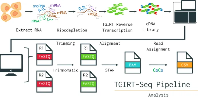
High-throughput RNA-Sequencing stands as a modern tool of choice to tackle many pressing biological inquiries. Its usefulness in assessing transcript abundance and splicing events has prompted researchers to continuously adapt the technique for various purposes, leaving us with a still growing number of different protocols suited to a plethora of analyses. Due to the sheer amount of data the technique generates, it also has crucial bioinformatic analytical steps with an equally if not more diverse landscape of methodologies. Despite all these variations, until recently, none of them allowed for the accurate detection of all snoRNAs (Boivin et al., 2018; Deschamps-Francoeur et al.,2019) (Figure 3). Thus, new protocols and tools suited for representative whole-transcriptome sequencing experiments have been elaborated to resolve unaddressed biases within RNA-Seq.
Figure 3: TGIRT-Seq + CoCo Pipeline for Holistic RNA Sequencing
Overview of the library preparation and bioinformatics analysis steps of an RNA-Seq experiment using TGIRT and CoCo.
RNA-Seq Part 1: Library Preparation and TGIRT
Sequencing experiments first begin with the isolation of total RNA from a population of cells. This RNA extract can then be treated in various ways to deplete rRNA, as their great abundance overshadows that of other RNA types. Alternatively, one may choose to ‘’pull’’ on the polyA tails, that feature mostly on mRNA as a sequence of adenine repeats at their 3’ end. This method does however create a detection bias in favor of those 3’ ends, which also makes it unsuitable for the detection of degraded or fragmented transcripts, and make it favor the detection of protein-coding RNA and polyA tail-bearing lncRNAs (Zhao et al., 2018).
Once the RNA extract has been suitably prepared, it can optionally be fragmented, using alkaline solutions or enzymes, to a specific length that fits the restriction imposed by some sequencing machines. The next step is often the reverse transcription of the RNA sample into a complementary DNA library (cDNA) (Hrdlickova et al., 2017).
TGIRT-Seq: Holistic RNA-Seq Addressing Structural Bias
RNA-Sequencing is often in fact DNA sequencing since the technology already exists, and because certain enzymes can affordably reverse transcribe RNA into DNA for sequencing to take place without new machinery being required (Wang et al., 2009). This reverse transcription step has traditionally always been carried out using retroviral reverse transcriptase (RT). This RT works well at room temperature but lacks the ability to run through highly structured RNA strands, such as long paired double stems-loops seen in H/ACA snoRNA (Figure 2) (Nottingham et al., 2016). Conducting this step at a higher temperature would denature and linearize highly structured RNAs that could then more easily be slid across by the enzyme. However, the retroviral RT (RRT) would also be denatured under these conditions rendering it unusable (Mohr et al., 2013). The laboratory of Professor Lambowitz from the University of Texas was the first to put forward an alternative enzyme to address this problem, known as TGIRT for Thermostable Group 2 Intron Reverse Transcriptase. As its name indicates, this bacterial enzyme is thermostable, a property that allows it to function at maximum efficiency at temperatures as high as 70 degrees Celsius. Unlike RRT, TGIRT also benefits from high processivity, that is the ability to polymerize long nucleic acid chains without releasing its substrate, and fidelity, which is the amount of mutations generated (Mohr et al., 2013; Nottingham et al., 2016). In a recent study, TGIRT-Seq was validated as detecting proportional abundances of highly structured RNAs, such as tRNAs and snoRNAs, without compromising the detection of other RNA types, such as mRNA or other ncRNAs, which often act as snoRNA host genes (Boivin et al., 2018).
After reverse transcription of RNA into cDNA, adaptors and primers are ligated to the ends of the cDNA fragments to differentiate their strand of origin (forward or reverse) and allow for their amplification via PCR. Typically, the amplification of cDNA is tracked by a computer in a process called base-calling, which tracks fluorescent signals, generated by the binding of labeled-nucleotides to the cDNA (Ledergerber and Dessimoz, 2011). As the amplification of PCR-generated cDNA-copies are performed in parallel, mixed signals are used to generate statistical quality scores for each base called. A popular algorithm for this is the Phred score (Ewing and
Green, 1998). The generated signals, and their quality scores, are translated to computerized data files which can then be analyzed using bioinformatic tools.
RNA-Seq Part 2: Computational Analysis & CoCo
The first processing step in the bioinformatic analysis of sequencing data is the evaluation of sequencing quality for each base, and the “trimming” of bad quality reads. This step also serves to remove the adaptor sequences used for amplification. There are several tools available for this task, one of them being Trimmomatic (Bolger et al., 2014). Typically, these tools remove reads with a Phred score below a threshold of 20, which equates to their being a 1 in 100 chance that the base was called incorrectly (Mbandi et al., 2014). Once trimming is complete, sequencing reads are often aligned to a reference genome annotation to identify which regions of the genome they correspond to. However, the presence of multiple references that don’t always agree with each other is problematic. De novo transcriptome assembly, which doesn`t use an annotation, is possible though it constitutes a more daunting task without guaranteeing better results (Hölzer and Marz, 2019; Salzberg, 2019; Ungaro et al., 2017). Once again, many tools are available for alignment as well with different properties, with a popular aligner being the “ultrafast” STAR (Dobin et al., 2013). Beyond its alignment speed, STAR is also noteworthy for being one of the first competent splice-aware aligner, a property that enables it to align reads belonging to known alternative transcripts based on a database of splice-junctions (Gatto et al., 2014; Williams et al., 2014). Splice-awareness also enables mapping to be done to the genome, which contains intronic sequences that aligners such as STAR can account for, instead of aligning to a more biased transcript-based annotation (Liu et al., 2018). Regardless of the method used, once alignment is complete, the reads can finally be quantified.
CoCo: Count Corrector for Nested & Multimapped Genes
No matter which RNA-Seq protocol is used, our ability to accurately quantify RNA transcripts relies on the proper assignment of sequencing reads. This step often relies on the genomic annotation used for alignment which has the start and end positions of genes mapped to specific nucleotide coordinates. Mapping sequenced nucleotides to those found in the annotation allows
us to calculate how many times a region or gene of the genome was transcribed. However, the use of annotations presents us with certain biases, notably when trying to quantify small non-coding RNA (<200 nucleotides) like most snoRNAs. These small non-non-coding RNAs (sncRNA) are often found within the introns of other genes, called host genes, and/or found in many copies across the genome (Dupuis-Sandoval et al., 2015). This leads to most tools assigning sncRNA reads to their host and/or assigning reads to only one of the multiple mapped regions respectively. In both cases, sncRNA reads are being mishandled, leading to under-detection of sncRNA. A recently published tool called CoCo, for Count Corrector, addresses this bias and correctly reassigns up to 15% of sequencing reads, allowing for a more accurate quantification of RNA transcripts (Deschamps-Francoeur et al., 2019).
CoCo, coupled with TGIRT-Seq, currently stands at the cutting edge for those interested in whole-transcriptome RNA-Sequencing, including highly structured, embedded and/or multimapped RNAs. One such RNA type that often fits all three criteria is small nucleolar RNAs. Although proportional quantification of snoRNAs, along with other RNA types, yields incredibly useful information, it is by no means the only type of data that can be leveraged from high-throughput sequencing experiments. Adaptations of the methodology beyond straight RNA quantification have been elaborated, as seen with high-throughput RNA-RNA interaction experiments.
High-throughput RNA-RNA Interaction Studies
Having covered the various canonical and non-canonical interactions involving snoRNAs, we turn now to a methodology with the potential to exponentially expand said interactome. Large scale RNA-RNA interaction protocols sequence a range of RNA interactions present in a total RNA sample. These experiments incorporate crosslinking, which covalently bonds RNA-interacting strands in the sample with a loop, followed by a degradation of non-RNA-interacting single strands. Finally, a linearization and sequencing of the RNA duplexes is performed, after the ligation of primers for amplification as in standard RNA-Seq. The interacting RNAs are hence contiguous in a single strand, and using bioinformatics pipelines, their identity, interaction and abundance is recorded (Lu et al., 2018; Sharma et al., 2016). A database regrouping results from
multiple RNA-RNA interaction studies with various protocols was published in 2017 under the name RISE: RNA Interactome from Sequencing Experiments (Gong et al., 2018). RISE houses 1671 snoRNA interactions. snoRNA interactors range from the classic rRNAs and snRNAs to an abundance of mRNAs, snoRNAs and more. Despite the high potential for discovery of these techniques, they can only provide a snapshot of interactions in the cells, which no doubt explains the heterogeneous results between different studies. As more of these experiments are conducted perhaps with further improvements, an even wider array of potential interactions waiting to be confirmed will be available for research.
So, as we can see, the current snoRNA interactome is much more extensive and diverse than what was once believed, and this is just the beginning. Modern snoRNA studies are still often focusing only on canonical C/D box snoRNA functions, such as the latest paper yielding a snoRNA database, which sought to discover new snoRNAs and ascribe them methylation targets in rRNA (Jorjani et al., 2016). Turning to PubMed for statistics points to a similar bias (C/D snoRNA yields 92 PubMed articles in the last 5 years vs 42 for H/ACA snoRNA. Adding methylation and pseudouridylation to those searches brings the numbers down to 46 and 14 respectively). As more and more research orients itself towards non-canonical snoRNA functions, more of their so far unknown yet critical roles will likely emerge and further our understanding of RNA biology and health. To help pave the way towards such a future, an updated and holistic snoRNA database regrouping the wealth of available information is needed.
Databases
Databases are powerful tools for storing, organizing and searching through large quantities of data. We are often most familiar with online visual representations of databases. However, these often give limited searching powers compared to the actual database engines used in the back-end of the website. That is because many of these databases use a relational database management system (RDBMS) with SQL (Structured Query Language) to manage some or all of their data.