Vibronic valence and Rydberg transitions in geminal chloro-fluoro-ethene
(1,1-C2H2FCl): a spectroscopic and quantum chemical investigation
R. Lochta, D. Deharengb and B. Leyha
a
Department of Chemistry, Molecular Dynamics Laboratory, University of Liège, Liège, Belgium; b
Department of Live Sciences, Centre for Protein Engineering, University of Liège, Liège, Belgium
Abstract
The vacuum ultraviolet photoabsorption spectrum of 1,1-C2H2FCl has been examined in detail between 5 and 15 eV photon energy by using synchrotron radiation dispersed by three different monochromators. Quantum chemical calculations are performed to help in the analysis of the valence/Rydberg transition region centered at 7.05 eV including the 3a"(π)→π* and the 3a"(π)→3s Rydberg transitions. Interactions between states involving transitions to the 3s, 4d and σ* orbitals are identified. A vibrational analysis is proposed for the structures belonging to these transitions. For the π(3a")→π* transition, one vibrational progression is observed with ω3 =
1410 ± 50 cm-1 and its lowest excitation energy is determined at about 6.398 ± 0.003 eV The π(3a")→3s Rydberg transition is characterized by a single progression with ω3 = 1410 ± 80 cm-1 likely starting at about 6.45 eV. These vibrations are ascribed to the C=C stretching motion. The abundant structure observed in the spectrum between 7.8 and 10.5 eV has been analyzed in terms of vibronic transitions to ns (δ = 0.97), np (δ = 0.63 and 0.40) and nd (δ = 0.13 and -0.11) Rydberg states, which belong to series converging to the
ionic ground state. The analysis of the vibrational structure of the individual Rydberg states has been attempted leading to average values of the wave numbers ω3 = 1420 ± 20 cm
-1
, ω7 = 720 ± 50 cm-1 and ω9 = 390 ± 50 cm-1. Between 10.5 and 12.5 eV, nine other Rydberg states converging to the first excited state were analyzed by the same way. The vibrational structure of these Rydberg states results from the excitation of one vibrational normal mode v7 with an average value of ω7 = 520 ± 20 cm-1, which is assigned to the C-Cl stretching vibration as inferred from quantum chemical calculations.
Keywords: vacuum UV photoabsorption; synchrotron radiation; quantum chemical calculations; valence and
Rydberg transitions; 1,1-C2H2FCl
1. Introduction
The influence of the position and nature of the substituent(s) on the dynamics of the molecular ions has been investigated on ethylene and a few of its halogenated derivatives, e.g. on C2H3F [1], C2H3Cl [2] and C2H3Br [3], but also on C2H2FCl (1,1- and 1,2-isomers) [4], and more recently on 1,1-C2H2F2 [5] using vacuum ultraviolet (VUV) photoabsorption and photoelectron spectroscopies. The ionization and dissociation dynamics were investigated by photoionization mass spectrometry and photoelectron-photoion coincidence spectroscopy.
Ab initio quantum chemical calculations were applied to support the different aspects of the interpretation of the
numerous experimental results.
To our knowledge the earliest VUV photoabsorption work on the 1,1-C2H2FCl molecule has been reported by Scott and Russell [6] together with two other fluorochloro-substituted ethylenes. This study was restricted to the 1200-2200 (5.635-10.332 eV) spectral range. A classification of the Rydberg transitions and their vibrational analysis has been proposed.
Tornow et al. [4] have investigated the three isomers of C2H2FCl between 6.0 and 13.0 eV by low-resolution VUV photoabsorption spectroscopy using synchrotron radiation as a light source. The observed transitions have been assigned to valence-valence and valence-Rydberg transitions. For a few Rydberg states, the vibrational structure could be observed and has been analyzed. In addition, the HeI (21.22 eV) and NeI (16.67-16.85 eV) photoelectron spectra of these compounds have been measured. The successive ionization energies have been interpreted with the help of MNDO calculations. Assignments of the observed vibrational structure have been reported.
The VUV photoabsorption spectroscopic data reported in the literature on 1,1-C2H2FCl remain very scarce. The present paper reports on the VUV photoabsorption spectrum (PAS) of the 1,1-C2H2FCl molecular system obtained at higher resolution in the 5.0-15.0 eV spectral region. Interpretation and assignments of several additional spectral features are supported by ab initio quantum chemical calculations.
2. Experimental
2.1. Experimental set-up
The experimental set-ups used in this work have already been described in detail previously [1,3]. Only the most salient features will be reported here. Furthermore, three monochromators were available in the laboratory and at the BESSY synchrotron radiation facilities.
Synchrotron radiation available from the BESSY I facility (Berlin, Germany) was dispersed with a modified VUV normal incidence 225 McPherson monochromator with a focal length of 1.5 m, instead of 1 m in the commercial version (1m-NIM-2 beam line). A laminar Zeiss grating is used for the efficient reduction of the second spectral order. It is gold coated with 1200 lines mm-1 and its transmission breaks down above 26 eV (210,000 cm-1 or 48 nm). The width of the entrance and exit slits of 100 µm ensures a 0.1-nm wavelength resolution corresponding to a resolving power of about 1200 at 10 eV (124 nm). This monochromator has been used for recording the low-resolution absolute photoabsorption spectra in the 5-13 eV photon energy range [4].
In the laboratory, a commercial version of the 1m-NIM 225 McPherson monochromator is equipped with an Al grating of 1200 lines mm-1. The light source is a commercial Ophthos Kr-microwave discharge lamp allowing us to scan between 125 nm (9.92 eV) and 165 nm (7.51 eV). The slit widths were adjusted at 25-50 µm providing a 0.02-nm wavelength resolution corresponding to a resolving power of about 5000 at 10 eV.
The 3m-NIM monochromator at the 3m-NIM2 beam line at BESSY II (Berlin, Germany) is positioned at a bending magnet front end. It is equipped with two spherical gratings, i.e. an Al/MgF2 grating of 600 lines mm-1 and a Pt grating of 2400 lines mm-1. The entrance and exit slits were adjusted between 10 and 40 µm leading to a resolving power of about 25,000-13,000 at 10 eV photon energy. This monochromator was used for recording high-resolution spectra. Most of the spectra discussed in the present work were measured with 40-µm entrance and 10-µm exit slits, using the 600-lines mm-1 Al/MgF2 grating.
In all the set-ups described above, the light has to travel through a 1-mm thick stainless steel microchannel plate located at the exit slit of the monochromator, in order to maintain a differential pressure of 1:1000, before entering a 30-cm long stainless steel absorption cell. The vapor pressure in the cell is measured by a Balzers capacitor manometer. The light is detected by a sodium salicylate sensitized photomultiplier located at the end of the absorption cell and in front of the absorption cell entrance slit. Output pulses are recorded by a 100-MHz counter. The recording of an absorption spectrum requires one scan with gas in the absorption cell and one with the evacuated cell. The stability of the synchrotron radiation and of the pressure in the cell ensured reliable absorption data. If necessary, the spectra presented in the following sections were corrected for any pressure drift.
Two different samples of 1,1-C2H2FCl have been used. A first sample was prepared in the laboratory as described earlier [4] with a 99.9% gas chromatographic purity and a boiling point of -24°C. The commercially available 1,1-C2H2FCl, purchased from ABCR GmbH and of 98% purity, was used without further purification. 2.2. Data handling and error estimation
As will be mentioned in the next sections, weak sharp peaks and diffuse structures are often superimposed on a strong continuum. To make the characterization of these features easier, a continuum subtraction procedure has been applied. The mathematical background of this digital data processing method has been set out in detail by Carbonneau et al. [7], Marchand [8] and Marmet [9]. The parameters of this filter have been evaluated by Morawski [10]. This digital filtering by 'straightening through smoothing' [8] was first applied to a large number of atomic and molecular spectra excited by electron impact [11-13]. Later, its application has been extended to e.g. photoionization mass spectrometry [14], photoelectron [15] and photoabsorption [5] spectroscopy. Briefly, for this purpose, the experimental curve is strongly smoothed to simulate the underlying continuum which is then subtracted from the original spectrum. The smoothing procedure consists of filtering the experimental curve by fast Fourier transform (FFT). The weak features then emerge from a remaining strongly attenuated background. The resulting diagram will be called plot in the forthcoming sections. To verify that no weak structure has been removed by this operation, the same procedure is applied to the subtracted continuum. The resulting residue is a signal oscillating around zero with about two orders of magnitude lower amplitude.
The wavelength calibration of the 1.5m-NIM and 3m-NIM monochromators has been performed by using the Ar absorption spectrum between the 2P3/2 and the 2P1/2 ionic states. The accuracy of this calibration is better than 2 meV. In the measurements between 5 and 15 eV photon energy, the PAS has been recorded with energy steps of about 10 meV. The uncertainty on the energy position of a feature is estimated to be 6 meV. In the photoabsorption spectra recorded between 6 and 10 eV and between 9 and 15 eV, an energy increment of 1 meV has been adopted. The uncertainty on the energy position of a feature is estimated to be of the order of 3
meV. This evaluation is confirmed by the reproducibility of energy positions measured in different spectra recorded over several years.
3. Experimental results
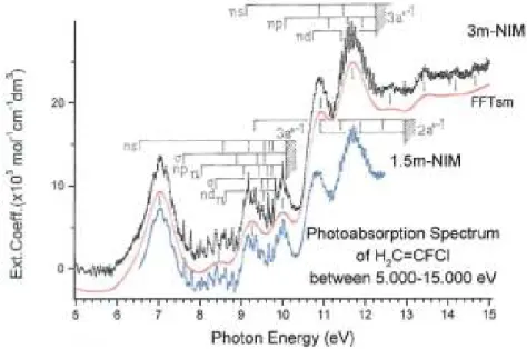
The VUV PAS of 1,1-C2H2FCl as measured between 5 and 15 eV photon energy (with 10-meV increments) with the 3m-NIM monochromator is shown in Figure 1. The good control of the experimental parameters allows us to display the spectrum in terms of the molecular extinction coefficient εhυ as a function of
the photon energy (eV). For comparison, the VUV PAS of the same compound as obtained earlier [4], but at lower resolution and slightly shifted on the εhv scale, is shown in the same figure. The gain in resolution is
obvious but the extinction coefficient is significantly larger for the high energy part of the spectrum. A continuous dotted (red) line drawn through the spectrum shows the underlying continuum obtained by FFT smoothing. Vertical lines indicate the valence (V) and Rydberg (R) transition energies. Their energy positions are listed in Table 1 and compared with previous results [4,6].
In contrast with our observations in the 1,1-C2H2F2 VUV-PAS [5], Figure 1 clearly shows a regular fine structure spread over the whole photon energy range investigated in this work.
Figure 1. VUV photoabsorption spectrum (in terms of the molecular extinction coefficient ) of 1,1-C2H2FCl between 5 and 15 eV photon energy obtained with the 3m-NIM monochromator. The same spectrum (blue line,
slightly shifted down the y-scale) measured with the 1.5m-NIM [4] is shown for comparison. Vertical bars and shaded areas locate Rydberg states and their convergence limits. The open dotted (red) curve represents the FFT
strongly smoothed spectrum. Vertical lines locate valence-valence transitions.
4. Ab initio calculations: methods and results
4.1. Computational tools
All the calculations were performed with the Gaussian 09 programme [17]. The basis set used for all the calculations is aug-cc-pVDZ [18], containing polarization as well as diffuse functions. Some calculations were also performed without diffuse functions (basic cc-pVDZ).
The geometry optimization of the neutral ground state has been performed at the CCSD(FC) [19,20], M06-2X(DFT) [21], PBEO(DFT) [22] and SAC-CI [23] levels in order to determine the vertical excitation energies for the low-lying excited states within different frameworks and cross-check them. The PBEO functional was already recognized as well suited for describing low-lying Rydberg and valence excited states [24], and the M06-2X functional is a new functional generally considered as providing very satisfactory agreement on thermodynamical properties. A geometry optimization of the first three excited states was performed at the UM06-2X and TDDFT [25] levels. The ground-state vibrational wave numbers were determined at the M06-2X level.
The geometry optimization of the cationic ground state and of the first two excited and states was performed at the M06-2X ( and states) and TDDFT ( state) level. The molecular orbital configuration of 1,1-C2H2FCl in the Cs symmetry group is described by
where the 1a' and 2a' are the first outer valence shell orbitals. The 3a" MO has a π character.
Table 1. Energy positions (eV), wave numbers (cm-1) (in brackets) and assignments proposed for valence and Rydberg transitions in the vacuum UV PAS of 1,1-C2H2FCl between 5.0 and 15 eV Comparison is made with
the results and assignments of [4, 6]. Conversion factor 1 eV = 8065.545 cm-1 [16].
[6] Valence transitions [4] This work 7.012 (56,556) 7.14 (57,588) 7.01 (56,539) 8.50 (68,557) 8.45 (68,154) 9.20 (74,203) 9.27 (74,768) 10.0 (80,655) 10.01 (80,736) 10.85 (87,511) 10.93 (88,156) 11.70 (94,367) 11.71 (94,448) - 12.65 (102,029) - 13.44 (108,401) - ~14.2 (114,531) - 14.6 (117,757) Rydberg transitions
Adiabatic convergence limit: [4] 3a"→ns 6.509 (52,500) - 6.645 (53,596) 8.406 (67,800) 8.60 (69,364) 8.535 (68,847) 9.108 (73,437) 9.21 (74,284) 9.186 (74,090) 9.420 (75,927) 9.49 (76,542) 9.482 (76,477) 9.584 (77,321) 9.65 (77,833) 9.654 (77,865) 9.688 (78,141) 9.75 (78,639) 3a"→npσ 9.758 (78,704) 7.61 (61,379) 7.608 (61,363) 8.88 (71,622) 8.839 (71,291) 9.33 (75,252) 9.316 (75,139) 9.57 (77,187) 9.551 (77,034) 9.66 (77,913) - 9.77 (78,800) 3a"→npπ - 8.02 (64,686) 8.012 (64,621) 8.92 (71,945) 9.065 (73,114) 9.37 (75,574) 9.408 (75,880) 9.57 (77,187) 9.597 (77,405) 9.70 (78,236) 9.705 (78,276) 9.77 (78,800) 9.841 (79,373) - - - 9.920 (80,010) 3a"→nd(σ) 8.38 (65,589) 8.387 (67,646) 9.15 (73,800) 9.151 (73,808) 9.44 (76,139) - 9.65 (77,832) 9.654 (77,865) 9.75 (78,639) 9.758 (78,704)
9.77 (78,800) 3a"→nd(π) - 8.601 9.211 9.501 9.654 Adiabatic convergence limit: 9a'→ns [4] 9.17 (73,916) - 10.85 (87,511) 10.838 (87,414) 11.38 (91,786) 11.448 (92,334) 11.72 (94,528) - 11.88 (95,819) - 11.97 (96,556) 9a' →np 10.052(81,075) 11.115 (89,649) 11.566 (93,286) 11.802 (95,190) 11.923 (96,165) 9a'→nd 10.626 (85,704) 11.377 (91,762) Vertical convergence limit: 13.22 eV — B2A" [4] 2a"→ns
[9.25] (74,606) [11.4] (91,950) 2a"→np
[10.84] (87,430) [11.9] (95,980) 4.2. Results of the calculations
The results of the geometry optimization are presented in Table 2, according to the numbering shown in the same table.
In the literature, the calculation level which is considered as the most accurate is CCSD(FC). However, the M06-2X is also recognized as a very good functional.
The wave numbers calculated for the vibrational normal modes represented in Figure 2 for the neutral ground state are listed in Table 3. In Table 4, the wave numbers related to the cationic ground and first two excited states are displayed. For the two excited states of the cation, a few vibrational modes are differing from the ground state of the ion. The ordering of the vibrational normal modes presented in this work follows the rules of nomenclature as proposed by Mulliken and Herzberg [26,27]. Experimental infrared [28,29] and Raman [30] spectroscopic results related to the neutral ground state are listed in Table 3 for comparison.
The vertical excitation energies to several valence states are presented in Table 5 at three calculation levels, all within the aug-cc-pVDZ basis set. For the first five excited states, all the calculation levels agree about the ordering of the states. It is therefore very likely that the three states around 7 eV are superimposed. From the two DFT calculations, the first two states are strongly mixed preventing to obtain an optimized geometry for the second excited state at the PBE0 level. Since this level provides good descriptions of low-lying Rydberg and valence states [25], these results can be considered as confirming the results of the M06-2X level [24] as well. It has to be pointed out that the excited valence σ* MO, which is antibonding on the C-Cl bond is strongly mixed with a Rydberg 4d MO. Such Rydberg-valence interactions involving σ* antibonding orbital and the associated 'rydbergisation' phenomenon have been recently reviewed [31].
To check this ordering of the excited states and the valence character of the [σ* + 4d] state, the first vertical excitation energies were also determined at the TDDFT/M06-2X level using a basis set without diffuse functions, i.e. cc-pVDZ basis set (see Table 5). These results suggest that a valence π→σ* and a Rydberg π →3s transitions should be below or close to the π→π* transition.
Table 2. Optimized geometry of the neutral and the ionic ground states and the first two cationic excited states in
the Cs symmetry group at different calculation levels. Internuclear distances in Å and angles in degrees. Neutral ground state
Level C1-C2 C2-H3 C2-H4 C1-F5 C1-C16 CCSD(FC) 1.3374 1.0917 1.0894 1.3498 1.7324 M06-2X 1.3231 1.0865 1.084 1.333 1.7251 H3-C2-C1 H4-C2-C1 F5-C1-C2 C16-C1-C2 CCSD(FC) 119.398 120.192 122.255 126.035 M06-2X 119.163 120.133 122.876 125.739
Cation ground state
Level C1-C2 C2-H3 C2-H4 C1-F5 C1-C16 CCSD(FC) 1.4214 1.0956 1.0938 1.293 1.6558 M06-2X 1.4098 1.0912 1.0893 1.2816 1.6497 H3-C2-C1 H4-C2-C1 F5-C1-C2 C16-C1-C2 CCSD(FC) 118.776 119.619 118.636 124.15 M06-2X 118.726 119.667 119.085 123.962
Cation first excited state
Level C1-C2 C2-H3 C2-H4 C1-F5 C1-C16 CCSD(FC) 1.3211 1.0965 1.0926 1.308 1.8741 M06-2X 1.3056 1.0942 1.0896 1.2891 1.879 H3-C2-C1 H4-C2-C1 F5-C1-C2 C16-C1-C2 CCSD(FC) 117.496 122.278 135.404 120.092 M06-2X 117.366 122.706 136.75 118.469
Cation second excited state
Level C1-C2 C2-H3 C2-H4 C1-F5 C1-C16
TD-DFT 1.3244 1.0959 1.0891 1.2617 2.0725
H3-C2-C1 H4-C2-C1 F5-C1-C2 C16-C1-C2
TD-DFT 116.99 121.78 135.46 120.36
The optimized geometries of the first three excited states are displayed in Table 6 together with the corresponding adiabatic excitation energies.
The optimized geometry of the first excited state is characterized by a very large C-Cl bond length, very likely related to the antibonding character of the MO populated by the excitation. The 3s-like Rydberg orbital does not show this feature, but the [σ* + 4d] MO involved in the excitation of the first two states does. The first excited state, showing a mixed character at the vertical excitation energy, is adiabatically correlated with the [σ*
+ 4d] state. This state exhibiting a dissociative character, its excitation would produce a broad continuous band.
The second state, correlating with the 3s Rydberg state, has to be observed but owing to its closeness with the π* state, the experimental determination of the adiabatic ionization energies is expected to be difficult. A schematic representation of the situation described by the present calculations is displayed in Figure 3.
5. Analysis of the experimental data and discussion.
We also measured the HeI-PES [4] and the threshold photoelectron spectrum (TPES) of 1,1-C2H2FCl, which will be reported in a forthcoming publication [32]. The first adiabatic ionization energy IEad(1,1-C2H2FCl+ 2A") is equal to 10.024 ± 0.003 eV [4] or 10.018 ± 0.007 eV [32]. The corresponding vertical value is equal to 10.201 ± 0.003 eV [4] or 10.198 ± 0.007 eV [32]. The first excited state of the cation exhibits an extended vibrational structure starting at = 12.236 ± 0.003 eV [4] or 12.238 ± 0.007 eV [32]. The corresponding vertical values are = 12.368 ± 0.003 eV [4] and 12.428 ± 0.007 eV [32]. This latter discrepancy corresponds to one vibrational
quantum.
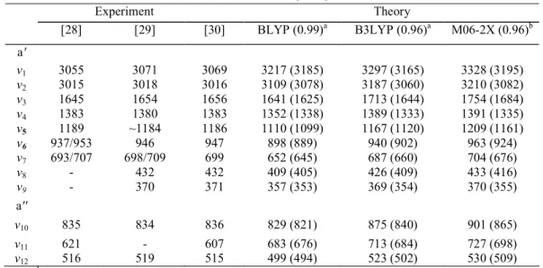
Table 3. Vibrational wave numbers of the neutral ground state as calculated at the BLYP, B3LYP and M06-2X
levels using the aug-cc-pVDZ basis set. The recommended scaling factors are given in parentheses. Comparison is made with experimental values obtained by IR [28, 29] and Raman [30] spectroscopy.
Wave numbers (cm-1) Experiment Theory [28] [29] [30] BLYP (0.99)a B3LYP (0.96)a M06-2X (0.96)b a' v1 3055 3071 3069 3217 (3185) 3297 (3165) 3328 (3195) v2 3015 3018 3016 3109 (3078) 3187 (3060) 3210 (3082) v3 1645 1654 1656 1641 (1625) 1713 (1644) 1754 (1684) v4 1383 1380 1383 1352 (1338) 1389 (1333) 1391 (1335) v5 1189 ~1184 1186 1110 (1099) 1167 (1120) 1209 (1161) v6 937/953 946 947 898 (889) 940 (902) 963 (924) v7 693/707 698/709 699 652 (645) 687 (660) 704 (676) v8 - 432 432 409 (405) 426 (409) 433 (416) v9 - 370 371 357 (353) 369 (354) 370 (355) a'' v10 835 834 836 829 (821) 875 (840) 901 (865) v11 621 - 607 683 (676) 713 (684) 727 (698) v12 516 519 515 499 (494) 523 (502) 530 (509) a See [35]. bSee [21].
Table 4. Wave numbers (cm-1) related to the vibrational normal modes of 1,1-C2H2FCl in its ground state and of 1,1- C2H2FCl+ in its ground and first two excited and states as calculated at the M06-2X level ( and states) and TDDFT ( state). In parentheses are given the values corrected for the chosen scale factor of
0.96 [21]. State
Vibrational normal mode a' symmetry a' symmetry v1 3328 (3195) 3326 (3193) 3253 (3123) 3310 (3178) v2 3210 (3082) 3190 (3062) 3126 (3001) 3208 (3080) v3 1754 (1684) 1541 (1479) 1840 (1766) 3082 (2959) v4 1391 (1335) 1429 (1372) 1371 (1316) 1394 (1338) v5 1209 (1161) 1352 (1298) 1105 (1061) 1306 (1254) v6 963 (924) 993 (953) 918 (881) 991 (951) v7 704 (676) 777 (746) 492 (472) 542 (520) v8 433 (416) 461 (443) 349 (335) 283 (272) v9 370 (355) 365 (350) 229 (220) 185 (178) a" symmetry v10 901 (865) 958 (920) 913 (876) 929 (892) v11 727 (698) 566 (543) 688 (660) 426 (409) v12 530 (509) 366 (351) 456 (438) 419 (402)
At higher energies, five bands are observed by HeI-PES [4] and are characterized by their vertical ionization energies at 13.22, 14.25, 14.93, 17.17 and 17.69 eV In the TPES measured between 9.9 and 25 eV, one additional maximum is observed at 20.208 eV [32].
In the absence of any prior information on the extent of possible Rydberg-Rydberg interactions, a first zero-order assignment of the spectral lines will be attempted fitting the simple Rydberg formula (1) for the positions in energy ERyd of the successive features:
Figure 2. Graphical representation of the 12 vibrational normal modes of 1,1-C2H2FCl in the Cs symmetry point group for which the associated wave numbers have been calculated.
where R is the Rydberg constant (R = 13.6057 eV) [16], δ is the quantum defect, n* is the effective quantum number and IE is the convergence limit of the considered Rydberg series. The successive ionization energies IE to be used in this work have been defined earlier in this section and are inserted in Figure 1. The fine structure observed in the spectrum will be assigned mainly to vibrational excitation associated with the successive Rydberg series rather than to Rydberg states with high principal quantum number n. In the former case, the intensity distribution follows the Franck-Condon distribution whereas in the latter the intensity follows the n-3 law. Robin [34] made an extensive and critical review of the analyses of Rydberg transitions and proposed rules and guidelines for assignments.
Figure 3. Schematic representation of the calculation results related to the first three excited states of
1,1-C2H2FCl.
5.1. The transitions in the 6.0-8.0 spectral region (see Figures 4 and 5)
The typical broadband of the ethylene derivatives has its maximum at 7.01 eV (56,540 cm-1) in 1,1-C2H2FCl. Contrarily to the observations made in the PAS of C2H4 [1] and 1,1-C2H2F2 [5], the vibrational structure essentially consists of broad and diffuse peaks. On the high energy side of the major band several weak but sharp structures appear. The comparison of the same band in three different related compounds, i.e. C2H4 [1], 1,1-C2H2F2 [5] and 1,1-C2H2FCl, as investigated in the present work, is very informative as shown in Figure 4, where the different contributions of valence and Rydberg transitions have been located.
Figure 4. VUV photoabsorption spectra of C2H4 (black), 1,1-C2H2F2 (red) and 1,1-C2H2FCl (green) on an expanded photon energy scale between 6.0 and 8.0 eV. The calculated vertical excitation energies to the valence
(σ*υ, σ
*
υ + 4d and π
*
υ) and the adiabatic excitation energy of the Rydberg (3s) orbitals are indicated for each
molecule.
.
shows a significant red shift of about 0.5 eV On the other hand the position of this maximum does not change significantly when the H atoms are substituted by one [1] or two F atoms [5]. However, the substitution of an F atom by a Cl atom seems to be crucial. This trend is confirmed by comparing the same spectral region in C2H3Cl [2] and C2H3Br [3], as performed in [5].
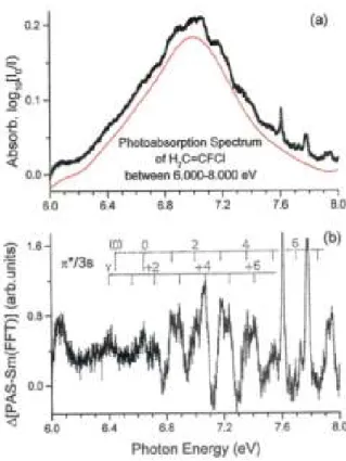
Figure 5. (a) VUV photoabsorption spectrum of 1,1-C2H2FCl on an expanded photon energy scale between 6.0 and 8.0 eV. The continuous (red) line is the FFT strongly smoothed spectrum, (b) ∆ plot resulting from the subtraction of this continuum. Long vertical bars indicate the vibrationless (0,0) transitions of the valence and of the Rydberg transitions. For each transition, the progression is identified by short vertical bars and only the even
quanta v are numbered.
Owing to the weakness and the diffuseness of the structures in the 7.01-eV band displayed in Figure 5(a), the subtraction method described in Section 2.2 has been applied to disentangle its vibrational structure. The subtracted continuum (dotted (red) curve in Figure 5(a)) is obtained by FFT smoothing of the band. The resulting plot is shown in Figure 5(b). It obviously shows a structure starting at about 6.4 eV and most of the features clearly exhibit a doublet structure. The proposed assignment to vibrational progressions is displayed in Figure 5(b). The intensity distribution of both progressions is bell shaped and is therefore related to the associated Franck-Condon factors, giving confidence in the proposed assignment.
As shown in Table 7, a first vibrational progression starts at 6.398 eV and extends at least up to υ + 6 with an average hcω = 175 ± 6 meV (1410 ± 50 cm-1). A second vibrational progression starts at 6.645 eV with at least seven vibrational levels with an average wave number ω = 1410 ± 80 cm-1 (175 ± 10 meV), too. The assignment of these transitions to the π(3a")→3s(R)(1A") and the π(3a")→π*(4a")(1A') next electronic states is
discussed below. For both states we assign the wave number of 1410 cm-1 to the vibration (see Figure 2 and Table 4). This is consistent with the excitation of one electron from the bonding π(3a") orbital. The comparison of Table 2 and 6 shows that the π→3s and the π→π* states are both characterized by a large increase of the bond length.
The calculated adiabatic excitation energies (see Table 6) also show that the two states are expected to be very close in energy, in qualitative agreement with the experiment. The π(3a")→π*(4a") state is slightly higher in energy than the π(3a")→3s(R) state. This ordering is identical for the calculated vertical excitation energies at the SAC-CI and TDDFT levels (see Table 5). We therefore assign the transitions with the
experimental (adiabatic) excitation energy at 6.398 eV to π(3a")→3s(R) (which is perturbed by its interaction with π(3a")→ [σ* + 4d]). The calculated adiabatic energy is indeed very close, i.e. 6.37 eV (see Table 6). The
transitions starting at an experimental adiabatic energy of 6.645 eV are then assigned to π(3a")→π*(4a"), for which the calculated adiabatic energy is in the 6.42-6.47 eV range.
Table 5. Vertical excitation energies (eV) of neutral states of 1,1-C2H2FCl obtained at three computation levels. All the results were obtained with the aug-cc-pVDZ basis sets. In parentheses are shown the results obtained
with the cc-pVDZ basis set. Descriptiona SAC-CI 6.34 π→3s 6.53 π→[σ* + 4d] 6.7 π→π* 7.38 π→3p 7.59 π→3p 8.18 σ→3pσ + [σ* + (Cl)] TDDFT M06-2X(M06-2X/cc-pVDZ) PBE0 Description Description 6.58 π→[σ* + 4d], π→3s 6.52 π→[σ* + 4d], π→3s 6.71 (6.96) π→3s, π→[σ* + 4d] 6.70 π→3s, π→[σ* + 4d] 6.96 (7.61) π→π* 6.87 π→π* 7.5 π→3p 7.46 π→3p 7.66 π→3p,π→[σ* + 4d] 7.68 π→3p 8.14 (8.43) σ→π* 7.77 σ→π* 8.21 σ→[σ* + 4d], σ→3p, σ→4d 8.35 π→4d 8.42 π→4d 8.37 σ→3s, σ→[σ* + 4d] 8.56 π→3pπ 8.63 π→3pπ 8.83 σ→3s, σ→[σ* + 4d] 8.74 σ→3s, σ→[σ* + 4d] 8.87 π→4d 8.94 9.15 πi→[σ* + 4d], πi→4d, 8.97 πi→π* πi→3p, πi→3s 9.29 πi→π* 9.14 πi→[σ* + 4d], πi→3s 9.31 π→4s 9.30 π→4s a
The ground state electronic configuration is represented as staying for The σ MO has a pronounced n(Cl) character. When two configurations are mentioned, the dominant one is reported first and the
πi,notation designates the internal π orbital.
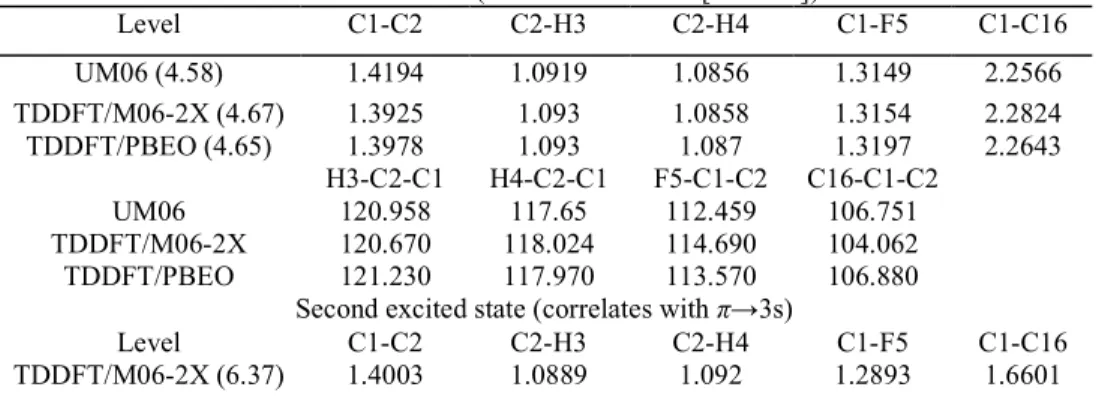
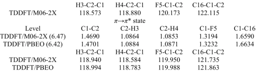
Table 6. Optimised geometries of the first two excited states at several calculation levels. The atomic numbering
refers to the figure in Table 2. Internuclear distances are given in and angles in degrees. The adiabatic excitation energies (eV) are listed in parentheses.
First excited state (correlates with π→[σ* + 4d])
Level C1-C2 C2-H3 C2-H4 C1-F5 C1-C16 UM06 (4.58) 1.4194 1.0919 1.0856 1.3149 2.2566 TDDFT/M06-2X (4.67) 1.3925 1.093 1.0858 1.3154 2.2824 TDDFT/PBEO (4.65) 1.3978 1.093 1.087 1.3197 2.2643 H3-C2-C1 H4-C2-C1 F5-C1-C2 C16-C1-C2 UM06 120.958 117.65 112.459 106.751 TDDFT/M06-2X 120.670 118.024 114.690 104.062 TDDFT/PBEO 121.230 117.970 113.570 106.880
Second excited state (correlates with π→3s)
Level C1-C2 C2-H3 C2-H4 C1-F5 C1-C16
H3-C2-C1 H4-C2-C1 F5-C1-C2 C16-C1-C2 TDDFT/M06-2X 118.573 118.880 π→π* state 120.173 122.115 Level C1-C2 C2-H3 C2-H4 C1-F5 C1-C16 TDDFT/M06-2X (6.47) 1.4690 1.0864 1.0853 1.3194 1.6590 TDDFT/PBEO (6.42) 1.4701 1.0884 1.0871 1.3232 1.6634 H3-C2-C1 H4-C2-C1 F5-C1-C2 C16-C1-C2 TDDFT/M06-2X 118.940 118.584 119.950 121.735 TDDFT/PBEO 118.994 118.783 119.988 121.863
The π(3a")→3s(R) electronic transition would then correspond to an effective quantum number n* =
1.95 (quantum defect δ = 1.05). This fairly low value of n* results most probably from the large coupling between the π(3a")→3s(R) and the π(3a")→[σ* +4d] configurations. Such Rydberg-valence interactions between 3s orbitals and antibonding σ* valence orbitals lead frequently to a so-called rydbergisation along given dissociation pathways. This phenomenon has been observed in many different excited molecules with important consequences for their photochemical behaviour [31].
Table 7. Positions in energy (eV), wave numbers (cm-1) and assignments proposed for the features observed in the 7.01-eV band in the vacuum UV PAS of 1,1-C2H2FCl. Comparison is made with the observation and
assignments of [6]. Conversion factor: 1 eV = 8065.545 cm-1 [16].
This work [6] Assignments
(eV) (cm-1) (cm-1) This work [6]
6.398 51,603 3sR, υ = 0 (6.447) 51,999 (1A", υ = 0)a (6.592) 53,168 (3sR, υ = 1) 6.645 53,596 (1A", υ = 0)a 6.719 54,192 3sR, υ = 2 6.839 55,160 1A", υ + 1 6.895 55,612 3sR, υ = 3 7.018 56,604 56,561 1A", υ + 2 C-Cl stretch 7.073 57,048 57,100 3sR, υ = 4 stretch 7.192 58,007 57,887 1A", υ + 3 stretch 7.240 58,395 58,411 3sR, υ = 5 stretch 7.360 59,362 59,249 1A", υ + 4 stretch 7.416 59,814 59,773 3sR, υ = 6 stretch 7.530 60,733 60,606 1A", υ + 5 7.694 62,056 1A", υ + 6 7.869 63,468 1A", υ + 7 a
For explanation and discussion, see text (Section 5.1).
The other state resulting from this configuration interaction, which is denoted as 'π→[σ* + 4d], π→3s' in Table 5 is calculated at lower energy (see Table 6 and Figures 3 and 4) and is most probably repulsive. It is therefore not surprising that no vibrational progression could be observed in the corresponding energy range. At least a part of the subtracted continuum could be assigned to this state.
From this analysis based on the ab initio calculations, the π(3a")→3s Rydberg vertical transition energy is red shifted by about 0.4 eV with respect to C2H4 by halogen substitution (in C2H2F2 and C2H2FCl) (see Figure 4). As shown in the figure, the π(3a")→π*(4a")(1A') vertical transition exhibits a small blue shift of about 0.1 eV
in C2H2F2 and a large red shift in C2H2FCl, i.e. of about 0.6 eV.
The wave number of 1410 cm-1 for ν3 (see Figure 2) as determined in the present work can readily be compared with the value of 1350 cm-1 measured by Scott and Russell [6]. The major difference in the interpretation of the band at 7.01 eV between Scott and Russell [6] and the present work is that in the former the entire band is assigned to the π →π* transition only. The vibrational structure is therefore accounted for by two vibrational wave numbers of about 1350 and 530 cm-1 assigned to and C-Cl stretching vibrations, respectively [6]. However, considering the intensity distribution pattern of the vibrational structures as observed in Figure 5(b), progressions of two different electronic states are more probable.
5.2. The transitions in the 7.8-10.4 eV spectral region (Figures 6 and 7)
Part of this spectral region is shown in Figure 6 and exhibits an abundant structure superimposed on a weak continuum with a maximum measured at about 8.45 eV (68,154 cm-1) in the present work (see Figure 1 and Table 1). This latter feature was assigned earlier to the n(Cl)→σ*(C-Cl) valence transition [4]. Furthermore, a n(Cl)→π* was expected at 9.27 eV [4]. The present calculations predict a quite more complex situation as it is shown in Table 5.
Figure 6. High-resolution VUV photoabsorption spectrum of 1,1-C2H2FCl between 7.5 and 10 eV photon energy obtained in the laboratory using the 1m-NIM monochromator and a discharge lamp (red line) and at BESSY using a 3m-NIM monochromator dispersing synchrotron radiation. The two spectra are slightly shifted
on the y-scale for clarity.
At the TDDFT level, a σ→π* transition (the σ MO has a n(Cl) character) is predicted around 8 eV,
slightly below the σ→[σ* + 4d] transition calculated at ~8.30 eV According to the same calculations, several valence excited and Rydberg states lay close together and will very likely overlap in this region (see Table 5).
Figure 6 also shows the comparison between the PAS measured using the lm-NIM at medium resolution and the 3m-NIM at higher resolution. This latter spectrum has been handled by the subtraction procedure (see Section 2.2) and the result is shown in Figure 7(a)-(c).
All narrow and sharp features observed in this spectral region are assigned to transitions to Rydberg states which belong to series converging to ionization energy limits associated with the different vibrational levels of a common electronic state, i.e. the ionic ground state. Figure1 and Table 1 show the energy positions of the observed vibrationless Rydberg transitions.
To assign the observed spectral features, we assumed as already mentioned that the Rydberg formula is valid. Any significant perturbation due to Rydberg-Rydberg couplings is expected to induce repulsion of the zero-order energies and should be detected as poor quality fits to the Rydberg formula. In addition, in the absence of coupling between Rydberg series, the quantum defect has typical values which are characteristic of the angular momentum of the Rydberg orbital. When couplings are turned on, these values are perturbed. Such a situation already appeared in some of our previous investigations, where we could then highlight and quantitatively analyze transition between Hund's coupling cases [33]. The quantum defects are therefore important information. Consistent with our first zero-order hypothesis, we assume also that the vibrational structure within a transition to a given Rydberg state should be close to that of the cationic state to which the Rydberg series converges [34]. This argument involves constraints on both the energy and the intensity distribution. Here again, any discrepancy in the fits should be identified as a possible vibronic interaction. We emphasize that this procedure has been used successfully for the analysis of the VUV spectra of C2H3F [1] and 1,1-C2H2F2[5].
The 3a"→ns (n > 4) Rydberg transitions are observed up to n = 8 with an average quantum defect δ = 0.97 ± 0.02 compared to 0.90 observed previously [4]. This is a typical value for an ns series. In the present photon energy range, at least the transitions corresponding to n = 4 and n = 5 are characterized by vibrational progressions. The energy values listed in Table 1 correspond to adiabatic excitation energies.
The vertical bars in Figure 7 and the data listed in Table 8 show our assignments of the structures to vibrational progressions as obtained by the comparison of the HeI-PES to the plot of the PAS. By this procedure, three vibrational normal modes are identified for the 3a"→4s transition and characterized by the wave numbers ωA = 1420 ± 50 cm -1 (176 ± 6 meV), ωB = 740 ± 20 cm -1 (92 ± 2 meV) and ωc = 395 ± 40 cm -1 (49 ± 5
meV). In the HeI-PES of the ionic state [4], the wave numbers of 1400 ± 40 cm-1, 720 ± 10 cm-1 and 380 ± 20 cm-1 were determined. By quantum chemical calculations (see Table 4), a' vibrational normal modes (see Figure 2) are predicted for ν3 at 1479 cm-1 ( stretching), ν7 at 746 cm
-1
(C-Cl stretching and H rocking) and
ν9 at 350 cm-1 (H rocking and C-F stretching). The good agreement between observed and predicted values gives us confidence in an assignment of the observed wave numbers ωA to ν3 ωB to ν7 and ωC to ν9 vibrational normal modes. In addition, the intensity ratios observed are similar to those of the photoelectron band.
Figure 7. plot of the VUV photoabsorption spectrum of 1,1-C2H2FCl on an expanded photon energy scale between 7.5 and 10.5 eV showing the details of the vibrational structure. The ν9 vibrational component is not
represented to avoid overcrowding.
The adiabatic excitation energy of the 3a"→5s Rydberg transition is observed at 9.186 eV. In this region, the spectrum becomes fairly too crowded. The short progression observed has been tentatively interpreted assuming the excitation of the same modes as for the 3a"→4s transition. Two wave numbers are identified at 1440 ± 20 cm-1 (179 ± 3 meV) and 660 ± 60 cm-1 (82 ± 8 meV). The first one corresponds very well to ω3 whereas the second one is not incompatible with ω7.
For the 3a"→np Rydberg (n = 3-6 or 8) transitions, two different quantum defects are obtained and their average values are δ(npσ) = 0.63 ± 0.04 and δ(npπ) = 0.40 ± 0.04. The dispersion of the quantum defect along the experimentally observable members of the series is fairly narrow. This should point towards the validity of the Rydberg formula and thus towards limited interstate couplings. To be rigorous on a symmetry basis (Cs point group), the npσ and npπ orbitals should be denoted as npa' and npa", respectively. However, if we assume that the molecular ion field is nearly cylindrical, i.e. diatomic like, it makes sense to use the σ, π, δ nomenclature. On this basis, the observed transitions are classified as shown in Table 1. In our earlier work [4] on the same system, δ values of 0.62 and 0.40 have been obtained. Similar observations have been reported in the study of the VUV spectrum of 1,1-C2H2F2 [5]. In this latter case, the 3pσ-3pπ energy splitting is 183 meV whereas this quantity becomes 404 meV in the present molecular system. This observation is not unexpected. The 3pσ-3pπ splitting results from the non-spherical nature of the molecular field which is obviously more
severe for the C2H2FCl molecule. Clearly, the npσ transitions are observed up to n = 6, whereas the npπ transitions appear up to n = 8. As will be discussed later in this section, a transition observed at 9.920 eV could likely be assigned to the 12pπ Rydberg state and characterized by δ = 0.451.
Vibrational progressions could be identified for the 3pσ, 3pπ and 4pπ Rydberg states. These are displayed in Figure 7 and the energies are listed in Table 8. For some spectral features, no clear unique assignment could be provided. In these cases, the energy values have been put in square brackets and possible tentative assignments have been suggested but the interpretation is, in such cases, clearly much less reliable. The intensity of overlapping transitions is accounted for by considering the sum of the individual contributions. For the 3pσ and 3pπ Rydberg states, well-defined progressions are observed and three different wave numbers can be inferred i.e. ω3 = 1450 ± 20 cm -1 (180 ± 3 meV), ω7 = 650 ± 60 cm -1 (81 ± 7 meV) and ω9 = 360 ± 30 cm -1 (45 ± 4 meV) for the 3pσ Rydberg state; and ω3 = 1390 ± 30 cm-1 (172 ± 4 meV), ω7 = 670 ± 20 cm-1 (83 ± 3 meV)
and ω9 = 330 ± 120 cm
-1
(41 ± 15 meV) for the 3pπ Rydberg state. The present measurements agree, but with a higher accuracy, with the observations reported earlier [4], i.e. with a vibrational spacing ranging from 1380 to 1460 cm-1 and a low energy spacing of 320 cm-1 in the 3pσ Rydberg state. However, for ω7, the agreement with
the theoretical predictions (see Table 4) is not as satisfactory, so that this assignment can be questioned.
Table 8. Positions in energy (eV), corresponding wave numbers (cm-1) and assignments proposed in the present work for the structures in the vacuum UV photoabsorption spectrum of 1,1-C2H2FCl in the 7.8-10.4 eV spectral
region. In the fourth column, the average values of the energy and the wave numbers associated with the observed vibrational mode are indicated. Conversion factor: 1 eV = 8065.545 cm-1 [16].
Energy (eV)a Wave number (cm-1) Assignments 3a"→3pσ 7.608 61,363 0 ω3 = 180 ± 3 meV 7.652 61,717 na 1450 ± 20 cm-1 7.67 61,863 v9 ω7 = 81 ± 7 meV 7.695 62,064 v7 650 ± 60 cm-1 7.730 62,347 3v9 ω9 = 45 ± 4 meV 7.785 62,790 v3 360 ± 30 cm-1 7.816 63,040 na 7.837 63,210 v3 + v9 7.873 63,500 v3 + v7 7.888 63,621 na 7.919 63,871 v3 + 3v9 7.949 64,113 na 7.967 64,258 2v3 [8.013] 64,629 2v3 + v9 8.037 64,823 2v3 + v7 [8.064] 65,040 na 8.116 65,460 2v3 + 2v9 [8.149] 65,726 3v3 3a"→3pπ 8.013 64,629 0 ω3 = 172 ± 4 meV [8.064] 65,040 v9 1390 ± 30 cm-1 8.100 65,331 v7 ω7 = 83 ± 3 meV 8.149 65,726 3v9 670 ± 20 cm-1 8.192 66,073 v3 ω9 = 41 ± 15 meV 8.223 66,323 na 330 ± 120 cm-1 8.235 66,420 v3 + v9 8.275 66,742 v3 + v7 8.314 67,057 v3 + 3v9 8.362 67,444 2v3 8.422 67,928 2v3 + v9 [8.446] 68,122 2v3 + v7 8.475 68,355 na 8.490 68,476 na [8.515] 68,678 2v3 + 3v9
[8.533] 68,823 3v3 8.553 68,985 na [8.569] 69,114 3v3 + v9 [8.601] 69,372 3v3 + v7 8.630 69,606 3v3 + 3v9 [8.664] 69,880 3v3 + 2v7 8.704 70,202 4v3 8.737 68,985 na 3a"→3dσ 8.387 67,646 0 ω3 = 177 ± 5 meV 8.437 68,049 v9 1430 ± 40 cm-1 [8.515] 68,678 3v9 ω9 = 45 ± 3 meV 8.566 69,089 v3 360 ± 20 cm-1 [8.601] 69,372 v3 + v9 [8.663] 69,872 v3 + 2v9 [8.703] 70,202 v3 + 3v9 [8.737] 70,469 2v3 8.848 71,363 2v3 + 2v9 8.893 71,646 2v3 + 3v9 [8.918] 71,928 3v3 3a"→4sσ 8.535 68,839 0 ω3 = 176 ± 6 meV 8.630 69,606 v7 1420 ± 50 cm-1 8.676 69,977 3v9 ω7 = 92 ± 2 meV 8.712 70,267 v3 740 ± 20 cm-1 [8.764] 70,687 v3 + v9 ω9 = 49 ± 5 meV 8.803 71,001 v3 + v7 395 ± 20 cm-1 8.846 71,348 v3 + 3v9 [8.882] 71,638 2v3 8.933 72,050 2v3 + v9 8.974 72,380 2v3 + v7 9.045 72,953 2v3 + 3v9 9.064 73,106 3v3 3a"→3dπ 8.601 69,372 0 8.690 70,090 v7 ω3 = 176 ± 3 meV 8.777 70,791 v3 1420 ± 20 cm-1 8.832 71,235 v3 + v9 ω7 = 89 ± 3 meV 8.869 71,533 v3 + v7 720 ± 20 cm -1 [8.918] 71,928 v3 + 3v9 ω9 = 50 ± 8 meV 8.950 72,187 2v3 400 ± 60 cm-1 9.010 72,670 2v3 + v9 9.035 72,872 2v3 + v7 9.129 73,630 3v3 3a"→4pπ 9.065 73,114 0 ω3 = 173 ± 7 meV 9.127 73,614 v9 1395 ± 60 cm-1 9.209 76,575 3v9 ω9 = 48 ± 6 meV [9.244] 76,913 v3 390 ± 50 cm-1 9.386 77,341 v3 + 3v9 9.423 77,824 2v3 9.564 78,107 2v3 + 3v9 9.594 78,550 3v3 9.757 78,905 4v3 3a"→5sσ 9.186 74,090 0 ω3 = 179 ± 3 meV 9.282 74,864 v7 1440 ± 20 cm-1 9.369 75,586 v3 ω9 = 82 ± 8 meV 9.448 76,203 v3 + v7 660 ± 60 cm -1 9.544 76,978 2v3
9.621 77,599 2v3 + v7 9.723 78,421 3v3 9.800 79,042 4v3 3a"→4dσ (doublet) 9.151 73,808 0 ω3 = 180 ± 2 meV 9.166 73,929 0 1450 ± 20 cm-1 9.209 74,276 v9 ω7 = 93 ± 1 meV 9.222 74,380 v9 750 ± 8 cm-1 9.244 74,558 3v9 ω9 = 59 ± 8 meV 9.260 74,687 3vv9 480 ± 60 cm-1 9.329 75,243 v3 Splitting ∆ 9.347 75,389 v3 ∆ = 18 ± 2 meV 9.381 75,663 v3 + v9 145 ± 20 cm -1 9.398 75,780 v3 + v9 9.422 75,994 v3 + v7 9.510 76,703 2v3 9.527 76,840 2v3 9.579 77,260 2v3 + v7 [9.596] 77,397 2v3 + v7 9.688 78,139 3v3 9.709 78,308 3v3 3a"→7sσ/6d 9.654 77,865 0 ω3 = 173 ± 7 meV 9.708 78,300 v9 1395 ± 60 cm -1 9.747 78,615 v7 ω7 = 93 ± 10 meV 9.776 78,849 3v9 750 ± 80 cm-1 9.829 79,276 v3 ω9 = 60 ± 5 meV 9.88 79,688 v3 + v9 480 ± 40 cm -1 9.933 80,115 v3 + v7 9.961 80,341 v3 + 3v9 10.006 80,704 2v3 10.069 80,212 2v3 + v9 10.089 81,373 2v3 + v7 10.156 81,914 2v3 + 3v9 10.184 82,140 4v3 10.347 83,454 5v2 3a"→8sσ/7d 9.758 78,704 0 ω3 = 178 ± 4 meV 9.802 79,058 v9 1440 ± 30 cm -1 9.851 79,454 v7 ω7 = 91 ± 3 meV 9.893 79,792 na 730 ± 20 cm-1 9.940 80,172 v3 ω9 = 46 ± 2 meV 9.987 80,551 v9 370 ± 20 cm-1 [10.030] 80,897 v7 10.075 81,260 3v9 10.113 81,567 2v3 10.291 83,003 3v3 10.441 84,212 4v3 3a"→12pπ 9.920 80,010 0 ω3 = 175 ± 7 meV [10.030] 80,889 v9 1410 ± 60 cm-1 10.098 81,446 v3 ω7 = 91 ± 17 meV 10.278 82,898 2v3 780 ± 100 cm-1 10.373 83,583 v9 ω9 50 meV 10.425 84,083 3v7 400 cm-1 10.445 84,246 3v7 a
At the end of the energy range considered in this section, weak but fairly sharp and close features are observed up from 9.920 eV (see Figure 7(c) and Table 8). These structures could be assigned to vibrational progressions where ω3 = 1410 ± 56 cm-1 (175 ± 7 meV), ω7 = 780 ± 80 cm-1 (97 ± 10 meV) and ω9 403 cm-1 (50 meV). Assuming the adiabatic excitation to be at 9.920 eV, an effective quantum number n* = 11.549 is
obtained and the transition is therefore assigned to 3a"→ 12pπ.
Compared to our earlier investigation [4], owing to the better resolution achieved in the present work, many sharp and very narrow features (about 5 meV full width half maximum [FWHM] at 9.7 eV) can be observed. Several 3a"→nd Rydberg transitions are observed. They have been classified on the basis of their respective quantum defects. As for the np series, at least two from the three nd series are likely to be observed i.e. an ndσ series with δ = 0.13 ± 0.03 and an ndπ series with δ = -0.11 ± 0.02. Both series are observed up to n = 6 or 7. However, for the higher n values, nd and ns states are very close in energy and are expected to interact more or less strongly. In our earlier work, only one quantum defect value of 0.12 could be obtained [4]. A similar situation has been observed in the VUV PAS of 1,1-C2H2F2 [5], where the energy difference between 3dσ and 3dπ is 109 meV. This splitting becomes 390 meV in the present 1,1-C2H2FCl molecular system. The propensity of an increase of the σ-π splitting with the increase of the atomic number of the substituent was already observed for the npσ- and npπ-Rydberg series. However, simultaneously, the symmetry lowering of the molecular system resulting from the chemical substitution breaks the spherical symmetry and induces an increase of the energy spacing between states differing by the projection of their orbital angular momentum.
From 9.151 eV upwards, closely lying very narrow doublets are observed. The first doublet corresponds to 9.151 and 9.166 eV (see Table 8). Using an average adiabatic excitation energy of 9.158 eV and IEad = 10.024 eV, an effective quantum number n* = 3.963 and a quantum defect δ = 0.047 are obtained indicating that we are
likely dealing with 3a"→4d Rydberg transition.
As a further proposal, these signals are classified into two different vibrational progressions likely corresponding to two 4d-Rydberg transitions separated by a constant splitting of 145 ± 20 cm-1 (18 ± 2 meV) and characterized by quantum defects δ = 0.052 and δ = 0.018, successively. Both components of this doublet exhibit a vibrational structure very clearly consisting of the wave numbers ω3 = 1450 ± 20 cm-1 (180 ± 2 meV), ω7 = 750
± 8cm-1 (93 ± 1 meV) and ω9 = 480 ± 60 cm-1 (59 ± 8 meV). This doublet could be assigned to 4dπ/4dδ Rydberg transitions characterized by small quantum defects. The core containing less π- and δ-type orbitals than σ orbitals is expected to interact less with π and δ Rydberg orbitals than with σ ones. They should therefore be characterized by smaller quantum defects.
Table 8 and Figure 7 show the proposed vibrational analysis pertaining to 7s/6d and 8s/7d Rydberg states. For these states, the vibrational structure consists of the three following wave numbers: ω3 = 1390 ± 60 cm-1 (173 ± 7 meV) and 1440 ± 30 cm-1 (178 ± 4 meV), ω7 = 750 ± 80cm-1 (93 ± 10 meV) and 730 ± 20 cm-1 (91 ± 3 meV) and ω9 = 480 ± 40 cm-1 (60 ± 5 meV) and 370 ± 20 cm-1 (46 ± 2 meV). Once again, we find very consistently the presence of the same modes as for the other Rydberg series. However, here, the excitation of the
ω9 mode in the 7s/6d state is questionable.
5.3. The transitions in the 10.4-12.5 eV spectral region (Figure 8)
As shown in Figure 1, this part of the spectrum exhibits two quite characteristic parts: (1) a broad and strong continuum underlying quite diffuse, very weak structures and (2) a very regular series of sharp but weak structures superimposed on a strong continuum.
Concerning the continua, their maximum can be measured on the FFT smoothed spectrum shown in Figure 1, i.e. at 10.93 eV (88,160 cm-1) and at 11.70 eV (94,450 cm-1), successively. The absorption at 10.93 eV has been assigned to the σCH→π* or nF→σ
*
transitions [4]. The continuum at 11.70 eV was assigned to an nF/σCH→3p Rydberg transition as predicted at 11.8 eV [4]. The narrow features between 11.4 and 12.4 eV have
not been analyzed in our earlier work. To allow us to perform a more detailed analysis of this spectral region the subtraction method has been applied (see section 2.2). The plot provided by this procedure is shown in Figure 8(a)-(c).
The Rydberg series observed in this energy range has to converge to excited electronic states of the 1,1-C2H2FCl+ cation. The first excited state has been detected at 12.236 eV by HeI-PES [4]. This state is characterized by a long vibrational progression with a small wave number ω = 520 ± 30 cm-1. Very
few Rydberg series converging to this ionization limit are actually observed. Those observed in the present spectrum are listed in Table 1.
In our earlier work, only the 9a'→ns transitions were mentioned [4]. In the present higher resolution work, the analysis also reveals additional series which could be assigned to transitions to np and nd Rydberg states. Their vibrational analysis allowed us to observe mainly 9a'→np type Rydberg transitions.
The lowest member of the 9a'→ns Rydberg series is predicted to be close to 9.15 eV, i.e. at about the same energy as the 3a"→4dσ transition (see Section 5.2). The vibrational analysis strongly suggests the latter assignment. Very likely the 9a'→3s Rydberg transition could be hidden in the 9.27-eV continuum. The two higher members of the ns series, exhibiting a vibrational progression, could be characterized by an average quantum defect δ = 0.87 ± 0.03. This value has to be compared with δ = 0.90 as determined earlier [4].
A longer Rydberg series is observed up to n = 7 with an average quantum defect δ = 0.46 ± 0.06 which suggests a 9a'→np assignment. Two additional members of a series are observed with their vibrational structure and correspond to an average quantum number of 0.06 ± 0.04. This suggests an assignment to an nd series.
Above 10.2 eV, the extinction coefficient sharply increases and peaks at about 10.8 eV (see Figure 1). In a term scheme based on experimental data, valence-valence excitation energies were predicted at 10.5 and 10.8 eV and assigned to nF→σ
*
C-Cl and π *
transitions, respectively [4]. Superimposed on this strong feature,
several very weak structures are observed. Above 11.0 eV the structure becomes stronger. In spite of their weakness and diffuseness, an analysis and assignments have been attempted.
Figure 8. plot of the VUV photoabsorption spectrum of 1,1-C2H2FCl on an expanded photon energy scale between 10.4 and 12.5 eV showing the details of the vibrational structure.
The plot in this energy range shows fairly clearly a vibrational structure for which several peaks seem to have a doublet structure. Using the HeI-PES data related to the band [4] as a reference, the 10.5-11.1 eV region could be decomposed into three vibrational progressions. The energy positions are listed in Table 9.
Starting at 10.534 eV, a vibrational progression of a single wave number is observed. A second progression with about the same vibrational spacing starts likely at 10.626 eV Using these two excitation energies and 12.236 eV as convergence limit, effective quantum numbers n* = 2.83 and 2.91 are obtained. The
latter value being close to that expected for a 9a'→3d type Rydberg transition, the former excitation energy at 10.534 eV is then assigned to a valence transition. Based on a qualitative simulation using the PES data information, a weak 9a'→4s Rydberg transition is very likely involved at 10.838 eV Owing to the crowding of the spectrum in this range only four vibrational spacing could be detected.
As mentioned in the fourth column of Table 9, the vibrational wave numbers deduced for the valence-to-valence transition and for the 9a'→3d and 9a'→4s Rydberg transitions are surprisingly quite similar, i.e. around 500 cm-1. The wave numbers characterizing the 12 vibrational normal modes of the state of the 1,1-C2H2FCl+ have been predicted by quantum chemical calculations (see Table 4). A normal mode corresponding to the stretching vibration of C-Cl combined with the F-CC and Cl-CC bending motions (v7) has been calculated at 492 cm-1, in good agreement with the values determined for the valence-valence and valence-Rydberg transitions. Furthermore, with respect to the neutral ground state, the predicted geometry of the state (see Table 2) shows a strong decrease of the C-F and a very strong increase of the C-Cl bond length. The F-CC and Cl-CC bond angles also undergo strong modifications. These results point to the likelihood of the above assignment. Unfortunately, no such information is available for the valence σ* state.
The extensive structure observed from 11.1 to 12.4 eV has been analyzed in detail by the same procedure as described above. The HeI-PES band of the cation state has been used as a reference. The result of the analysis is shown in Figure 8(b) and 8(c) and the energy positions are listed in Table 9.
The entire spectral region could be accounted for by a series of Rydberg transitions characterized by their effective quantum number n* as listed in Table 9. The successive adiabatic excitation energies are identified
as well as the vibrational progressions, each consisting of a unique wave number which is observed in all states at an average of 515 ± 15 cm-1 (64 ± 2 meV). It is assigned to the v7 normal mode (see Table 4).
Table 9. Energy positions (eV), corresponding wave numbers (cm-1) and assignments proposed in the present work for the structures in the vacuum UV photoabsorption spectrum of 1,1-C2H2FCl in the 10.4-12.5 eV spectral
region. In the fourth column, the averaged values of the energy and the wave numbers associated with the observed vibrational modes are indicated. Conversion factor: 1 eV = 8065.545 cm-1 [16].
Energy (eV)a Wave number (cm-1) Assignments Valence-valence transition 10.534 84,962 0 ω7 = 61 ± 8 meV 10.584 85,366 v7 490 ± 60 cm-1 10.638 85,801 2v7 10.700 86,301 3v7 10.770 86,866 4v7 10.838 87,414 5v7 10.906 87,963 6v7 9a'→3p (n* = 2.495) 10.050 81,059 0 ω7 = 61 ± 8 meV [10.113] 81,567 v7 490 ± 60 cm-1 [10.184] 82,140 2v7 10.250 82,672 3v7 10.313 83,180 4v7 10.378 83,704 5v7 10.444 84,236 6v7 9a'→4s (n* = 3.119) 10.838 87,414 0 ω7 = 67 ± 5 meV [10.905] 87,955 v7 540 ± 40 cm-1 10.977 88,535 2v7 11.037 89,019 3v7 11.108 89,592 4v7 9a'→3d (n* = 2.907) 10.626 85,704 0 ω7 = 66 ± 5 meV 10.687 86,196 v7 530 ± 40 cm-1 10.757 86,761 2v7
10.818 87,253 3v7 10.883 87,777 4v7 10.948 88,302 5v7 11.022 88,898 6v7 9a'→4p (n* = 3.484) 11.115 89,649 0 ω7 = 64 ± 4 meV 11.175 90,132 v7 520 ± 30 cm-1 11.244 90,689 2v7 11.307 91,197 3v7 11.368 91,762 4v7 [11.438] 92,254 5v7 [11.500] 92,753 6v7 9a'→4d (n* = 3.980) 11.377 91,762 0 ω7 = 63 ± 2 meV [11.438] 92,254 v7 510 ± 20 cm-1 [11.500] 92,754 2v7 11.567 93,294 3v7 [11.631] 93,810 4v7 [11.692] 94,302 5v7 11.754 94,802 6v7 9a'→5p (n* = 4.506) 11.563 93,262 0 ω7 = 65 ± 4 meV [11.631] 93,810 v7 520 ± 30 cm-1 11.692] 94,302 2v7 11.755 94,810 3v7 11.827 95,391 4v7 11.889 95,891 5v7 9a'→5s (n* = 4.155) 11.448 74,090 0 ω7 = 65 ± 8 meV 11.511 74,864 v7 520 ± 60 cm-1 11.572 75,586 2v7 11.650 75,203 3v7 11.709 76,978 4v7 11.767 77,599 5v7 11.839 78,421 6v7 9a'→6p (n* = 5.599) 11.804 95,206 0 ω7 = 64 ± 6 meV 11.862 95,673 v7 520 ± 50 cm-1 11.923 96,165 2v7 11.995 96,746 3v7 12.064 97,303 4v7 12.125 97,795 5v7 9a'→7p (n* = 6.593) 11.928 96,205 0 ω7 = 63 ± 5 meV 11.988 96,690 v7 510 ± 40 cm-1 12.049 97,182 2v7 12.12 97,754 3v7 12.185 98,279 4v7 12.244 98,755 5v7 12.313 99,311 6v7 12.371 99,779 7v7 a
Energy positions assigned to two or more transitions are given in square brackets.
All our analyses have been performed using zero-order formula and based on ab initio calculations, as discussed in the first part of Section 5. We were not able to identify couplings which would result in energy perturbations exceeding the experimental uncertainties. A higher resolution is probably required for this purpose. In addition, in the present spectra, no unexplained doublet appears, so that there is no indication that spin-orbit coupling might play a role. This is not unexpected since, first of all, all detected Rydberg states
presumably are singlet states. Despite the presence of a chlorine atom, the spin-conserving rule is expected to remain a strong propensity rule in electronic transitions. Therefore, singlet-triplet transitions, if any, are expected to be very weak. Second, as the C2H2FCl molecule and its cation belong to the Cs point group, the cation electronic states to which the Rydberg states converge are non-degenerate, and therefore, possess no electronic orbital angular momentum. The angular momentum associated with the rotational motion is expected to bring about only very small splitting of the spin states, in any case not detectable under our experimental resolution conditions.
6. Conclusions
The measurement of the VUV PAS of 1,1-C2H2FCl at higher resolution by using synchrotron radiation enabled us to re-examine in greater detail the data from 5 to 15 eV photon energy. Valence-valence [π(3a.")→π*] and valence-Rydberg [(π)3a"→3s] transitions are involved at the low-energy end of the spectrum, i.e. between 6 and 7.5 eV Interactions with the σ* valence orbital and with the 4d Rydberg orbital are also highlighted.
Quantum chemical calculation results are used to help the interpretation of this complex region. An assignment is proposed for the vibrational structure of both states. A comparison is made with previous interpretations proposed for the same type of transitions observed in C2H4 [1] and 1,1-C2H2F2 [5].
In the 7.8-10.5 eV photon energy range, the abundant fine structure has been assigned to vibronic Rydberg transitions, i.e. 3a"→ns (n = 8), npσ and npπ (n = 6 and 8) and the three ndσ, ndπ and ndδ (n = 3-7 and 3-6). All involved Rydberg states belong to series which converge to the ionic ground state at 10.024 eV [4]. The vibrational structures associated with these transitions have been analyzed as based on the first band of the 1,1-C2H2FCl+ HeI-PES results [4]. This procedure allowed us to assign the observed structure to three vibrational modes (and their harmonics and combination): v3 ( and C-F stretching), v7 (C-Cl stretching) and
v9 (F-CC and Cl-CC bending).
In the 10.5-12.5 eV spectral range, the structures have been assigned to Rydberg series converging to the second cationic state at 12.236 eV [4]. The detailed analyses allowed us to assign most of the features to vibrational excitation of the v7 motion (C-Cl stretching combined with F-CC and Cl-CC
bending) (Table 4).
Acknowledgements
R. Locht and B. Leyh gratefully acknowledge the European Community for its support through its TMR (Contract EU-HPRI-1999CT-00028) and 13 (Contract R II 3 CT-2004-506008). D. Dehareng's contribution was supported by the Belgian programme on Interuniversity Attraction Poles of the Belgian Science Policy (IAP n°P6/19).
Funding
We are indebted to the University of Liège and the Fonds de la Recherche Fondamentale Collective (FRFC) for financial support.
References
[1] R. Locht, B. Leyh, D. Dehareng, H.-W. Jochims, and H. Baumgärtel, Chem. Phys. 362, 97 (2009).
[2] R. Locht, B. Leyh, K. Hottmann, and H. Baumgärtel, Chem. Phys. 220, 207 (1997); H. Keller-Rudek, O.K. Moortgat, MPI-Mainz-UV-VIS Spectral Atlas of Gaseous Molecules. www.atmosphere.mpg.de/spectral-atlas-mainz.
[3] A. Hoxha, R. Locht, B. Leyh, D. Dehareng, K. Hottmann, H.-W. Jochims, and H. Baumgärtel, Chem. Phys. 260, 237 (2000). [4] G. Tornow, R. Locht, R. Kaufel, H. Baumgärtel, and H.-W. Jochims, Chem. Phys. 146, 115 (1990).
[5] R. Locht, H.-W. Jochims, and B. Leyh, Chem. Phys. 405, 124 (2012). [6] ID. Scott and B.R. Russell, J.Am. Chem. Soc. 94, 2634 (1972). [7] R. Carbonneau, E. Bolduc, and P. Marmet, Can. J. Phys. 51, 505 (1973). [8] P. Marchand and P. Veillette, Can. J. Phys. 54, 1309 (1976).
[9] P. Marmet, Rev. Sci. Instrum. 50, 79 (1979). [10] R.Z. Morawski, Rev. Sci. Instrum. 53, 540 (1982).
[11] P.D. Marchand, Can. J. Phys. 51, 814 (1973).
[12] R. Carbonneau and P. Marmet, Can. J. Phys. 51,2202 (1973); Phys. Rev. A 9, 1898 (1974); and references therein. [13] P. Marmet and H.K. Nasrallah, Can. J. Phys. 63,1015 (1985).
[14] R. Locht, B. Leyh, W. Denzer, G. Hagenow, and H. Baumgärtel, Chem. Phys. 155, 407 (1991). [15] R. Locht, B. Leyh, D. Dehareng, K. Hottmann, and H. Baumgärtel, J. Phys. B 43, 015102 (2010). [16] P.J. Mohr, B.N. Taylor, and D.B. Newell, Rev. Mod. Phys. 80, 633 (2008).
[17] M.J. Frisch, G.W. Trucks, H.B. Schlegel, G.E. Scuseria, M.A. Robb, J.R. Cheeseman, G. Scalmani, V. Barone, B. Mennucci, G.A. Petersson, H. Nakatsuji, M. Caricato, X. Li, H.P. Hratchian, A.F. Izmaylov, J. Bloino, G. Zheng, J.L. Sonnenberg, M. Hada, M. Ehara, K. Toyota, R. Fukuda, J. Hasegawa, M. Ishida, T. Nakajima, Y. Honda, O. Kitao, H. Nakai, T. Vreven, J.A. Montgomery, Jr, J.E. Peralta, F. Ogliaro, M. Bearpark, J.J. Heyd, E. Brothers, K.N. Kudin, VN. Staroverov, R. Kobayashi, J. Normand, K. Raghavachari, A. Rendell, J.C. Burant, S.S. Iyengar, J. Tomasi, M. Cossi, N. Rega, J.M. Millam, M. Klene, IE. Knox, J.B. Cross, V. Bakken, C. Adamo, J. Jaramillo, R. Gomperts, R.E. Stratmann, O. Yazyev, A.J. Austin, R. Cammi, C. Pomelli, J.W. Ochterski, R.L. Martin, K. Morokuma, V.G. Zakrzewski, G.A. Voth, P. Salvador, J.J. Dannenberg, S. Dapprich, A.D. Daniels, O. Farkas, J.B. Foresman, J.V. Ortiz, J. Cioslowski, and D.J. Fox,
Gaussian 09, Revision A.02 (Gaussian Inc., Wallingford, CT, 2009).
[18] T.H. Dunning, Jr, J. Chem. Phys. 90, 1007 (1989). [19] J. Cizek, Adv. Chem. Phys. 14, 35 (1969).
[20] G.E. Scuzeria and H.F. Schaefer, III, J. Chem. Phys. 90, 3700 (1989). [21] Y. Zhao and D.G. Truhlar, Theor. Chem. Acc. 120, 215 (2008). [22] M. Ernzerhof and G.E. Scuseria, Chem. Phys. 110, 5029 (1999). [23] H. Nakatsuji and K. Hirao, J. Chem. Phys. 68, 2053 (1978).
[24] C. Adamo and V.J. Barone, Chem. Phys. 110, 6158 (1999); I. Ciofini and C. Adamo, J. Phys. Chem. A 111, 5549 (2007). [25] C. Van Caillie and R.D. Amos, Chem. Phys. Lett. 317, 159 (2000).
[26] G. Herzberg, Molecular Spectra and Molecular Structure. Vol 2: Infrared and Raman Spectroscopy (D. Van Nostrand Co Ltd, Princeton, NJ, 1949).
[27] G. Herzberg, Molecular Spectra and Molecular structure. Vol 3: Electronic Spectra and Electronic Structure of Polyatomic Molecules (D. Van Nostrand Co Ltd, Princeton, NJ, 1967).
[28] P. Torkington and H.W. Thompson, Trans. Faraday Soc. 41, 236 (1945). [29] D.E. Mann, N. Acquista, and E. Plyler, J. Chem. Phys. 23, 2122 (1955). [30] J.R. Nielsen and J.C. Albright, J. Chem. Phys. 26, 1566 (1957).
[31] M.N.R. Ashfold, G.A. King, M. Murdock, M.G.D. Nix, T.A.A. Oliver and A.G. Sage, Phys. Chem. Chem. Phys. 12, 1218(2010). [32] R. Locht, D. Dehareng, and Leyh B., unpublished work.
[33] R. Locht, B. Leyh, A. Hoxha, D. Dehareng, H.W. Jochims, and H. Baumgärtel, Chem. Phys. 257, 283 (2000); R. Locht, B. Leyh, A. Hoxha, H.W. Jochims, and H. Bumgärtel, Chem. Phys. 272, 259 (2001); R. Locht, B. Leyh, H.W. Jochims, and H. Baumgärtel, Chem. Phys. 317, 73 (2005); Chem. Phys. 365, 109 (2009).
[34] M.B. Robin, Higher Excited States of Poly atomic Molecules,Vol 1. (Academic Press, New York, 1974). [35] K.K. Irikura, R.D. Johnson III, and R.N. Kacker, J. Phys. Chem. A 109, 8430 (2005).