HAL Id: hal-02304112
https://hal.archives-ouvertes.fr/hal-02304112
Submitted on 4 Oct 2019HAL is a multi-disciplinary open access archive for the deposit and dissemination of sci-entific research documents, whether they are pub-lished or not. The documents may come from teaching and research institutions in France or abroad, or from public or private research centers.
L’archive ouverte pluridisciplinaire HAL, est destinée au dépôt et à la diffusion de documents scientifiques de niveau recherche, publiés ou non, émanant des établissements d’enseignement et de recherche français ou étrangers, des laboratoires publics ou privés.
Distributed under a Creative Commons Attribution| 4.0 International License
Pigments and Lipids
Parisa Heydarizadeh, Isabelle Poirier, Damien Loizeau, Lionel Ulmann,
Virginie Mimouni, Benoît Schoefs, Martine Bertrand
To cite this version:
Parisa Heydarizadeh, Isabelle Poirier, Damien Loizeau, Lionel Ulmann, Virginie Mimouni, et al.. Plastids of Marine Phytoplankton Produce Bioactive Pigments and Lipids. Marine drugs, MDPI, 2013, 11 (9), pp.3425-3471. �10.3390/md11093425�. �hal-02304112�
marine drugs
ISSN 1660-3397
www.mdpi.com/journal/marinedrugs
Review
Plastids of Marine Phytoplankton Produce Bioactive Pigments
and Lipids
Parisa Heydarizadeh 1,2, Isabelle Poirier 2, Damien Loizeau 2, Lionel Ulmann 3, Virginie Mimouni 3, Benoît Schoefs 1 and Martine Bertrand 2,*
1 MicroMar, Mer Molécules Santé/Sea Molecules & Health, IUML-FR 3473 CNRS, LUNAM,
University of Le Mans, Avenue Olivier Messiaen, Le Mans 72000, France; E-Mails: [email protected] (P.H.); [email protected] (B.S.)
2 Microorganisms, Metals and Toxicity, Cnam SITI CASER STM, BP 324, Cherbourg Cedex 50103,
France; E-Mails: [email protected] (I.P.); [email protected] (D.L.)
3 MicroMar, Mer Molécules Santé/Sea Molecules & Health, IUML-FR 3473 CNRS, LUNAM,
University of Le Mans, IUT Génie Biologique, Laval Cedex 53020, France;
E-Mails: [email protected] (L.U.); [email protected] (V.M.)
* Author to whom correspondence should be addressed; E-Mail: [email protected];
Tel.: +33-233-88-73-43; Fax: +33-233-88-73-39.
Received: 15 May 2013; in revised form: 2 July 2013 / Accepted: 24 July 2013 / Published: 9 September 2013
Abstract: Phytoplankton is acknowledged to be a very diverse source of bioactive
molecules. These compounds play physiological roles that allow cells to deal with changes of the environmental constrains. For example, the diversity of light harvesting pigments allows efficient photosynthesis at different depths in the seawater column. Identically, lipid composition of cell membranes can vary according to environmental factors. This, together with the heterogenous evolutionary origin of taxa, makes the chemical diversity of phytoplankton compounds much larger than in terrestrial plants. This contribution is dedicated to pigments and lipids synthesized within or from plastids/photosynthetic membranes. It starts with a short review of cyanobacteria and microalgae phylogeny. Then the bioactivity of pigments and lipids (anti-oxidant, anti-inflammatory, anti-mutagenic, anti-cancer, anti-obesity, anti-allergic activities, and cardio- neuro-, hepato- and photoprotective effects), alone or in combination, is detailed. To increase the cellular production of bioactive compounds, specific culture conditions may be applied (e.g., high light intensity, nitrogen starvation). Regardless of the progress made in blue biotechnologies,
the production of bioactive compounds is still limited. However, some examples of large scale production are given, and perspectives are suggested in the final section.
Keywords: plastids; carotenoids; cyanobacteria; microalgae; polyunsaturated fatty
acids; tetrapyrroles
1. Introduction
Phytoplankton was coined in 1897 using the Greek words φυτόν (phyton), meaning “plant”, and πλαγκτός (planktos), meaning “wanderer” or “drifter” [1]. Phytoplankton is therefore a generic term designing unicellular prokaryotic and eukaryotic organisms. Most of the taxa are autotrophic but taxa such as Polytoma sp., Polytomella sp., Prototheca wickerhamii, etc., have been described with degenerated chloroplasts [2,3]. These taxa grow heterotrophically in the dark whereas other algae ordinarily grow phototrophically in the light. Despite the fact that the number of newly described species is increasing yearly [4–6] only a small percentage of species belonging to phytoplankton have been described so far [7]. Phytoplankton has colonized every type of ecological niche, and it constitutes the dominant group of living organisms in term of species diversity in marine waters [8].
Phytoplankton was playing crucial roles during Earth evolution. For instance, cyanobacteria were at the origin of the rise of the atmospheric oxygen, an event known as the Great Oxidation Event (GOE) [9–11]. Today, the contribution of phytoplankton to the biosphere continues to be unique because this group largely contributes to the renewal of the atmospheric oxygen and acts as a tremendous sink for CO2, which is used for the synthesis of organic compounds through
photosynthesis [12]. This function makes phytoplankton crucial primary producers on which every food chain relies. While accounting for less than 1% of Earth’s biomass, phytoplankton is responsible for more than 50% annual net biomass production [13,14]. It is known for ages that marine organisms, including phytoplankton, constitute a pretty diversified source of compounds interesting for health, well-being, cosmetics, new fuels etc. [7,15–18]. Actually, it is estimated that approximately 20,000 active substances originating from marine organisms are used in the industry and can be used as an alternative source of chemically synthesized medicine [19]. As mentioned earlier, phytoplankton differentiates from other planktonic taxa by the presence of photosynthetic membranes. In prokaryotic species, the photosynthetic membranes are located within the cytosol whereas in eukaryotic species, they are enclosed in a cell compartment denoted as the chloroplast (for a review, see [20]). Beside the photosynthetic activity, chloroplasts are involved in many biosynthetic pathways and constitute as much as real cellular factories synthesizing crucial cell components such as lipids and pigments [21].
This manuscript starts with reminders on plastid phyllogeny. Then two families of compounds are considered, i.e., pigments and lipids; their diversity is described and their health benefits are detailled. Tetrapyrroles and carotenoids have strong anti-oxidant, anti-inflammatory and anti-mutagenetic activities, whereas lipids (essentially polyunsaturated fatty acids) have anti-cholesterol and cardioprotective effects. The following section deals with the cellular increased production of the bioactive molecules by variation of environmental parameters. Because plastids derived from photosynthetic prokaryotes, cyanobacteria have been also considered in this review.
2. Plastid Phylogeny
According to the fossil record cyanobacteria were already present over 2 billion years ago [22]. Based on the assumption that cyanobacteria were responsible of the GOE, it is thought that this phylum existed already 2.45–2.32 Bya [9–11]. The impact of GOE cannot be emphasized enough as this event changed Earth’s history by enabling the evolution of aerobic life [23]. Oxygen accumulation in the atmosphere was the result of oxygenic photosynthesis. This process required four macrocomplexes, namely the water-oxidizing photosystem II (PSII), cytochrome b6/f, photosystem I
(PSI), and the H+-translocating ATP synthetase (CF0F1) [24]. They supply ATP and NADPH for
synthesis of many essential compounds for autotrophic growth and the synthesis of many compounds including those discussed in this contribution.
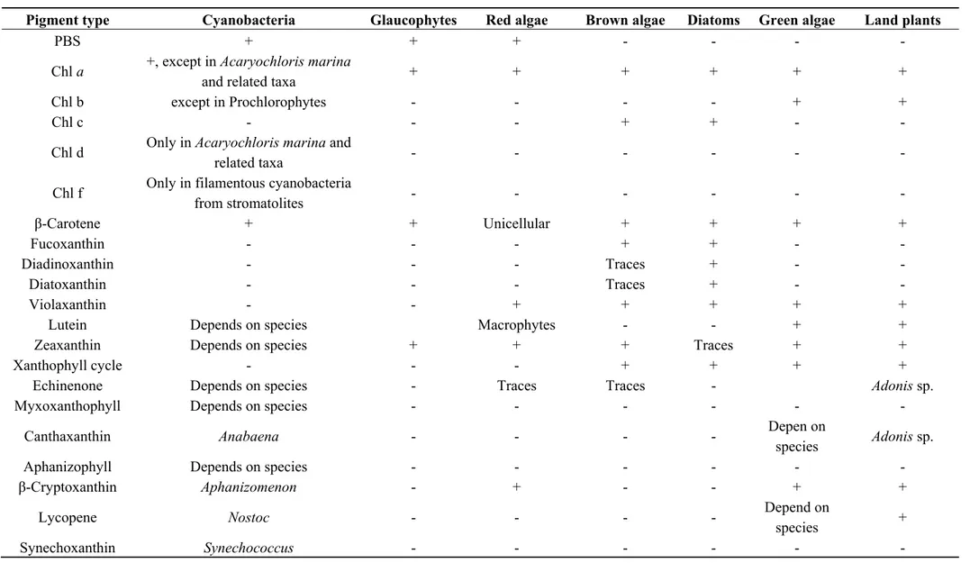
A photosystem is a pigment-protein complex composed of a reaction center (RC) and a light-harvesting complex (LHC). Two families of pigments are found in LHCs: the tetrapyrroles and carotenoids [25,26]. Besides the typical closed tetrapyrrole chlorophyll (Chl) a and β-carotene, cyanobacteria harbour usually two types of accessory pigments, i.e., open tetrapyrroles (phycobilins) and glycosylated xanthophylls [27]. Phycobilins (PBS) are also accessory pigments in red algae and Glaucophytes. In the other eukaryotic phytoplanktonic taxa, Chla a is accompanied by another type of Chl (Table 1). Chlorophyllin (Chlin) is a water-soluble semi-synthetic derivative of Chl that is widely used as a food colorant, and is commercially available as an herbal “remedy”. Therefore, although not natural, this compound was also considered in this review.
On the basis of molecular, biochemical and cellular arguments, it is classically assumed that eukaryotic phytoplankton derives from the internalization of a cyanobacterium-type organism into a eukaryotic heterotrophic cell, the so-called first endosymbiotic act (for a review, see [28]). The reconstitution of true phylogenetic history of organisms has become a very important issue in biology. The availability of numerous molecular sequences allowed biologists to propose new hypotheses for evolutionary histories of taxa that lead eventually to unresolved or conflicting interpretations and the real history seems more complicated than thought initially (see for instance [29–31]) and will not be discussed here. This primary endosymbiotic act explains why today’s chloroplasts have two envelopes (for a review on the photosynthetic membranes in phytoplankton, see [20]). To explain the presence of additional membranes around the chloroplasts, additional endosymbiotic acts are invoked [32], for a review, see [33]. Actually, the recent phylogenetic studies carried out indicated that the history of secondary endosymbionts is more complicated than originally thought because a detailed study of the diatom genes support a putative green algal origin of several genes [14,20,30–34]. The members of the red lineage of secondary endosymbionts constitute a most diverse group of organisms. In these organisms, the gene mixing that occurred along evolution allowed a tremendous diversification of the metabolism and the most important from the pharmaceutical point of view being the diatoms (Heterokonta) and the dinoflagellates (Alveolata).
Table 1. Main chlorophyll and carotenoid types in the various taxa of photosynthetic organisms. “+” and “-” mean that the pigment was
detected or not in the taxa.
Pigment type Cyanobacteria Glaucophytes Red algae Brown algae Diatoms Green algae Land plants
PBS + + + - - - -
Chl a +, except in Acaryochloris marina
and related taxa + + + + + +
Chl b except in Prochlorophytes - - - - + +
Chl c - - - + + - -
Chl d Only in Acaryochloris marina and
related taxa - - - - - -
Chl f Only in filamentous cyanobacteria
from stromatolites - - - - - - β-Carotene + + Unicellular + + + + Fucoxanthin - - - + + - - Diadinoxanthin - - - Traces + - - Diatoxanthin - - - Traces + - - Violaxanthin - - + + + + +
Lutein Depends on species Macrophytes - - + +
Zeaxanthin Depends on species + + + Traces + +
Xanthophyll cycle - - - + + + +
Echinenone Depends on species - Traces Traces - Adonis sp.
Myxoxanthophyll Depends on species - - - -
Canthaxanthin Anabaena - - - - Depen on
species Adonis sp.
Aphanizophyll Depends on species - - - -
β-Cryptoxanthin Aphanizomenon - + - - + +
Lycopene Nostoc - - - - Depend on
species +
3. Bioactivity of Pigments and Lipids from Plastids of Marine Phytoplankton
As mentioned earlier, most of phytoplanktonic species are photoautotrophs. Consequently, they are rich in pigments. Three major classes of photosynthetic pigments are recognized, i.e., (1) Chls and Chl-related molecules, (2) Cars (carotenes and xanthophylls) and (3) PBS. Altogether the pigment family represents several hundreds of purified molecules [25,35]. All these molecules share the unique property to absorb light thanks to their characteristic conjugated double bond network [25,36,37]. Considering the high structural diversity of pigments and the possibility to chemically modify these molecules to obtain better pharmacological properties, the potential of phytoplankton as a source of molecules of therapeutical interest is obviously very high. Unfortunately, the lability and difficult purification of these molecules make the biological activity of most of these molecules unstudied. However, when purified pigments were obtained, they presented a high activity on pharmacological and cellular effectors even at very low concentrations. These molecules can therefore be used for chemoprevention.
The superiority of marine molecules, as compared to analogue terrestrial resources, can be explained by the simultaneous presence of a wider variety of substances in marine organisms. These substances would act synergentically, providing an enhanced bioactivity [38]. Very often, synergistic actions between pigments and lipids are described. For instance, Rao & Rao [39] and Micallef & Garg [40] found a synergistic action between pigments, fatty acids and phytosterols on plasma lipid concentration decrease, on inflammatory response and thus on cardiovascular disease risk prevention.
3.1. Pigments
3.1.1. Tetrapyrroles
Human populations regularly have been consuming green vegetables since the discovery of agriculture practices regularly some 15,000 years ago. This traditional and long practice made in the collective unconscious green pigments as save compounds. Indeed, Chl derivatives such as pheophytin have been found in some plants known in traditional medicine to be active against diseases such as
Clinacanthus nutans-herpes simplex virus [41] and Lonicar hypoglauca-hepatitis C virus [42]. Until
recently, almost nothing was known about the assimilation of closed tetrapyrroles: it was simply thought that Chl absorption was limited (1%–3%). Actually, the phytol chain of the native Chl molecules would be cleaved whereas the Mg ion is leached upon ingestion, the resulting pheophorbide and pheophytine being excreted in the feces [43]. Major progress in studying tetrapyrrole assimilation was possible when intestinal cell could be grown in vitro. Using such a model, i.e., Caco-2 human cells, [44] demonstrated that pheophytin can be absorbed as well. The anti-genotoxicity of natural Chls was revealed by Negishi et al. [45] whereas others studies reported the anti-oxidant activity, the modulation of xenobiotic enzyme activity, and the induction of apoptotic event in cancer cells of Chl [46,47]. This anti-cytotoxicity seems specific to natural Chl and could not be demonstrated with non-natural Chl molecules (see below) [48]. Chl precursors, Chl derivatives, or Chl analogs are also used in medicine for photodynamic treatments of cancers, e.g., [49].
The harmlessness of tetrapyrroles, especially Chl does not mean that the ingestion of tetrapyrroles is without danger. For instance, Chl intolerance can occur due to the liberation of the Chl phytol moity
in patients suffering from Refsum’s disease. In this pathology, phytanic acid accumulates in serum and other tissues due to a defect of the metabolic pathway for phytanic acid [43]. Lohrey et al. [50] demonstrated that albino rats fed with lucerne—but not with ryegrass—protein concentrate, develop photosensitization. This allergy to sunlight could be due to the accumulation of pheophorbide and related pigments in tissues [51]. Similar sunlight allergic reactions were reported in humans having ingested dried laver enriched with pheophorbides and pyropheophorbides [52]. Gandul-Rojas et al. [53] have shown that these deesterified tetrapyrroles are more strongly absorbed than the phytylated ones from the food matrix by the intestinal epithelial cells, allowing a larger accumulation in the patient body. Therefore, there is a need for information on the effects of processes on tetrapyrrole bioaccessibility and bioavailability. For instance, conservation industrial processes such as freezing and canning along with cooking have a positive effect on Chl bioavailability [54,55], making the pigment’s quantity after processing exceeding 100% of the value measured before processing (e.g., [56,57]). Non-natural Chls, such as Cu-Chls, Cu-pheophorbide, Cu-pheophytin, or Chlins, are digested similarly to natural Chl molecules, that is, the phytol chain is cleaved but the Cu ion is not leached from the porphyrin ring because the chelate is much more stable. The stability is only relative because an increase in Cu has been measured in the plasma following Chlin ingestion [58]. Several tests suggested that oral or parenteral administration of Chlins did not produce any gross adverse effect on health [59,60].
Phytoplankton contains also other types but usually minor tetrapyrroles such as hemes. Heme-protein complexes are degraded in the lumen of the stomach and small intestine [61]. Hemes being soluble in alkaline environment, they do not need to bind proteins for the luminal absorption, which is indeed facilitated by the presence of specific heme transporters such as the heme carrier protein 1 (HCP1) at the apical surface of enterocytes [62]. Once in the erythrocytes, hemes are cleaved by heme oxygenases yielding to iron, biliverdin and carbone monoxide formation [63]. Iron is driven to a labile iron pool and used for metabolism. Open tetrapyrroles derived from hemes through the oxygenation of the tetrapyrrole ring [64].
3.1.1.1. Anti-Oxidant, Anti-Inflammatory and Anti-Mutagenic Activities
Many diseases such as cancer, diabetes and, neurodegenerative and cardiovascular diseases have in common oxidative stress as a major cause of inflammatory events. The anti-oxidant and anti-inflammatory activities of phytoplanktonic pigments are widely demonstrated and evidenced in numerous in vitro free and in vivo radical scavenging assays. The presence of a metal-chelate strongly enhances the anti-radical capacity closed tetrapyrroles. Protoporphyrin methyl ester and its magnesium chelated derivative, as well as pheophorbide b and pheophytin b, were also identified as strong anti-oxidant molecules [65]. The ability of the porphyrin ring to transfer electrons explains the anti-oxidant activity of Chls and derivatives. The highest anti-oxidant activity was obtained with of pheophorbide b, compared to pheophorbide a, suggesting that the presence of the aldehyde function may be critical to this activity [47]. This was confirmed in a study that investigated the action mechanisms of natural and synthetic porphyrins regarding their anti-inflammatory effects in vitro and
in vivo [66]. Unfortunately, no structure related to the Chl familly of compounds has been tested
alpha production, the Fyn tyrosine kinase inhibitors [66]. However, on the basis of the study, some potential for Chl and Chl-derivatives can be hypothesized. More generally, it should be recall that the antioxidant properties of Chls and Chl-derivatives disappear in the presence of light. Actually, the accumulation of free tetrapyrroles in photosynthetic cells leads to a dramatic oxidative stress upon light illumination [67–69]. Metal-free and metallo-Chl derivatives have also anti-mutagenic activities, as demonstrated using a bacterial mutagenesis assay [47,70]. Similar properties are exhibited by open-chain tetrapyrroles (anti-oxidative: allophycocyanin [71], phycocyanin [72], phycoerythrin [73] and anti-inflammatory: phycocyanin [74]) in vitro. As a conclusion, most microalgal pigments exert strong in vitro anti-oxidant activity, but additional intervention trials are required to precise their absorption, metabolism and potential as natural anti-oxidant, anti-inflammatory and anti-mutagenic compounds in vivo. Very importantly, the capacity of phycocyanin to quench oxygen radical species is preserved in food supplement [75].
3.1.1.2. Anti-Cancer
Chlin has been reported to have potent anti-mutagenic and anti-carcinogenic effects in short-term tests in vitro and in vivo [45,46]. There is an abundant literature about the interest of food molecules to preserve health, for reviews see [76]. However, it may happen that food, for instance stored in poor conditions, gets infected by molds such as Aspergillus flavus and A. parasiticus. These fungi secrete mycotoxin aflatoxin B1 (AFB), one of the most known human carcinogenesis linked to diet. The action mechanism of AFB is pretty well known. However, it is out of the scope of this contribution to present the details of these mechanisms and it is sufficient to known the following facts to understand the interest of tetrapyrroles in thwarting AFB action. Briefly, AFB is metabolized by cytochrome P450 (CYP) enzymes to the exo-AFB-8,9-epoxide (AFBO), which constitutes the main metabolites of the reaction. (for details, see recent reviews, e.g., [77]). AFBO is highly reactive, and can further bind covalently to cellular macromolecules such DNA forming AFB-N7-guanine adducts. These adducts are chemically unstable and rapidly excreted but they are stabilized through rearranging to a ring-opened formamidopyramidine structure. The AFB-DNA-adducts can trigger somatic alterations at transcriptionally active DNA regions when not removed by DNA repair enzymes [78]. The ability of Chlin to form complex with carcinogens is not restricted to AFB (for a review, see [79]) as AFB could also bind heterocyclic amines that favour the development of colorectal adenoma [80]. Heterocyclic amines are for instance produced when meat is cooked at high temperature [80]. Thus, Chlin exhibits the property to chelate AFB yielding to the formation of chemically stable Chlin-AFB complexes, and those complexes are no longer absorbed by intestine and excreted with feces [81]. Moreover, inhibitory effects of Chlin on human CYP have been reported [82]. Based on these effects, Chlin was used recently in a chemointervention trial in China, resulting in a 55% reduction of the urinary AFB-N7-guanine adduct [83]. Shaughnessy et al. [84] proposed arguments for a protective action of Chl on DNA damages. The efficiency of dephytylated compounds is less than the phytylated ones [85]. In addition, Chlin has been reported to exhibit an anti-proliferative effect on various cancer cell lines [86–88]. Priyadarsini et al. [70]) reported that Chlin alterates the expression of genes coding for proteins involved in the transformation of normal to malignant phenotype of buccal pouch cells.
However, Nelson [89] reported that commercially available Chlin can have a tumor promoting effect and at present the Chlin content in food is regulated [55].
Microalgae pigments may have interest to restore drug sensitivity or reverse multi-drug resistance (MDR) in cancer cells, as some of them inhibit or down-regulate drug efflux pumps. For instance, a significant reduction of P-glycoprotein expression in drug-resistant hepatocellular carcinoma (R-HepG2) cells, at both transcriptional and translational levels, was observed when cells were treated with pheophorbide a [90]. Recently, a natural Chl of the Phaeophyceae, Chl c2, was found to trigger the degranulation of a rat basophilic leukemia cell line (RBL-2H3), whereas Chl a and b were unable to do it [91]. In addition, Chl c2 has an anti-allergic power.
3.1.1.3. Miscellaneous
Because of their antioxidant and anti-inflammatory activity, most microalga pigments have neuroprotective effects in cultures rat cerebellar neurons, and hepatoprotective effects in hepatocytes grown in vitro (e.g., phycocyanin, phycoerythrin [92]). Besides, some studies have demonstrated anti-viral and anti-fungal activities for some pigments (e.g., allophycocyanin, phycocyanin [92]). The physicochemical characteristics of PBS, Chl and Chl catabolites make them suitable for use as fluorescent probes for cellular and tissular analysis (e.g., cell sorting, cytofluorescence, flow cytometry, histofluorescence, binding assays, reactive oxygen species (ROS) detection, labeling of pathological or apoptotic cells, etc.). Phycocyanin- or phycoerythrin-coupled anti-bodies are common reagents available for research and medical use, in which PBS act as powerful and highly sensitive fluorescent probes (for reviews, see [92]).
3.1.2. Carotenoids
Many xanthophylls (violaxanthin, antheraxanthin, zeaxanthin, neoxanthin, lutein, loroxanthin, astaxanthin and canthaxanthin) can be synthesized by microalgae while others (diatoxanthin, diadinoxanthin and fucoxanthin) can also be derived by brown algae ([93]; Table 1). In cyanobacteria carotenoids are much diversified: β-carotene; echinone, synechoxanthin, myxoxanthophyll, canthaxanthin, zeaxanthin, isozeaxanthin, aphanizophyll, lutein, lycopene, β-cryptoxanthin and Orange Carotenoid Protein (OCP) can be found (Table 1). This blue-green light photoactive protein was described very recently; it binds the carotenoid 3′-hydroxyechinenone as its only pigment [94,95]. Cyanobacteria are rich sources of lycopene, and also provide higher ratios of cis/all-trans lycopene which is very important in increasing the bioavailability of health beneficial lycopene [96].
As tetrapyrroles, carotenoids are not synthesized by humans and other animals, and moreover they are required for their metabolism. Table 2 illustrates the health benefits obtained with carotenoids from phytoplankton.
Table 2. Bioactivities of carotenoids from microalgae and cyanobacteria.
Pigments Bioactivities References
Aphanizophyll Photoprotection [97–99] Astaxanthin
Anti-allergic, anti-cancer, anti cardiovascular diseases Anti-oxidant Photoprotection [100–102] [100,103,104] [38,105] Canthaxanthin Anti-cancer Anti-oxidant [106] [107] β-Carotene Anti-allergic Anti-cancer Anti-oxidant [108,109] [110,111] [112] β-Cryptoxanthin Anti-inflammatory
Improvement of skin health
[113] [114] Diadinoxanthin/Diatoxanthin Photoprotection [115,116] Fucoxanthin Anti-allergic Anti-cancer Anti-inflammatory Anti-obesity Anti-oxidant Photoprotection [109,117,118] [119,120] [121–123] [124–127] [128,129] [130] Fucoxanthinol Anti-cancer Anti-obesity [131] [124–126] Lutein Anti-inflammatory Protection of eyes [113] [131–133] Myxoxanthophyll Anti-oxidant Photoprotection [134,135] [97–99] Neoxanthin Anti-cancer [134]
Orange Carotenoid Protein Photoprotection [136–139]
Peridin Anti-cancer [140] Sporopollenin Photoprotection [141] Synechoxanthin Anti-oxidant [142] Violaxanthin Anti-cancer [111,112,134] Violeaxanthin Anti-cancer [111,112,134] Zeaxanthin Anti-allergic Antioxidant Photoprotection [109] [135,143] [139] 3.1.2.1. Anti-Oxidant, Anti-Inflammatory and Anti-Mutagenic Activities
Number of works report anti-oxidant properties of microalgae and cyanobacteria [107,110,143]. Most microalgal and cyanobacterial pigments exert strong anti-oxidant activities in vitro and
in vivo in animal models (Table 2). It was postulated that circulating anti-oxidants also have a role
in the prevention of cardiovascular disease: C-reactive protein and oxidized low-density lipoprotein-cholesterol concentrations, are inversely related to plasmatic concentrations of vitamin C, carotenoids and phenols [144–146].
Therefore, dietary anti-oxidants may be protective against the development of inflammatory disease. However, additional intervention trials are required with carotenoids to precise their
absorption, metabolism and potential as natural anti-oxidant, anti-inflammatory and anti-mutagenic compounds in vivo. Carotenoids have also a role in regulating gene expression and in inducing cell-to-cell communications [147].
Astaxanthin (Asta) is a nonprovitamin A found in Haematococcus pluvialis, up to 2–20 g kg−1 on a dry weight basis [148]. It is a powerful anti-oxidant, more effective than vitamins C and E or other carotenoids [149,150]. In particular, it has been reported that the anti-oxidant properties of Asta are about 10 times greater than those of β-carotene, lutein, zeaxanthin, canthaxanthin and over 500 greater than that of α-tocopherol. Thus the term “super vitamin E” has been proposed for Asta [100,103]. Asta has also a great potential to prevent cancer, diabetes and cardiovascular diseases [100,101]. The presence of the hydroxyl and keto endings on each ionone ring explains Asta unique features, such as the ability to be esterified [115], a higher anti-oxidant activity and a more polar configuration than other carotenoids [38,102,128]. Because of its anti-oxidant and membrane preservation properties, Asta has a considerable potential in the prevention and treatment of various chronic inflammatory disorders, such as cancers, rheumatoid arthritis, metabolic syndrome, diabetes, diabetic nephropathy, and gastrointestinal liver and neurodegenerative diseases, and could provide benefits not only for the cardiovascular system, but also in other inflammatory diseases. Therefore, its daily consumption is a practical and beneficial strategy in human health management [101]. Despite of these studies the molecular mechanism related to the biological activities of Asta is not yet fully understood. In a recent study, Sayo et al. [114] investigated whether the actions of nonprovitamin A carotenoids including Asta are mediated via retinoid signaling in cultured human keratinocytes. They found that β-carotene, β-cryptoxanthin, lutein, zeaxanthin, and Asta, upregulated HAS3 gene expression and stimulated hyaluronan synthesis. The results suggest that nonprovitamin A can be substituted for retinoids and should be considered as a potential means of improving skin health. The several examples of benefits brought by Asta on health have however to be verified using human trials. To our knowledge, such trials have been started. For instance, Djordjevic et al. [104] tested the effect of Asta supplementation on muscle enzymes, oxidative stress markers and antioxidant response in elite young soccer players in Serbia. The results suggest that soccer is associated with excessive production of ROS and oxidative stress, which are prevented in Asta-supplemented in young soccer players.
Fucoxanthin (Fuco) is a brown pigment belonging to the class of xanthophylls, with anti-oxidant properties [128] under anoxic conditions, whereas other carotenoids have practically no quenching abilities, donating electrons as a part of its free-radical quenching function [129]. Fuco protects cells and provides other health benefits [38]: it improves cardiovascular health, blood pressure, and liver functions; it reduces inflammation, cholesterol and triglyceride levels [119,121,124]. Moreover, Fuco has anti-inflammatory effects via inhibitory effects of nitric oxide production [122,123].
Future clinical studies should determine the effectiveness of Asta and Fuco, not only on the vascular structure, but also on cartilage and joint health in at-risk patients or in those with established osteoarthritis [38].
β-Carotene isan important carotenoid because it displays a pro-vitamin A activity. Because animal cells cannot synthesize this molecule, it is considered as an essential compound in the human body [151].
Canthaxanthin has also anti-cancer properties [106] and a potent antioxidant. In particular, it inhibits the oxidation of lipids in liposomes [107].
Lutein and β-cryptoxanthin may down-regulate factors involved in inflammation associated with osteoarthritis and rheumatoid arthritis [113]. Lutein is currently considered as effective agent for the prevention of a variety of human diseases. Synechoxanthin, isolated in 2008 from the cyanobacterium
Synechococcus sp. strain PCC 7002, has exceptional strong antioxidant properties [142].
3.1.2.2. Anti-Cancer
Epidemiologic studies demonstrate an inverse relationship between cancer incidence and dietary carotene intake or blood carotenoid levels, provided the carotene intake is not too high [140,152]. For instance, Moreau et al. [120] works focalized to carotenoids extracts of the marine diatom
Odontella aurita that showed an anti-proliferative effect on cultures of bronchopulmonary and
epithelial cells when extracts were added to cell medium. Carotenoids such as astaxanthin or lycopene are active inducers of communication between cells at the cell-gap junctions [100,102,153]. DNA has receptors for specific carotenoids in controlling connexin gene transcription (connexin being a protein that mediates inter-cellular permeability and communication). The immune system cells need intercellular communication to function effectively, so they can be affected by the DNA regulating mechanism of carotenoids. For example, high doses of β-carotene increase the CD4 to CD8 lymphocyte ratio [154]. MDR reversion is often observed. Microalgae pigments may have interest to restore drug sensitivity or reverse MDR in cancer cells, as some of them inhibit or down-regulate drug efflux pumps. As examples, neoxanthin increases rhodamine accumulation in MDR colon cancer cells [134], inhibits the P346 glycoprotein efflux pump and reverses MDR in doxorubicin-resistant MCF-7 cells and hmdr1-transfected L1210, at 4 and 40 μg mL−1, respectively [155]. Violaxanthin and violeoxanthin are effective MDR modulators in colon, at 4 and 40 μg mL−1, respectively [111,112,134]. 3.1.2.3. Anti-Allergic
Some experiments performed on mices demonstrated that carotenoids (α- and β-carotene) can reduce sharply the immunoglobulin E production. For instance, the reduction of food allergy was observed on animals sensitized to ovalbumine [156]. Another work reported a similar effect of β-carotene combined with vitamin E on splenocytes [108]. Fuco, especially, but also Asta, zeaxanthin and β-carotene have anti-allergic propreties; they can block the aggregation of high affinity receptor for immunoglobin E via inhibiting their translocation to lipid rafts [109]. Fuco anti-allergic activity was recently evidenced using a rodent mast cells model [117,118].
3.1.2.4. Anti-Obesity
Fuco and its metabolite fucoxanthinol [135] limit the risk of obesity [124,125]. Actually, these two marine carotenoids inhibit lipase activity in the gastrointestinal lumen and suppress triglyceride absorption [126]. During normal metabolism, the body produces heat [38]: fuco affects many enzymes involved in fat metabolism determining an increase in the release of energy from fat [127], thus an increase of thermogenesis.
Treatment with neoxanthin significantly reduces lipid accumulation. Examination of structures and functions suggests that carotenoids containing an allene bond and an additional hydroxyl sustituent on the side group may show suppressive effects on adipocyte differentiation [157].
3.1.2.5. Protection against Light
To cope with the deleterious effects of excess illumination, photosynthetic organisms have developed photoprotective mechanisms that dissipate the absorbed excess energy as heat from the antenna system. Certain bloom-forming microalgae are “sun-adapted” and have the capacity to synthesize sunscreen pigments [158]. In many microalgae, the cell is made more resistant to UV light by the accumulation in the cell wall of sporopollenin [141], a carotene-polymer absorbing UV light. Lutein is one of the major xanthophylls found in green microalgae. It selectively accumulates in the macula of the human retina, protects the eyes from oxidative stress, and acts as a filter of the blue light involved in macular degeneration and age-related cataract [131–133]. Asta protects against UV light; it is true for microalgae, animals and human beings [38,105]. Fuco inhibits tyrosinase activity, melanogenesis in melanoma and UVB-induced skin pigmentation by topical or oral application [130].
In cyanobacteria, strong blue-green (or white) light activates a soluble carotenoid protein, the OCP which, by interacting with the phycobilisome, increases energy dissipation in the form of heat, thereby decreasing the amount of energy arriving at the RCs. Consequently, OCP plays a key role in the photoprotective mechanism in cyanobacteria [136–139] and it is the first photoactive protein identified containing a carotenoid as the photoresponsive chromophore [139]. The fluorescence decrease associated with this energy dissipation in the antenna was called qEcya to differentiate this mechanism from that existing in plants. The qEcya mechanism is not the only one that protects cyanobacteria cells from high irradiance. Mutants lacking zeaxanthin are very sensitive to strong light, even more than mutants lacking the qEcya mechanism, indicating an important role of this carotenoid in photoprotection [139].
Another protein, called the Fluorescence Recovery Protein (FRP) encoded by the slr1964 gene in
Synechocystis PCC 6803, plays a key role in dislodging the red OCP from the phycobilisome and in
the conversion of the free red OCP to the orange, inactive, form. As a result, under low light conditions, the FRP mediates the recovery of the full antenna capacity by accelerating the deactivation of the OCP [159]. The amplitude of photoprotection depends on both OCP concentration and FRP concentration but also on the OCP/FRP ratio. Therefore, the synthesis of FRP and OCP is probably strictly regulated to accomodate both concentration and OCP/FRP ratio to the right level of photoprotection.
3.2. Lipids
Marine and freshwater microalgae are known to be characterized by a large diversity in their lipid and fatty acid composition. Glycerides, phospholipids (PLs) and glycolipids are major classes produced and stored in different subcellular compartments of microalgae such as globules, endoplasmic reticulum and chloroplast, respectively. The lipid composition in marine microalgae reveals that the major lipid classes are neutral lipids and glycolipids but also, even in a low range, the phospholipid class. According to this wide variety in lipid class, these microorganisms are good
candidates to produce these lipids for which the amounts can vary according to culture and environmental factors.
Triacylglycerols (TAG) and diacylglycerols (DAG), monogalactosyldiacylglycerols (MGDG) and digalactosyldiacylglycerols (DGDG), and phosphatidylcholine (PC) and phosphatidylethanolamine (PE) are the main classes as constitutive of neutral lipids, glycolipids, and phospholipids, respectively. In this section will be described a non-exhaustive information concerning the use and the effects of these lipids in human or animal models in relation with their health benefits.
3.2.1. Omega-3 Polyunsaturated Fatty Acids
Omega-3 Polyunsaturated Fatty Acids (PUFA) are well known to be beneficial for human health. The most recognized omega-3 fatty acid source is fish oil but its quality depends on several parameters such as species, seasons and quality of consumed food. Moreover, the content of lipid-soluble pollutants in fish oil may be high because of the biomagnification of such compounds in food chains. Microalgae, and specifically the marine strains, are considered as more interesting alternative sources for omega-3 PUFA production. Indeed, they have PUFA of higher and more constant quality than the ones from fish because they are at the bottom of food chains.
Among the marine strains, the prymnesophytes as Isochrysis galbana and Pavlova lutheri are rich sources of docosahexaenoic acid (DHA; 6%–14% total fatty acids) and eicosapentaenoic acid (EPA; 5%–26% of the total fatty acids [160–164], while diatoms as Phaeodactylum tricornutum or
Odontella aurita are rich sources of EPA (26% of the total fatty acids) [160,161,165].
Due to its content of DHA and EPA, Isochrysis galbana was used in alloxan-induced diabetic rats. This microalga induced a decreased glucose, TAG and cholesterol blood levels. However, an increased light-density lipoprotein level and a decreased high-density lipoprotein level were observed in the diabetic rats, but also in the healthy ones [166]. The marine diatom Odontella aurita is the only microalga known to be rich in EPA and currently approved as a dietary supplement [120].
Omega-3 fatty acids act as important mediators of gene expression working via the peroxisome proliferator-activated receptors that control the expression of the genes involved in the lipid and glucose metabolism and adipogenesis [167]. Ethyl-EPA markedly reduced the fatty droplets in the liver cells of mice fed a high-fat diet, also lowering plasma levels of total cholesterol and TAG [168]. Kajikawa et al. [169] showed that oral administration of highly purified EPA ethyl ester improved hepatic fat accumulation in high fat/high sucrose diet-fed mice by suppressing the TAG synthesis enzymes regulated by sterol regulatory element binding protein-1 and decreased the accumulation of hepatic monounsaturated fatty acids produced by stearoyl-coenzyme A desaturase 1. Numerous studies showed biochemical functions of Chlorella, especially when administered on the presence of underlying disorders such as in streptozotocin induced diabetes rats [170]. Recent studies in hypercholesterolemic rats have shown that DHA supplementation reduced total weight gain, adiposity index, high-density lipoprotein (HDL)-cholesterol and glucose plasmatic concentration regardless of the dose and form of supplementation [171].
3.2.1.1. Cardioprotective Effects
The cardioprotective effects of the omega-3 fatty acids such as EPA and DHA has been reported through epidemiological studies in human and animal models but also in cell culture studies [172–175]. Fish oil supplements, and other sources such as microalgae provide EPA and DHA usually used for human diets [176,177]. Omega-3 fatty acids are known to reduce the risk of cardiovascular disease, through a reduction of factors involved in metabolic syndrome development, by modification of serum lipid profile [176].
3.2.1.2. Anti-Obesity
EPA and DHA are involved in a reduction of TAG synthesis and adiposity. During obesity, the body mass index and waist circumference are inversely correlated with EPA and DHA intakes [178]. In rabbits fed with a high cholesterol diet for 10 weeks, Chlorella vulgaris, one species of Chlorella, showed anti-lipidemic and anti-atherosclerotic actions [176]. Cherng et al.[179] also showed that
Chlorella pyrenoidosa has the ability to prevent dyslipidemia in rat and hamster models that were fed
chronic high fat.
3.2.1.3. Anti-Cholesterol
In type 2 diabetes patients, dietary EPA and DHA have been shown to reduce TAG and very low-density lipoprotein-cholesterol levels and to increase the HDL-cholesterol level, in blood [180]. Patients with dyslipidemia have been shown to have significant decrease in blood TAG and increase in HDL-cholesterol after a consumption of EPA and DHA during 3 and 6 months [181]. Adan et al. [182] showed that EPA and DHA feeding reduces serum cholesterol and TAG levels, and decreases platelet aggregation in hypercholesterolemic rats. Ethyl esters of DHA extracted from microalgae have been reported to decrease the plasma TAG and total cholesterol levels in rats fed high-fructose diet [183]. Another observation has indicated that Chlorella intake can reduce cholesterol levels in patients with hypercholesterolemia [184].
3.2.1.4. Normalizing Platelet Hyper-Aggregability
EPA and DHA also play a key role in normalizing platelet hyper-aggregability. When added to the diet, EPA and DHA can alter the phospholipid membrane composition of the cells, and therefore impact on the synthesis and action of eicosanoids, and regulate transcription factor activity and abundance [177]. During dyslipidemia in rats, the use of the marine diatom Odontella aurita has shown beneficial effects as a reduction in the risk factors for high-fat induced metabolic syndrome such as hyperlipidemia, platelet aggregation and oxidative stress [177].
3.2.2. Glycerides
TAG are beneficial to health provided they are rich in PUFA. This is the case for instance in the marine dinoflagellate Crypthecodinium cohnii that can accumulate a high % of DHA (25%–60% of the total fatty acids) in its TAG (oil) with only trivial amounts of other PUFA [185,186]. DAG may be
also interessant although considered as minor constituent [187]. Several studies have been conducted using a DAG-oil (producted during high-temperature process), in which the 1,3-DAG isoform was the major component. In this case, and by comparison with a TAG rich oil, a suppression of hypertriglyceridemia during the post-prandial period was observed. Moreover, a decreased body fat mass was obtained [188–190]. In rodent models, the anti-obesity effect of DAG oil was attributed to an increased fatty acid beta-oxidation and to a suppression of TAG synthesis [191,192].
3.2.3. Phospholipids
Phospholipids are the most important storage forms of the biologically active PUFA. In marine sources such as fish or microalgae, the omega-3 fatty acids are usually found both in triacylgycerol form and as phospholipids. Many studies have reported high amounts of omega-3 fatty acids such as EPA and DHA in phospholipids from fish and other marine sources [193–196]. Omega-3 fatty acid rich PC by comparison with other PC improves disorders related with obesity. Indeed, these phospholipids increase the fatty acid beta-oxidation and the adiponectin levels in serum of obese rats [197].
In addition, Lemaitre-Delaunay et al. [198] have shown the potential interest of PC-DHA over TAG-DHA, which might favor the DHA uptake by erythrocytes and putatively by the brain, providing that phospholipid sources of DHA are available. Indeed, the metabolic fate of DHA differs substantially when ingested in TAG or PC both in terms of bioavailability in plasma and accumulation in target tissues.
The intake of dietary phospholipids such as PC and PE are estimated to 3–4 g/day, which amounts 5%–8% of total dietary lipids [199,200]. These amounts are enough to have beneficial effects on health, by comparison with dietary TAG. PE intake has been reported to have a cholesterol-lowering effect in rats in relation with fecal neutral steroid excretion [201,202]. Cholesterol liver metabolism is also improved by PC. Indeed, this phospholipid has protective effects on liver metabolism; it has been observed a decreased cholesterol level by enhancing bile cholesterol secretion, decreasing lymphatic cholesterol absorption and reducing fatty acid synthesis in different animal models [203,204]. Minor phospholipids such as phosphatidylinositol has been reported to increase the HDL-cholesterol levels in mammals [205–207], by mobilization of cellular sterol.
4. Changing the Cultivation Conditions for Optimizing the Production of Bioactive Compounds
Abiotic stresses, such as high irradiance, nutrient starvation, UV irradiation, trigger metabolic reorientations ending with the production of other bioactive compounds such as omega-3 fatty acids or carotenoids.
4.1. Pigments
In natural conditions, the pigment contents vary with hydrographic factors of the ocean and the seasons (e.g., [208]). It means that cultivated algae can produce large quantities of pigments if some environmental growth conditions, such as temperature, light intensity and nutrients, are controlled [209].
4.1.1. Tetrapyrroles
In the photosynthetic membranes, closed tetrapyrroles form complex with carotenoids. In these complexes, they are photoprotected from photooxidation by carotenoids [68,210]. This protection is absolutely required because the absolute yield of singlet oxygen formation through tetrapyrrole irradiation (via its triplet excited state) is between 0.3 and 0.9, depending on the type of tetrapyrrole and solvent conditions (reviewed by Spikes and Bommer [211]. Singlet oxygen formation triggers a cascade of reactions in which other ROS are formed [212], reviewed by Spikes and Bommer [211]). Photodynamic herbicides (reviewed by [213]) and photodynamic treatments of cancers (reviewed by [214,215]) are based on this mechanism. Therefore, tetrapyrrole synthesis and accumulation in photosynthetic cell was always strictly regulated and even for short illumination time, tetrapyrroles should be protected [37,216,217]. Consequently, the tetrapyrrole biosynthetic pathway will not be reviewed in this contribution.
4.1.2. Carotenoids
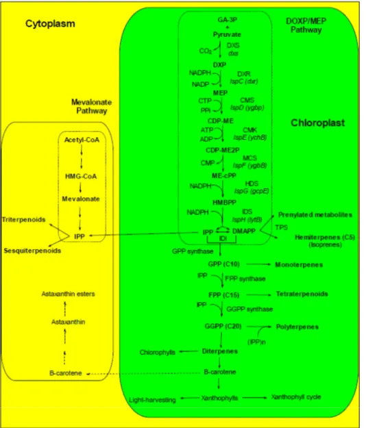
In microalgae, photosynthesis is efficiently performed if carotenoids are bound to peptides to form pigment–protein complexes in the thylakoid membrane. In algae, carotenoids function essentially as accessory light harvesting pigments during the light phase of photosynthesis and as agents to photoprotect the photosynthetic apparatus from excess light dissipation by scavenging ROS such as singlet oxygen and free radicals [218–220]. Carotenoids in the majority of green algae are synthesized and accumulate within plastids, while secondary xanthophylls (e.g., Asta in Haematococcus pluvialis) accumulate in the cytosol. However, these xanthophylls are synthesized in the chloroplast, but they accumulate in the cytosol (Figure 1). Thus xanthophylls can be found in all cellular compartments [221]). NADPH is required in the early steps of carotenoid biosynthesis [222]; consequently, for an efficient carotenoid biosynthesis, efficient light-dependent reactions of photosynthesis are needed (NADP+ must be photoreduced) (Figure 1).
Most of the carotenoids of cyanobacteria are embedded in the photosynthetic membrane which is both, the site of their synthesis and the site of their major functions of light harvesting and photoprotection. Some carotenoids are exported to the periphery of the cell, especially during high intensity light exposure, where it may provide additional photoprotection [223,224]. Other carotenoid proteins may be involved in the transport of carotenoid from the photosynthetic membrane to the exterior.
Control of photosynthetic gene expression and enzyme activity is coordinated by a number of molecular triggers including photosynthetic metabolites, hormones, Chl and carotenoid biosynthesis precursors, photoreceptors, stress-generated ROS, and soluble redox active compounds [225,226]. All known enzymes involved in carotenoid synthesis are nuclear encoded, but the carotenoid synthesis itself takes place in chloroplasts [222,227,228] (Figure 1). Most of xanthophylls are structural and functional components of the cellular photosynthetic apparatus and for this reason are considered as primary carotenoids essential for survival [93] whereas it is not the case for secondary carotenoids produced in large amounts [93,229,230]. Carotenoid content in some microalgae can be increased up to 140 mg g−1 biomass by manipulating environmental conditions [149].
Figure 1. Scheme of the biochemical pathways leading to carotenoids and other
terpenoids. The DOXP/MEP pathway produces the isoprenoid precursors DMAPP and IPP, that are required for the production of carotenoids and other chloroplast-produced terpenoids. Some of the intermediates of the DOXP/MEP pathway can be exported to the cytoplasm and used in the mevalonate pathway of terpenoid synthesis. The cytoplasmic mevalonate pathway does not provide IPP to the chloroplastic DOPX/MEP pathway neither in chloroplast nor in chromoplasts [231]. In contrast, the chloroplastic pathway delivers IPP to the cytoplasmic pathway, allowing the incorporation of photosynthetically fixed C into sterols [231]. Dashed arrows indicate the Asta biosynthetic pathway from β-carotene.
CDP-ME: 4-diphosphocytidyl-2-C-methylerythritol; CDP-ME2P: 4-diphosphocytidyl-2-C-methyl-D
-erythritol-2-phosphate; CMK: CDP-ME kinase; CMS: CDP-ME synthase; DMAPP: dimethylallyl diphosphate; DOXP: 1-deoxy-D-xylulose 5-phosphate; DXR: DOXP reductoisomerase; DXS: DOXP
synthase; FPP: farnesyl diphosphate; GA-3P: glyceraldehyde-3-phosphate; GGPP: geranylgeranyl diphosphate/pyrophosphate; GPP: geranyl diphosphate; GPPS: geranyl diphosphate synthase; HDS: HMBPP synthase; HMBPP: (E)-4-Hydroxy-3-methyl-but-2-enyl pyrophosphate; IDI: isopentenyl/dimethylallyl diphosphate isomerase; IDS: IPP/DMAPP synthase; IPP: isopentenyl diphosphate; MCS : ME-cPP synthase; ME-cPP: 2-C-methyl-D-erythritol 2,4-cyclopyrophosphate; MEP: 2-C-methyl-D-erythritol-4-phosphate.
4.1.2.1. Temperature
Increasing the temperature is a mean to change the polar to non-polar carotenoid ratio. Examples can be found more especially with cyanobacteria. For instance Klodawska et al. [232] demonstrated it on the cyanobacterium Synochocystis sp. PCC6803, for which polar to non-polar xanthophyll ratio increased from 25 °C to 35 °C. Another example is given by Mehnert et al. [97] who found a specific increase of echinenone content in the cyanobacteria Cylindrospermopsis raciborskii and Aphanizomenon
gracile when cultivated from 10 °C to 35 °C. In both examples, the membranes of the living cells
became more rigid with the increasing of temperature. 4.1.2.2. Light Intensity
Microalgae and cyanobacteria are confronted with variations of light by movements in the water column and emersion for coastal benthic species. For these reasons, more than terrestrial plants, marine microorganisms acclimate to light level and light quality by optimizing pigment content and composition [208,233–235]. Therefore they display a wide diversity of light harvesting pigments (Table 1) allowing optimal photosynthesis at different depths in seawater.
Autotroph microorganisms sometimes experience high levels of UV-radiation, in shallow areas and during low tides when they are deposited on intertidal flats. To cope with high sunlight intensities, microalgae have developed different photoprotective mechanisms. One of these, the xanthophyll cycles (Figure 1), consists in the reversible conversion of violaxanthin, antheraxanthin and zeaxanthin in green algae and in the reversible conversion diadinoxanthin and diatoxanthin in brown algae [115,234,236]. Several authors have shown that UV exposure increases carotenoid content in the following hours. It is true for microalgae [237–239] and cyanobacteria [98,240]. In cyanobacteria, carotenoid glycosides play an important role in photoprotection [97]). In cyanobacteria submitted to high light and nitrogen deprivation, the carotenoid content can increase [241]. A high light stress alone leads to an increase of myxoxanthophyll and aphanizophyll, and to a decrease of β-carotene content [97–99]. The increase of myxoxanthophyll amount is in agreement with a coordinated activity of the enzymes of the myxoxanthophyll biosynthetic pathway, with no rate-limiting intermediate steps [99].
Acclimation of microalgae or cyanobacteria to low irradiance intensity or blue enriched light increases the synthesis of specific carotenoids (e.g., fuco in phaeophyceae [196,208,241]). Generally, the photoprotection or the low photoacclimation leads carbon to carotenoid synthesis whereas in nonstressfull conditions, carbon serves mainly to growth (cell wall edification). In the marine diatom
Haslea ostrearia, carbon fixation by β-carboxylation is almost equal to that in the C3 pathway whereas
under low irradiation C3 fixation dominates [242]. 4.1.2.3. Nutrients
Nutrient starvation is generally efficient for the overproduction of carotenoids in cyanobacteria [243]. It is also the case for microalgae. For instance, nitrogen starvation triggers β-carotene in
Eustigmatos cf. polyphem (Eustigmatophyceae) [244] and in Dunaliella salina [245]. β-carotene can
Actually, Lamers et al. [245] applied a combined high light and nitrogen stress and observed a β-carotene accumulation which negatively correlated with the degree of insaturation of the total fatty acid pool. In a frame of an optimal production of biofuels by microalgae, nitrogen starvation is often applied and leads at the same time to an overproduction of β-carotene. Nitrogen starvation leads to a modification of the photosynthetic apparatus. The linear electron flow from water to NADP+ was slowed down while the cyclic electron flow was increased [246]. An overproduction of β-carotene can also be obtained with high nitrate contents (1–3 mM) in Dunaliella salina [247,248].
Low nitrate level, coupled with high light intensity, was the key to high cellular accumulation of Asta in Haematococcus sp [249,250] or Chlorella sp [251]. Chloroplasts of H. pluvialis degenerate in the transition from green coccoid cells to red cyst cells during encystment. Oil droplets containing Asta accounted for more than 50% of the cyst cell volume [252].
Varying the salt concentration is an alternative to get more carotenoids. For instance, when grown under optimized conditions of salinity and light intensity, Dunaliella sp produces up to 14% β-carotene [149,253,254]. The culture of the cyanobacterium Synechocystis sp. PCC 6803 in high-salt conditions (0.7 M NaCl) triggers the up-regulation of protective carotenoid pigments, especially zeaxanthin and myxoxanthophyll [255]. The presence of externally added trehalose at the same time of the salt stress increased even harder the carotenoid to Chl a ratio in this strain. The high accumulation of carotenoids in the presence of trehalose could allow a best cell protection against salt induced production of ROS, and thus a best cell protection against oxidative damage [255].
4.1.2.4. Other Stress
As carotenoids are thought to play an important role in the protection of cellular membrane lipoproteins against oxidative damage by scavenging several active oxygen species, heavy metal stress may stimulate carotenogenesis in cyanobacteria. Actually, elevated levels of β-carotene and Asta were observed in the cyanobacteria Hapalosiphon fontinalis in response to chromium (VI) [256].
Cells of Anabaena sp. PCC 7120, a low desiccation tolerant cyanobacterium, was subjected to prolonged desiccation and effect of loss of water was examined on antioxidant response as well as on overall viability in terms of photosynthetic activity. In desiccated state, Chl a and carotenoid contents increased after 1 h of incubation under desiccation. The oxygen-evolving activity declined in desiccated cyanobacterial biomass while rehydration led to instant recovery, indicating that cells protect the photosynthetic machinery against desiccation [257]. As desiccation leads to oxidative stress, an enhanced level of carotenoids most likely facilitates quenching of ROS.
4.2. Lipids
4.2.1. Temperature
The effects of temperature on growth and on lipid and fatty acid metabolism have been studied on several microalgae. Indeed, in natural conditions, the temperature is one of the main environmental factors that can influence the biology of microalgae, and thus, influencing the carbon metabolism.
In microalga Pavlova lutheri, it has been shown that the decrease of growth temperature (from 25 to 15 °C) has the consequence to increase the polar lipids such the betaine lipids [163]. In these
conditions, the levels of sulfoquinovosyldiacylglycerol and phosphatidylglycerol were increased while the MGDG and TAG levels were decreased. Concerning the omega-3 fatty acids, the EPA and DHA levels were also increased. These results are considered as an adaptative response to temperature with a modulation of omega-3 fatty acids incorporated into lipids involved in the membrane building.
In the diatom Phaeodactylum tricornutum, the decrease of growth temperature (from 25 to 10 °C) showed that the representative fatty acids in the total fatty acids decreased proportionally by about 30% in palmitic acid and 20% in palmitoleic acid but increased about 85% in EPA [258]. Other species such as the Prymnesiophyte Isochrysis galbana, when cultured at temperatures from 15 to 30 °C showed a global lipid accumulation, with a slight decrease of the neutral lipids, by comparison with other polar lipids. The glycosylglyceride levels were increased while no change was observed concerning the phosholipid classes. At 15 °C, as observed for other strain, higher levels of omega-3 fatty acids were obtained (α-linolenate and DHA) while the monounsaturated and saturated fatty acid levels were decreased [259]. Concerning the omega-3 fatty acid levels (EPA and DHA), they were lower at the highest temperatures [260]. The diatom Chaetoceros sp., a tropical Australian microalga, was studied at different growth temperatures, from 25 to 35 °C. The highest lipid percentage was obtained when the cells were cultured at 25 °C. Authors showed that the highest omega-3 fatty acid levels were slightly lower at the highest temperatures [260,261]. The same observations were made concerning Phaeodactylum tricornutum in which the saturated, monounsaturated but also the omega-6 fatty acids were increased with temperature, while the EPA level was decreased [262]. Lipids produced at varing temperatures can have varing biological activities such as antibacterial and cytotoxic activitiesas reported by Gacheva et al. [263] for the fatty acids from
Gloeocapsa sp.
4.2.2. Light Intensity
Light intensity level is the major parameter involved in the microalgal metabolism, in relation with photosynthesis activity. Several studies have been conducted in Pavlova lutheri, a prymnesiophyte rich in both EPA and DHA. The proportions of PUFA, especially EPA, have been shown to be significantly higher under low light, and saturated fatty acids and DHA levels were significantly higher under high light. In this microalga, the galactolipids, a major component of chloroplast lipid membranes, made up approximately 54%–66% of total lipids. The highest PUFA levels, such as those of EPA, were predominantly found in the galactolipid fraction when the cells were grown at low light [264]. Using radiolabelling carbon sources as substrate, the response to light intensity was specific of the use of hydrogenocarbonate or acetate [265]. Indeed, it has been shown that the lipid synthesis, including galactolipids and phospholipids, was increased with light intensity when the microalgae used inorganic carbon (hydrogenocarbonate) but a less sensitivity was observed to the light intensity changes when acetate, as carbon source, was used. Concerning the omega-3 fatty acids, EPA and DHA synthesis has been shown to be lower with high light intensities with hydrogenocarbonate but not with acetate. These results led the authors to hypothesize the fact that Pavlova lutheri was able to synthesize LC-PUFA by two distinct pathways: one inside the chloroplast, depending on light intensity, and another one outside the chloroplast, independent of light.
Studies of light intensity were made with the diatom Thalassiosira pseudonana showing significant changes in fatty acid and lipid composition. Cells grown under moderate continuous light level showed high accumulation of TAG and a reduced level of polar lipids. In the diatom Skeletonema costatum, the highest levels of EPA are observed with the highest light levels during late exponential phase and during the stationary phase [266]. In Nitzschia closterium and Isochrysis zhangjiangensis, after a 3 day-UVA-stress period, the PUFA content increased too [239]. By opposition, in
Nannochloropsis sp., the degree of unsaturation of fatty acids decreased with increasing irradiance,
with a 3-fold decrease of the percentage of total omega-3 fatty acids, caused mainly by a decrease of EPA [267]. Dark exposure can also impact the lipid content; for instance, the dinoflagellate
Prorocentrum minimum reduces its content of TAG and galactolipids, whereas the total content of
phospholipids changed little (PC, PE and phosphatidylglycerol decreased; phosphatidylserine, phosphatidic acid and phosphatidylinositol increased) [268].
4.2.3. Nutrients
Other factors such as nutrients are involved in lipid and fatty acid synthesis. A well-documented description has been reviewed by Guschina [269]. The effect of phosphorus-limitation has been studied in different species of marine algae [270]. It has been reported that phosphorus-limitation led to an increased lipid content in several species such Phaeodactylum tricornutum, Chaetoceros sp. and
Pavlova lutheri, while a decreased lipid content in the green flagellates, Nannochloris atomus and Tetraselmis sp. was observed. The effect of nitrogen-limitation has been studied in the microalga Phaeodactylum tricornutum [271]. A decrease of the nitrogen concentration caused the decrease of the
galactolipid fraction from 21% to 12%. In contrast, both neutral lipids and phospolipids increased from about 73% to 79% and from 6% to 8%, respectively in nitrogen starved cells.
The effect of CO2 on the content and composition of lipid fatty acids and chloroplast lipids has been
studied in Dunaliella salina [272]. An increase in the CO2 concentration from 2% to 10% was shown
to increase the total fatty acid levels. Differences in the fatty acid content and composition indicated increased fatty acid synthesis de novo but an inhibition of their elongation and desaturation steps. This led to an increase in the relative content of saturated fatty acids at the highest levels of CO2. After CO2
stress, the MGDG/DGDG ratio was increased while the ratio of omega-3/omega-6 fatty acids, was clearly increased in the phosphatidylglycerol class.
Dunaliella salina is also tolerant to high salt. It has been shown that the expression of
β-ketoacyl-coenzyme A synthase (which catalyzes the first and rate-limiting step in fatty acid elongation) increased in Dunaliella salina cells transferred from 0.5 to 3.5 M NaCl [273]. It has been suggested that the salt adaptation in this microalga induced modifications of the fatty acid composition of algal membranes, and lipid analysis indicated that microsomes, but not plasma membranes or thylakoids, from cells grown in 3.5 M NaCl contained a considerably higher ratio of 18C (mostly unsaturated) to 16C (mostly saturated) fatty acids compared with cells grown in 0.5 M NaCl. The salt-induced ketoacyl-CoA synthase, jointly with fatty aciddesaturases, was thought to play a role in adapting intracellular membrane compartments to function in the high internal glycerol concentrations used to balance the external osmotic pressure created by high salt [273].
5. Role of Chloroplast and Reticulum Compartments in Lipid and Fatty Acid Synthesis in Marine Microalgae
Marine microalgae can be considered as good models in lipid and fatty acid synthesis studies. Indeed, different works have described that in microalgae, the lipid and fatty acid metabolic pathways were distributed according to the location of enzymatic activities involved in their synthesis. The main compartments involved in lipid and fatty acid synthesis are chloroplast and endoplasmic reticulum in which the glycerolipid and phospholipid classes are recovered respectively. Concerning the phospholipid classes, their presence depends on the microalgal species. Indeed, phospholipids can be replaced by betaine lipids and thus gives some specificity in the concerned species. However, as for the phopholipids, the betaine lipids are also recovered outside the chloroplast, in the reticulum.
In this section, we will describe the location of the different metabolic pathways by the use or data reported on microalgae. Moreover, and specifically in the marine microalgae, the influence of environmental factors on omega-3 fatty acid metabolism will be described.
5.1. Lipid and Fatty Acid Plastid Synthesis in Microalgae
Main reports have been conducted in plant models from which lipid trafficking pathways have been proposed with a difference in lipid location. Even if the lipid composition of plant cell membranes can vary according to environmental factors, cell type culture or development state, it is recognized that glycolipids such as MGDG, DGDG or sulfoquinovosyldiacylglycerol are exclusively lipid elements of plastidial membranes while the phospholipids have a less typical distribution. Among the phosholipid classes, the PC, absent from the thylakoids, are only present in the outer membrane of plasts. Concerning the PE and phosphatidylserine, these classes are totally absent of the plasts [120]. From these results, it can be suggested that the enzyme complex involved in the lipid synthesis are located in specific cellular compartments or that lipid transport can occur in cell to transfer lipids from one compartment to another one.
It is well-recognized that the major lipid class in the chloroplast compartment is the glycolipid class even if phosphatidylglycerol is the main phospholipid in this subcellular fraction. Fatty acids incorporated into glycolipids are related with their metabolism. Indeed, according to the chain length, fatty acids are synthesized during a de novo plastid metabolism or are imported from the endoplasmic reticulum before being incorporated into the glycolipids [274]. According to the identification of the enzymes involved in the glycolipid synthesis in the chloroplast, and their resemblance with the bacterial enzymes, the plast metabolic pathways were named prokaryotic pathway while the reactions involved in the endoplasmic reticulum were named the eukaryotic pathway. The prokaryotic pathway is characterized by properties to incoporate C16 fatty acids at the sn-1 position. Some fatty acids synthesized in the plast compartment are exported to the endoplasmic reticulum and these are involved in the synthesis of lipids of non chloroplastic membranes. These fatty acids, and specifically the C18 ones, are more incorporated into the sn-2 position, and are used to synthesize longer and more unsaturated fatty acids through the eukaryotic pathway [274].
Another example of fatty acid and lipid traffic between the endoplasmic reticulum and the chloroplast is that in some algal species, fatty acids incorporated into diacylglycerols of the

![Figure 2. Proposed lipid synthesis pathway in the marine microalgae. Role of chloroplast and endoplasmic reticulum compartments in lipid synthesis and location of omega-3 fatty acid incorporation [265,277–280]](https://thumb-eu.123doks.com/thumbv2/123doknet/11584742.298294/25.892.147.747.309.768/proposed-synthesis-microalgae-chloroplast-endoplasmic-reticulum-compartments-incorporation.webp)
