medium reveals an unexpected impact of a low N:P
ratio on vegetative growth
Pierre Tocquin
∗, Anthony Fratamico
∗and Fabrice Franck
† Laboratory of Plant Biochemistry and Photobiology,Institute of Plant Biology, University of Li`ege, 4000-Li`ege, Belgium Accepted for publication in Journal of Applied Phycology, November 16, 2011
The original publication is available atwww.springerlink.com
Abstract
Haematococcus pluvialis is the current better source of natural astaxanthin, a high-value carotenoid. Traditionally, the production process of astaxanthin by this algae is achieved by a two-stage system: during the first stage, vegetative “green” cells are produced and then converted, in the second stage, into cysts that accumulate astaxanthin. In this work, a medium screening strategy based on the mixing of a 3-component hydroponic fertilizer was applied to identify a new formulation optimized for the vegetative stage. A maximal and high cell density of 2 x 10⁶ cells mL−1
was obtained in a medium containing a high level of phosphate relative to nitrate, resulting in a N:P ratio much lower than commonly used media for H. pluvialis. In this medium, cells remained at the vegetative and motile stage during a prolonged period of time. Both high cell density culture and motile stage persistence was proved to be related to the N:P feature of this medium. We conclude that the macrozoid stage of H. pluvialis is favored under high-P and low-N supply and that low-cost hydroponic fertilizers can be successfully used for achieving high density cultures of vegetative cells of H. pluvialis.
Keywords Haematococcus pluvialis, Culture media
opti-mization, Phosphate supply, Nutrient-induced fluorescence transients
Introduction
Haematococcus pluvialis Flotow (Chlorophyceae) is a unicellular green alga, which is in a bi-flagellated motile form under optimal environmental condi-tions. In response to adverse conditions, it enters a resting stage and transforms into cysts, which are enlarged cells with a thick and resistant cell-wall and accumulate large amounts of carotenoids, espe-cially astaxanthin.
Astaxanthin (3-3’-dihydroxy-β,β’-carotene-4,4’-dione) is a high-value secondary carotenoid tradi-tionally used as a red dye in aquaculture feed in-dustry. Because of its high antioxidant potential, greater than β-carotene and tocopherol (Naguib,
2000), this ketocarotenoid has also received an in-creased interest for clinical applications: natural as-taxanthin has been proved useful for cancer treat-ment (Palozza et al., 2009) and to have an
anti-∗These authors contributed equally to this work †Corresponding author: Fabrice Franck, email: f.franck@ulg.ac.be, Tel: +32-4-366-39-04, Fax: +32-4-366-29-60
inflammatory action in cardiovascular disease ( Fas-sett and Coombes,2009). Today, most of commer-cially available astaxanthin is a synthetic product. However, the growing demand for natural foods and the high cost of chemical synthesis has en-couraged the research on production from natural sources (Lorenz and Cysewski, 2000). Among the few micro-organisms able to synthesize astaxanthin, H. pluvialis is one that accumulates the most, up to 4 % of its biomass (Aflalo et al.,2007).
Accumulation of astaxanthin in H. pluvialis oc-curs in response to photo-oxidative stress, i.e. when energy input exceeds growth capacity (for review see Lemoine and Schoefs, 2010). Astaxanthin syn-thesis has long been associated with growth arrest and cyst development. Although the feasibility of continuous production of astaxanthin by vegetative motile cells (macrozoids) has been recently demon-strated (Del R´ıo et al.,2008), the most common pro-duction process consists in separating the biomass production phase and the astaxanthin accumulation phase. Finding optimal culture conditions for each of these 2 phases has been the topic of numerous studies (Borowitzka et al., 1991; Cifuentes et al.,
2003;Dom´ınguez-Bocanegra et al.,2004; F´abregas et al.,2000;Garc´ıa-Malea et al.,2005;Harker et al.,
1995;Choi et al.,2002). Even if the exact influence or efficiency of some specific factors on growth -N-source (Borowitzka et al.,1991;Cifuentes et al.,
2003), acetate addition (Cifuentes et al.,2003;Jeon et al., 2006), light level (Borowitzka et al., 1991;
Garc´ıa-Malea et al.,2005;Harker et al.,1995) - and on astaxanthin induction - salt stress, phosphate de-ficiency (Borowitzka et al.,1991;Choi et al.,2002), light requirement (Cifuentes et al.,2003;Choi et al.,
2002;Garc´ıa-Malea et al.,2005) - are still unclear or debated, the general rule governing the green and red stages is widely accepted: a low C:N ratio is favorable to the production of green biomass and, conversely, the production of astaxanthin occurs preferentially when C:N is high (Kakizono et al.,
1992).
In practice, low C:N ratio for green biomass pro-duction is generally achieved by growing the cells at low light in media containing saturating levels of ni-trate. The Bold Basal Medium (BBM,Nichols and Bold,1969) has been often used in its initial formu-lation or modified to contain up to 4-fold more ni-trate (Dom´ınguez-Bocanegra et al.,2004;F´abregas et al., 2000). Since biomass accumulation is the major bottleneck in the 2-stage process of astaxan-thin production, further optimizations of the grow-ing medium have been undertaken (Cifuentes et al.,
2003; F´abregas et al., 2000; Harker et al., 1995;
Gong and Chen,1997; Sarada et al.,2002). A common approach to determine the factors for optimal biomass production has been to start from a known medium and change relative con-centrations of different macro-elements and micro-elements in the medium. In an attempt to opti-mize the elemental composition of an Haematocco-cus growing medium, F´abregas et al.(2000) evalu-ated the contribution of 18 components on the final yield of vegetative biomass using a single-variable optimization strategy. In a semi-continuous culture, the steady-state cell density they obtained with their optimized medium (OHM) was three times higher than with the often used BBM. However, as noticed by the authors, this “one-factor-at-a-time” strategy probably lacks to identify positive or neg-ative interactions between nutrients or between nu-trient availability and other environmental condi-tions.
In a comparative study, Dalay et al. (2007) observed, comparing 9 common medium formula-tions, that biomass accumulation was maximal in a medium prepared with a common agricultural fer-tilizer. As a starting point of our study, we looked for a low-cost and optimized growing medium for H. pluvialis based on the use of a commercial fertilizer. Our optimization strategy relied on the use of a 3-component hydroponic fertilizer which allowed to vary the composition of the media by using different ratios of the 3 core solutions. By screening 18
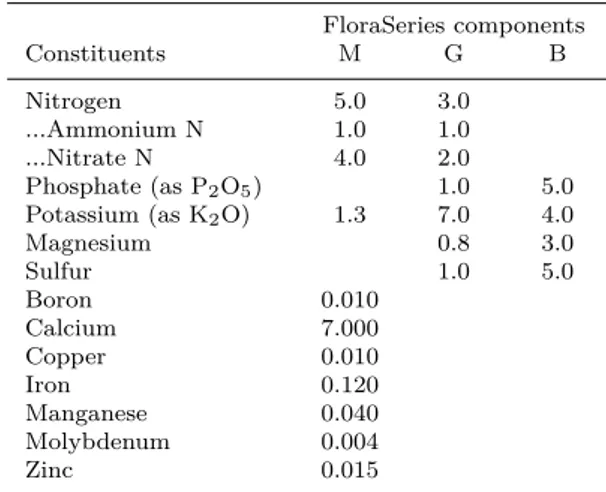
dif-Table 1: Composition of the Floraseries components
Nutrient concentrations (guaranteed minimum concentra-tions) in % (w/v) as described by the producer (GHE, Fleu-rance, France) for each of the 3 components of the FloraSeries fertilizer. M: FloraMicro, G: FloraGro, B: FloraBloom.
FloraSeries components
Constituents M G B
Nitrogen 5.0 3.0
...Ammonium N 1.0 1.0
...Nitrate N 4.0 2.0
Phosphate (as P2O5) 1.0 5.0
Potassium (as K2O) 1.3 7.0 4.0 Magnesium 0.8 3.0 Sulfur 1.0 5.0 Boron 0.010 Calcium 7.000 Copper 0.010 Iron 0.120 Manganese 0.040 Molybdenum 0.004 Zinc 0.015
ferent combinations for vegetative high density cul-tures, we selected the best-performing formulation and compared growth, nutrients (NO3, PO4)
up-take and carotenoid accumulation in batch culture of H. pluvialis in this medium and two other com-mon formulations. The relationship between the unusual N and P content of the identified medium and growth of vegetative cells were further investi-gated.
Materials and Methods
Algal strain and culture conditions Haema-tococcus pluvialis (strain 34/1D) was obtained from the Culture Collection of Algae and Protozoa of the Center for Hydrology and Ecology, Amble-side, UK. Stock cultures were grown in 100 mL of modified BBM3N in 250 mL cell culture flask (Cellstar Suspension Culture Flasks, Greiner Bio-One, Belgium). Modified BBM3N was prepared from a concentrated Bold modified Basal Freshwa-ter Nutrient solution (Sigma-Aldrich, Belgium) to which NaNO3 was added up to 8.82 mM. Flasks
were placed horizontally to increase the gas ex-change surface and optimize exposition to light. They were gently shaken twice-a-day. Cultures were not supplied with an extra source of CO2.
Temperature was maintained at 25◦C, cultures were continuously illuminated by Phillips Master TL5 HO 54W/827 fluorescent lamps. Light in-tensity was either 30 µmol m−2 s−1 (low-light) or 150 µmol m−2 s−1 (high-light).
Media used in the screening were prepared by mixing 3 components of the Floraseries hydroponic fertilizer (GHE, Fleurance, France, guaranteed min-imum concentrations, see Table1). Cells from stock culture were collected at the vegetative stage,
cen-trifuged (3 min, ± 3000 x g, room temperature) and resuspended in 50 mL flasks (Cellstar Suspen-sion Culture Flasks, Greiner Bio-One, Belgium) at the specified density in 10 mL of each of the media formulations tested.
Analytical procedures Cell densities were es-tablished by counting under the microscope, using an improved Neubauer hematocytometer. For pig-ments analysis, cells were collected by centrifuging culture aliquots (3 min, 16000 x g, room temper-ature). The supernatant was discarded and the pellet was homogenized in 100 % methanol. The extraction procedure was repeated until cell de-bris was almost colorless. Chlorophylls and total carotenoids were quantified with a UV-VIS spec-trophotometer (PerkinElmer UV-Vis spectropho-tometer Lambda 20) by recording absorbance at 470, 652 and 665 nm and using the equations of
Lichtenthaler(1987).
Investigations of Nutrient Induced Fluorescence Transients (NIFT) involved the recording of chloro-phyll a fluorescence by Pulse Modulated Fluorime-ter (PAM, MFMS, Hansatech). 2 mL of culture sample were placed into a glass cuvette connected to the PAM and containing a magnetic stirrer. The ac-tinic light (λ=650 nm) was set at 30 µmol m−2s−1. After the sample had been left to stabilize for 10 min, H2O (control), 500 µM of either NaNO3
or K2HPO4 were added to the cuvette. Any
sig-nificant change in fluorescence measured at 680 nm (the analytic modulated light was provided by light-emitting diodes with a central emission wavelength at 580 nm and a photon flux of 0.5 µmol m−2 s−1) following nutrient addition was considered as a NIFT.
Nitrate in media was assayed by the colorimetric reaction with salicylic acid as described byCataldo et al. (1975). Briefly, 20 µL of diluted sampled medium were incubated with 2.5 % (w/v) salicylic acid in H2SO4 98 % for 20 minutes before
neutral-ization with 500 µL NaOH 3.8 M. Concentration in NO3was then assayed by measuring absorbance
at 405 nm with a spectrophotometer (Victor X3, Multilabel Plate Reader, Perkin-Elmer) and com-parison with a NaNO3standard curve.
Phosphate was assayed by reaction with ascor-bic acid as described by Chen et al. (1956). Briefly, 50 µL of diluted sampled medium were mixed with 50µL of a freshly prepared reac-tion solureac-tion containing ascorbic acid 2 % (w/v), (NH4)6Mo7O24.4H2O 0.5 % (w/v) and H2SO4
0.6 M. Concentration in PO4 was then assayed by
measuring absorbance at 750 nm with a spectropho-tometer and comparison with a KH2PO4 standard
curve.
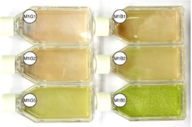
Figure 1: 9-day-old cultures of a screening experi-ment Representative picture showing 6 of the 18 cultures
of a screening experiment. On the left-hand side, from the top to the bottom, cultures M1G1, M1G2 and M1G5 and, on the right-hand side, M1B1, M1B2 and M1B5 (see Table2
for details). The reddening observed from M1G5 to M1G1 and M1B5 to M1B1 is a typical illustration of the devel-opmental shift from the vegetative stage to the resting and astaxanthin accumulating stage. The M1B5 culture shows the distinguishing reticulated-pattern of motile cells group-ing together.
Results
Screening for a medium formulation leading to high vegetative cell density A screening for an optimized Haematococcus pluvialis medium was carried out using a blind approach: 18 media were prepared by mixing the 3 components of the Floraseries hydroponic fertilizer (GHE, Fleurance, France) following the experimental design of Ta-ble2. A pre-culture in modified Bold Basal medium (BBM3N) was used to inoculate 10 mL of each cul-ture media at 2 x 10⁴ cells mL−1. Cells were grown
in continuous light at 30 µmol m−2 s−1 during 9 days.
Maximum cell densities are presented in Table2: among the 18 screened media, a maximum cell den-sity of 1 x 10⁶ cells mL−1, corresponding to a
mean growth rate of 0.41 day−1, was recorded in M1B5 medium (i.e. 1 µL mL−1 FloraMicro and 5 µL mL−1FloraBloom) on day 9 while for all other media, except M5B5, the maximum cell density was reached earlier and was 2 to 10-fold lower. M1B5 was the only medium allowing to keep the cells in the green and motile stage after day 9 (Fig.1) and to sustain growth during 14 days, up to a cell den-sity of 1.73 x 10⁶cells mL−1 (data not shown).
One specificity of M1B5 medium, and other ’B5 media, is to have a very high content in phos-phate (P), magnesium (Mg) and sulfur (S) which are specifically provided at a high level by the B component (Tables1and2). As for potassium (K), it is provided at highest level in the media prepared with the G component and was thus not supposed to contribute in M1B5 efficiency. Since the B com-ponent does not contain a nitrogen (N) source,
an-Table 2: Maximal cell densities obtained in the 18 media Maximal cell densities (mean ± standard deviation,
x 105cells mL−1, n=16) measured in a 9-day screening experiment of 18 media obtained by mixing concentrated FloraSeries stock solutions. G B µL.mL−1 1 2 5 1 2 5 1 1.9 ± 0.6 1.4 ± 0.5 1.4 ± 0.5 1.6 ± 0.6 1.6 ± 0.7 10.0 ± 1.6 M 2 1.2 ± 0.6 1.2 ± 0.5 5.0 ± 1.0 1.9 ± 0.8 5.2 ± 1.5 5.4 ± 1.4 5 2.3 ± 0.3 2.9 ± 0.7 5.3 ± 1.1 4.2 ± 0.8 3.5 ± 0.9 6.9 ± 1.4
other feature of M1B5 is to have a basal content in N. We thus evaluated which of these 4 nutrients (P, Mg, S and N) could account, alone or inter-acting with others, for the high macrozoid density obtained in M1B5. Starting from the BBM3N for-mulation, we decreased the N content by a factor 3, increased P content by a factor 3, and the Mg and S content (as MgSO4) by a factor 5 or 10. Twelve media were generated and used for the culture of H. pluvialis in the same experimental conditions as for the previous screening experiment. As shown in Fig.2a, the highest cell density at day 6 was mea-sured in the media in which P was increased 3 times, other nutrients remaining at their basal BBM3N level. Increasing Mg/S supply was found to have a negative impact on cell growth, independently of N and P supply, while increasing N concentration did not stimulate vegetative cell production and even, in some experiments (data not shown), counterbal-ance the positive effect of high P supply.
This observation suggested that N and P are interacting in controlling production of vegetative cells. When modifying BBM3N formulation to mimic N and P content of M1B5 (M1B5-like), we indeed observed that it was sufficient to prevent pre-cocious encystment and to stimulate green cells pro-duction (Fig.2b). Moreover, when plotting the 18 screened media on a graph as a function of their initial NO3 and PO4 content, the best performing
medium, M1B5, appeared as the one with the low-est N:P ratio (± 0.6, Fig. 3). We further observed, by analyzing nitrate and phosphate contents of the media during growth, that there was a very strong relationship between the phosphate consumption by the algae and initial phosphate content of the medium (Fig. 4a). This correlation was not ob-served for nitrate (Fig.4b) for which measured de-pletions in media were low and not related to the initial content. Note that ammonium was not as-sayed but could act as an N-source at the beginning of the culture.
Evaluation of nutrient limitations in screened media by chlorophyll a fluorescence analyzes
The role of phosphate as a growth-limiting factor was further evaluated by a non-invasive method
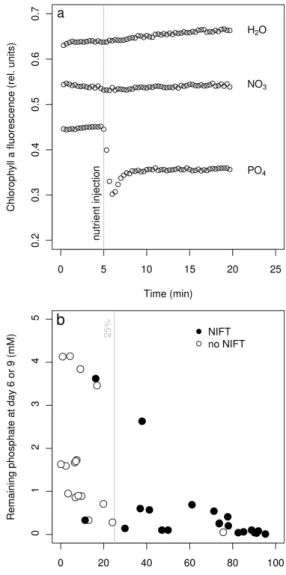
based on chlorophyll a fluorescence measurement. It is known that, if a nutrient such as phosphate, ni-trate or ammonium is added to algal culture limited in that particular nutrient, a change in fluorescence is observable within minutes. This phenomenon has been referred to as “nutrient-induced fluores-cence transients” (NIFT) (Beardall et al.,2001) and proved useful to detect true, in situ, nutrient lim-itations (Holland et al., 2004). In a preliminary experiment, we checked for NIFT occurrence in a 3-week old culture of H. pluvialis in BBM3N and observed phosphate-induced NIFT but no nitrate-induced NIFT (Fig.5a).
The chlorophyll a fluorescence analysis at day 6 and day 9 in all 18 screened media revealed that NIFT were induced by phosphate addition in al-most all media at day 6 and/or day 9 (data not shown). Conversely, as observed in the preliminary experiment, no NIFT were induced by nitrate ad-dition. When results were analyzed independently of media and age but rather plotted against either relative phosphate uptake or remaining phosphate concentration in medium, the NIFT were observed to occur preferentially when phosphate uptake was greater than 25% of initial content (Fig.5b).
Evaluation of growth in selected medium M1B5 and in two other media To the best of our knowledge, high-phosphate containing me-dia, such as M1B5, were never used for growing H. pluvialis. To evaluate the benefit of such a media on the growth of H. pluvialis, we compared growth, in terms of cell density, in M1B5 and two other com-mon media, BBM3N and OHM, containing respec-tively about 2.5 and 25 times less phosphate than M1B5 and whose NO3/PO4 ratio are 4.6 and 23.9,
respectively (Fig. 3). Cultures were initiated in 100 ml fresh media at a density of 5 x 10⁴cells mL−1
and were run at 30 µmol m−2 s−1 (low light) and 150 µmol m−2 s−1 (high light). We observed that, in both BBM3N and OHM, the cells shifted rapidly (around days 4-6) to the palmelloid stage while, in M1B5, they remained in the macrozoid form during the whole experimental period. The developmental shift observed in BBM3N and OHM occurred at both light intensities and coincided with a
signif-Figure 2: Growth of H. pluvialis in modified BBM3N-based media (a) H. pluvialis was grown in BBM3N medium
modified to contain 3 fold less nitrate (2.9 mM vs 8.8 mM), 3 fold more phosphate (5.1 mM vs 1.7 mM), 5 or 10 fold more MgSO4 (1.5 and 3 mM vs 0.3 mM), either separately or in combination. (b) Starting from the BBM3N formulation, NO3, NH4 and PO4 concentrations (respectively 8.7 mM, 0 mM and 1.7 mM in BBM3N) were modified to mimic M1B5. The resulting medium M1B5-like contained 2.7 mM NO3, 0.7 mM NH4 and 4.6 mM PO4. The observed de-crease of cell density in BBM3N is due to adsorption of cysts to walls of flask. These figures are representative results of 3 independent experiments.
icant increase in the carotenoid-to-chlorophyll ra-tio above 0.5 which is a maximum for H. pluvi-alis green cells (this study, see also Solovchenko et al. (2011)). These observations correlated well with growth measurements: as shown on Fig. 6, a significant increase in cell density was only ob-served in M1B5 medium. A maximum of 1.4 and 2.1 x 10⁶ cells mL−1 was measured after 14 days at low and high light, respectively.
Figure 3: Nitrate-to-phosphate content and relative N:P ratio of the 18 screened media 2D-graph of the
ini-tial measured nitrate (y-axis) and phosphate (x-axis) content of 18 screened media. The 2 other common media (OHM and BBM3N) used in this study are also plotted. The plot areas where N:P ratio are below 1, between 1 and 10, and above 10 are filled with dark-grey, light-grey and white, respectively.
Effect of N & P deficiencies on M1B5 cul-tures The above observations tend to show that the M1B5 medium has specific impacts on growth and development of H. pluvialis by stimulating cell division and preventing precocious cysts formation. Nutrient uptake measurements and NIFT assays in-dicated that the high-phosphate content and/or the low NO3/PO4 ratio of M1B5 could be at the origin
of these responses. To further test this hypothesis, we transferred M1B5 exponentially growing cells to media containing lower phosphate (high N:P ra-tio) or lower nitrate (low N:P rara-tio) and evaluated the quantitative (cell density) and developmental (cell types, carotenoid-to-chlorophyll ratio) conse-quences of these treatments.
Independently of light intensity, transfer to BBM3N or phosphate-deprived BBM3N led to a dramatic decrease in cell densities due to induced cell death as observed under the microscope (Fig.7a & b). In high light, cell death occurred rapidly and was almost complete after 2 days while, in low light, the surviving cells shifted to the palmelloid stage: 95% of them were palmella at the end of the ex-periment. Conversely, when cells are transferred to fresh M1B5 or nitrate-deprived BBM3N, cell den-sity still increased. Furthermore, more than 90% of the cells stayed in motile macrozoid stage at ei-ther low or high light. Strikingly, the maximum cell density was observed in the nitrate-deprived BBM3N media in high light. In these conditions, cells accumulated carotenoids, they became red and carotenoid-to-chlorophyll ratio increased up to 0.8,
Figure 4: Phosphate and nitrate uptake at day 6 in relation with initial content Phosphate and nitrate
concentrations in culture media were measured in fresh me-dia (initial content) and at day 6. Phosphate and nitrate depletion were respectively plotted against initial phosphate content (a) and initial nitrate content (b) of screened media.
while remaining at the macrozoid stage (Fig. 7b). In this specific case, carotenoids accumulation did not occur simultaneously with growth arrest as it is the case in the BBM3N and phosphate-deprived media at high light.
Discussion
In this study, medium optimization was performed by screening three-component hydroponic fertilizer based media. This strategy allowed us to evaluate, on a limited number of media (18), the effect of a large range of individual nutrient supplies together with a large range of macro-nutrients ratios. For
Figure 5: NIFT occurrence at day 6 or day 9 in screened media in relation with nutrient uptake (a)
Representative result of a preliminary experiment where NIFT occurrence was analyzed in 3-week old BBM3N stock culture following addition of either 20 µL H2O, 500 µM NaNO3 (NO3) or 500 µM K2HPO4 (PO4). (b) Phosphate-induced NIFTs plotted as 2-D graph of actual phosphate con-tent vs phosphate uptake. Empty dots: no NIFT observed. Filled dots: phosphate-induced NIFT.
example, nitrate and phosphate ranges were 2.20-28.85 mM and 0.18-4.33 mM, respectively, while their relative N:P ratio ranged from 0.6 to 111.8. With this experimental set-up, the relative supply of micro-nutrients was not allowed to be optimized since micro-elements are provided by only one of the three fertilizer components. However, accord-ing to the work of F´abregas et al. (2000) micro-elements do not contribute much in optimization and even their omission has been shown to have a limited growth-inhibitory impact on H. pluvialis (Dom´ınguez-Bocanegra et al., 2004; Hagen et al.,
2001).
Figure 6: Growth of H. pluvialis in M1B5, BBM3N and OHM media at two light intensities Cell
den-sities, chlorophyll and carotenoids content were measured at 2 days interval during a 14 days culture in M1B5, BBM3N and OHM media either at 30 µmol m−2 s−1 (a) or 150 µmol m−2 s−1. The carotenoid-to-chlorophyll ratio of each sample is represented on a color-coded scale from light-green (0) to red (3). A non-linear scale was used to vi-sualize “green” stage (car:chl < 0.5) as green. This figure is a representative result of two independent experiments. Data are means ± standard deviations (n=8). The fluctuating cell densities measured in BBM3N and OHM from day 6 are due to partial adsorption of cyst to walls of flask and production of macrozoids from cyst (concomitant to a drop in Car/Chl ratio).
of one optimal formulation, which is composed of 1 µL ml−1 FloraMicro and 5 µL ml−1 FloraBloom (see Materials & Methods for composition) and was labeled “M1B5”. During 9 days screening ex-periments, growth was indeed sustained until day 9 in only 2 media (M1B5 and M5B5, data not shown), but cell density was the highest in M1B5 (1 x 10⁶ cells mL−1, Table 2). When we
com-pared growth in M1B5 with BBM3N and OHM, we observed that vegetative growth was sustained at least during 2 weeks and a cell density of
Figure 7: Impact of nitrate or phosphate starvation on the growth of H. pluvialis Cell densities, chlorophyll
and carotenoid contents were measured at 24-hours interval during 3 days following the transfer of M1B5 growing cells to Bold Basal medium (BBM3N), N-depleted BBM3N (-N) and P-depleted BBM3N (-P) at either at 30 µmol m−2s−1(a) or 150 µmol m−2s−1(b). The carotenoid-to-chlorophyll ratio of each sample is represented on a color-coded scale from light-green (0) to red (3). A non-linear scale was used to visualize “green” stage (car:chl < 0.5) as green. This figure is a representative result of 2 independent experiments. Data are means ± standard deviations (n=8).
2 x 10⁶ cells mL−1 was reached under high light
intensity. These results represent a minimum 5-fold improvement compared with previous stud-ies in similar batch-mode conditions (Borowitzka et al., 1991; Cifuentes et al., 2003; Dom´ınguez-Bocanegra et al., 2004; Kaewpintong et al., 2007) and with the maximal cell densities of 1.8 and 3.2 x 10⁶ cells mL−1 obtained in this study with
BBM3N and OHM, respectively. For BBM3N, recorded maximal cell density was in the range of published data obtained in similar growing
condi-tions (Harker et al., 1996; Tripathi et al., 1999;
Dom´ınguez-Bocanegra et al.,2004), while for OHM, it correspond to steady-state density obtained by
F´abregas et al. (2000) in a semi-continuous mode with daily addition of fresh medium. The efficiency of M1B5 can not be explained by absolute availabil-ity of nutrients since, in our screening, other formu-lations contained as much nutrients, like nitrate or phosphate, or even more. Rather, we observed that M1B5 had a specific physiological effect by delaying the shift to encystment. This effect was neither ob-served for any of the other screened media, nor for BBM3N or OHM, where a maximum in cell density observed after 3 or 6 days preceded a developmental shift to the red resting stage.
Although phosphate is not the only nutrient that is at high level in M1B5, our results indicate that high phosphate is the key feature leading to its ben-eficial effect on cell growth. Firstly, we showed that among nutrients that are present in higher amount in M1B5, phosphate accounted, alone, for the in-crease in cell density. Secondly, we observed that phosphate uptake increased with available phos-phate in medium and, finally, we also demonstrated, using the NIFT approach, that cells are rapidly behaving as phosphate-limited whatever the initial phosphate content of the medium. Compared to ni-trate, phosphate has received less attention in opti-mization approaches and has long been considered to promote growth at moderate or low concentra-tion (± 0.5 mM,Borowitzka et al.(1991);F´abregas et al.(2000)) while it could promote carotenogenesis at higher concentration (up to 0.9 mM,Borowitzka et al.(1991)). However, in other reports, carotenoid accumulation has been shown to be reduced when phosphate supply was increased above 0.85 mM (up to 3.4 mM, Harker et al. (1996)). Harker et al.
(1996) even reported a positive impact of a high phosphate concentration (>3 mM) on the growth of H. pluvialis.
In addition to have a high phosphate content, M1B5 also is remarkable for having the low-est nitrate-to-phosphate ratio (0.6) among the 18 screened media. Both features are rarely found in already known H. pluvialis media: most of them have a moderate phosphate content (± 0.2-2 mM) and a N:P ratio largely in favor of N (± 7-20) (Dom´ınguez-Bocanegra et al.,2004;F´abregas et al.,
2000). There are only a few reports on H. pluvi-alis growing in N:P ratio close or below one but, in all cases that we identified, the low N:P condi-tions were beneficial to growth (Dalay et al.,2007;
Harker et al., 1996; Hagen et al., 2001). In the present study, we showed that the transfer from the M1B5 medium to low-phosphate containing BBM3N medium led to a rapid cell death while it was not the case if nitrate was omitted in media. In this case where N:P ratio was null, growth was not
inhibited during the time-course of the experiment and the cells stayed at the vegetative motile stage and accumulated carotenoids under increased irra-diance without shifting to resting stage as it was al-ready observed in similar conditions byHagen et al.
(2001). Moreover, we observed that increasing N-supply together with P-N-supply reduced the positive effect of high P on macrozoids production. The low N:P ratio thus appeared to be a critical parameter that maintains actively dividing cells at the macro-zoid stage.
In most organisms, phosphate accumulates in-side the cell as a linear polymer forming intra-cellular polyphosphate granules (PolyP) (Kulaev and Kulakovskaya,2000). In a preliminary experi-ment, we looked for the presence of PolyP granules in H. pluvialis by microscopy with the fluorescent dye 4’,6-diamidino-2-phenylindole (DAPI), follow-ing the method by Tijssen et al. (1982) and we observed that granules were visible in M1B5 grow-ing macrozoids (data not shown). The function of PolyP, which has been essentially studied in pro-caryotes, are numerous, including stress response, quorum sensing, motility, etc... (for review, see
Rao et al.). In microalgae, roles in osmotic stress response, in heavy metal tolerance or phosphate storage have been already proposed but the iden-tification of more functions will undoubtedly occur as the interest for PolyP in eucaryote increases. In this context, the relation between phosphate-supply, PolyP accumulation and cell cycle control in H. pluvialis is of particular interest and requires deeper investigations.
In conclusion, by a medium screening strategy based on a 3-component commercial fertilizer, we obtained a new medium formulation that proved to be efficient, at least in batch culture, in promoting high density culture of H. pluvialis macrozoids and in post-poning the development shift to the resting stage. We demonstrated that these effects were a response to both the unusual phosphate content and the below-one N:P ratio of this medium. Even if the exact impact of these parameters on the physiology of H. pluvialis remains to be determined, achieve-ment of high vegetative cell density culture in this easy to prepare and low-cost media could be evalu-ated for the production of astaxanthin in either 1-or 2-step strategies.
Acknowledgements
This research was funded by WINNOMAT2 program, Service Public de Wallonie, DGO6, Grant N◦71/6662. A.F. is grate-ful to F.R.I.A. for the award of research fellowships. F.F. is a Senior Research Associate of the Fonds de la Recherche Scientifique, F.R.S-FNRS. Authors are grateful to Dr. P. Cardol and anonymous reviewers for helpful comments on an earlier draft of this manuscript.
References
C. Aflalo, Y. Meshulam, A. Zarka, and S. Boussiba. On the relative efficiency of two- vs. one-stage production of astaxanthin by the green alga Haematococcus pluvialis. Biotechnology and Bioengineering, 98(1):300–305, 2007. J. Beardall, T. Berman, P. Heraud, M. Omo Kadiri, B. R.
Light, G. Patterson, S. Roberts, B. Sulzberger, E. Sa-han, U. Uehlinger, and B. Wood. A comparison of meth-ods for detection of phosphate limitation in microalgae. Aquatic Sciences - Research Across Boundaries, 63(1): 107–121, 2001.
M. Borowitzka, J. Huisman, and A. Osborn. Culture of the astaxanthin-producing green alga Haematococcus pluvi-alis 1. Effects of nutrients on growth and cell type. Journal of Applied Phycology, 3(4):295–304, 1991.
D. A. Cataldo, M. Maroon, L. E. Schrader, and V. L. Youngs. Rapid colorimetric determination of nitrate in plant tissue by nitration of salicylic acid. Communications in Soil Science and Plant Analysis, 6(1):71, 1975.
P. S. Chen, T. Y. Toribara, and Huber. Warner. Microde-termination of phosphorus. Analytical Chemistry, 28(11): 1756–1758, 1956.
Y. E Choi, Y. S Yun, and J. M Park. Evaluation of factors promoting astaxanthin production by a unicellular green alga, Haematococcus pluvialis, with fractional factorial de-sign. Biotechnology Progress, 18(6):1170–1175, 2002. A. S. Cifuentes, M. A. Gonz´alez, S. Vargas, M. Hoeneisen,
and N. Gonz´alez. Optimization of biomass, total carotenoids and astaxanthin production in Haematococ-cus pluvialis flotow strain steptoe (Nevada, USA) under laboratory conditions. Biological Research, 36(3-4):343– 57, 2003.
M. C. Dalay, E. Imamoglu, and Z. Demirel. Agricultural fer-tilizers as economical alternative for cultivation of Haema-tococcus pluvialis. Journal of Microbiology and Biotech-nology, 17(3):393–7, 2007.
E. Del R´ıo, F. G. Aci´en, M. C. Garc´ıa-Malea, J. Rivas, E. Molina-Grima, and M. G. Guerrero. Efficiency assess-ment of the one-step production of astaxanthin by the mi-croalga Haematococcus pluvialis. Biotechnology and Bio-engineering, 100(2):397–402, 2008.
A. R. Dom´ınguez-Bocanegra, I. Guerrero Legarreta, F. Mar-tinez Jeronimo, and A. Tomasini Campocosio. Influence of environmental and nutritional factors in the production of astaxanthin from Haematococcus pluvialis. Bioresource Technology, 92(2):209–214, 2004.
R. G. Fassett and J. S. Coombes. Astaxanthin, oxidative stress, inflammation and cardiovascular disease. Future Cardiology, 5:333–342, 2009.
J. F´abregas, A. Dominguez, M. Regueiro, A. Maseda, and A. Otero. Optimization of culture medium for the contin-uous cultivation of the microalga Haematococcus pluvialis. Applied Microbiology and Biotechnology, 53(5):530–535, 2000.
M.C. Garc´ıa-Malea, C. Brindley, E. Del R´ıo, F.G. Aci´en, J.M. Fernandez, and E. Molina. Modelling of growth and accumulation of carotenoids in Haematococcus pluvialis as a function of irradiance and nutrients supply. Biochemical Engineering Journal, 26(2-3):107–114, 2005.
X. Gong and F. Chen. Optimization of culture medium for growth of Haematococcus pluvialis. Journal of Applied Phycology, 9(5):437–444, 1997.
C. Hagen, K. Gr¨unewald, M. Xyl¨ander, and E. Rothe. Effect of cultivation parameters on growth and pigment biosyn-thesis in flagellated cells of Haematococcus pluvialis. Jour-nal of Applied Phycology, 13(1):79–87, 2001.
M. Harker, A. J. Tsavalos, and A. J. Young. Use of response surface methodology to optimise carotenogenesis in the microalga, Haematococcus pluvialis. Journal of Applied Phycology, 7(4):399–406, 1995.
M. Harker, A. J. Tsavalos, and A. J. Young. Factors re-sponsible for astaxanthin formation in the Chlorophyte Haematococcus pluvialis. Bioresource Technology, 55(3): 207–214, 1996.
D. Holland, S. Roberts, and J. Beardall. Assessment of the nutrient status of phytoplankton: a comparison between conventional bioassays and nutrient-induced fluorescence transients (NIFTs). Ecological Indicators, (3):149–159, 2004.
Y-C. Jeon, C-W. Cho, and Y-S. Yun. Combined effects of light intensity and acetate concentration on the growth of unicellular microalga Haematococcus pluvialis. Enzyme and Microbial Technology, 39(3):490–495, 2006.
K. Kaewpintong, A. Shotipruk, S. Powtongsook, and P. Pavasant. Photoautotrophic high-density cultivation of vegetative cells of Haematococcus pluvialis in airlift biore-actor. Bioresource Technology, 98(2):288–295, 2007. T. Kakizono, M. Kobayashi, and S. Nagai. Effect of
car-bon/nitrogen ratio on encystment accompanied with as-taxanthin formation in a green alga, Haematococcus plu-vialis. Journal of Fermentation and Bioengineering, 74 (6):403–405, 1992.
I. Kulaev and T. Kulakovskaya. Polyphosphate and phos-phate pump. Annual Review of Microbiology, 54(1):709– 734, 2000.
Y. Lemoine and B. Schoefs. Secondary ketocarotenoid astax-anthin biosynthesis in algae: a multifunctional response to stress. Photosynthesis Research, 106:155–177, 2010. H. K. Lichtenthaler. Chlorophylls and carotenoids: Pigments
of photosynthetic biomembranes. Methods in Enzymology, pages 350–382, 1987.
R. T. Lorenz and G. R. Cysewski. Commercial potential for Haematococcus microalgae as a natural source of astaxan-thin. Trends in Biotechnology, 18(4):160–7, 2000. Y. M. A. Naguib. Antioxidant activities of astaxanthin and
related carotenoids. Journal of Agricultural and Food Chemistry, 48(4):1150–1154, 2000.
H.W. Nichols and H.C. Bold. Trichsarcina polyinorpha gen. et sp. nov. J. Phycol., pages 34–38, 1969.
P. Palozza, C. Torelli, A. Boninsegna, R. Simone, A. Cata-lano, M. C. Mele, and N. Picci. Growth-inhibitory effects of the astaxanthin-rich alga Haematococcus pluvialis in human colon cancer cells. Cancer Letters, 283(1):108–117, 2009.
N. N. Rao, M. R. G´omez-Garc´ıa, and A. Kornberg. Inorganic polyphosphate: Essential for growth and survival. Annual Review of Biochemistry, 78(1):605–647.
R. Sarada, S. Bhattacharya, and G.A. Ravishankar. Opti-mization of culture conditions for growth of the green alga Haematococcus pluvialis. World Journal of Microbiology and Biotechnology, 18(6):517–521, 2002.
A. E. Solovchenko, O. B. Chivkunova, and I. P. Maslova. Pigment composition, optical properties, and resistance to photodamage of the microalga Haematococcus pluvi-alis cultivated under high light. Russian Journal of Plant Physiology, 58(1):9–17, 2011.
J.P.F. Tijssen, H.W. Beekes, and J. Van Steveninck. Lo-calization of polyphosphates in Saccharomyces fragilis, as revealed by 4’,6-diamidino-2-phenylindole fluorescence. Biochimica et Biophysica Acta (BBA) - Molecular Cell Research, 721(4):394–398, 1982.
U. Tripathi, R. Sarada, S.R. Rao, and G.A. Ravishankar. Production of astaxanthin in Haematococcus pluvialis cul-tured in various media. Bioresource Technology, 68(2): 197–199, 1999.