Clinical outcome after surgical correction of cleft palate by laryngeal
tie-forward in 2 horses
Tosi I.
1,
Arévalo J.
2, Salciccia A.
2, Grulke S.
2, Art T.
1, de la Rebière G.
21 Center of Equine Sports Medicine, Faculty of Veterinary Medicine, University of Liege, Belgium 2 Equine Clinic, Department of Companion Animals and Equids, University of Liège, Liège, Belgium
INTRODUCTION
Cleft palate is a rare congenital defect in horses. Its description in literature is generally limited to cases of young foals referred for milk regurgitation from nostrils, coughing and dysphagia, with a guarded to poor prognosis if not treated. Surgical intervention consists mainly in palatoplasty that is considered a salvage procedure and complications are frequent. Only few cases of cleft palate in adult horses are described in literature. Laryngeal tie-forward, a surgical technique generally used to treat dorsal displacement of the soft palate, has been described only in one 4-year-old pony with cleft palate but clinical improvement after surgery was partial and temporary
.
OBJECTIVES
To present the history, clinical records and punctual follow-up of two sports horses, diagnosed with a defect of soft palate and treated surgically by laryngeal tie-forward technique.
METHODS
Clinical records of two 8 years old jumping horses referred for respiratory noise, exercise intolerance, cough and alimentary nasal discharge. In both cases resting endoscopy revealed a large, asymmetrical defect of the soft palate (Fig.1). In horse 2 a tracheal wash was performed revealing presence of food particles, inflammatory cells and bacteria. Laryngeal tie-forward was performed to reduce the gap of the soft palate: a polyblend suture (Fiberwire) was passed twice into the thyroid cartilage on each side, forming a loop in the thyroid lamina. Then dorsal and ventral sutures on each side were tied over the lingual process of the basihyoid bone, so as to lead the thyroid cartilage immediately dorsal and rostral to the caudal aspect of the basihyoid bone. Bilateral partial sternothyroidectomy was performed.
RESULTS
In both horses nasal discharge decreased quickly after surgery. Post-operative endoscopy revealed a good advancement of the larynx, the point of the epiglottis covering the border of the soft palate defect. Horses were discharged from the clinic with home instructions. Respective owners were contacted again on February 2017, that means respectively 3 years and 4 months after surgery for horses 1 and 2. Both owners were satisfied with the results of the intervention, as initial symptoms had completely disappeared. Horse 1 had started to show again intermittent cough 6 months after surgery, improving under better bedding conditions. Its performances had improved after surgery but no further increase in workload had been attempted because of lack of time. Horse 2 gained weight (from 4/9 to 5/9 of BCS) and the horse was being successfully prepared for a higher competition level. Tracheal wash in both horses were free of food particles and bacteria. Control endoscopy in both horses revealed the re-apparition of a small gap between the apex of the epiglottis and the caudal border of the soft palate, but smaller than before surgery, probably due to some degree of sutures release.
Fig. 1. Resting endoscopy showing a defect in the soft palate (cleft palate or palatoschisis) in two 8 years old sports horses (horse 1 and 2), presented for exercise intolerance, cough and alimentary nasal discharge. In both cases there is a large asymmetrical defect (right>left) in the soft palate; caudal pillars of the soft palate seem underdeveloped in horse 1 and cover partially the epiglottis in horse 2. The communication with the oral cavity is visible.
1 2
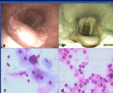
Fig. 2: Control endoscopy of horse 2, 24 hours (a) and 4 months (b)after surgery and microscopic appearance of its tracheal wash cytology before (c) and 4 months after surgery (d). In figure a there is a good advancement of the larynx, the epiglottis is in close contact with the caudal border of the soft palate; in figure b there is a small gap between the two but smaller than before surgery. An improvement in tracheal wash cytology and absence of contaminating food/bacteria was noticed.
CONCLUSIONS: Some horses can grow up to adult age with moderate defects of the soft palate; they are
likely to show clinical signs of variable severity. In such cases laryngeal tie-forward should be considered as
an option if the defect is too large or asymmetrical to be corrected with palatoplasty.
a b