Dissection génétique de la résistance végétale contre les
virus
Par
Xiaofang Ma
thèse en cotutelle
présentée au Département de biologie en vue de l’obtention du grade de docteur ès sciences (Ph.D.)
FACULTÉ DES SCIENCES,
UNIVERSITÉ DE SHERBROOKE
au College of Plant Science and Technology, en vue de l’obtention du grade de docteur ès sciences (Ph.D.)
HUAZHONG AGRICULTURAL UNIVERSITY
Genetic dissection of plant-virus interactions
By
Xiaofang Ma
co-supervised thesis
submitted to the Department of Biology in fulfillment of requirements for the degree of Doctor Philosophiae (Ph.D.)
FACULTÉ DES SCIENCES,UNIVERSITÉ DE SHERBROOKE
and to the College of Plant Science and Technology in fulfillment of requirements for the degree of Doctor Philosophiae (Ph.D.)
Le 25 âout 2015
le jury a accepté la thèse de Madame Xiaofang Ma dans sa version finale.
Membres du jury Professeur Peter Moffett
Directrice de recherche
Département de biologie, Université de Sherbrooke Professeur Guoping Wang
Codirecteur de recherche
College of Plant Science and Technology, Huazhong Agricultural University Professeur Zhongqi Qin
Évaluateur externe
Institute of Fruit and Tea, Hubei Academy of Agricultural Sciences Associated Professeur Wenxing Xu
Évaluateur interne
College of Plant Science and Technology, Huazhong Agricultural University Professeur Kamal Bouarab
Président-rapporteur
Acknowledgements
I would like to express my sincerest gratitude to the following people and institutions: Dr. Peter Moffett, I can not express in words how much I appreciate your help during
my time (20120918-20140909) in Sherbrooke, especially at my arrival in this new and interesting country. You helped me where you could, on a more scientific side, thank you for your invaluable scientific input, your patience and your help with presentation and thesis correcting.
Dr. Guoping Wang, I am so appreciated your and Dr Ni Hong’s help during my time in you lab. You helped me so much, also thank you for your invaluable scientific input, your patience and your help with presentation and thesis correcting.
Dr. Kamal Bouarab and Dr. Daniel Lafontaine, thank you for your scientific suggestions during my study time in University of Sherbrooke.
Members in Peter Moffett’s Lab, who includes: Chantal Brosseau, Therese Wallon, Shawkat Ali, Louis Valentin Meteignier, Mohamed EI Oirdi, Louis Philippe Hamel, Goretty Caamal. Thank you your guys help me a lot when I was in Canada.
Daniel Garneau, thank you for help me the p-bodies data analysis.
Also thank you all the members in Guoping Wang’s Lab. Your guys help me a lot when I was there, I would not forget your kindness.
The Chinese Scholarship Council, thank you for giving me the financial support to study in Canada for two years.
And most importantly, thank you my husband (Chao Liu), for your love, support and understanding, especially during my time in Canada.
Summary
To live in host cells or to escape from host immunity, plant viruses involved a series of defense strategies. Here we investigated Apple stem pitting virus (ASPV) population structures and molecular diversity of ASPV pear isolates based on its function important gene CP and TGB in China, so as to infer the evolution mechanisms of ASPV. Our study showed that mutations (including insertions or deletions), purifying selection, and recombination were important factors driving ASPV evolutions in China or maybe even in the world. And also ASPV defends against it hosts by encoding a VSR. We also showed that ASPV molecular diversity not only induced different biological properties on its herbaceous host N. occidentails but also resulted in antigenic variation of different ASPV CP isolates, which leaded to differences in serological reactivity among rCPs of different ASPV isolates.
Plants have developed a series of mechanisms to defend themselves against viruses. Here we how Arabidopsis defend against. We show that virus susceptibility, recovery, and virus induced gene silencing (VIGS) appear to be separable phenomena, with AGO2 and AGO4 playing important roles in the initial susceptibility to TRV, AGO1 playing an important role in VIGS, and as yet unidentifid players mediating recovery. These results suggest the existence of distinct RNA-induced silencing complexes that target different RNA populations within the cell and over time. Furthermore, we showed that translational repression of viral RNA is likely to play an important role in virus recovery and that decapping function plays an important role in clearing viral RNA from the cell. We also showed that a decapping mutant (DCP2) displayed an increased VIGS and virus RNA accumulation, an important role for PBs in eliminating viral RNA.
Keywords Apple stem pitting virus, CP, pear, VSR, Argonaute, VIGS, RNA silencing,
Sommaire
Pour se propager dans les cellules de son hôte et évader les réponses immunitaires, les virus végétaux ont développé plusieurs stratégies de défense. Ici, nous avons investigué les structures génétiques du Apple stem pitting virus (ASPV). Nous avons aussi étudié la diversité moléculaire des isolats d’ASPV provenant des poires en regardant les séquences des gènes CP et TGB afin de mieux comprendre les mécanismes évolutionnaires utilisés par ASPV. Nos études ont démontré que les mutations, incluant les insertions et les délétions, la sélection purificatrice et la recombinaison furent des facteurs importants dans l’évolution du l’ASPV en Chine et possiblement mondialement. Comme tous les virus végétaux, l’ASPV se défend contre le RNA silencing de l’hôte grâce à un suppresseur de RNA silencing (VSR) et nous avons montré que le VSR de l’ASPV est la protéine de capside (CP) du virus. Nous avons aussi établi que la diversité moléculaire cause non seulement une variété de symptômes chez son hôte, Nicotiana occidentalis. Cependant elle cause aussi de la variabilité antigénique chez différents isolats, ce qui mène à des écarts de réactivité sérologique entre isolats.
Les plantes ont développé plusieurs stratégies pour se défendre contre les virus. Ici, nous avons étudié comment la plante Arabidopsis se défend contre le Tobacco rattle virus (TRV) via le RNA silencing. Nous avons constaté que les phénomènes de susceptibilité, récupération et virus induced gene silencing (VIGS) sont des mécanismes séparables. Nous avons démontré que les protéines AGO2 et AGO4 sont nécessaires à la susceptibilité initiale au TRV, tandis qu’AGO1 est importante pour les VIGS, tandis que la récupération est médiée par d’autres acteurs qui n’ont pas encore été identifiés. Nos résultats suggèrent l’existence de complexes distincts ciblant différentes populations d’ARN viral et cellulaire. De plus, nous avons montré que la répression de la traduction est un mécanisme important durant la récupération de la plante suite à une infection virale, et que les complexes de décoiffage et de RNA processing jouent des rôles importants dans la dégradation des ARNs viraux. Finalement, nous avons montré que les plantes ayant une mutation dans le gène DCP2 présentent un niveaux de VIGS accrue, ainsi qu’une augmentation des niveaux d’ARN viral. Puisque DCP2 fait partie des complexes de décoiffage qui se trouvent dans des granules spécialisés nommés processing bodies (PBs), cela suggère que les PBs jouent un rôle important dans l’élimination les virus.
Keywords Apple stem pitting virus, CP, pear, VSR, Argonaute, VIGS, RNA silencing,
Table of Contents
Acknowledgements... I Summary ... II Sommaire ... III Table of Contents ... i List of Abbreviations ... ivList of Figures ... vii
List of Tables ... ix
Chapter 1 - Plant immune responses against viruses ... 1
1.1 RNA Silencing in plants ... 2
1.1.1 Definition of RNA Silencing ... 2
1.1.2 The non-cell-autonomous nature of RNA Silencing ... 3
1.1.3 Different types of small RNAs in plants ... 3
1.2 RNA silencing-associated proteins in Arabidopsis thaliana ... 5
1.3 The antiviral role of RNA silencing ... 13
1.4 Virus-encoded VSRs ... 15
1.5 Tobacco rattle virus (TRV) ... 16
1.6 Recovery ... 17
1.7 Virus-induced gene silencing (VIGS) ... 19
1.8 Foveavirus ... 21
1.9 Purposes and objectives of our projects ... 23
Chapter 2 - Genetic diversity and evolution of Apple stem pitting virus isolates from pear in China ... 24
2.1 Abstract ... 24
2.2 Introduction... 25
2.3 Materials and methods ... 26
2.3.1 Sample collection... 26
2.3.2 RT-PCR, cloning and sequencing ... 27
2.3.3 Sequence alignments, phylogenetic and recombination analysis ... 29
2.2.4 Selection pressure and neutrality tests analysis ... 29
2.4 Results ... 31
2.4.1 Genetic diversity of ASPV pear isolates from China ... 31
2.4.3 A new type of continuous insertion in the 5' terminal of CP ... 36
2.4.4 Novel recombination events in the ASPV CP and TGB ... 38
2.4.5 Selection pressure and neutrality tests analysis ... 40
2.5 Discussion ... 41
Chapter 3 - Diversity analysis of ASPV CP ... 44
3.1 Abstract ... 44
3.2 Introduction... 45
3.3 Materials and Methods ... 45
3.3.1 RT-PCR, cloning and sequencing ... 45
3.3.2 Sample preparation and deep sequencing ... 48
3.3.4 Expression of ASPV CP in Escherichia coli ... 48
3.3.5 Preparation of antiserum against ASPV rCP and Western blot ... 49
3.3.6 Expression of ASPV CP in planta ... 49
3.4 Results and Discussion ... 51
3.4.1 Analysis of the whole genome of HB-HN1 ... 51
3.4.2 Analysis of vsiRNAs derived from ASPV pear isolate HB-HN1 ... 54
3.4.3 Differences in symptoms induced by different ASPV isolates in N. occidentails .... 56
3.4.4 Differences in serological reactivity among rCPs of different ASPV isolates ... 57
3.4.5 ASPV CP possesses VSR activity ... 59
Chapter 4 - Different roles for RNA silencing and RNA processing components in virus recovery and virus-induced gene silencing in plants ... 60
4.1 Abstract ... 60
4.2 Introduction... 61
4.3 Materials and Methods ... 63
4.3.1 Plants and viruses ... 63
4.3.2 RNA extraction and northern blotting ... 66
4.3.3 SDS-PAGE Western Blotting ... 67
4.3.4 Polysome RNA isolation ... 68
4.3.5 Microscopy and quantification ... 69
4.4 Results ... 69
4.4.1 Recovery from TRV in Arabidopsis RNA silencing mutants ... 69
4.4.2 AGO2 and AGO4 mutants show increased TRV susceptibility ... 76
4.4.4 VIGS intensity of dcl2/dcl3/dcl4 plants is temperature dependent ... 81
4.4.5 TRV recovery involves translational repression and PB formation... 82
4.5 Discussion ... 87
Chapter 5 - Conclusions and Perspectives ... 92
Appendix 1 Genome structures of viruses or virus vector used in this study... 96
Appendix 2 Statistical Table for detection of three often occurred viruseson pome fruit trees by RT-PCR ... 98
Appendix 3 Summary of GenBank Acession number of sequenced ASPV CP ‘unique sequences’ in this study ... 109
Appendix 4 Summary of GenBank Acession number of sequenced ASPV TGB ‘unique sequences’ in this study ... 111
Appendix 5 Prediction of B cell epitope(s) of ASPV CP obtained from six isolates by using on line software ABCPred ... 113
Appendix 6 Determination of work concentration of the polyclonal antibodies made in our study by indirect ELISA ... 115
Appendix 7 Determination of work concentration of PAb-HB-HN6-8 to detected ASPV CP fused proteins expressed in prokaryote by indirect ELISA ... 116
Appendix 8 Determination of work concentration of PAb-HB-HN9-3 to detected ASPV CP fused proteins expressed in prokaryote by indirect ELISA ... 117
Appendix 9 Determination of work concentration of PAb-YN-MRS-17 to detected ASPV CP fused proteins expressed in prokaryote by indirect ELISA ... 118
Appendix 10 pGEM®-T vector information ... 119
Appendix 11 pET-28a (+) vector information ... 120
Appendix 12 All formulations of solutions in this study ... 121
Appendix 13 Papers published during pHD study ... 124
List of Abbreviations
Abbreviations Full name
+ssRNA positive-sense single-strand RNA ACLSV Apple chlorotic leaf spot virus
AGO ARGONAUTE(S)
AlMV Altroemeria mosaic virus
ALSV Apple latent spherical virus
ApLV Apricot latent virus
ApMV Apple mosaic virus
APV-1 Asian prunus virus 1
ASGV Apple stem grooving virus
ASPV Apple stem pittingvirus
At Arabidopsis thaliana
BMMV Banana mild mosaic virus
bp base pair
CaLCuV Cabbage leaf curl virus
CaMV Cauliflower mosaic virus
cDNA complementary DNA
CGRMV Cherry green ring mottle virus
CMV Cucumber mosaic virus
CNRMV Cherry necrotic rusty mottle virus
CP Coat protein
CRP Cysteine-rich protein
CTAB Cetyl-Triethylammonium Bromide CymRSV Cymbidium ring spot tombusvirus
DCL Dicer-like
DEPC Diethypyrocarbonate DNA Deoxyribonucleic acid dpi day post-inoculation
DRB double stranded RNA binding protein dsRNA double-stranded RNA
EMS Ethyl Methanesulfonate ETI Effector triggered immunity GFP Green fluorescent protein
GLRaV Grapevine leafroll associated virus
gRNAs genomic RNAs
GRSPaV Grapevine rupestris stem pitting-associated virus
GVA Grapevine virus A
GVB Grapevine virus B
GVCC Grapevine vein-clearing complex HEN1 Hua Enhancer
HR Hypersensitive Response LB and RB left and right borders
MAMPs Microbe-associated molecular patterns Mcs Multiple cloning site
MID Middle
miRNA microRNA
Abbreviations Full name
NAT-siRNAs Natural antisense transcript siRNAs Nb Nicotiana benthamiana
NOSt Nopaline synthase terminator
Nt Nucloetide
PAGE Polyacrylamide Gel Electrophoresis PAMPs Pathogen associated molecular patterns PAZ Piwi Argaonaut and Zwille
P-bodies Processing bodies PDS Phytoene desaturase PEBV Pea early-browning virus PRR Pattern recognition receptor PTGS Post-transciptional gene silencing PTI PAMP triggered immunity
PVT Potato virus T
PVX Potato virus X
PVYV Pear vine yellow virus
R proteins Resistant proteins
RDR RNA Dependent RNA polymerase (in plant) RDR1 RNA-depedent RNA polymerase1
RDR2 RNA-depedent RNA polymerase 2 RDR6 RNA-depedent RNA polymerase 6
RdRP RNA Dependent RNA polymerase (in virus) RISC RNA-induced silencing complex
RNA Ribonucleic Acid RNAi RNA interference RuCV-1 Rubus canadensis virus 1
SAR Systemic acquired resistance SDE1 Silencing Defective1 SDS Sodium Dodecyl Sulfate sgRNAs subgenomic RNAs
SGS2 Suppressor of Gene Silencing 3 SGS3 Suppressor of Gene Silencing 3 siRNA small interferent RNA
sRNA small RNA
SSMaV Sugarcane striate mosaic-associated virus
ta-siRNA trans-acting RNA TBSV Tomato bushy stunt virus
TCV Turnip crinkle virus
T-DNA Transferred DNA TGB Triple Gene Block
TGMV Tomato golden mosaic virus
TGS Transcriptional gene silencing
TILLING Targeting induced local lesions in genome
TMV Tobacco mosaic virus
ToRSV Tomato ringspot virus
TRV Tobacco rattle virus
TSV Tobacco streak virus
Abbreviations Full name
TVCV Turnip vein clearing virus
UTR Untranslated Region
VIGS Virus induced gene silencing
vRNA viral RNA
vsiRNA viral small interferent RNA VSR Viral Suppressor of RNA silencing
List of Figures
Fig. 1-1 Simplified schematic representation of the plant immune system ... 1
Fig. 1-2 Simplified model of plant RNA silencing pathway ... 2
Fig. 1-3 Information on Dicer-like Enzymes ... 7
Fig. 1-4 Information on Argonaute proteins ... 12
Fig. 1-5 Current model of antiviral RNA silencing in plants and its VSRs ... 15
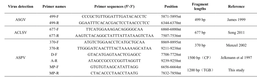
Fig. 1-6 The genome structure of Tobacco rattle virus (TRV) and derivative vectors 17 Fig. 2-1 1.0% agarose gel electrophoresis of RT-PCR products of fragments amplified by primers pair in Table 2-2 ... 28
Fig. 2-2 Phylogenetic tree of complete CP (A) and TGB (B) sequences of ASPV isolates ... 35
Fig. 2-3 Multiple alignment of amino acids of 16 representative ASPV CP sequences ... 37
Fig. 2-4 Recombinant events ASPV genes by using RDP software ... 38
Fig. 3-1 Different symptoms induced by Apple stem pitting virus on pear ... 45
Fig. 3-2 Full genome amplification of ASPV ... 47
Fig.3-3 Small RNAs sequencing strategy for plant ... 48
Fig.3-4 Phylogenetic tree of whole genome sequences of HB-HN1 and global isolates ... 53
Fig.3-5 Phylogenetic tree of whole genome sequences of HB-HN1 and global isolates ... 54
Fig.3-6 ASPV-vsiRNAs analysis obtained from pear plant HB-HN1 ... 55
Fig. 3-7 Symptoms induced by different ASPV isolates on N. occidentails ... 56
Fig. 3-8 SDS-PAGE and Western blot analyzed the fused CP from different ASPV isolates expressed in Escherichia Coli BL21 (DE3) ... 58
Fig. 3-9 VSR activity test of ASPV CP ... 59
Fig. 4-1 Schematic representation of the various stages of recovery experiment ... 65
Figure 22Fig. 4-2 Schematic representation of the various stages involved in VIGS with the TRV-PDS ... 66
Fig. 4-3 1.2% agarose gel electrophoresis for detecting PRC labeling DNA probes that detects TRV viral Northern blot process (Table 4-2) ... 67
Fig. 4-4 Schematic representation of the protocol allowing isolation of polysome-bound mRNAs from Arabidopsis samples ... 69
Fig. 4-5 TRV-GFP as a model for virus recovery in Arabidopsis ... 72
Fig. 4-6 Wild-type Arabidopsis were co-infected with TRV-GFP and TCV ... 72
Fig. 4-7 TRV-GFP susceptibility and recovery in Arabidopsis RNA silencing mutants ... 74
Fig. 4-8 AGO2 and AGO4 play anti-viral roles against TRV-GFP ... 77
Fig. 4-9 AGO1 is required for optimal VIGS by TRV-PDS ... 80
Fig. 4-10 VIGS phenotypes in ago1 and compound mutants ... 80
Fig. 4-11 Reduced TRV-GFP viral RNA in the ago1-27 mutant ... 81
Fig. 4-12 TRV-PDS VIGS in the triple DICER mutant is temperature-dependent .... 82
Fig. 4-13 Reduction in ribosome association of TRV RNAs after recovery ... 84
Fig. 4-14 TRV recovery induces an increase in PB formation ... 86
Fig. 4-15 VIGS and recovery in the Arabidopsis its1 mutant ... 87
List of Tables
Table1-1 Hierarchical classification system for endogenous plant small RNAs ... 4
Table 1-2 The 5' terminal nucleotide and size preferences of Arabidopsis AGOs ... 11
Table 1-3 Other proteins involved in Arabidopsis sRNA pathways ... 14
Table 2-1 Incidence of ASPV in different areas in China surveyed in this study ... 26
Table 2-2 Primers used in this study for detecting the often occurred virus on apple and pear ... 28
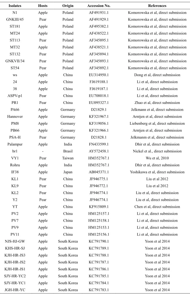
Table 2-3 Origin and GenBank accession numbers of ASPV isolates analyzed in our study ... 30
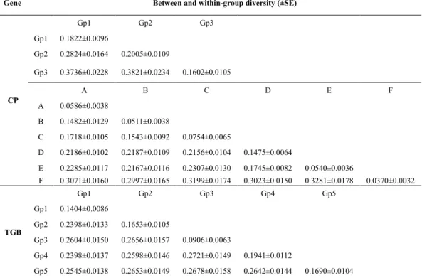
Table 2-4 Genetic distance between and within groups and subgroup clustered in phylogenetic trees based on ASPV CP and TGB sequences ... 35
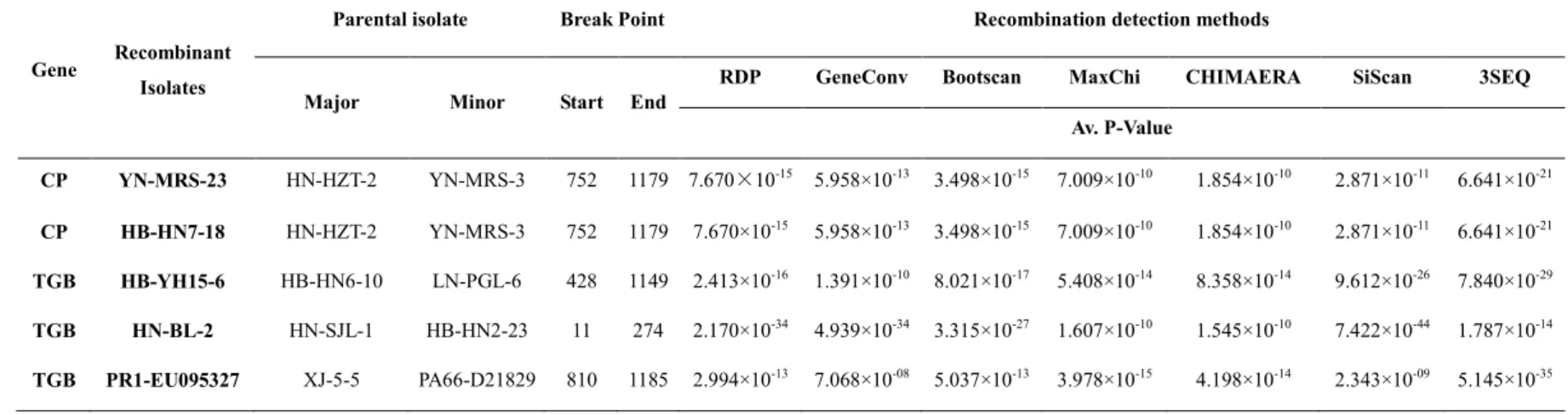
Table 2-5 List of putative recombination events among ASPV CP and TGB sequences .. 39
Table 2-6 Selection pressures (dN/dS) of different ASPV genes ... 40
Table 2-7 Population genetic parameters and neutrality tests calculated for ASPV CP and TGB based on geographic origins or variant groups ... 40
Table 3-1 The primers used for amplifying fragments of ASPV (HB-HN1) full genome 47 Table 3-2 List of primers with Restriction Enzyme cutting site used for constructing vectors that could express fused CP with a His tag in Escherichia Coli BL21 (DE3) ... 49
Table 3-3 List of primers with Restriction Enzyme cutting site used for constructing PVX vectors that could express ASPV coat protein in N. benthamiana ... 50
Table 3-4 The Host, origin, full length of ASPV genome and different genes on GneBank ... 52
Nucleotides and amino acids similarity among the six selected CP sequences ... 57
Table 3-6 Secondary structures prediction of the six selected CP sequences encoded proteins by using software SOPMA ... 57
Table 4-1 The Arabidopsis mutants and its origin used in this study ... 64
Table 4-2 Primers used for making Northern blot probes to detect TRV ... 67
Chapter 1 - Plant immune responses against viruses
There are two layers of plant immune responses against pathogens such as viruses, bacteria, fungi and oomycetes. First, certain conserved pathogen- or microbe-associated molecular patterns (P/MAMPs) recognized plant pattern recognition receptors (PRRs), which is called PAMP or MAMP-triggered immune (PTI). In many cases, PTI likely contributes to the so-called non-host resistance of plants. Second, to avoid plant PTI defenses, adapted microbes develop specific effector proteins to suppress PTI. To further defend the action of the microbial effectors, plants evolved specific surveillance systems involving resistance (R) proteins that directly or indirectly recognize the microbial effectors or monitor their activities in the cell to trigger the so-called effector-triggered immune (ETI) (Pieterse et al 2009) (Fig. 1-1).
Plants have developed several mechanisms to defend against viruses, such as Resistance (R) gene-mediated response, RNA silencing (Soosaar et al 2005), Lectin protein-mediated responses, and other host proteins such as Translational initiation factors, Endoplasmic Reticulum (ER) (Mandadi and Scholthof 2013). However, RNA silencing was thought to be the primary plant defense against viruses (Ding and Voinnet 2007). Recently, significant progress has been made in understanding RNA silencing and how viruses counter this ubiquitous antiviral defense.
Fig. 1-1 Simplified schematic representation of the plant immune system
A, upon pathogen attack, Pathogen-or Microbial-associated molecular patterns (PAMPs or MAMPs) activate pattern-recognition receptors (PRRs) in the host, resulting in a downstream signaling cascade
that leads to PAMP-triggered immunity (PTI); B, Virulent pathogens have acquired effectors (purple stars) that suppress PTI, resulting in effector-triggered susceptibility (ETS); C, In turn, plants have evolved resistance (R) proteins that recognize these attacker-specific effectors, resulting in a secondary
immune response called effector-triggered immunity (ETI) (Pieterse et al 2009)
1.1 RNA Silencing in plant
1.1.1 Definition of RNA Silencing
RNA silencing is a sequence-specific RNA degradation mechanism that occurs in a broad range of eukaryotic organisms including fungi (quelling), animals (RNA interference, RNAi), and plants (post-transcriptional gene silencing, PTGS). In plants, RNA silencing is a fundamental regulator of the expression of endogenous genes and exogenous molecular parasites such as viruses, transgenes, and transposable elements (Dunoyer et al 2013). And so how does RNA silencing function?
In the past ten years, by using genetic and molecular analysis, several RNA silencing pathways in plants have been revealed (Baulcombe 2004; Brodersen and Voinnet 2006). However, those pathways share some common biochemical features: 1) formation of double-stranded RNA (dsRNA); regardless of its origin, the appearance of dsRNA in the cytoplasm of plant cells triggers RNA silencing, 2) longer precursor molecules of either perfectly or imperfectly dsRNA is cut by an enzyme, Dicer, that has RNase III domains, into small 20–25 nt dsRNAs with 2 nucleotide overhangs at the 3’ ends, 3) these small dsRNA molecules are unwound and a selected sRNA strand binds to ‘slicing’ complexes called RISC (RNA-induced silencing complexes), which contain Argonaute (AGO) proteins to act on partially or fully complementary RNA or DNA (Fig. 1-2) (Brodersen and Voinnet 2006). Despite common features of RNA silencing in plants, there are differences between different RNA silencing pathways as reviewed by Brodersen and Voinnet in 2006 (Brodersen and Voinnet 2006). In next several paragraphs I will briefly talk about the different small RNAs produced by different RNA silencing pathways mechanism.
1.1.2 The non-cell-autonomous nature of RNA Silencing
Plant virus infections begin with virus entry into plant cells through a wound created either by mechanical ways or a vector organism, followed by viral replicase gene expression and replication in infected cells. Then the virus begins cell-to-cell movement through plasmodesmata and long distance movement through the vascular system. It was then found that the RNA silencing shares the same movement pathway with the virus (Voinnet 2005a).
Systemic RNA silencing was firstly discovered in transgenic tobacco exhibiting spontaneous co-suppression of nitrate reductase (Nia) of both the transgene and the host gene (Palauqui et al 1996). Subsequently, graft experiments demonstrated the Nia co-suppressed state was transmitted with 100% efficiency from silenced rootstocks to non-silenced scions expressing the corresponding transgene (Palauqui et al 1997). Soon after this discovery, leaf agro-infiltration Agrobacterium tumefaciens carrying a GFP reporter gene into GFP transgenic Nicotiana benthamiana was found to trigger a systemic, sequence-specific loss of GFP expression (Voinnet and Baulcombe 1997). Since then, the ‘non-cell-autonomous’ character of RNA silencing has been documented, which was one of the most important properties of RNA silencing found in plants and animals. Non-cell-autonomous, in other words, is its ability to move from the cell where it has been initiated to the neighboring cells (Mlotshwa et al 2002; Yoo et al 2004; Voinnet, 2005a; Kehr and Buhtz 2008). The degree of movement of silencing signals depends on the physiological conditions and surrounding environment of the tissue.
The next question was what triggered RNA silencing moving systemically? Although virtually any RNA that shares the required sequence homology with target RNAs can trigger systemic silencing in transgenic plants, the mobile and spreading of RNA silencing signals finally was demonstrated to be mediated by small noncoding RNA (sRNA, 21-25 nucleotides in length) (Klahre et al 2002; Hewezi et al 2005; Dunoyer et al 2010; Molnar et al 2010).The functions of small RNA movement include developmental patterning, viral resistance, epigenetic changes, etc (Dunoyer et al 2013).
1.1.3 Different types of small RNAs in plants
Plant endogenous small RNAs (sRNAs) involved in regulating gene expression at transcriptional or post transcriptional level, so as to regulate plants growth and devolepment. sRNAs sizes range from 20 to 24 nt, and include microRNAs (miRNAs), small interfering RNAs (siRNAs) and so on (Table 1-1) (Axtell 2013). These sRNAs can
be distinguished based on their origin, biogenesis pathways, molecular features and their fuctions in regulationg gene expression (Brodersen and Voinnet 2006). SiRNAs can function in plants in a non-cell-autonomous manner in that they are able to move from the cell where it has been initiated to neighboring cells (Voinnet, 2005a; Melnyk et al 2011). In contrast, most plant miRNAs are relatively immobile and cell autonomous (Parizotto et al 2004; Alvarez et al 2006), although some miRNAs have been recently reported to function in non-cell-autonomous way, for instance, miR165/166 is produced in specific root cells and moves to adjacent cells, movement of miRNAs over a set number of cells could generate gradients of gene expression in meristems and primordial (Himber et al 2003).
Table1-1 Hierarchical classification system for endogenous plant small RNAs
Primary classifications Secondary classifications Tertiary classifications
hpRNAs
small RNAs whose precursor is single-stranded hpRNA
MiRNAs
precisely processed precursor hairpins yielding just one or a few functional small
RNAs
Lineage-specific miRNAs
miRNAs that are found in only one species or a few closely related species
Long miRNAs
23–24-nt miRNAs that function similarly to heterochromatic siRNAs to deposit repressive
chromatin marks
Other hpRNAs
Imprecisely processed precursor hairpins that do not qualify as miRNAs
siRNAs
small RNAs whose precursor is dsRNA
Heterochromatic siRNAs
siRNAs produced chiefl from intergenic and/or repetitive regions; typically 23–24 nt
in length and associated with de novo deposition of repressive chromatin marks
Secondary siRNAs
siRNAs whose precursor dsRNA synthesis depends on an upstream small RNA trigger
and subsequent RDR activity
Phased siRNAs
secondary siRNA loci whose dsRNA precursor has a uniformly defied terminus, resulting in the
production of a phased set of siRNAs
trans-Acting siRNAs
secondary siRNAs that have one or more targets distinct from their locus of origin
Natural antisense transcript siRNAs (NAT-siRNAs)
siRNAs whose precursor dsRNA is formed by the hybridization of complementary and
independently transcribed RNAs
cis-NAT-siRNAs
NAT-siRNAs whose precursors were transcribed from overlapping genes in opposite polarities
trans-NAT-siRNAs
NAT-siRNAs whose precursors were transcribed from nonoverlapping genes whose mRNAs have
complementarity
1.2 RNA silencing-associated proteins in Arabidopsis thaliana
Dicer-like enzymes (DCLs), Argonaute (AGO) proteins, RNA-dependent RNA polymerase (RDR) proteins, and dsRNA binding proteins (DRBs) are core components of RNA silencing pathways involved in siRNA biogenesis. In in the following sections I will discuss in detail some of these proteins.
I.2.1 Dicer-like enzymes
The Dicer or Dicer-like (DCL) (a kind of ribonuclease III enzymes) proteins found in animals, fungal and plants are large proteins (~200 kDa), which consist of seven functional domains that interact with RNA in different ways (Fig. 1-3A). These seven domains are: DExD-helicase domain, helicase-C domain, Duf283 domain (unknown function but is strongly conserved among Dicers), PAZ domain, two RNaseIII domains and double stranded RNA-binding (dsRB) domain. Dicer proteins are encoded by most eukaryotes, but plants contain all the seven characteristic domains selectively (Margis et al 2006; Chapman and Carrington 2007).
Dicer-like or DCL proteins play an important role in the miRNA and siRNA biogenesis by processing double-stranded RNAs into sRNAs, which make them essential for eukaryote organism development and viral defense. The number of Dicer or DCL proteins encoded by different organisms is different, for instance, humans, mice and nematodes each possess only one Dicer gene, insects and fungi each possess two Dicer genes, in plants, both rice and Arabidopsis thaliana have been reported to have four Dicer-like genes (Tijsterman and Plasterk 2004; Margis et al 2006; Liu et al 2009). The four Dicer-like (DCL) RNaseIII proteins in Arabidopsis are named DCL1, DCL2, DCL3, DCL4, respectively. DCL1 locates in the chromosome 1 of Arabidopsis thaliana genome, DCL2 and DCL3 locate in the chromosome 3, whereas DCL4 located in chromosome 5 (Fig. 1-3B).
In Arabidopsis, DCLs are ubiquitously, but not evenly expressed in different tissues. Different expression patterns of DCL genes were also found at different developmental stages, DCL1, 2, 3, and 4 have relatively higher expression level in flowers, but lower in rosette leaf and stem (Liu et al 2009) (Fig. 1-3C). The four Dicer-like proteins are found to have roles in generating different siRNAs. DCL1 mainly processes miRNAs (Vazquez et al 2004) that guide cleavage of homologous cellular transcripts involved in development and probably many other functions, 21 nt secondary nat-siRNAs (Vazquez et al 2004) and also has a role in the production of small RNAs from endogenous inverted
repeats. DCL2 synthesizes 24 nt primer nat-siRNAs, viral siRNAs and in dcl4 mutant plants, it alternately processed 22 nt long siRNAs from ta-siRNA precursors (Bouche et al 2006; Deleris et al 2006). DCL3 produces 24 nt long, DNA repeat–associated siRNAs as guides for chromatin modification, RDR2-dependent siRNAs. DCL3 also produces RDR6-dependent trans-acting siRNAs. DCL4 processes dsRNA into 21nt-long siRNAs that mediate trans-acting RNA silencing (Vazquez et al 2004; Dunoyer et al 2005; Gasciolli et al 2005; Xie et al 2005; Adenot et al 2006), transgene RNA interference and some miRNAs biosynthesis.
There is functional redundancy of the four DCL in Arabidopsis thaliana. For instance, the functions of DCL1 and DCL3 overlap to promote Arabidopsis flowering. In dcl3 mutants DCL2 and DCL4 have access to DCL3 substrates and produce 22-nucleotide and 21-nucleotide siRNAs from RDR2-dependent precursors (Gasciolli et al 2005; Moissiard et al 2007; Yang et al 2007). Some results show that in Arabidopsis thaliana DCL proteins interact with the HYL1/DRB family of dsRNA-binding proteins, such as that DRB4 interacts specifically with DCL4, and HYL1 most strongly interacts with DCL1. These results indicate that each HYL1/DRB family protein interacts with one specific partner among the four Dicer-like proteins (Hiraguri et al 2005).
DCL4 is the primary antiviral Dicer against (+) ssRNA viruses and produces 21 nt long viRNAs (Deleris et al 2006). viRNA synthesis by DCL2 is hardly detectable when DCL4 is functional, but DCL2 can produces 22 nt long viRNAs if DCL4 is genetically inactivated or suppressed. For instance, the coat protein of Turnip crinkle virus (TCV) functions as a suppressor by indirectly inhibiting DCL4 expression, which leads to hyper-accumulation of DCL2 dependent 22 nt long siRNAs (Thomas et al 2003; Deleris et al 2006). DCL3 seems play a more important role in DNA virus defense, however, loss of the Cauliflower mosaic virus-derived 24 nt siRNA in dcl3 mutants is accompanied by an increased accumulation of virus-derived 21 nt long siRNAs (Moissiard and Voinnet 2006). The contribution of the DCL1 to immunity against some viruses is negligible because dcl2/dcl3/dcl4 and dcl1/dcl2/dcl3/dcl4 mutants showed similar susceptibility to
Cucumber mosaic virus (CMV) or TCV, and DCL1-dependent viRNAs were hardly
detectable even in the dcl2/dcl3/dcl4 mutant (Deleris et al 2006). These findings indicate that DCL proteins collectively contribute to the plant’s defense against different viruses, and that loss or suppression of activity of one DCL can be compensated for by the activities of other DCL proteins.
A
B C
Fig. 1-3 Information on Dicer-like Enzymes
A, The linear arrangement of domains typically found in DCL (Margis et al 2006); B, The chromosome locations of DCL genes in Arabidopsis. Each chromosome is depicted approximately to
scale, the number under each gene is the position on the pseudomolecule of the start of the gene (Margis et al 2006); C, Gene expression patterns of Arabidopsis DCLs in different tissues (Liu et al
2009) 1.2.2 Argonaute (AGO) proteins
The Argonaute (AGO) proteins are executor components of RISCs, bringing about degradation or translational inhibition of targeted RNAs specified by incorporated sRNAs. The word ‘Argonaute’ was firstly used by Karen Bohmert in 1998 to describe Arabidopsis thaliana ago1 mutant, in which morphology of the leaves closely resembled the tentacles of a small squid of Argonauta genus (Bohmert et al 1998; Hutvagner and Simard 2008). The first three founding members of AGO proteins family were: element-induced wimpy testis (PIWI) of Drosophila P (Lin and Spradling 1997), ARGONAUTE1 (AGO1) of Arabidopsis (Bohmert et al 1998) and ZWILLE (ZLL) of Arabidopsis (Moussian et al 1998). AGOs are large proteins (90-100 kDa) consisting of a variable N-terminal domain and conserved C-terminal PAZ, MID and PIWI domains (Fig. 1-4B, C), in which the PIWI domain makes AGO proteins different from DICER proteins that also have a PAZ domain. Argonaute proteins are highly specialized small RNA-binding modules and are considered to be the key components of RNA silencing pathways. The N-terminal domain is thought to facilitate the separation of the sRNA/target transcript duplex after cleavage. The MID domain binds to the 5' phosphate of sRNAs, whereas the PAZ domain recognizes the 3' end of sRNAs. The PIWI domain resembles that of bacterial RNaseH
enzymes and exhibits endonuclease activity (Vaucheret 2008).
All AGO proteins are divided into three groups on the basis of both their phylogenetic relationships and their capacity to binding sRNAs. Group 1 members bind miRNAs and siRNAs are referred to as AGO-like proteins. Group 2 members bind PIWI-interacting RNAs (piRNAs) are referred to as PIWI-like proteins. Group 3 members have been described only in Caenorhabditis elegans, where they bind secondary siRNAs (Yigit et al 2006). The number of AGO proteins encoded by different organisms varies greatly, Saccharomyces cerevisiae does not encode any AGO proteins and does not seem to encode other small RNA pathway factors, Schizosaccharomyces pombe expresses one AGO-like protein, Caenorhabditis elegans expresses 27 AGO proteins that fall into both AGO-like and PIWI-like subfamilies, Homo sapiens express 8 AGO proteins that fall into both AGO-like and PIWI-like subfamilies subfamilies, plant AGO proteins are all in the AGO-like subfamily, Oryza sativa eoncodes 18 AGO proteins while Arabidopsis thaliana encodes 10 AGO proteins. (Vaucheret 2008; Ender and Meister 2010; Mallory and Vaucheret 2010; Kim et al 2011). A phylogenetic analysis of the 10 Arabidopsis AGO proteins placed them in three major clades: the AGO1, AGO5, and AGO10 clade; the AGO2, AGO3, and AGO7 clade; and the AGO4, AGO6, AGO8, and AGO9 clade (Fig. 1-4A). It is important to note that the distribution of the 10 Arabidopsis AGO proteins into three distinct clades is based on amino acid sequence similarity, and does not necessarily directly infer similarities in activity or redundancies in function.
Several studies reveal that AGO1 is ubiquitously expressed at high levels throughout developmental stages and different tissues, but its expression appears to be highest in meristem and provascular cells. However, AGO10 is initially expressed throughout the embryo but becomes limited to provascular strands and the adaxial sides of the cotyledons at about the globular stage. Thus, AGO1 and AGO10 expression patterns overlap partially, with the AGO1 expression pattern being broader than that of AGO10. Moreover, fusion of the AGO10 coding sequence to the AGO1 promoter revealed that AGO10 could partially compensate for AGO1 activity, but ago10 mutants were not impaired in S-PTGS and show no reduction in the accumulation miRNAs, tasiRNAs or any other siRNA. By contrast, the expression profile for AGO5 is highly limited to reproductive tissues, accumulating in the sperm cell cytoplasm in mature pollen and growing pollen tubes (Schmid et al 2005; Vaucheret et al 2006; Vaucheret 2008; Mallory et al 2009; Mallory and Vaucheret 2010) (Fig. 1-4D).
Array data reveals that all three family members in the AGO2/3/7 clade have overlapping expression domains. AGO2 and 3 have high level of sequence similarity, proximal genomic positioning and same expression patterns (both AGO2 and 3 are most highly expressed in developing seeds and siliques, and at lower levels in senescing leaves and flowers). All of this strongly suggests that they have the same or similar RNA silencing roles in Arabidopsis, however, to date, no function has been reported for AGO3 nor has redundancy has been reported for these two proteins, suggesting that AGO3 may be a pseudogene. AGO7 is involved in tasi-RNA production, which regulates the expression of many developmentally important genes. AGO7 expression is very importance for normal leaf development, which is predominantly expressed in the vasculature of seedlings and in the cells and tissues immediately surrounding the SAM (Schmid et al 2005; Mallory and Vaucheret 2010) (Fig. 1-4D).
Results from fusing the AGO4, 6, 9 proteins to GUS reporter gene revealed that, AGO4 has widespread expression in embryos, leaves, and flowers. AGO4 is not only required for sRNA-directed DNA methylation, but also for the maintenance of heterochromatin (Irvine et al 2006). By contrast, AGO6 expression is restricted to shoot and root growth points and the vascular tissue connecting these domains, and AGO9 expression is restricted to the embryonic shoot apex region and developing ovules (Havecker 2010) (Fig. 1-5D). The amino-acid sequences of these AGO8 and AGO9 are very similar and the two genes are almost adjacent to one another on chromosome 5 of the Arabidopsis genome. The tissue-specific expression patterns of AGO8 and AGO9 mRNAs are also highly similar (Schmid et al 2005). However, the AGO8 transcript is expressed at a much lower level than AGO9 and it has been proposed that AGO8 is a pseudogene (Takahashi et al 2008).
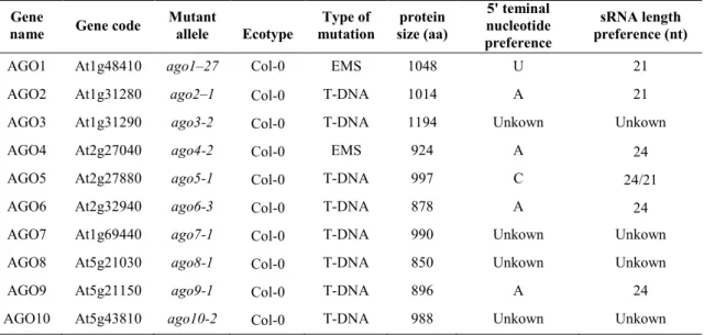
AGO proteins are often named ‘slicer proteins’ because they cleave target ssRNAs at the duplex formed with the guide-strand small RNA. AGO proteins directly bind sRNAs, and different AGOs bind different sRNAs of specific length and preferred 5' nucleotide. For example, AGO1 binds sRNAs that are predominantly of the 21 nt size class with a 5' terminal uracil, whereas AGO2 preferentially binds 21 nt size class with a 5' terminal adenine. Usually AGO5 preferentially binds sRNAs of the 24 nt size class with a cytosine at their 5' terminal, however, AGO5 is also able to bind miR169, which is a 21 nt long miRNA with a uracil as the 5' terminal nucleotide (Irvine et al 2006; Mi et al 2008). AGOs 4, 6 and 9 also preferentially bind sRNAs of the 24 nt size class with 5' adenine
residues. The 5' terminal nucleotide preference for AGOs 3, 7, 8 and 10 remain to be determined (Mi et al 2008) (Table1-2). Because of the sRNA binding ability, most of the Arabidopsis AGO proteins have been observed to bind miRNA involved in plant development, for instance, AGO5/miR169 (Takeda et al 2008), AGO10/miR165 and miR166 (Liu et al 2009; Zhu et al 2011), AGO7/ miR390 (Montgomery et al 2008).
Among the ten Arabidopsis AGO proteins, AGO1 is the best studied. By using ago1 mutant alleles in genetic screens for reactivation of post transcriptionally silenced sense transgenes (S-PTGS), AGO1 was implicated in miRNA biogenesis. Indeed, in ago1 mutant alleles, miRNA accumulation is reduced and, at the same time miRNA target mRNA accumulation is increased, further indicating that AGO1 is necessary in the miRNA pathway (Vaucheret et al 2006). The majority of miRNAs have a 5' terminal uracil residue and are preferentially loaded by AGO1, and AGO1 has been shown to direct sRNA-mediated gene expression regulation for all currently characterized Arabidopsis miRNAs. Because AGO1 has important role of in the regulation of other genes in plants through RNA silencing, there are a lot of studies associated with how AGO1 is regulated in planta. In 2006, Hervé Vaucheret and colleagues reported that AGO1 homeostasis is maintained through the repressive action of miR168 on AGO1 mRNA and the stabilizing effect of AGO1 protein on miR168 (Vaucheret et al 2004; Vaucheret et al 2006). In 2009, the same group found that AGO1 homeostasis not only regulated by the action of the microRNA but also siRNA pathways, in this report they showed that AGO1-derived siRNAs trigger AGO1 silencing in an RDR6-, SDE5- and SGS3-dependent manner, and that production of AGO1-derived siRNAs requires the action of DCL2 and DCL4, similar to viruses and inverted repeat transgenes (Mallory and Vaucheret 2009). Recent studies have described the increased expression of miR168 and AGO1 mRNA in virus-infected plants, miR168-driven control of AGO1 can persist for a long time in virus-infected plants and can be an important component of symptom development (Varallyay et al 2010; Varallyay and Havelda 2013).
Several studies have demonstrated a role for AGO1 in RNA silencing based antiviral defense, including the following observations: (1) ago1 hypomorphic mutants are more susceptible to CMV and TCV (Morel et al 2002; Qu et al 2008; Wang et al 2011); (2) several VSRs are able to interact directly with AGO1 (Zhang et al 2006; Baumberger et al 2007; Chiu et al 2010; Csorba et al 2010; Feng et al 2013; Varallyay and Havelda 2013); (3) AGO1 binds virus-specific siRNAs (Zhang et al 2006). In addition to AGO1, other
AGO proteins such as AGO2 (Rand et al 2005; Harvey et al 2011; Jaubert et al 2011; Scholthof et al 2011; Wang et al 2011) and AGO7 was observed in the antiviral defense (Qu et al 2008), while ago4 plants are observed hyper-susceptibile to the bacterial pathogen Pseudomonas syringae (Agorio and Vera 2007). AGO2 has also been shown to act downstream of the viral secondary siRNA biogenesis together with AGO1 in a non-redundant manner, essential for defense against CMV infection (Wang et al 2011). Knock-down of Ago2 in N. benthamiana by VIGS allows high accumulation of a version of Tomato bushy stunt virus (TBSV) lacking a VSR. At the same time, of all ten AGOs, only mutations in AGO2 allow high level accumulation of PVX in Arabidopsis, a host that it does not normally infect. These results suggest that different AGO family members are engaged by basal and induced anti-viral responses, respectively.
Table 1-2 The 5' terminal nucleotide and size preferences of Arabidopsis AGOs (Kim et al 2011, Vaucheret 2008)
Gene
name Gene code
Mutant allele Ecotype Type of mutation protein size (aa) 5' teminal nucleotide preference sRNA length preference (nt)
AGO1 At1g48410 ago1–27 Col-0 EMS 1048 U 21
AGO2 At1g31280 ago2–1 Col-0 T-DNA 1014 A 21
AGO3 At1g31290 ago3-2 Col-0 T-DNA 1194 Unkown Unkown
AGO4 At2g27040 ago4-2 Col-0 EMS 924 A 24
AGO5 At2g27880 ago5-1 Col-0 T-DNA 997 C 24/21
AGO6 At2g32940 ago6-3 Col-0 T-DNA 878 A 24
AGO7 At1g69440 ago7-1 Col-0 T-DNA 990 Unkown Unkown
AGO8 At5g21030 ago8-1 Col-0 T-DNA 850 Unkown Unkown
AGO9 At5g21150 ago9-1 Col-0 T-DNA 896 A 24
Fig. 1-4 Information on Argonaute proteins
A, Phylogenetic classification of Arabidopsis AGO proteins into three clades (Vaucheret 2008); B, Domain structure of an Argonaute protein (EnderMeister 2010); C, Crystal structure of the Argonaute
protein from Thermos thermophile.s (EnderMeister 2010); D, Expression Intensities of the 10
Arabidopsis AGO genes at various developmental stages and in different tissues (MalloryVaucheret 2010)
1.2.3 RDR proteins and other proteins involved in RNA silencing
The activity of plant RNA-dependent RNA polymerases (RdRPs) was first reported in Chinese cabbage in 1971 (Astier-Manifacier et al 1971). Since then, RdRPs have been found in a number of plants including Arabidopsis (Dalmay et al 2000; Mourrain et al 2000). Arabidopsis plants contain at least six active RdRP genes, termed RDR1-RDR6 (RDR6 also known as SDE1/SGS2) (Schwach et al 2005) and they function both differently and redundantly (Yu et al 2003). A likely role of RDRs in RNA silencing is to produce dsRNA that is cleaved by DCL proteins in plants. SDE1, an RNA-dependent RNA polymerase gene in Arabidopsis is required for PTGS mediated by a transgene not by a virus. The role of SDE1 is to produce a dsRNA activator of PTGS (Dalmay et al 2000). Arabidopsis SGS2 encode a protein that is similar to an RdRPs and SGS3 encodes a novel plant-specific protein. The isolation of sgs2 and sgs3 Arabidopsis mutants impaired in PTGS, SGS2/SDE1 and SGS3 were also reported to be required for juvenile
2000; Peragine et al 2004). Thus RDR6 is thought to recognize, and to use as templates, certain transgene transcripts with aberrant features that include a lack of 5’ capping. This favours their conversion into dsRNA by RDR6 and the subsequent degradation of all transgene transcripts through the S-PTGS pathway (Brodersen and Voinnet 2006). RDR2 is required for the production of siRNAs from endogenous transcripts, but is not required for virus resistance. RDR1 is induced upon viral infection and limits virus replication, but has no apparent role in the silencing of endogenous transcripts (Xie et al 2004). RdRPs were first implicated in regulating virus accumulation in plants through the analysis of the sgs2 and sgs3 mutant Arabidopsis lines. The sgs2 and sgs3 mutants show enhanced susceptibility to CMV but not to TuMV or TVCV Infection (Mourrain et al 2000). Plants with compromised RDR6 function are hyper-susceptible to several viruses (Brodersen and Voinnet 2006). The ortholog of RDR1 has been shown to contribute to defense against Tobacco mosaic virus (TMV) and PVX in N. tabacum (Mandadi and Scholthof 2013) and the lack of a functional ortholog of this enzyme is at least partly responsible for the hyper-susceptibility of N. benthamiana plants to TMV. SGS2/SDE1 and SGS3 were reported to be required for virus induced gene silencing (VIGS) of endogenous genes using a geminivirus CaLCuV vector.
Except for the proteins described above, a number of other proteins in Arabidopsis have also been shown to play roles in RNA silencing pathways (Table1-3).
1.3The antiviral role of RNA silencing
Plants have developed diverse mechanisms to defend virus infections. One mechanism is through RNA silencing. Two lines of evidence indicate that RNA silencing has a role for antiviral defense in plants (Baulcombe 2004). Firstly, virus-derived siRNAs (viRNAs) accumulate during virus infections. Secondly, Plant viruses have elaborated a variety of counter-defensive measures to overcome the host silencing response. One of these strategies is to produce proteins called viral suppressors of RNA silencing (VSRs) that target different steps of RNA silencing such as DCL, RISC, or small RNA. Thirdly, Plants lacking of RNA silencing components are more susceptible to some virus infection (Voinnet et al 1999).
Table 1-3 Other proteins involved in Arabidopsis sRNA pathways (BrodersenVoinnet 2006)
Proteins Domains and motifs Biochemical activity Pathway HYL1 dsRNA binding domnian dsRNA binding miRNA pathway
HST RanGTP binding Putative exportin miRNA pathway
HEN1
sRNA binding domnian Lupus La RNA binding S-adenosyl binding
sRNA Methyltranseferase All sRNA pathways
WEX 3'–5' exonuclease Putative 3'–5' exonuclease S-PTGS SDE3 DEAD Helicase Putative RNA helicase S-PTGS /Transitivity NRPD1a RNA Polymerase Referred DNA dependent
RNA Polymerase Chromosome/nat-siRNA NRPD1b RNA Polymerase Referred DNA dependent
RNA Polymerase Chromosome NRPD2 RNA Polymerase Referred DNA dependent
RNA Polymerase Chromosome
HDA6 Deacetylase Putative histone
deacetylase Chromosome DRD1 SNF2-related DNA and
ATP binding Helicase
Putative chromatin
remodeling Chromosome CMT3 Cytosine DNA Methyltranseferase
Chromodomain Brom adjacent domain
Cytosine DNA
Methyltranseferase Chromosome DRM1/2 Cytosine DNA Methyltranseferase Cytosine DNA
Methyltranseferase Chromosome MET1 Cytosine DNA Methyltranseferase
Bromo-adjacent domain Cytosine DNA Methyltranseferase Chromosome KYP SET domain ZnII-binding domain Pre-SET domain Post-SET domain YDG domain EF-hand H3 K9 Methyltranseferase Chromosome SUVH SET domain ZnII-binding pre-SET domain
YDG domain
1.4 Virus-encoded VSRs
RNA silencing suppressors were first identified because of the study of the potexvirus and potyvirus synergistic interaction, which led to the identification of the potyviral Helper HC-Pro of potyvirus as the synergism determinant in this interaction (Pruss et al 1997). Subsequently HC-Pro and CMV-2b were identified as the first silencing suppressor proteins (Anandalakshmi et al 1998; Brigneti et al 1998; Kasschau and Carrington 1998). A subsequent survey of more than 15 viruses further confirmed that suppression of RNA silencing is a general property for plant viruses to counter-plant defense (Voinnet et al 1999). Interestingly, all the suppressors identified from plant viruses have a high diversity in sequence and protein structure, suggesting that they function by diverse mechanisms. Different suppressors suppress different steps of the RNA silencing pathway and a number of recent reviews have discussed plant RNA silencing suppressors and their different behavior in plant (Dalmay et al 2000; Voinnet 2001; MacDiarmid 2005; Qu and Morris 2005; Voinnet 2005a; Scholthof 2006; Ding and Voinnet 2007; Song et al 2011; Incarbone and Dunoyer 2013) (Fig. 1-5).
Fig. 1-5 Current model of antiviral RNA silencing in plants and its VSRs
Viral-silencing suppressors can disrupt RNA silencing pathways at multiple points, thereby preventing the assembly of different effectors or inhibiting their actions. The points at which certain VSRs (i.e. P14, P38, V2, 2b, P19, HC-Pro, P21, P0 and P1) interact with the silencing pathways are depicted (Burgyán
1.5 Tobacco rattle virus TRV
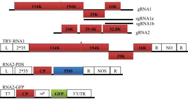
TRV, which forms rod-shaped particles (MacFarlane 1999) and is transmitted by nematodes (Taylor and Brown 1997), has a bipartite, positive-sense single-stranded RNA (+ssRNA) genome and is a type member of the genus Tobravirus (family Virgaviridae). TRV genome consists of two RNA molecules, RNA1 and RNA2, in which RNA1 is conserved in size and gene content while RNA2 displays a high sequence variation (MacFarlane 1999). RNA1 encodes four proteins, 134 and 194 kDa replicase proteins (including the methyltransferase, helicase, and RNA-dependent RNA polymerase domains), a 29 kDa movement protein and a 16 kDa cysteine-rich protein (CRP), respectively. The replicase proteins are translated directly from the genomic RNA while the 29 K and 16 K are expressed from respective subgenomic RNAs (sgRNA) and both of them function as a suppressor of RNA silencing (Ghazala et al 2008; Martinez-Priego et al., 2008 Deng et al 2013). Depending on the strains, TRV RNA2 can vary in size and gene content and serial passage of TRV under different selection conditions results in deletion of structural and nonstructural genes in RNA2 (Hernandez et al 1996). However, it always encodes a coat protein (CP), which encapsidates RNA1 and RNA2 into different rod-shaped particles (MacFarlane 1999). RNA2 also encodes the proteins 2b and 2c, which are involved in transmission of TRV by root nematodes (Hernandez et al 1997; MacFarlane 1999), and 2b also involved in invasion of meristems in roots (Valentine et al 2004). All proteins encoded by RNA2 are translated from sgRNAs (MacFarlane 1999). The host range of TRV is very wide, natural infection has been reported in more than 100 species, when inoculation with sap, about 400 species in more than 50 dicotyledonous and monocotyledonous families can be infected. The disease symptoms induced by TRV are affected by environmental conditions, which include necrotic or chlorotic spots, rings, vein necrosis, etc.
Fig. 1-6 The genome structure of Tobacco rattle virus (TRV) and derivative vectors
1) Wild type TRV; 2) In TRV-RNA1 vector, TRV cDNA clones were placed in between the duplicated CaMV 35S promoter (2×35S) and the nopaline synthase terminator (NOSt) in a T-DNA vector. Rz
refers to self-cleaving ribozyme, LB and RB refer to left and right borders of T-DNA; 3) In TRV-RNA2-PDS, whereas the two genes involved in nematode transmission of TRV (2b and 2c) have
been removed and replaced with a multiple cloning site (Mcs), PDS was inserted into the Mcs (Liu et al 2002); 4) In RNA2-GFP, the 2b and 2c genes encode nonstructural proteins involved in nematode
transmission and are deleted from the vector constructs, GFP was inserted into the Mcs, and the CP subgenomic RNA promoter (sgP) derived from Pea early-browning virus has been added upstream of
GFP (MacFarlane and Popovich 2000)
1.6 Recovery
Recovery was first described by Wingard S. A. in 1928 and is typified by systemic virus infection with associated symptoms followed by decrease and disappearance of symptoms in young leaves (MacDiarmid 2005). Recovery occurs within some natural virus infections (e.g, nepoviruses, AlMV, TRV, CaMV and geminiviruses) (Covey et al 1997; Ratcliff et al 1997; Ratcliff et al 1999; Szittya et al 2002) and transgenic plants engineered for viral resistance (Smith et al 1994; Carlsbecker et al 2010). The recovered plants are immune to the same or closely related strains of the initially infecting virus and this resistance shown by the recovered tissue is sequence specific (Covey et al 1997; Ratcliff et al 1997; Ratcliff et al 1999).
For a long time recovery of plants from natural virus infection or transgenic plants following the primary virus infection is described as a consequence of RNA silencing
* 134K 194K 29K 16K gRNA1 sgRNA1a sgRNA1b 29.4K 32.8K 24K gRNA2 134K 194K 29K 16K * 2*35 S L B R B R Z NO St TRV-RNA1 GFP CP T7 P sgP 3’UTR RNA2-GFP RNA2-PDS PDS CP 2*35 S L B R B NOS t R Z
(Ratcliff et al 1997; Baulcombe 2004), and there are two lines of evidence for this. One is that the recovered plants are observed to associate with activation of RNA silencing and increased viral siRNA (Szittya et al 2002; Jovel et al 2007). Another is that loss of expression of viral silencing suppressors in mutant viruses results in a recovery-like phenotype instead of nonrecovery (Ding et al 1995; Szittya et al 2002). However, several lines of evidence suggest that activation of virus RNA silencing in infected plants is insufficient to ensure recovery: 1) TRV and Potato virus X (PVX) induced RNA silencing provides a sequence-specific cross-protection in Nicotiana benthamiana plants, however, only the TRV-infected plants recover (Ratcliff et al 1999); 2) many viruses encode VRSs to counter defense the host RNA silencing antiviral defense including the recover and nonrecovery viruses; 3) for some viruses (e.g., Tomato ringspot virus, Tobacco streak virus) viral clearance by RNA silencing may not be essential for the initiation of host recovery (Xin and Ding 2003; Jovel et al 2007). Thus, these data collectively argue that host recovery is not an inevitable consequence of activation of the RNA silencing in infected plants and that other factors may play a key role in the initiation of recovery.
There is an association between recovery and meristem exclusion in that most natural recovery viruses can invade the apical growing point while the nonrecovery viruses cannot (Ratcliff et al 1997; Schwach et al 2005). Meristem exclusion is thought to be a variation of the recovery process; classical meristem exclusion would be recovery that is restricted to the growing point of the infected plant, whereas recovery would be meristem exclusion that operated not only in the meristem but also in the uppermost leaves of the plant (Schwach et al 2005). There is also evidence that meristem exclusion is involved in RNA silencing. Firstly, based on the study of the virus-derived gene transgenic plants challenged with the original virus (Smith et al 1994; Carlsbecker et al 2010), scientists found that different transgenic lines exhibited differently, recovery or the normal response to virus infection, which could be influenced by the different expression level of the virus-derived gene in different transgene lines. Frank Schwach et.al (2006) attributed this different exhibition to the amount of silencing signal produced by the virus and the nature of the viral silencing suppressor protein(s) (Schwach et al 2005). Secondly, the nonrecovery virus PVX acquires the ability to invade meristems if it is inoculated on plants expressing viral suppressors of silencing (Foster et al 2002). Thirdly, PVX acquires the ability to invade meristems in N. benthamiana line in which NbRDR6 was down regulated, and the RDR6 is involved RNA silencing (Schwach et al 2005).
Little is known about how host recovery is initiated in virus infected plants, which occurs only in certain host-virus interaction systems. The entire mechanism of recovery is not understood but may simply be an example of completely successful RNA silencing antiviral defense or maybe the ability of viruses to infect the meristem of the plant may also trigger an additional host response that results in recovery.
1.7 Virus-induced gene silencing VIGS
The term Virus-induced gene silencing (VIGS) was first used to describe the phenomenon of recovery from virus infection (Van Kammen et al 1997), so VIGS may in fact be a kind of recovery. Nowadays, VIGS is often used as an RNA silencing based technique used for down regulating a host gene through the use of a recombinant virus. The dsRNA replication intermediates of the viruses is processed into small interfering siRNA in the infected cell that correspond to different parts of the viral vector genome, including the insertion fragments. The host gene derived-siRNAs can mediate degradation of related endogenous gene transcripts, resulting in silencing of target gene expression. This virus-based technique can also be used to explore the plant defense system against viruses. VIGS also was developed as a reverse genetics approach for gene function study. There are several advantages of VIGS when compared to the traditionally approaches, such as chemical or physical mutagenesis, tilling, T-DNA insertion, and transposon tagging. Firstly, VIGS is labour and time saving. It can identify a specific gene function within a single plant generation in a month. Also, VIGS avoids plant transformation, which is labour and time consuming and sometimes, unpredictable. Second, VIGS overcomes gene functional redundancy. By using an inserted sequence derived from the most highly conserved region of a gene family, it is possible to silence all or most of members of a given family. In contrast, specific member of a gene family can be targeted by selecting unique sequence of this member. Third, VIGS allows rapid comparisons of gene function between species because some VIGS vector can infect different plant species (Godge et al 2008). Despite its advantages, there are some limitations for VIGS. Firstly, you need to know the gene sequence of the targets, it is hard to work with plant species that lack sequenced genomes. Second, VIGS does not always result in the complete loss of expression of a target gene and it is hard to say with certainty that the reduced expression of target the gene is not enough to produce at least some functional protein. Third, some VIGS vectors produce disease symptoms and sometimes it can be hard to tell the silenced gene phenotypes from the disease symptoms. Finally, the levels
of silencing can vary between plants and experiments, which introduces an element of variability (Godge et al 2008).
So far, several viruses have been developed as VIGS vectors in plants (Godge et al 2008; Igarashi et al 2009), including TMV (Kumagai et al 1995), PVX (Ruiz et al 1998), Tomato golden mosaic virus (TGMV) (Kjemtrup et al 1998), TRV (Ratcliff et al 2001), and Apple latent spherical virus (ALSV) (Igarashi et al 2009). These VIGS vectors have successfully silenced endogenous genes like phytoene desaturase (PDS) in N.benthamiana plants. Except for being a VIGS vectors, many of these VIGS vector-viruses could also be used as expression vectors to highly express a gene by inserting the gene to the viral genome, but the recombinant viruses often express foreign gene in 2-4 days after inoculation while VIGS a host gene often costs one month. For TRV VIGS and GFP vector, the 2b and 2c genes are removed from RNA2, and the GFP gene and the PDS gene was introduced to the RNA2 (Fig. 1-6) (MacFarlane and Popovich 2000; Ratcliff et al 2001; Liu et al 2002). TRV-derived vectors can be used for VIGS in Solanum species including tomato, potato, N. benthamiana, as well as Arabidopsis thaliana. ALSV is a new VIGS vector and does not induce any obvious symptoms in most host plants, and can effectively VIGS endogenous genes among a broad range of plant including different Nicotiana species, Solanum lycopersicum, Arabidopsis thaliana, different cucurbit species, and different legume species (Igarashi et al 2009).
To be a good VIGS vector, first, the recombinant virus should have a broad range of hosts. Second, they should not cause obvious disease symptoms on inoculated plants, which make interpretation of some genes VIGS phenotypes difficult. Third, these viruses should not be excluded from the growing points or meristems of their hosts, if not, which will preclude effective silencing of genes in those tissues. Finally, they should not suppress RNA silencing too efficiently, because that would negatively affect gene silencing (Godge et al 2008; Igarashi et al 2009). The limitations of host range and meristem exclusion were overcome by the TRV and ALSV based VIGS vectors (Ratcliff et al 2001; Igarashi et al 2009). TRV and ALSV is able to spread more vigorously throughout the entire plant, including meristem tissue, yet the overall symptoms of infection are mild compared with other viruses, and also they both have a broad range of hosts.
1.8 Foveavirus
Foveavirus, Allexivirus, Potexvirus, Carlavirus, Trichovirus, Vitivirus, Capillovirus, Mandarivirus, Citrivirus, and some genuses of undefined viruses such as Banana mild mosaic virus (BMMV), Cherry green ring mottle virus (CGRMV), Cherry necrotic rusty mottle virus (CNRMV), Potato virus T (PVT) and Sugarcane striate mosaic-associated virus (SSMaV) were classified into the family of Flexiviridae in 2007 (Martelli et al 2007). However, in 2009 the family of Flexiviridae were further divided into Alphaflexiviridae (α-flexiviridae), Betaflexiviridae (β-flexiviridae) and Gammaflexiviridae (г-flexiviridae) accorrding to the documents of International Committee on Taxonomy of Viruses (ICTV), these three family and Tymoviridae were classified into Tymovirales (Carstens 2009). Foveavirus was belong to the family of Betaflexiviridae, which included Apple stem pitting virus (ASPV) (Jelkmann et al 1994), Apricot latent virus (ApLV) (Nemchinov et al 2000), Grapevine rupestris stem pitting-associated virus (GRSPaV) (Martelli and Jelkmann 1998), Asian prunus virus 1 (APV-1) (ICTV, 2013), Rubus canadensis virus 1 (RuCV-1) (ICTV, 2014).
Viruses in Foveavirus genuspositive single-stranded RNA with polyA tail, and virus particles of Foveavirus filamentous, 800~1000 nm long and 12~15 nm wide. Usually their genome sizes ranged from 8.4 to 9.3 kb, which contained five open reading frame (ORF1- ORF5) and 5' and 3' untranslated region (UTR) and with CP sizes from 28 to 44 KDa. ORF1 encoded replicase, ORF2-ORF4 encoded Triple gene block (TGB1-TGB3) movement proteins, and ORF5 encoded coat proteins (CP).
TGB (TGB1-TGB3) genes of Foveavirus encoded TGBp1 (24~26 KDa), TGBp2 (12~24 KDa), TGBp3 (7~11 KDa), respectively (Solovyev et al 2000), those of whom involved in transporting viral RNAs to the adjacent cells. TGBp1 contained a conservative ATP-GTP binding domain (Wong et al 1998). Both TGBp2 and TGBp3 consisted a hydrophobic functional domains, wherein TGBp2 encoded by all TGB containing viruses has a conserbative core area with two hydrophobic domains aside, however, domains composition of TGBp3 by different viruses were relatively variable, for example, TGBp3 encoded by Foveavirus only has one hydrophobic domain at N . TGBp1 functioned as binding single strand RNA (ssRNA) (Morozov and Solovyev 2003), suppressing RNA silencing (Verchot-Lubicz 2005) and increasing the plasmodesmata permeability to help virus particles transport, whereas TGBp2 and TGBp3 functioned as membrane-bound proteins (Krishnamurthy et al 2003).