O
pen
A
rchive
T
OULOUSE
A
rchive
O
uverte (
OATAO
)
OATAO is an open access repository that collects the work of Toulouse researchers and
makes it freely available over the web where possible.
This is an author-deposited version published in :
http://oatao.univ-toulouse.fr/
Eprints ID : 16546
To cite this version : Macha, Innocent J. and Charvillat, Cédric and
Cazalbou, Sophie and Grossin, David and Boonyang, Upsorn and
Ben-Nissan, Besim Comparative study of coral conversion, Part 3:
Intermediate products in the first half an hour. (2016) Journal of The
Australian Ceramic Society, vol. 52 (n° 1). pp. 177-182. ISSN
0004-881X
Any correspondence concerning this service should be sent to the repository
administrator:
[email protected]
Comparative study of coral conversion, Part 3:
Intermediate products in the first half an hour
Innocent J. Macha
1, Cedric Charvillat
2, Sophie Cazalbou
2, David Grossin
2, Upsorn Boonyang
3and Besim Ben-Nissan
11) Department of Chemistry Materials and Forensic Science, University of Technology Sydney, Broadway NSW 2007
2) CIRIMAT Carnot Institute, University of Toulouse, UPS-INPT-CNRS UMR 5085, France
3) School of Science, Walailak University, 222 Thaiburi, Thasala District, Nakhonsithammarat, 80160 Thailand. Email: [email protected]
Abstract
Different methods to produce calcium phosphate materials have been well established and are currently used by both scientific and industrial community. While other new and more economical production techniques are under development, the actual reactions mechanisms involve in these techniques are not well understood. Understanding what really happen during reaction will pave a way to tune the final product for well-defined morphology and purity. We focused into improving in-depth understanding of the reaction mechanisms and the intermediates products participating in the reaction of coralline materials with orthophosphoric and ammonium phosphate solutions under mechano-chemical reaction technique. The results suggest that within 30 minutes of reaction under ammonium phosphate solution only HAp phase is produced through solid-state iron exchange reaction. On the other hand, under orthophosphoric acid solution, intermediate phases such as octacalcium phosphate (OCP) and monetite form and convert to hydroxyapatite HAp at different times. Other phase that formed as an intermediate was identified as brushite. It was also observed that pH plays a big role in the formation of these phases due to their different pH stability. The results also confirm our previous hypothesis that under orthophosphoric acid phosphate solution the reaction mechanism is dissolution-recrystallization while under ammonium phosphate solution is solid-state topotactic ion exchange reaction mechanism. It is envisaged that there are possibilities of the formation of intermediate products within or before the first 5 minutes of reaction.
Keywords: Calcium phosphate, mechano-chemical, dissolution, precipitation, solid state, ion exchange,
topotactic
1. INTRODUCTION
Calcium phosphate materials are of interest in biomedical field due to their biocompatibility and bioactivity. Among these, hydroxyapatite (Ca10(PO4)6(OH)2, HAp) is the most used materials
and has attracted lots of attentions for further studies on its productions and characterizations [1-3]. Much effort has been focused into synthesis from less expensive raw materials using low cost methods. Different materials both synthetic and natural calcium sources have been used to synthesize HAp. Different phosphate sources have been investigated under different reaction conditions in order to improve in-depth understanding of the reaction mechanism, which will help to control and refine the final products. Several biological calcium sources such as eggshells, sea urchins, coral, nacre, mussel and land
snails [4-6] have been used to synthesize calcium phosphate materials.
We have recently reported the possibility of synthesizing HAp from coralline materials using phosphoric acid or ammonium hydro-phosphate solutions under mechano-chemical method [7]. Two conversion routes were observed, first, solid state topotactic ion exchange reaction mechanism using ammonium phosphate solution which mainly produce HAp and the second, dissolution-recrystallization using orthophosphoric acid phosphate solution which resulted into DCPA, HAp and whitlockite phases. It was noted that the formation and evolution of calcium phosphate at a fast rate and within 30 minutes after mixing calcium and phosphate sources full development of HAp was observed. It is believed that the reaction
happens in series or chain that evolve different intermediate products that could possibly be controlled to refine the final products. There are number of factors influence morphological and crystal growth and chemical changes which include pH, particle size and shape, solution addition rate, stirring speed, reaction temperature etc and we have kept some of these factors constant in our experimental work.
In this research, the effort was focused on the understanding of the reaction mechanism of coralline materials to phosphoric and ammonium phosphates solution in the first half an hour of the reaction. The influence of pH on the microstructural evolution of calcium phosphate materials was also studied.
2. MATERIALS AND METHODS
Coral skeleton samples were obtained from the Great Barrier Reef shoreline (QLD Australia), and from artificially grown corals from Prof. R. Vago of the Ben-Gurion University of Negev (Israel). Di-ammonium hydrogen phosphate ((NH4)2HPO4,
98%), ortho phosphoric acid (H3PO4) and sodium
hypochlorite (NaClO) were obtained from Sigma Aldrich (Castle Hill, Australia). The methods of coral pre-preparation were covered in our previous publications [7, 8].
The methods aimed to convert corals to HAp by using H3PO4 and (NH4)2HPO4 under
mechano-chemical method describe in [9, 10] and were coded HA-P and HA-A, respectively. Samples from HA-P and HA-A were taken and immediately quenched in dry ice after every 5 minutes after the first drop of phosphate solutions mixed with coral-distilled water mixture at 80 oCfor half an hour. These samples were coded HA-P1, HA-P2, HA-P3, HA-P4, HA-P5, HA-P6, HA-A1, HA-A2, HA-A3, HA-A4, HA-A5, HA-A6 respectively. Samples before mixing were taken and coded HA-P0 and HA-A0 respectively.
Phase analysis of the products were carried out by
X-ray powder diffraction using D8 DISCOVER (BRUKER AXS), radiation Cu Kalpha1+2 = 0.15418 nm, no monochromator, from 3 to 60° (2 theta), steps of 0.021°, 0.7 seconds/step, divergence slit 0.5°, Sollers slit 2.5°, detector linear: LynxEye (2.955°). The phase quantification and crystal size were evaluated in the 20-60 range (2 theta) using Rietveld analysis performed by Topaz software and crystal size was determined by Scherer with Topas software (BRUKER AXS).
The SEM analysis was performed using a Zeiss Supra 55VP SEM system (Carl Zeiss AG, Oberkochen, Germany) equipped with RAITH E-beam Lithography System & EBSD for the secondary electron imaging (SEI) the energy was kept at 15 kV, working distance and magnification were varied to obtain best possible pictures. The powder samples were fixed by mutual conductive adhesive tape on aluminium stubs and coated with gold using a sputter coater (Leica EM ACE600 Sputtering and Carbon Thread Coater, Leica, German)
3. RESULTS AND DISCUSSION
XRD studies were carried out to identify the phases and assess the phase purity and crystallinity of the HAp crystal and other calcium phosphate materials. The results suggest that HAp formation in the first half an hour of the reaction between coralline materials and ortho phosphoric acid (H3PO4)
preceded with the formation of intermediates such as octacalcium phosphate, and monetite with small amount of brushite that formed at only one time (samples HA-P2). On the other hand the reaction that involves ammonium phosphate solution ((NH4)2HPO4)there were no intermediate products
but direct conversion of coralline materials to HAp (samples HA-A(1 – 6)). In both experiments as shown in Table 1 and 2, it was observed that the reactivity of aragonite doesn’t seem to depend on pH of reaction media as it displays faster reaction than calcite while calcite seemed to react slower in basic conditions.
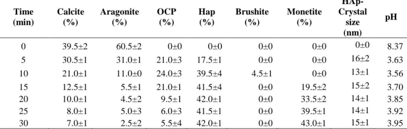
Table 1: Quantification for orthophosphoric solution (HA-P) experiment showing the amount of precipitated phases and growth of HAp phase
Time (min) Calcite (%) Aragonite (%) OCP (%) Hap (%) Brushite (%) Monetite (%) HAp-Crystal size (nm) pH 0 39.5±2 60.5±2 0±0 0±0 0±0 0±0 0±0 8.37 5 30.5±1 31.0±1 21.0±3 17.5±1 0±0 0±0 16±2 3.63 10 21.0±1 11.0±0 24.0±3 39.5±4 4.5±1 0±0 13±1 3.56 15 12.5±1 5.5±1 21.0±1 41.5±4 0±0 19.5±2 15±2 3.70 20 10.0±1 4.5±2 9.5±1 42.0±1 0±0 33.5±2 14±1 3.85 25 8.0±1 5.0±3 6.0±3 41.5±1 0±0 39.5±1 14±1 3.92 30 7.0±1 2.5±2 5.5±4 42.0±1 0±0 43.0±1 15±1 3.95
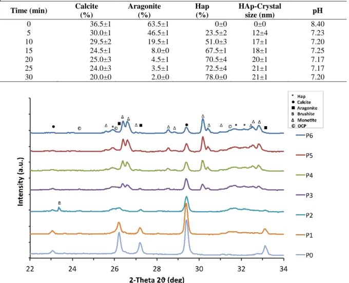
Table 2: Quantification for HA-A experiment showing the amount of precipitated phases and growth of HAp phase
Time (min) Calcite
(%) Aragonite (%) Hap (%) HAp-Crystal size (nm) pH 0 36.5±1 63.5±1 0±0 0±0 8.40 5 30.0±1 46.5±1 23.5±2 12±4 7.23 10 29.5±2 19.5±1 51.0±3 17±1 7.20 15 24.5±1 8.0±0 67.5±1 18±1 7.25 20 25.0±3 4.5±1 70.5±4 20±1 7.17 25 24.0±3 3.5±1 72.5±4 21±1 7.17 30 20.0±0 2.0±0 78.0±0 21±1 7.20
Figure 1: XRD pattern of the sample products from the reaction of coral with ortho-phosphoric acid (HA-P) for the first 30 minutes showing phase evolution of calcium phosphate materials.
Figure 2: XRD pattern of the sample products from the reaction of coral with ammonium phosphate solution (HA-A) for the first 30 minutes showing phase evolution of calcium phosphate materials.
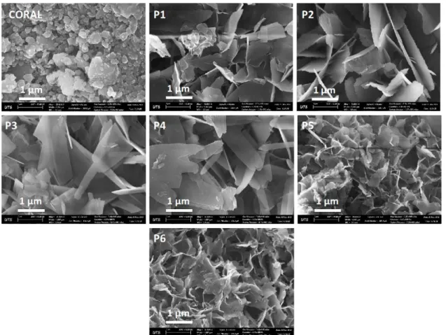
Figure 3: SEM pictures showing morphology and microstructural evolution of calcium phosphate under ortho-phosphoric acid solution, HA-P.
Figure 3: SEM pictures showing morphology and microstructural evolution of calcium phosphate under ammonium phosphate solution HA-A.
Figure 1 and 2 show the XRD patterns of the product samples from HA-P and HA-A respectively. The evolution of calcium phosphates materials whereas HA-P shows three mainly different phases (Monetite, Octacalcium phosphate and Hydroxyapatite) and small amount of brushite in the product samples at different time of experiment during the first 30 minutes.
We recently reported in our previous publication [7] that different phase formation under acidic conditions is mainly influenced by different pH stability of these phases. HA-A experiment resulted into a single product mainly HAp.
Figure 3 and 4 show the morphology of product samples from HA-P and HA-A experiment respectively. Under HA-P, the results indicate changes of morphology from the first 5 minutes from irregular morphology of coral particles to well-defined platelets morphology of calcium phosphate materials. Furthermore, the results indicate growth of these platelets up to 20 minutes
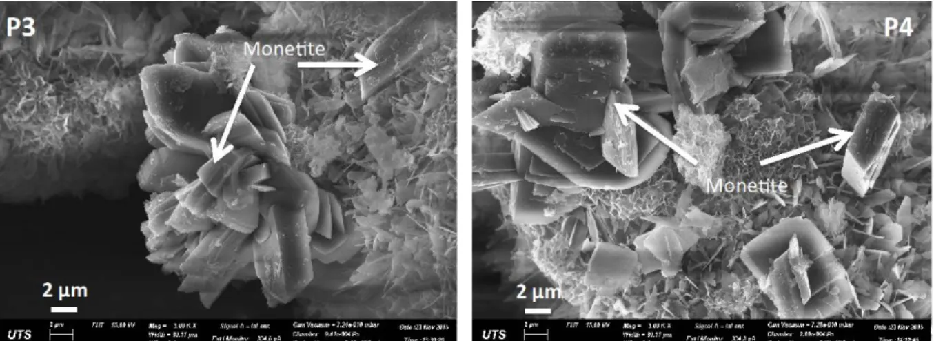
of reaction. It was postulated in our previous research [10] that there is a possibility of OCP conversion into HAp without changing of morphology. Evolution of monetite took place after 15 minutes with rectangular block morphology as shown in Figure 5 which convert to HAp with different morphology of mesh-like thin platelets. On the other hand HA-A experimental sample products show similar platelets morphology for the first 30 minutes with an increase in their size and number from time 0 to 30 minutes. The first sample HA-A1 shows the mixture of coralline materials and HAp and the ration of HAp: Coral keeps on increasing over time. There are no significant changes of platelets morphology except from coral to HAp which in consistent with the phase quantification results Table 2.
At the end of 30 minutes reactions results from both experiments suggest that with HA-P two phases are observed in the product samples with 42 % HAp and 43% Monetite while with HA-A only single phase of HAp was observed with around 71 %HAp.
Figure 4: SEM pictures showing morphology of monetite
4. CONCLUSION
This study focused at evaluating evolution of calcium phosphate materials in comparison to phosphoric and ammonium phosphate solutions. Particularly, the interest was to study these evolutions in the first 30 minutes of reaction in order to enhance more understanding on what are intermediate phases in the formation of HAp. It was clear that octacalcium phosphate formed within 5 minutes of reaction while brushite forms within 5-10 minutes and being consumed in the formation of HAp and monetite. Even though monetite formed within 10-15 minutes, the competition between HAp and Monetite depend entirely on the pH conditions of the reaction medium as we reported earlier [7]. We also reported that as pH increases monetite becomes unstable and converts to HAp, which becomes more stable. The results in this study confirm the reaction mechanisms suggested in our earlier publications [7, 8]. First, under
orthophosphoric acid phosphate solution the reaction mechanism is dissolution-recrystallization resulted into mainly OCP, monetite, HAp and small amount of brushite while under ammonium phosphate solution is solid-state topotactic ion exchange reaction mechanism mainly produced HAp. It can also be concluded that production of single and may be more pure HAp ammonium phosphate solution could be the better choice. The production of 21% OCP and 17% HAp after 5 minutes under orthophosphoric acid solutions leaves us with a question whether there are other intermediate products formed before or within the first 5 minutes of reaction.
5. REFERENCES
[1] B. Ben-Nissan, A.H. Choi, D.W. Green,
B.A. Latella, J. Chou, A. Bendavid,
hydroxyapatite nanocoatings by sol-gel method for clinical applications, in: S. Zhang (Ed.) Biological and Biomedical Coatings Handbook: Processing and Characterization, CRC Press, Boca Raton, FL U.S.A, 2011, pp. 37-79.
[2] P. Hui, S.L. Meena, S. Gurbhinder, R.D.
Agarawal, P. Satya, Synthesis of
Hydroxyapatite Bio-Ceramic Powder by Hydrothermal Method, Journal of Minerals & Materials Characterization & Engineering, Vol. [9], (2010), 683-692.
[3] A. Engin, İ. Girgin, Synthesis of hydroxyapatite by using calcium carbonate and phosphoric acid in various water-ethanol solvent systems, Central European Journal of Chemistry, Vol. [7], (2009), 745-751.
[4] I.J. Macha, L.S. Ozyegin, J. Chou, R.
Samur, F.N. Oktar, B. Ben-Nissan, An
Alternative Synthesis Method for Di Calcium Phosphate (Monetite) Powders from Mediterranean Mussel (Mytilus galloprovincialis) Shells, Journal of The Australian Ceramic Society, Vol. [49], (2013), 122-128.
[5] F.N. Oktar, Microstructure and mechanical properties of sintered enamel hydroxyapatite, Ceramics International, Vol. [33], (2007), 1309-1314.
[6] F. Oktar, U. Tuyel, N. Demirkol, O.
Gunduz, R. Samur, S. Kannan, S.
Agathopoulos, A new safe method to produce
bioceramic nano-powders from nacre venus verrucosa, Artificial Organs Conference, FYROM, 2010, International Journal of Artificial Organs, (Special Issue), (2010), pp. 467-467.
[7] I.J. Macha, U. Boonyang, S. Cazalbou, B.
Ben-Nissan, C. Charvillat, F.N. Oktar, D.
Grossin, Comparative study of Coral
Conversion, Part 2: Microstructural evolution of calcium phosphate, Journal of the Australian Ceramic Society Vol. [51], (2015), 149-159.
[8] R.-N.R. Cegla, I.J. Macha, B. Ben-Nissan,
D. Grossin, G. Heness, R.-J. Chung,
Comparative Study of Conversion of Coral with Ammonium Dihydrogen Phosphate and Orthophosphoric Acid to Produce Calcium Phosphates, Journal of the Australian Ceramics Society, Vol. [50], (2014), 154-161. [9] E. Avvakumov, L. Karakchiev,
Mechanochemical Synthesis as a Method for the Preparation of Nanodisperse Particles of Oxide Materials, Chemistry of sustainable development, Vol. [12], (2004), 287-291. [10] I.J. Macha, L.S. Ozyegin, F.N. Oktar, B.
Ben-Nissan, Conversion of Ostrich Eggshells
(Struthio camelus) to Calcium Phosphates, Journal of The Australian Ceramic Society, Vol. [51], (2015), 9.