Pulmonary lipid homeostasis in cigarette
smoke-associated lung diseases
Thèse
Eric Jubinville
Doctorat en médecine expérimentale
Philosophiæ doctor (Ph. D.)
Pulmonary lipid homeostasis in cigarette
smoke-associated lung diseases
Thèse
Éric Jubinville
Sous la direction de :
Mathieu, Morissette, directeur de recherche
Benoît, Arsenault, codirecteur de recherche
Résumé
Introduction. Les effets du tabagisme demeurent problématiques dans notre société. Les
mécanismes initiateurs de la réponse immunitaire pulmonaire induits par la fumée de cigarette sont peu caractérisés. Un des phénomènes dominants en contexte tabagique est l’augmentation de la taille des macrophages alvéolaires. Ce changement phénotypique se distingue par une accumulation intracellulaire de lipides suggérant que le transport lipidique pulmonaire des macrophages alvéolaires est modifié en contexte tabagique. Le transport lipidique pulmonaire est composé de plusieurs étapes, dont la capture, le remaniement et l’export de lipides par les macrophages alvéolaires. Les impacts du tabagisme sur le transport lipidique pulmonaire sont actuellement inconnus.
Hypothèse. Le tabagisme altère le transport lipidique pulmonaire. Objectifs : Chapitre 1) Investiguer
l’impact de l’exposition à la fumée de cigarette sur le transport lipidique pulmonaire dans un modèle murin et chez l’humain ainsi qu’évaluer l’impact d’une thérapie d’augmentation des high-density
lipoproteins (HDLs) dans un modèle murin. Chapitre 2) Investiguer les effets d’une thérapie avec un
agoniste du récepteur nucléaire liver X receptor (LXR) activant l’export lipidique dans un modèle murin.
Chapitre 3) Caractériser les répercussions d’une carence alimentaire sur la santé pulmonaire et sur la
réponse pulmonaire en contexte tabagique dans un modèle murin.
Méthodes. 1. Le transcriptome pulmonaire de souris exposées à la fumée de cigarette et de sujets
non-fumeurs, fumeurs et ex-fumeurs a été étudié. La capacité d’efflux de cholestérol a été mesurée dans le sérum et dans le lavage bronchoalvéolaire (LBA) de souris fumeuses et non fumeuses. Une HDL recombinante, le MDCO-216, a été administrée à des souris fumeuses et non-fumeuses et leurs réponses immunitaires, leurs fonctions pulmonaires et leurs compositions corporelles ont été analysées.
2. Un agoniste du LXR, le T0901317, a été administré à des souris en contexte tabagique. Le
transcriptome pulmonaire relié au transport lipidique, la réponse immunitaire pulmonaire et du macrophage alvéolaire ainsi que les impacts sur le surfactant pulmonaire ont été investigués.
3. Des souris fumeuses et non fumeuses ont été nourries avec des diètes déficientes en méthionine
(MD), choline (CD) et méthionine et choline (MCD) et leurs fonctions pulmonaires, leurs réponses immunitaires pulmonaires et leurs expressions géniques pulmonaires ont été caractérisées.
Résultats. Chapitre 1. L’expression des gènes impliqués dans le transport lipidique pulmonaire murin
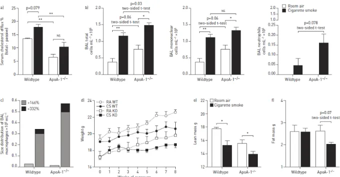
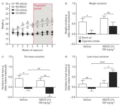
et humain est modifiée en contexte tabagique. La capacité du sérum et du LBA à promouvoir la sortie de cholestérol est augmentée après une seule exposition à la fumée de cigarette. L’administration du MDCO-216 réduit la réponse inflammatoire pulmonaire et la taille des macrophages alvéolaires dans un modèle d’exposition aiguë à la fumée de cigarette. Le MDCO-216 semble protéger les fonctions pulmonaires et induit une augmentation de la quantité de masse maigre chez les souris fumeuses.
Chapitre 2. L’agoniste du LXR augmente l’expression des gènes de transport lipidique pulmonaire,
cependant il exacerbe la réponse immunitaire pulmonaire en contexte tabagique. Les macrophages alvéolaires ont aussi un phénotype inflammatoire exacerbé et ont davantage de stress au réticulum endoplasmique lorsqu’ils sont traités en contexte tabagique. L’activation de LXR mène à une réduction des niveaux de surfactant pulmonaire.
Chapitre 3. La diète MCD altère les fonctions pulmonaires en induisant un profil restrictif pulmonaire
et abolit la réponse immunitaire pulmonaire à la fumée de cigarette. En histologie, ces souris nourries avec la diète MCD n’ont toutefois aucun foyer fibrotique pulmonaire. L’expression génique de plusieurs gènes associés à la matrice extracellulaire et les niveaux de surfactant pulmonaire sont réduits chez les souris nourries avec la diète MCD. Les phénotypes pulmonaires de la diète MCD sont toutefois réversibles après un retour d’une semaine sur la diète contrôle. La diète CD induit un profil pulmonaire de type emphysémateux et la diète MD mène à un profil restrictif.
Conclusions. Ces travaux démontrent que le transport lipidique pulmonaire a un rôle majeur en
contexte tabagique et qu’il est modulé rapidement. La thérapie d’augmentation des HDLs, avec le MDCO-216, propose une nouvelle voie de traitement envisageable pour les ex-fumeurs. La thérapie ciblant LXR suggère qu’il pourrait y avoir des effets délétères chez les sujets fumeurs actifs. Les carences alimentaires en méthionine et en choline démontrent d’importants changements sur la
physiologie pulmonaire. Ce tout nouveau domaine de recherche, le nutri-respiratoire, requiert davantage d’études afin de mieux comprendre l’impact d’une mauvaise nutrition sur la santé pulmonaire.
Abstract
Introduction. Cigarette smoking remains a major problem in our society. While a lot of cigarette smoke
impacts are actually known, few data are available on initiating mechanisms involved in the pulmonary immune response to cigarette smoke. One of the most intriguing phenomena under cigarette smoke exposure conditions is the presence of enlarged alveolar macrophages. This phenotypic change is characterized by an intracellular lipid accumulation which may be a sign of inadequate lipid export by alveolar macrophages induced by cigarette smoking. Pulmonary lipid transport begins with lipid capture, lipid reorganization and lipid droplet formation followed by lipid export by alveolar macrophages. Cigarette smoke impacts on these steps are actually unknown.
Hypothesis. Cigarette smoking alters pulmonary lipid transport. Objectives: Chapter 1) To investigate
the effect of cigarette smoke exposure on pulmonary lipid transport in cigarette smoke-exposed mice and in healthy controls, smokers and former smokers. To investigate the impact of high-density lipoprotein (HDLs) therapeutic potential in cigarette smoke-exposed mice. Chapter 2) To investigate, in mice, the therapeutic potential of an agonist activating the nuclear receptor liver X receptor (LXR) involved in the transcription of lipid export genes. Chapter 3) To explore, in mice, if a dietary deficiency alters the pulmonary health and the pulmonary response to cigarette smoke.
Methods. 1. The pulmonary transcriptome of cigarette smoke-exposed mice and healthy controls,
smokers and former smokers was assessed. Cholesterol efflux capacity of serum and bronchoalveolar lavage (BAL) was measured in unexposed and cigarette smoke-exposed mice. MDCO-216, a recombinant HDL, was administered to unexposed and cigarette smoke-exposed mice and analyzed their pulmonary immune response, lung functions and body composition.
2. T0901317, an LXR agonist, was systemically given to mice under cigarette smoke exposure
conditions. Pulmonary genes associated with lipid transport, lungs and alveolar macrophage immune pulmonary response to cigarette smoke and the impact of T0901317 on the pulmonary surfactant were assessed.
3. Unexposed and cigarette smoke-exposed mice were fed with methionine deficient (MD), choline
deficient (CD) or methionine and choline deficient (MCD) diet. Diets impact on lung functions, pulmonary immune response to cigarette smoke and pulmonary transcriptome were characterized.
Results. Chapter 1. Cigarette smoking altered the expression of pulmonary lipid transport genes in
mice and in humans. Serum and BALF cholesterol efflux capacities were increased following a two-hour cigarette smoke exposure. MDCO-216 dampened the pulmonary inflammatory response and reduced the size of alveolar macrophages in our acute cigarette smoke exposure model. MDCO-216 also seemed to be beneficial to lung functions and induced an increase in lean mass in cigarette smoke-exposed treated mice.
Chapter 2. T0901317 treatments led to an increase in the expression of pulmonary lipid transport
genes. However, it also induced an exacerbated pulmonary immune response during cigarette smoking. Cigarette smoke-exposed treated-alveolar macrophages displayed an exacerbated inflammatory phenotype and showed an augmented endoplasmic reticulum stress. Furthermore, LXR activation led to pulmonary surfactant depletion under cigarette smoke exposure conditions.
Chapter 3. The MCD diet altered lung function displaying a restrictive profile and almost abolished the
pulmonary immune response to cigarette smoke. Lung histology showed no signs of fibrosis, a phenotype usually associated with restrictive pulmonary functions. MCD diet led to a dramatic change in the pulmonary expression of extracellular matrix genes and also reduced pulmonary surfactant levels. Nevertheless, these pulmonary phenotypes were reversible within a week when mice were re-fed a control diet. Interestingly, the CD diet induced an emphysema-like profile, while MD diet showed similar pulmonary functions to the MCD diet.
Conclusions. The present thesis adds major data to an underestimated field of research and
demonstrates the importance of pulmonary lipid transport, especially during cigarette smoking. Recombinant HDL therapy with MDCO-216 may be a new opportunity to overcome adverse effects of cigarette smoking, while activating LXR seems rather deleterious. Nutrient deficiencies, such as methionine and choline led to unprecedented impacts on the pulmonary health and on the pulmonary response to cigarette smoke. This completely new field of research, “nutri-respiratory”, requires additional studies to fully decipher the impact of unhealthy nutrition on the respiratory system.
Table of contents
Résumé ... ii
Abstract ... v
Table of contents ... vii
List of Figures... xii
List of abbreviations and symbols... xiv
Dedication ... xvii
Acknowledgements... xix
Foreword ... xxii
Introduction... 1
Cigarette smoking and socioeconomic outcomes ... 1
Cigarette smoking and general impacts on environment and health ... 2
Cigarette smoking and associated lung diseases ... 4
Cigarette smoking and lung cancer ... 4
Cigarette smoking and interstitial lung diseases ... 7
Cigarette smoking and chronic obstructive lung disease ... 9
Inflammation and pulmonary macrophage responses to cigarette smoke ... 14
What is inflammation? ... 15
The pulmonary response to cigarette smoke ... 16
Pulmonary macrophages ... 21
Pulmonary macrophage responses to cigarette smoke ... 22
Foamy pulmonary macrophages following cigarette smoke exposure ... 23
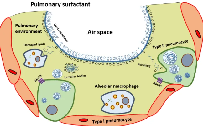
Pulmonary surfactant ... 24
Surfactant proteins ... 25
Surfactant synthesis and turnover ... 26
Cigarette smoke and pulmonary surfactant ... 29
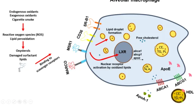
Lipid uptake to lipid export by macrophages ... 30
Macrophages and scavenger receptors ... 30
Macrophage and lipid droplet formation ... 40
Nuclear receptors and their implication in lipid transport ... 41
Lipid export by macrophages ... 46
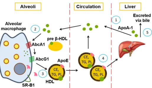
Systemic lipid transport ... 52
High-density lipoproteins ... 53
HDL composition and structure ... 53
Apolipoprotein A-1 ... 54
Apolipoprotein E ... 55
HDL biosynthesis ... 58
Impact of cigarette smoke on HDLs ... 60
Therapeutic approaches to increasing lipid transport ... 61
First ApoA-1 therapies and recombinant HDLs ... 61
ApoA-1 mimetic peptides ... 64
ApoE mimetic peptides... 65
HDL delipidation or phospholipidation ... 65
Pharmacological LXR agonists ... 66
Nutrition and respiratory health ... 67
Lung health and benefits from nutrition? ... 68
Choline ... 70
Methionine choline deficient diet ... 70
Methionine... 71
Rationale, hypothesis, and objectives ... 73
Chapter 1: Interplay between cigarette smoking and pulmonary reverse lipid transport ... 77
1.1 Foreword ... 78 1.1.1 Author contributions ... 78 1.2 Résumé ... 79 1.3 Abstract ... 80 1.4 Introduction ... 82 1.5 Methods ... 84
1.5.1 Human lung gene expression cohort and analyses ... 84
1.5.2 Mice, cigarette smoke exposure and treatments ... 84
1.5.3 Lung harvesting and processing ... 84
1.5.4 Human ApoA-1 ELISA ... 85
1.5.5 Quantitative PCR (qPCR) ... 85
1.5.6 Lung function assessment ... 86
1.5.7 Body composition analyses ... 86
1.5.8 In vitro cholesterol efflux assay ... 86
1.5.9 Statistical analysis ... 87
1.6 Results ... 88
1.6.1 Impact of cigarette exposure on key genes involved in the regulation of reverse lipid transport ... 88
1.6.2 Impact of cigarette smoke exposure on pulmonary and circulating lipid export capacity ... 88
1.6.3 ApoA1 deficiency exacerbates pulmonary and systemic responses to cigarette smoke exposure .. 89
1.6.4 Impact of MDCO-216 therapy on the pulmonary response to cigarette smoke ... 89
1.6.5 Impact of MDCO-216 therapy on lung functional alterations caused by cigarette smoke ... 90
1.6.6 Impact of MDCO-216 therapy on body composition alterations caused by cigarette smoke ... 90
1.7 Discussion ... 92
1.8 Acknowledgements ... 95
1.9 References ... 96
1.11 Figures ...100
1.12 Supplementary data...103
1.12.1 Supplementary Methods ...103
1.12.2 Supplementary Figures Legends ...103
1.12.3 Supplementary Figures ...104
Chapter 2: Pharmacological activation of Liver X Receptor during cigarette smoke exposure adversely affects alveolar macrophages and pulmonary surfactant homeostasis...106
2.1 Foreword ...107 2.1.1 Author contributions ...107 2.2 Résumé ...108 2.3 Abstract ...109 2.4 Introduction ...111 2.5 Methods ...113 2.5.1 Animals ...113
2.5.2 Cigarette smoke exposure and T0901317 administration ...113
2.5.3 Bronchoalveolar lavage and lung processing...113
2.5.4 Bronchoalveolar lavage and alveolar macrophages culture ...114
2.5.5 Alveolar macrophage size ...114
2.5.6 Serum sampling and processing ...114
2.5.7 Mouse IL-1α, CCL2 and G-CSF ELISA ...114
2.5.8 Lungs and alveolar macrophages RNA extractions and cDNA synthesis ...115
2.5.9 Quantitative PCR (qPCR) gene expression of lung tissue and alveolar macrophages ...115
2.5.10 Measurement of phosphatidylcholine in the bronchoalveolar lavage fluid and serum ...116
2.5.11 Bronchoalveolar lavage fluid SP-B Western blot analysis ...116
2.5.12 In vitro and ex vivo cholesterol efflux assay ...116
2.5.13 Statistical analysis ...117
2.6 Results ...118
2.6.1 Pharmacological activation of LXR modulates the expression of key lipid transport genes in alveolar macrophages and lung tissue ...118
2.6.2 Pharmacological activation of LXR promotes expression of lipid transport genes during acute cigarette smoke exposure ...118
2.6.3 LXR activation exacerbates the pulmonary immune response to acute cigarette smoke exposure ...119
2.6.4 LXR activation affects alveolar macrophages during cigarette smoke exposure ...119
2.6.5 LXR activation induces pulmonary surfactant depletion during cigarette smoke exposure ...120
2.7 Discussion ...121
2.8 Table ...124
2.10 Figure legends ...128
Chapter 3: Critical importance of dietary methionine and choline in maintenance of lung homeostasis during
normal and cigarette smoke exposure conditions ...134
3.1 Foreword ...135
3.1.1 Author contributions ...135
3.2 Résumé ...136
3.3 Abstract ...137
Critical importance of dietary methionine and choline in maintenance of lung homeostasis during normal and cigarette smoke exposure conditions ...138
3.4 Introduction ...139
3.5 Methods ...141
3.5.1 Animals, cigarette smoke exposure and dietary protocols ...141
3.5.2 Blood collection and processing ...141
3.5.3 Lung function assessment ...141
3.5.4 Bronchoalveolar lavage and lung tissue collection ...142
3.5.5 RNA extraction, quantitative PCR and gene expression microarray analysis ...142
3.5.6 BAL fluid and serum biomarkers ...143
3.5.7 Statistical analysis ...144
3.6 Results ...145
3.6.1 Dietary deficiency in choline and methionine reversibly affects lung function ...145
3.6.2 Dietary choline and methionine deficiency strongly reduces pulmonary expression of extracellular matrix-related genes ...145
3.6.3 Dietary choline and methionine deficiency markedly affects cigarette smoke-induced changes in lung function and inflammation ...146
3.6.4 Dietary choline and methionine deficiency predisposes mice to small airway atelectasis due to reduced pulmonary surfactant levels and structural alterations independent of pulmonary surfactant ...147
3.6.5 Choline or methionine dietary deficiencies have different effects on lung function and lung inflammation ...147
3.7 Discussion ...149
3.7.1 The pulmonary response to an ‘essential nutrient starvation state’. ...149
3.7.2 Interaction between cigarette smoke exposure and dietary methionine/choline deficiency. ...150
3.7.3 Implications for respiratory health in non-smokers and smokers. ...151
3.8 References ...152
3.9 Figure legends ...154
3.10 Figures ...156
General discussion, limitations and perspectives ...161
Are alveolar macrophages or is the pulmonary environment responsible for disrupted lipid transport? ...162
The pulmonary environment ...162
The alveolar macrophage ...163
What is the best option to promote pulmonary lipid transport in the context of cigarette smoking? ...166
Can HDLs be used to prevent lung damage induced by cigarette smoke? ...166
Can LXR activation be beneficial during cigarette smoking?...168
Promoting RCT via dietary modification and other pharmacological treatments ...169
Limitations ...171
Perspectives ...171
The underestimated role of pulmonary surfactant ...173
Liver X receptor and type 2 pneumocytes ...173
Pulmonary surfactant and nutrition ...174
Limitations ...175
Perspectives ...175
Conclusion...177
List of Figures
Introduction
Figure I: Proposed classification of many smoking-related interstitial lung diseases by Vassallo and
Ryu. ... 7
Figure II: ABCD assessment tool for COPD patients. ... 11
Figure III: Cigarette smoking is a major risk factor for numerous systemic and pulmonary diseases.14 Figure IV: Cigarette smoke-induced lung alterations... 21
Figure V: The pulmonary surfactant. ... 28
Figure VI: From lipid capture to lipid export by alveolar macrophages... 51
Figure VII: Schematic representation of reverse cholesterol transport (RCT). ... 59
Chapter 1
Figure 1. 1: Cigarette smoking affects pulmonary expression levels of key genes involved in reverse lipid transport in both humans and mice. ... 100Figure 1. 2: Impact of cigarette smoke exposure on pulmonary and systemic reverse lipid export capacity. ... 100
Figure 1. 3: Deficiency in ApoA1 exacerbates the response to cigarette smoke. ... 101
Figure 1. 4: Impacts of prophylactic and therapeutic MDCO-216 treatment on the pulmonary response to cigarette smoke. ... 101
Figure 1. 5: Therapeutic MDCO-216 treatment reduces aspects of cigarette smoke-induced lung function alterations. ... 102
Figure 1. 6: Administration of MDCO-216 improves aspects of cigarette smoke-induced changes in body composition. ... 102
Figure 1. 7 Supplementary 1: Impact of ApoA-1 deficiency and MDCO-216 administration on the pulmonary expression levels of key genes involved in reverse lipid transport in response to cigarette smoke exposure. ... 104
Figure 1. 8 Supplementary 2: Presence of antibodies against MDCO-216 in mice chronically injected with compound. ... 105
Chapter 2
Figure 2. 1: T0901317 treatment impacts key lipid transport genes in lung tissue and alveolar macrophages without causing pulmonary inflammation. ... 131Figure 2. 2: T0901317 treatment restores the expression of key pulmonary lipid transport genes altered by cigarette smoke exposures. ... 131
Figure 2. 3: Pharmacological activation of LXR during acute cigarette smoke exposure exacerbates the neutrophilic inflammatory response. ... 132
Figure 2. 4: Alveolar macrophages from T0901317-treated mice exhibit an exacerbated
inflammatory phenotypes as well as signs of endoplasmic reticulum stress... 132
Figure 2. 5: T0901317 treatment causes pulmonary surfactant depletion. ... 133 Figure 2. 6: Schematic representation of the distinctive impact of pharmacological LXR
activation under normal homeostatic conditions and during exposure to cigarette smoke. .. 133
Chapter 3
Figure 3. 1: Dietary methionine and choline deficiency progressively and reversibly affects lung
functions... 156
Figure 3. 2: Dietary methionine and choline deficiency reduces pulmonary expression of
extracellular matrix genes. ... 157
Figure 3. 3: Dietary methionine and choline deficiency progressively alters cigarette
smoke-induced lung function changes and inflammation. ... 158
Figure 3. 4: Dietary methionine and choline deficiency reduces pulmonary surfactant levels as
well as lung tissue mechanical properties. ... 159
Figure 3. 5: Dietary deficiencies in choline and methionine each affect differently the lungs. 160 Figure 3. 6: Dietary deficiencies in choline and methionine each affect differently the pulmonary
List of abbreviations and symbols
4-HNE: 4-hydroxynonenal AAT1: alpha-1 antitrypsin
ACAT1: acyl-CoA:cholesterol acyltransferase 1 APCs: antigen-presenting cells
Aβ: amyloid beta
ABCA1: ATP-Binding Cassette subfamily A member 1 ABCA3: ATP-Binding Cassette subfamily A member 3 ABCG1: ATP-Binding Cassette subfamily G member 1 Apo: apolipoprotein
ApoA-1: apolipoprotein A-1 ApoC2: apolipoprotein C-2 ApoE: apolipoprotein E
ATGL: adipose triglyceride lipase ATP: adenosine triphosphate ATS: American thoracic society BALF: bronchoalveolar lavage
cAMP: cyclic adenosine monophosphate
CAT: chronic obstructive pulmonary disease assessment test CCL2: C-C motif chemokine ligand 2
CCL7: C-C motif chemokine ligand 7 CD36: cluster of differentiation 36 CETP: cholesterol ester transfer protein COPD: chronic obstructive pulmonary disease CRD: carbohydrate recognition domain CRP: C reactive protein
CT: computed tomography
CXCL5: C-X-C motif chemokine motif 5 DAMPs: danger-associated molecular patterns DC: dendritic cell
DIP: desquamative interstitial pneumonia DNA: deoxyribonucleic acid
DPPC: dipalmitoyl phosphatidylcholine EGFR: epidermal growth factor receptor ER: endoplasmic reticulum
FEV1: forced expiratory volume in one second
FVC: forced vital capacity
G-CSF: granulocyte-colony stimulating factor
GM-CSF: granulocyte-macrophage colony-stimulating factor HDLc: high-density lipoprotein cholesterol
HDLs: high-density lipoproteins HDM: house dust mite
ICAM-1: intracellular adhesion molecule-1 IDLs: intermediate density lipoproteins
IFN-γ: interferon gamma IL-1: interleukin 1 IL-1α: interleukin 1 alpha ILDs: interstitial lung diseases IPF: idiopathic pulmonary fibrosis IVUS: intravascular ultrasound kDa: kilodaltons
LCAT: lecithin-cholesterol acyltransferase LDLr: low-density lipoprotein receptor LDLs: low-density lipoproteins LPS: lipopolysaccharide
LXRα,β: liver X receptor alpha and beta MAA: malondialdehyde-aldehyde
MARCO: macrophage receptor with collagenous structure MAPK: mitogen-activated protein kinase
MCD: methionine choline deficient
MCP-1: monocyte chemoattractant protein 1 MIP1α: macrophage inflammatory protein 1 alpha MIP1β: macrophage inflammatory protein 1 beta MIP1γ: macrophage inflammatory protein 1 gamma MIP2: macrophage inflammatory protein 2
MIP3β: macrophage inflammatory protein 3 MMP9: matrix metalloproteinase 9
MMP12: matrix metalloproteinase 12
mMRC: Modified British medical research council MPO: myeloperoxidase
mRNA: messenger ribonucleic acid MSR1: macrophage scavenger receptor 1 NAFLD: non-alcoholic fatty liver disease NETs: neutrophil extracellular traps NK: natural killer
NNK: nicotine-derived nitrosaminoketone NOS: nitric oxide synthase
NRDS: newborn respiratory distress syndrome NSCLC: non-small-cell lung carcinoma NTHi: nontypeable Haemophilus influenza NrF2: nuclear erythroid-related factor 2 O2: oxygen
PAP: pulmonary alveolar proteinosis
PAMPs: pathogen-associated molecular patterns PC: phosphatidylcholine
PE: phosphatidylethanolamine
PIK3CA: phosphoinositide-3-kinase catalytic alpha polypeptides PLCH: pulmonary Langerhans cell histiocytosis
PLIN2: perilipin 2
PLTP: phospholipid transfer protein PON1: paraxonase 1
PPARα: peroxisome proliferator-activated receptor alpha
PPARβ/δ: peroxisome proliferator-activated receptor beta and delta PPARγ: peroxisome proliferator-activated receptor gamma
PRRs: pattern recognition receptors
RB-ILD: bronchiolitis-associated interstitial lung disease RCT: reverse cholesterol transport
ROS: reactive oxygen species RXR: retinoid X receptor
SREBP: sterol regulatory element binding proteins SNPs: single nucleotide polymorphisms
SP-A: surfactant protein A SP-B: surfactant protein B SP-C: surfactant protein C SP-D: surfactant protein D
SR-B1: scavenger receptor class B type 1 TAMs: tumour-associated macrophages TGF-β: transforming growth factor beta TLRs: Toll-like receptors
TLTs: tertiary lymphoid tissues TNF: tumor necrosis factor US: United States
VCAM-1: vascular cell adhesion molecule VLDLs: very low-density lipoproteins WHO: World Health Organization
Dedication
Acknowledgements
I want to thank my committee for taking their precious time reading my thesis. A French message will follow…
D’abord j’aimerais remercier le Dr. Mathieu Morissette de m’avoir accueilli dans son équipe en tant que premier étudiant gradué. Nous étions jeunes et fous à l’époque du commencement de ce laboratoire. T-shirts troués et expressions qui sortent de nulle part, Mathieu a été un très bon mentor pendant ces quatre années de doctorat. Étant assez proche en âge avec Mathieu, nous avions une très bonne complicité, c’est une des raisons pourquoi beaucoup de liquide malté a coulé pendant ces années et je suis très heureux d’avoir fait partie de ton équipe.
Je remercie aussi mes parents, la fameuse Marjo et mon père Pierre, qui m’ont encouragé depuis mon secondaire a étudié, étudié et a étudié. Ils ne pensaient probablement pas que j’allais être encore en train d’étudier à mon âge, mais bon… vous y êtes un peu pour dequoi. Vous m’avez demandé, presque harcelé, si je finissais bientôt mes études et bien je peux vous dire que c’est la fin maintenant. Un merci spécial à Caroline Duchaine qui sera toujours une amie et une confidente.
Un autre merci spécial à Steve Charette qui a été un phare me guidant dans des moments plus difficiles.
Je remercie aussi mon codirecteur Benoît Arsenault pour ces précieux conseils, ainsi que Marjorie Boyer pour son aide.
Je remercie tous les anciens membres et membres actuels du laboratoire Morissette. Merci, Josée, pour ce bref moment dans l’équipe. Merci à tous les stagiaires qui ont été parmi nous : Moïra Dion,
Michaël-Maranda Robitaille, Marc-Alexandre Lafrance (aka le MAL), Gabrielle Bouffard, Gabrielle Pageau et Jennifer Lamothe.
Je remercie aussi «l’original squad », Maude Talbot, Mélanie Hamel-Auger, Ariane Lechasseur, Joanie Routhier, sans qui cette thèse aurait été un fardeau énorme. Nous formions une belle gang. Nous avons tellement eu de beaux moments au laboratoire, en congrès ou à l’extérieur. Je m’ennuie des vendredis Bières dans le local du M et des bières aux Cactus à 13h30 PM because why not?
Je remercie aussi la nouvelle squad Marie Pineault (Mariss) et sa bonne humeur (Bon bon bon le ptit Ériss), Joanie Routhier la stagiaire, l’étudiante à la maîtrise, la professionnelle de recherche et la doyenne du labo (Joaniss et ses bloody ceasars), Nadia Milad (the french and english versions) merci d’avoir pris le temps de lire ma thèse, c’est fortement apprécié.
Merci énormément à Marie-Josée Beaulieu, Marie-Ève Paré et Sophie Aubin de m’avoir tellement aidé pendant toutes ces années. Marie-Ève, tu es probablement la personne la plus calme, gentille et aidante que j’ai eu la chance de rencontrer. Sophie tu es tellement enthousiaste, généreuse, dynamique et colorée ne change pas et merci pour toutes ces puffs de boucane. MJB plusieurs heures radioactives nous avons eu. Plusieurs chansons sur le DMSO nous avons entendu. Plusieurs injections nous avons fait. Plusieurs souris nous avons endormi. Merci pour ton aide et ta générosité. Sincèrement, vous êtes les meilleures et je ne vais jamais vous oublier.
Pendant toutes ces années, j’ai eu la chance de rencontrer tellement de bonnes personnes au centre de recherche. Je vais essayer de ne pas en oublier. Merci, Chantale Simard (pour m’avoir fait gagner à chaque fois que tu m’évaluais haha), Sylvie Pilote (merci pour tous, nous avions de beaux rideaux lipidiques), Dany Patoine et Annie Dubé (merci pour tous les conseils et les rires), merci Sandra Martineau (pour ces roulés au chocolat OMG que c’est bon). Merci à mes anciens ou actuels confrères et consoeurs d’étude Magali Boucher, Morgan Gazzola (câââlisse), David Gendron, JC Bérubé, Katherine Lortie, Catherine Maheux, Baboune, Carol-Ann Huppé, Émilie Bernatchez, Julianne Brassard, Alexandra Porlier, Samuel Mailhot-Larouche et Kim Santerre. Merci à toute l’équipe Duchaine, Hamza M’Barèche, Marie-Ève Dubuis, mes anciens collègues. Merci à tous les chercheurs
qui m’ont aidé pendant mon doctorat, Ynuk Bossé, Yohan Bossé, David Marsolais, Élyse Bissonnette et Mathieu Laplante. Merci à tous les gens de l’animalerie et aux employés du centre de recherche. J’aimerais m’excuser pour toutes les toasts qui ont cuit lentement, dégageant ainsi une odeur particulièrement parfaite dans l’air du pavillon M et du E, mais aussi pour celles qui ont trépassés et brûlés laissant planer une odeur plutôt tenace. Je remercie par le fait même, le beurre de peanut et la musique de m’avoir gardé en vie pendant toutes ces années. Désolé MHA et Mariss pour mes tiroirs de bench toujours ouverts haha.
Caffeine-free PhD.
Je tiens aussi à remercier ma petite dame pour son courage, son aide, son appuie et son amour, Mélissa tu es la meilleure.
Foreword
The results presented in this thesis were generated by me, under the supervision of Dr. Mathieu Morissette (supervisor) and Dr. Benoit Arsenault (co-supervisor). An introduction on how cigarette smoke alters systemic and pulmonary health is presented, focusing on the alveolar macrophage and its unique foamy phenotype during cigarette smoke exposure, followed by a description of lipid transport mechanisms used by alveolar macrophages. Several actual and under development reverse cholesterol transport therapies are also introduced. To conclude, this thesis explores the role of nutrition on pulmonary health.
A total of two studies were published and one submitted. Chapter 1 (published results) focuses on how cigarette smoke interacts with pulmonary lipid transport and if we can promote it via HDL supplementation therapy. Chapter 2 (published results) investigates if the activation of LXR, a nuclear receptor, can increase lipid export mechanisms. Chapter 3 (submitted results) explores the impact of a dietary deficiency on the pulmonary health and the pulmonary response to cigarette smoke. Please refer to the beginning of each chapter for additional descriptions.
Introduction
Although cigarette smoking has declined since the 1960s, the number of smokers worldwide remains impressive [1] and continues to have an important impact on socioeconomic, environmental, and health outcomes. Exposure to tobacco smoke is known to be a major risk factor for the development of many systemic and pulmonary diseases, such as lung cancer, interstitial lung diseases and chronic obstructive pulmonary disease (COPD). In this section, the general effects of cigarette smoking will be introduced followed by a description of lung diseases directly associated with cigarette smoke exposure.
Cigarette smoking and socioeconomic outcomes
Worldwide, the number of smokers is approximately 1.1 billion, 80% of which live in low- or middle-income countries. Cigarette smoking is a key societal problem, causing the death of 7 million people each year. More than 6 million of these deaths are directly associated with cigarette smoking, while the remaining 890 000 deaths are due to second-hand smoke exposure [2]. To summarize these statistics, worldwide cigarette smoking kills every 4.5 seconds.
Roughly 5.2 million Canadians aged 12 or older were daily or occasional smokers, representing 16.9% of the Canadian population in 2016. There was a higher number of smokers among men (19.4%) compared to women (14.5%). Men between 20 and 34 years of age had the highest proportion of smokers, reaching as high as one in four men [3]. The latter statistic is a sign that cigarette smoking still affects young people despite its well-known negative health effects.
Although the prevalence of smoking has decreased since the 1990s in young and adult Quebecers, one in six Quebecers are still current smokers, which represents approximately 1.4 million people. Studies conducted in 2013-2014 showed that 13 000 deaths each year can be attributed to tobacco use in Quebec alone [4].
While the number of smokers is higher in the male Canadian population, in the United States (US) the risk of death in women due to cigarette smoking is increasing and is close to be equal to the risk of men. This is mainly due to a lower number of female smokers during the 1940-50s compared to men. However, the number of women who smoked increased after World War II, resulting in a delay between the negative impact of cigarette smoke between men and women [5]. Studies characterizing the health of female smokers were scarce in the 1960s, mostly because they were not included in the first prospective epidemiological studies [6-8]. It also appears that the risk of lung cancer due to cigarette smoking among men has plateaued in the 1980s while the risk is actually increasing in women. This may be because women also have a harder time quitting compared to men [5]. Besides the effects on mortality rates, cigarette smoking also has important economic implications.
The economic impact of tobacco smoking is significant. Each year, more than 1.4 trillion US dollars is spent worldwide in the health system, for sick days, or due to premature deaths [9]. In the US, approximately $289 billion is spent in the health system because of cigarette smoking [1]. Developing countries are also fighting against the use of tobacco; [10] however, the legislative controls are inappropriate compared to developed countries. This is in part due to the fact that governments in developing countries often have other priorities, such as high infant mortality or communicable diseases and due to the arrival of transnational tobacco companies [10]. One in five smokers, roughly 226 million people, is found in low-income countries [11]. The World Health Organization (WHO) has reported that some households spent up to 10% of their revenue on tobacco products suggesting that additional strategies must be deployed in low-income countries to overcome the harmful aspects of cigarette smoking [11]. Overall, cigarette smoking is a complex and deleterious phenomenon that has substantial economic and societal effects.
Cigarette smoking and general impacts on environment and
health
The anthropogenic effects on the environment are a hot topic of the 21st century. The WHO has stated,
more than 10 billion cigarettes are disposed of in the environment every day [12]. It was reported that 30-40% of coastal and urban garbage is composed of cigarette butts [12]. On average, cigarette butts
still had more than 60% of their initial mass after two years of decomposition [13] and it is known that cigarette butts are toxic to microbes [14], insects [15], fish [13] and mammals [16]. The emission of cigarette smoke also releases toxicants and greenhouse gases into the atmosphere [12]. To summarize, cigarette smoking has clear impacts on the environment.
Cigarette smoke has deleterious effects on human health. It is composed of more than 7000 toxic compounds, 250 of them are known to be harmful while 50 are known carcinogens [2] [1]. Of importance, acetaldehyde, acrolein, benzene, 1,3-butadiene, carbon monoxide, formaldehyde and polycyclic aromatic hydrocarbons, such as nicotine-derived nitroasminoketone (NNK) and benzo(a)pyrene are found in cigarette smoke [17, 18]. It is widely accepted that these chemicals are the main reason why cigarette smoking is a major risk factor of cardiovascular diseases, cancer and chronic diseases.
Exposure to tobacco smoke is associated with cardiovascular diseases [1]. The Surgeon General in 1983 stated that cigarette smoking accelerates atherosclerosis, a disease characterized by thicker and irregular artery walls, mostly due to the deposition of cholesterol. Tobacco smoke also precipitates thrombosis, hemorrhage and vasoconstriction, which may lead to a narrowing of the arteries resulting in ischemia (less blood and oxygen reaching the heart muscle) and vascular occlusion [1].
Cigarette smoking is also a risk factor for type 2 diabetes. It was shown that smoking is associated with abdominal obesity [19-22], even though smokers tend to be thinner than non-smokers, a well-established risk factor for insulin resistance and the development of diabetes. Smoking also leads to increased inflammatory markers in the blood, oxidative stress and endothelial dysfunction, key contributing factors linked to diabetes. Lastly, nicotine on its own may lead to a reduced release of insulin due to the presence of neuronal nicotinic acetylcholine receptors on beta islet cells, responsible for the regulation of secretion of insulin [1].
Cigarette smoking is also associated with autoimmune diseases like rheumatoid arthritis, reproductive complications, such as congenital malformation, ectopic pregnancy and male sexual function, ocular diseases like age-related macular degeneration, dental diseases, as well as a variety of cancers, such
as liver, pancreas, colorectal, kidney, etc. [1]. However, the most important effects of cigarette smoking are on the pulmonary health and the development of lung disease.
Cigarette smoking and associated lung diseases
Since cigarette smoke is inhaled, it is primarily associated with lung disease. Cigarette smoking is a major risk factor for oropharynx, larynx, trachea, bronchus and lung cancer [1]. Smoking can also lead to interstitial lung diseases (ILDs) and COPD [23, 24]. Among other pulmonary diseases, cigarette smoking increases the risk of pulmonary infections due to bacteria or virus [1].
Cigarette smoking and lung cancer
Lung cancer is the leading cause of cancer death and it is the deadliest cancer among all cancers [25] [26], killing more Americans than prostate, breast and colon cancers combined in the US [25]. In 2012, close to 1.6 million deaths worldwide were due to lung cancer [27]. Interestingly, half of the new cases of lung cancer are diagnosed in developing countries [25]. Approximately 90% of all lung cancers are caused by cigarette smoke [17], whereas occupational and environmental exposures to asbestos, radon, arsenic and polyaromatic hydrocarbons from air pollution are also important determinants of lung cancer [28-30]. Smokers have a striking 30-fold increase chance of developing lung cancer compared to non-smokers [31-33]. In addition, half of lung cancer patients die within 1 year after diagnosis and the survival rate after 5 years is approximately 18% [34].
Lung cancers are divided into different two main subtypes: small-cell lung carcinoma and non-small-cell lung carcinoma (NSCLC). They represent 15% and 85% of all lung cancers respectively [35] and are usually diagnosed by histology or genome analysis. NSCLC is further classified into three different subtypes: adenocarcinoma, squamous cell carcinoma and large-cell carcinoma [36]. Adenocarcinoma is the most common type of lung cancer representing around 40-50% of lung cancer cases and usually develops in glandular structures [37]. This type of cancer is mostly found in the periphery of the lung [38]. Squamous cell carcinoma represents 30% of NSCLCs [37]. This type of NSCLC is associated with lung squamous cells and is highly correlated with cigarette smoking [39]. Large cell carcinomas
represent the remaining 10-15% of lung cancers [37]. This type of NSCLC is diagnosed when the two other types cannot be confirmed and it is mostly found in central parts of the lung near lymph nodes [36]. The latter type is also highly associated with cigarette smoking [40]. Several different mechanisms have been linked to the development of lung cancer, the most important being direct mutagenesis induced by carcinogens found in cigarette smoke.
In 1961, it was described that smokers had more premalignant lesions in their respiratory epithelium compared to non-smokers [41]. This finding was named the field cancerization effect, which associates tobacco smoke and its capacity to induce mutagenesis of the lung epithelium [42]. One well-accepted mechanism leading to lung cancer is the covalent binding of carcinogens found in cigarette smoke, such as NNK, to deoxyribonucleic acid (DNA) [17, 43, 44] [1]. In the presence of carcinogens, the
system is usually detoxified via cytochrome P450 enzymes, glutathione S-transferases,
UDP-glucuronosyl transferases and sulfotranferases, etc. [1]. As an example, cytochrome P450 enzymes oxidize carcinogens, making them more soluble and easier to excrete from the body. It is now known that some intermediate compounds that react with these enzymes render them more electrophilic, which gives them the ability to bind DNA [17]. These newly formed compounds are called DNA-adducts and are crucial to carcinogenesis [1, 45]. Cells have different systems in place for DNA repair in order to remove DNA-adducts [46, 47]. However, some DNA-adducts are able to evade DNA repair mechanisms. This may lead to the addition of an incorrect nucleotide during DNA replication resulting in a permanent mutation in the DNA sequence [17]. For example, if the mutation occurs in an oncogene, a gene that has the potential to cause cancer, such as KRAS or p53, it could consequently affect key pathways involved in cellular growth and proliferation ultimately causing cancerous cell replication [43]. Other mechanisms, such as inflammation [48] and genetic susceptibility [28, 49, 50] are also associated with lung cancer.
More recently, human genome analysis has led us to a better understanding of lung cancer. It is now known that carriers of mutated p53 genes are three times more likely to develop lung cancer [51]. For instance, multiple mutations are now associated with adenocarcinomas. In order of importance,
unknown mutations (40%), Kirsten rat sarcoma 2 viral oncogene homology (KRAS) (30%), epidermal
growth factor receptor (EGFR) (15%), anaplastic lymphoma kinase (5%), BRAF are the most common mutations associated with lung adenocarcinomas [37]. In comparison, few squamous cell carcinomas
are due to unknown mutations (20%). Phosphoinositide-3-kniase catalytic alpha polypeptides (PIK3CA) amplifications (35%), fibroblast growth factor receptor 1 (20%), PIK3CA mutations (15%), phosphatase and tensin homolog (10%) are the most common oncogenic drivers of squamous cell carcinoma [37]. These genes are involved in many pathways (such as extracellular regulated kinase, mitogen-activated protein kinase (MAPK), mammalian target of rapamycin, etc.) leading to uncontrolled cell growth, proliferation, and survival. These findings led to the development of personalized biomarker testing but also targeted therapeutic options for patients with certain lung cancers. These therapies mostly use inhibitors or monoclonal antibodies which block these upregulated pathways.
Different treatment options are available for patients diagnosed with NSCLC. If the tumour is resectable, surgeons could remove a lobe or a section of the lung in order to get rid of the tumour [52]. This option is usually used in the early stage of lung cancers. Some patients may also require adjuvant therapies after a resection surgery to reduce the risk of lung cancer re-emergence. These treatments include chemotherapy, radiation and targeted, pharmacological therapy. Chemotherapy [53] is usually proposed after a resection therapy [54]. However, approximately 40% of newly diagnosed lung cancer patients are in an advanced stage [36]. For these patients, chemotherapy using platinum as the cytotoxic agent is typically prescribed [55]. Other lung cancer patients use radiotherapy, which uses high-energy beams to target DNA within cancer cells to eventually killing them [56, 57]. This treatment is offered to patients that would not benefit from surgical resection. Based on known mutations associated with NSCLC, many new therapies focus on personalized medicine which targets known oncogenes and proto-oncogenes. For example, EGFR, a tyrosine kinase receptor, is a well-known mutation leading to uncontrolled cellular division. Inhibitors targeting the mutation in exon 19, the most commonly mutated region in EGFR, have been shown to be effective in lung cancer patients with a response rate of 70% to gefitinib or erlotinib [58].
Lung cancers are a burden on our society and cigarette smoking is one of the main risk factors for its development. Carcinogens found in cigarette lead to mutagenesis and may ultimately induce uncontrolled cellular growth. Many therapies are available and additional therapies are continually being investigated to treat lung cancer, for instance immunotherapy and vaccines [36]. Other than lung cancers, smokers may also develop devastating interstitial lung diseases.
Cigarette smoking and interstitial lung diseases
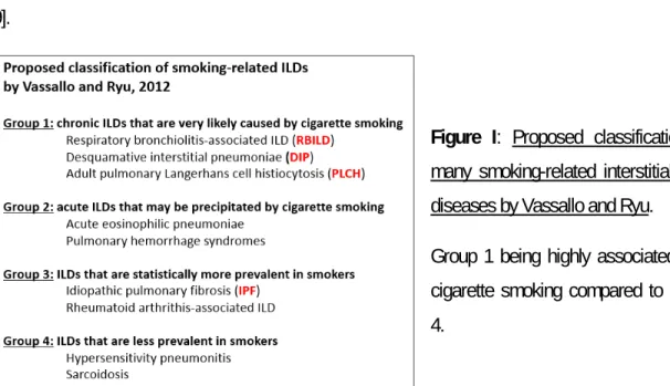
Individuals who smoke may also develop diverse diffuse interstitial and bronchiolar disorders named interstitial lung diseases. In 2012, Vassallo and Ryu decided to classify various smoking-related ILDs (Figure I). They characterized the different ILDs as follows: very likely to be caused by cigarette smoking (group 1), may be precipitated by cigarette smoking (group 2) and more prevalent in smokers (group 3) [59]. Cigarette smoking is broadly known to be the primary cause of different ILDs such as bronchiolitis-associated ILD (RB-ILD), desquamative interstitial pneumonia (DIP) and pulmonary Langerhans cell histiocytosis (PLCH) found in the group 1 classification [23, 60-63]. In group 2, we can find acute eosinophilic pneumonia and pulmonary hemorrhage syndrome. Cigarette smoking is also a risk factor of idiopathic pulmonary fibrosis (IPF) and rheumatoid arthritis associated ILD, two ILDs in group 3 [64, 65]. However, ILDs are rare diseases considering the high prevalence of smoking, suggesting that cigarette smoking is not the only factor leading to the development of most diffuse lung diseases. Genetics factors, microbial infections or allergens could also be required to induce such ILDs [59].
Figure I: Proposed classification of
many smoking-related interstitial lung diseases by Vassallo and Ryu.
Group 1 being highly associated with cigarette smoking compared to group 4.
A key feature of most ILDs, such as RB-ILD is that this disease affects mainly young smokers, between 30 and 40 years of age. RB-ILD is slightly more predominant in men [66]. Classic symptoms of RB-ILD are chronic couch, dyspnea, while inspiratory crackles, abnormal lung sounds during inhalation, may also be present in approximately half of cases [23]. High-resolution computed tomography (CT) is used to characterize most ILD patients. Usual RB-ILD CT findings are centrilobular nodules, ground-glass opacities and a thickening of the bronchial walls [59, 66]. Lung slices of RB-ILD patients show
pigmented macrophages and inflammation near bronchioles and alveoli [66]. As RB-ILD patients usually have a good prognosis, smoking cessation remains the only management option. Corticosteroids may also be prescribed although there is little evidence of any significant beneficial effect [59, 66].
In the case of DIP, smokers are usually diagnosed in their 4th or 5th decade of life. Compared to
RB-ILD, men have twice the risk of developing DIP compared to women [66]. Similar to RB-RB-ILD, DIP symptoms include dry cough, dyspnea and respiratory crackles in half of patients [23, 59]. DIP is characterized by abnormal CT findings such as diffuse ground-glass opacity compared to RB-ILD and often the presence of cysts [67]. DIP patients have an increased number of pigmented macrophages in alveolar spaces. RB-ILD and DIP share common features but the main difference being the diffuse versus centric ground-glass opacities [66]. Smoking cessation is also the most important therapeutic strategy for DIP and corticosteroid may also be given to patients. Survival rates after 5 years and 10 years are 95.2% and 69.6% respectively [23].
PLCH patients are usually diagnosed around 20 to 40 years of age. Men and women have equal risks of developing this type of ILD [68]. Most common symptoms are dyspnea and non-productive cough. In some patients, fever, night sweats and weight loss may also occur [69]. In 10 to 15% PLCH patients will develop a pneumothorax [59, 66]. CT analysis reveals the presence of nodules and cysts in upper or mid airways [66]. PLCH is characterized by an increase in CD1a-positive dendritic cells [59]. Physicians also use aggressive tobacco cessation strategies in order to stop the progression of this ILD [59]. Corticosteroids can be given to patients. Chemotherapeutic agents may be used if multiple organs are involved [68].
Briefly, IPF is also an ILD related to cigarette smoking. Compared to the previous IDLs, this disease is usually diagnosed in patients aged 60 to 70 [66]. Progressive symptoms such as cough, dyspnea, and respiratory crackles are seen in IPF patients [70]. The key radiological features of IPF is the presence of honeycombing due to progressive fibrosis. Histologic findings can be summarized as follows: the presence of fibroblast clusters and immature connective tissue in the lung [71]. The most important clinical feature of IPF is the deterioration of the patient’s condition associated with an average survival
of only 2-3 years post-diagnosis [72] and treatments are mostly to manage symptoms. This is due to the poor responsiveness to corticosteroids and the lack of effective pharmacological therapies available [66].
The mechanisms linking cigarette smoking and the development of ILDs are still unknown. However, excessive inflammation seems to be a hallmark of the ILDs related to cigarette smoking [73, 74]. As stated before, some ILDs are marked by an accumulation of pigmented macrophages in the lung [75, 76]. The cause of this increase in macrophage number is still unknown, though potentially due to intense recruitment and increased secretion of differentiating factors by epithelial cells, improved survivability of macrophages or reduced apoptosis [77]. In addition, a key feature of ILD patients is that their epithelial cells produce disproportionate levels of granulocyte-macrophage colony-stimulating factor (GM-CSF), a cytokine involved in the proliferation and activation of macrophages and dendritic cells [78, 79].
Although these diseases are rare, they clearly have a significant impact on the quality of life and they contribute to the burden of smoking on the health system. It has been estimated that ILDs will become more frequent worldwide with the rise of cigarette smoking prevalence in most developing countries.
Cigarette smoking and chronic obstructive lung disease
Besides lung cancer and interstitial lung diseases, smokers may also develop a COPD. COPD is a preventable and treatable disease marked by persistent respiratory symptoms and irreversible obstruction of airflow usually caused by exposure to chronic noxious particles or gases [24]. Classic symptoms of COPD are coughing, dyspnea and sputum production. COPD patients also often face periods of acute symptoms worsening called exacerbations known to be associated with increased hospitalization rate [24]. Most exacerbations are due to microbial infections, either bacterial or viral [80, 81], and lead to alteration of the pulmonary microbiome [82, 83]. An important negative impact of exacerbations is their effect on the patient’s quality of life [84].
COPD is currently the 4th leading cause of death worldwide. In 2012, more than 3 million deaths per
year, representing approximately 6% of worldwide deaths, were attributed to COPD [24]. According to the Burden of Obstructive Chronic Diseases, a program built to assess the prevalence of COPD, yearly worldwide deaths associated with COPD will reach approximately 4.5 million by 2030 [24]. It was also estimated in 2010 that the worldwide prevalence of COPD was 11.7% [85]. However, the prevalence of COPD is probably underestimated as this disease is underdiagnosed in the general population [86]. Morbidity from COPD is also affected by other chronic diseases such as cardiovascular disease [87], musculoskeletal impairments [88, 89] and diabetes mellitus [24]. The economic burden associated with COPD is worrisome. In the European Union, 56% of total healthcare budget is due to COPD (38.6 billion euros), while in the US approximately 30 billion and 20 billion dollars are disbursed due to the direct and indirect medical costs of COPD, respectively [90, 91].
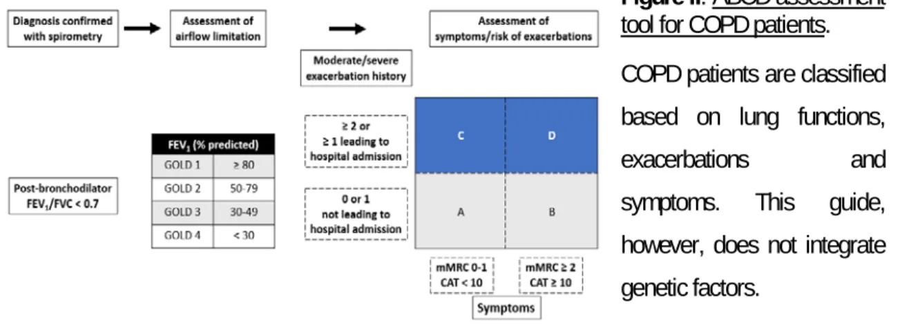
Cigarette smoking is the primary causal agent of COPD in North America [92]. However, in the Eastern world, exposure to air pollution and biomass fuels are the predominant cause [93]. Other factors including genetic, sex, respiratory infections such as tuberculosis, airway hyper-responsiveness, aging and poor lung development during childhood are also associated with the development and progression of COPD [24, 94-100]. Nonetheless, the exact factors leading to the development of COPD are still unknown since only up to 50% of smokers will be diagnosed with this disease [101-105]. Suspected COPD patients that exhibit shortness of breath, cough and mucus hypersecretion are diagnosed using spirometry, the gold standard diagnostic tool [24]. Spirometry is a non-invasive method used to measure the airflow limitation/obstruction. Post-bronchodilator spirometry allows for the measurement of two clinical parameters: the volume of air exhaled during the first second (forced expiratory volume in one second, FEV1) and the volume of air exhaled by force at maximal inspiration
(forced vital capacity, FVC). The ratio FEV1/FVC, also called the Tiffeneau index, is considered normal
when the value is higher than 0.7, while a ratio under 0.7 indicates airflow obstructions [24, 106]. COPD patients are also asked to answer questionnaires on breathlessness and symptoms for instance the Modified British medical research council (mMRC) questionnaire and the COPD assessment test, respectively (CAT; [24]. COPD is further classified by severity: mild, moderate, severe and very severe following the Global initiative for chronic Obstructive Lung Disease (GOLD) guidelines. This classification is based on the patient’s FEV compared to that of the general population, expressed as
a percentage of their predicted FEV1. Patients are categorized as follows: GOLD 1 FEV1 ≥ 80%, GOLD
2 50% ≤ FEV1 < 80%, GOLD 3 30% ≤ FEV1 < 50% and GOLD 4 FEV1 < 30% [24] (Figure II).
Figure II: ABCD assessment
tool for COPD patients. COPD patients are classified based on lung functions,
exacerbations and symptoms. This guide, however, does not integrate genetic factors.
Genetic analysis is a great tool which has led to the discovery of genes associated with COPD and may ultimately lead to new therapeutic possibilities [107]. It is now known that a rare autosomal mutation to alpha-1 antitrypsin is found in 1-2% of COPD patients. Deficiency in alpha-1 antitrypsin (AAT1), a key circulating protease inhibitors, was the first mutation associated with COPD [108, 109]. During cigarette smoke exposure, AAT1 inhibits serine proteases from different immune cells, such as neutrophils. Other single genes, for instance matrix metalloproteinase 12 (MMP12), have been associated with a decline in lung function [110]. Similarly, genetic loci near the alpha-nicotiαnic acetylcholine receptor and the hedgehog interacting protein may also be associated with the development of COPD [111, 112]. Siblings of severe COPD patients have a higher risk of developing airflow limitations, suggesting that a combination of genetic and environmental factors may influence the risk of developing COPD [113].
Chronic airflow limitation in COPD patients is usually due to two different airway diseases: chronic bronchitis and destruction of lung parenchyma (emphysema). Interestingly, these clinical presentations vary among COPD patients; while some patients only exhibit signs of emphysema, other patients have pronounced obstructive bronchiolitis [24]. Chronic bronchitis is characterized by an exaggerated production of mucus in the respiratory tract mainly caused by the remodeling of the bronchial epithelium, the replacement of ciliated cells with mucus cells and the hypertrophy of secretory mucus glands [114]. Emphysema is characterized by the destruction and the loss of elasticity of alveoli, which
is the main area for gas exchange [115, 116]. This phenomenon leads to non-ventilated pulmonary zones. Patients with emphysema must breathe more often in order to meet the necessary levels of circulating oxygen required for metabolic processes but also to expel toxic carbon dioxide [117]. Another important characteristic of COPD is the presence of chronic inflammation and its persistence even after smoking cessation. The mechanisms responsible for the intensified response to cigarette smoke in COPD patients remain poorly understood [24]. However, the oxidative stress and the surplus of proteinase activity may influence lung inflammation [118-120]. Cigarette smoke and activated immune cells such as macrophages and neutrophils are the main pulmonary sources of reactive oxygen species (ROS) in COPD patients. It was shown that oxidative stress markers are increased in the systemic circulation, exhaled breath condensate and sputum of COPD patients. Exacerbations also lead to increased levels of oxidative stress [121]. In addition, excessive protease secretion from immune cells and epithelial cells have been observed in COPD patients. Their role in the destruction of elastin, a vital connective tissue in lung parenchyma, is thought to be a key mechanism leading to the development of emphysema [108, 109, 122].
Another hallmark of COPD is the presence of an increased number of inflammatory cells, most importantly macrophages, neutrophils and activated lymphocytes [123]. These cells, combined with epithelial and structural cells, release a variety of inflammatory mediators including chemotactic factors, proinflammatory cytokines and growth factors [118, 124]. Inflammation might also precede the progression towards fibrosis. COPD patients may show signs of peribronchiolar fibrosis and interstitial opacities [125] and exhibit increased levels of growth factors. The repeated injury/repair of lung parenchyma induced by cigarette smoking could underlie the excessive proliferation of muscle and fibrous tissue [126].
Other than smoking cessation, most available COPD treatments aim to reduce symptoms (coughing, sputum, and dyspnea) and exacerbations [24]. Pharmacological therapies rely mostly on bronchodilators and corticosteroids [24]. Bronchodilators such as beta2-agonists are used to improve
the FEV1 of COPD patients by relaxing the airway smooth muscle and thus blocking
bronchoconstriction [129]. Furthermore, COPD patients have access to inhaled corticosteroids which are thought to reduce the frequency and severity of exacerbations. The combination of bronchodilators and corticosteroids is more effective than either components alone at improving the health status, ameliorating lung function and reducing the number of exacerbations [130-132]. Phosphodiesterase-4 inhibitors may also be given to COPD patients as they reduce inflammation by inhibiting the breakdown of cyclic adenosine monophosphate (cAMP) [133]. These inhibitors have been shown to reduce exacerbations and improve lung function when combined with corticosteroids and bronchodilators, respectively [134, 135]. It is recommended that COPD patients receive the influenza vaccine as it was shown to significantly reduce the number of exacerbations and deaths caused by the flu [136, 137]. Additionally, pneumococcal vaccines are suggested to COPD patients, especially the 13-valent and 23-valent pneumococcal vaccines [24, 138].
COPD is a major societal burden and we will likely see a rise in the incidence of this disease in developing countries due to the increased prevalence of cigarette smoking. There are still a lot of unanswered questions regarding COPD such as susceptibility factors, initiating mechanisms, etc. Currently, a broad range of treatments are in use or under investigation. However, most of the therapies available to patients today merely attempt to reduce symptoms.
As you can see, cigarette smoking is a global burden leading to unprecedented societal, economic and health problems. The exposure to cigarette smoke leads to the development of major pulmonary diseases such as lung cancer, interstitial lung diseases and chronic obstructive pulmonary disease, as
well as several other systemic diseases (Figure III, adapted from
https://www.cdc.gov/tobacco/infographics/health-effects/index.htm#smoking-risks). Many of these diseases are progressive, life-threatening or even fatal. This thesis will focus on macrophages, one of the main inflammatory cells involved in the response to cigarette smoke exposure that have been shown to be associated with most of the aforementioned diseases.
Figure III: Cigarette smoking is a major risk factor for numerous systemic and pulmonary diseases.
Generally, this habit leads to unprecedented health problems. To date cigarette smoking is associated with up to 15 different cancers and multiple chronic diseases.
Inflammation and pulmonary macrophage responses to
cigarette smoke
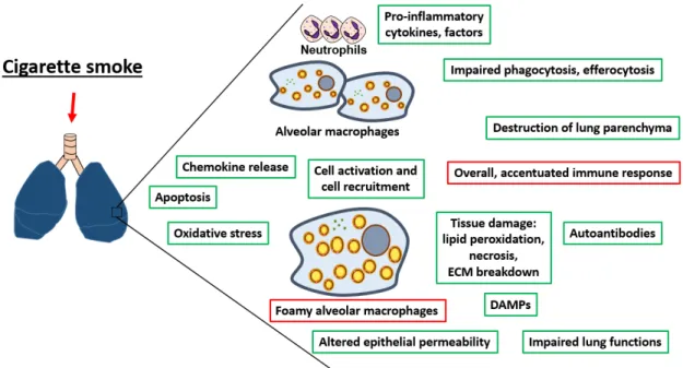
Inflammation is a process found in all of the pulmonary diseases described above. One of the main characteristics of lung inflammation induced by cigarette smoke exposure is the increased number of pulmonary macrophages [139]. This phagocyte has many immune functions (for instance efferocytosis [140], phagocytosis [141], microbicidal activities [142], recognition functions [143], etc.) which are all negatively affected by cigarette smoking. In this section, general information related to inflammation will be presented. The acute and chronic pulmonary effects of cigarette smoke exposure will be
described, followed by its impact specifically on pulmonary macrophages. To conclude, the development of a “foamy” pulmonary macrophage phenotype induced by cigarette smoke exposure will be introduced.
What is inflammation?
Inflammation is a vital process that has been recognized since ancient times. Our ancestors already knew that inflammation involved four different mechanisms: rubor (redness), tumor (swelling), calor (heat), and dolor (pain) [144]. By the 1850s, Virchow introduced Functio laesa, or the dysfunction of organs, as the fifth feature [144]. Inflammation can be found throughout the body, for instance in the lung when facing diverse insults [145].
Following a variety of pulmonary insults, e.g. bacteria, viruses, air pollution, allergens, tissue injury or cigarette smoke, etc., there will be the activation of the immune system which is crucial for host protection and for the initiation of repair mechanisms [146]. Depending on the nature of the insult, the lung will mount an appropriate immune response against microorganisms or against pollution particles, allergens, cigarette smoke, etc. Responses to microorganisms are orchestrated via the detection of pathogen-associated molecular patterns (PAMPs) by pattern recognition receptors (PRRs; [147], which will rapidly activate the innate immune response and later, the adaptive immune response.
Briefly, the innate immune response is a rapid response coordinated by resident pulmonary cells like pulmonary macrophages, epithelial cells, neutrophils and antigen-presenting cells (APCs) like dendritic cells [148] [149]. PRRs are found on macrophages, for instance toll-like receptors (TLRs), and are crucial in pathogen recognition [150]. Diverse PAMPs such as lipopolysaccharide (LPS) and modified low-density lipoproteins (LDLs) can bind TLRs [151]. Hallmarks of the innate immune response are the recruitment of pulmonary macrophages and neutrophils and the production of inflammatory cytokines and chemokines, for instance interleukin-1 (IL-1) and tumour necrosis factor alpha (TNFa) [147]. The main goal of the innate immune response is to mount an efficient response in order to eliminate diverse microbial threats and, if required, ultimately activate the adaptive immune response [152]. Adaptive immune response is mainly activated via APCs and the presentation of diverse antigens to naive T
cells [153]. Contrary to the innate immune response, the adaptive immune response is a slow and well-organized response relying on the production of specific antibodies against the antigen and the ability to build an immunological memory [144, 148]. The latter feature is crucial during reinfection with the same microorganism.
Interestingly, these PRRs can also be activated by non-microbial signals called danger-associated molecular patterns (DAMPs). DAMPs are the molecular signatures of sterile inflammation [147]. Cell death and tissue injury led to the release of DAMPs in the environment triggering a sterile inflammation response [147]. Binding of DAMPs to PRRs will activate downstream signaling pathways for instance the nuclear factor-κb (NFkB) and mitogen-activated protein kinase (MAPK), which will result in the upregulation of pro-inflammatory cytokines and chemokines [147]. Cigarette smoke exposure is a sterile stimulus that causes the release of DAMPs [154-156]. DAMPs can also be located extracellularly [147]. For instance, extracellular matrix degradation generates DAMPs, such as hyalorunan, biglycan, etc. These molecules are the products of proteolysis by enzymes released during cell death or via proteases secreted by pulmonary macrophages and neutrophils [157]. The latter mechanism becomes important under cigarette smoke conditions [158]. Similar to the immune response against microorganisms, responses to sterile stimuli lead to the recruitment of neutrophils and macrophages as well as the release of pro-inflammatory cytokines such as IL-1α [159, 160]. IL-1α, a pro-inflammatory cytokine, is also released in the pulmonary environment during necrosis, further promoting inflammation [161]. IL-1α is a key player involved in the initiation of sterile inflammation [162] and cigarette smoke-induced inflammation [163]. Overall, cigarette smoke triggers a sterile lung inflammation response with multiple consequences. The next section will focus on the pulmonary response to cigarette smoke and on pulmonary macrophages, a major player involved in maintaining lung homeostasis during inflammation.
The pulmonary response to cigarette smoke
Scientific and medical knowledge regarding the effects of cigarette smoke has greatly grown since the first Surgeon General in 1964 [164]. It is now known that nicotine is a lipophilic, addictive molecule found in cigarette smoke that is detected in the respiratory tract during cigarette smoke inhalation. Furthermore, nicotine binds within 10 second to the nicotinic acetylcholine receptor (nAChR) in the
brain, passing via the blood-brain barrier, to activate dopamine release [1, 165]. Dopamine leads to appetite loss, lower stress, anxiety, pain and mild euphoria [1, 165]. Other than the effects of nicotine and cigarette smoke on body composition [166], birth weight [167, 168], fetal development [169], central nervous system [170-173], metabolic and fertility problems [169], this section will focus on the cigarette smoke exposure systems available for animal research and the pulmonary outcomes of acute and chronic cigarette smoke exposure in these models.
Cigarette smoke exposure models
As acute cigarette smoke impact studies in humans are few due to ethic issues, one must turn to animal models. Animal models are therefore used to test new hypothesis, drug toxicity, side effects, and efficiency, but also to establish solid proofs of concepts. Cigarette smoke impacts are mostly studied in mice [174-177] but also in rats and guinea pigs [178]. However, molecular engineering is far more advanced in mice conferring an easier access to genetically modified mice strains [179].
Many cigarette smoke exposure systems are available. They differ in the type of exposure, which can be either nose only or whole-body exposure. Furthermore, mice can be exposed to mainstream primary smoke or secondary smoke. Most studies use acute, short-term exposure times or chronic protocols, with up to six months of cigarette smoke exposure. The former is used to characterize the pulmonary inflammatory response to cigarette smoke and to understand the initiating mechanisms leading to inflammation [180, 181]. On the contrary, subchronic and chronic models are primarily used to investigate the impact of cigarette smoke on the pulmonary structure and the long-term immune response [182, 183]. While acute cigarette smoke exposure models are known to generate an observable inflammatory response [180, 181], long-term exposure to cigarette smoke will result in a milder pulmonary inflammatory response. Though many variables can interact with the pulmonary outcomes of cigarette smoke exposure, several studies, utilizing various cigarette smoke exposure protocols, have established that cigarette smoke exposure has a negative impact on the lung.